Open Access
S H O R T R E P O R T
Bio
Med
Central
© 2010 Archer et al; licensee BioMed Central Ltd. This is an Open Access article distributed under the terms of the Creative CommonsAttribution License (http://creativecommons.org/licenses/by/2.0), which permits unrestricted use, distribution, and reproduction in any medium, provided the original work is properly cited.Short Report
Effect of probe characteristics on the subtractive
hybridization efficiency of human genomic DNA
Marie J Archer*
1, Nina Long
2and Baochuan Lin
1Abstract
Background: The detection sensitivity of low abundance pathogenic species by polymerase chain reaction (PCR) can be significantly enhanced by removing host nucleic acids. This selective removal can be performed using a magnetic bead-based solid phase with covalently immobilized capture probes. One of the requirements to attain efficient host background nucleic acids subtraction is the capture probe characteristics.
Findings: In this study we investigate how various capture probe characteristics influence the subtraction efficiency. While the primary focus of this report is the impact of probe length, we also studied the impact of probe conformation as well as the amount of capture probe attached to the solid phase. The probes were immobilized on magnetic microbeads functionalized with a phosphorous dendrimer. The subtraction efficiency was assessed by quantitative real time PCR using a single-step capture protocol and genomic DNA as target. Our results indicate that short probes (100 to 200 bp) exhibit the best subtraction efficiency. Additionally, higher subtraction efficiencies with these probes were obtained as the amount of probe immobilized on the solid phase decreased. Under optimal probes condition, our protocol showed a 90 - 95% subtraction efficiency of human genomic DNA.
Conclusions: The characteristics of the capture probe are important for the design of efficient solid phases. The length, conformation and abundance of the probes determine the capture efficiency of the solid phase.
Findings
The presence of a large excess of non-target nucleic acids (NA), i.e. host genomic NA, sometimes is inevitable and can cause false positive or negative results when using molecular diagnostic technologies, such as PCR. The sep-aration of background from low abundance pathogenic targets that coexist in complex matrices is necessary to ensure optimal detection sensitivity and specificity [1-4] and can be done using a solid phase with covalently attached probes. The characteristics of the probe, such as length, conformation and abundance on the solid phase, are of relevance in determining the capture efficiency. Therefore it is necessary to understand how such charac-teristics affect the capture efficiency in order to design efficient solid phases.
Information on the significance of probe diversification is limited. Most of the results reported to date deal with short single stranded oligonucleotides (20-70 nucleotides
(nt)) that may not be suitable for capturing genomic tar-gets [2,5-10]. Mathematical models combined with experimental data, aimed at understanding the hybridiza-tion dynamics of DNA to surface-bound probes, have revealed that the kinetics are determined by the amount of probes immobilized on the surface, the length, the con-centration, and the size of the target [6,11-15]. Zammateo et al. utilized capture probes of various lengths (56-255 base pair (bp)) immobilized on magnetic microparticles to characterize the capture efficiency of a 435 bp target. They found that the longer probes, which correlated with higher immobilization efficiency, exhibited the best sub-traction efficiency and determined the reaction yield. However, these observations were based solely on the size of the probe [16]. These results differ from the observa-tions of other studies which suggest that low probe densi-ties favour hybridization kinetics and that there is a trade-off between the length of the probes and their density on the surface [7,11,13]. These contradictory results high-light the complexity in which solid phase hybridization occurs and further support the need to optimize the cap-ture probe depending on particular application needs. * Correspondence: marie.archer@nrl.navy.mil
1 US Naval Research Laboratory, Center for Bio/Molecular Science &
Engineering, 4555 Overlook Avenue, S W, Washington, DC, 20375, USA
In this work, we focused on the effects of the length, conformation and the amount of probe on the selective capture of human genomic DNA using a previously developed magnetic bead based solid phase that enables capture of genomic targets in a single step [17-19]. Cap-ture probes of various lengths were synthesized using strand displacement, isothermal amplification and PCR. Our results demonstrated that better capture efficiency was achieved using shorter probes and correlates to the amount of immobilized probes. Furthermore, fragmented human genomic DNA targets (100-5000 bp) can be cap-tured efficiently even with probes as short as 100 to 200 bp. This finding disagrees with the existing literature which suggests that the target should be shorter than 100 bp for efficient solid phase hybridization [11].
Methods
Magnetic bead based solid support preparation
Preparation and functionalization of magnetic beads with generation 4.5 phosphorous dendrimer was as described in a previous publication [17] with slight modifications. Briefly, the magnetic beads were prepared in batch mode and the volumes of solvent used were adjusted accord-ingly [see Additional file 1 for details].
Preparation of capture probes
Capture probes were prepared by three different meth-ods: Sequenase DNA Polymerase, Klenow fragment, and PCR. The Sequenase probes were prepared in a 30 μl vol-ume containing 40 mM Tris-HCl (pH 7.5), 20 mM MgCl2, 50 mM NaCl, 7 μM Primer D [See additional file 1: Sup-plemental Table S1], 0.67 mM dNTPs, 13 U of Seque-nase™ Version 2.0 DNA Polymerase (USB, Cleveland, OH), and 4 μg of COT human DNA with preliminary denaturation at 95°C for 2 min., followed by incubation at 10°C for 5 min., then at 37°C for 60 min. The Klenow probes were prepared in a 50 μl reaction volume contain-ing 10 mM Tris-HCl (pH 7.9), 10 mM MgCl2, 50 mM NaCl, 1 mM DTT, 20 μM primer D, 0.2 mM dNTPs, 10 U of Klenow fragment (3' T 5' exo-) (NEB, Ipswich, MA) with initial denaturation at 95°C for 5 min. followed by immediate cool down at 4°C, incubation at 37°C for 6 hours and enzyme inactivation at 75°C for 20 min. For reactions using Sequenase DNA polymerase and Klenow fragment, the enzymes and the dNTPs were added after the denaturation step. The PCR probes were prepared in a 50 μl reaction volume with GoTaq® DNA Polymerase (Promega, Madison, WI) using 2 μM Primer NL and 0.8 μM Primer NLN [See additional file 1: Supplemental Table S1] and 400 ng of human genomic DNA (Roche, Indianapolis, IN) as template. The amplification reaction was carried out with preliminary denaturation at 94°C for 2 min. followed by 40 cycles of 94°C for 30 s, 40°C for 30
s, 50°C for 30 s, and 72°C for 2 min., and a final extension step at 72°C for 7 min.
All the products were purified using QIAquick PCR purification kit (Qiagen, Valencia, CA) following the manufacturer's instructions. Prior to immobilization, the PCR probes were denatured at 95°C for 5 min. and cooled to 4°C. Verification of the probes as single strands was performed using a Qubit® fluorometer with the Quant-IT™ ssDNA assay kit (Invitrogen, Carlsbad, CA).
Immobilization of capture probes
The probe immobilization was performed as described in a previous publication with minor modifications [17]. Briefly, magnetic beads functionalized with a generation 4.5 phosphorous dendrimer were re-suspended in 750 μl of 0.3 M sodium phosphate buffer at pH 9.0 (Na2HPO4) and sonicated for 10-15 seconds to ensure the beads were dispersed. The beads were incubated overnight at room temperature with three additional buffer changes. Immo-bilization was performed at room temperature by adding 150 μl of capture probe solution at a concentration of 3, 6, 9, 12 and 15 ng/μl with periodic re-suspension. After immobilization, reduction and blocking for non-specific adsorption was carried out as previously described [17] with slight modifications in the sodium borohydride solution (25 mg instead of 12.5 mg of NaBH4 was used) and incubation time (extended to 15 min). The beads were washed with 0.2% SDS, resuspended in 300 μl of stripping buffer (1 × SSC/0.1% SDS) and incubated for 20-24 min. at 95°C with one buffer exchange and re-sus-pension at the half way point. The beads were then washed with nuclease free water pre-warmed at 95°C for 20-24 min., with re-suspension at the half way point fol-lowed by a 30 min. wash in 5 × SCC/0.1% SDS and 0.6 × SCC/0.03% SDS at 55°C with re-suspension at the half way point. Finally, the beads were pooled into 1200 μg ali-quots in 0.1% SDS and stored at 4°C after discarding the SDS solution. The immobilization efficiency was deter-mined to be between 95-98% by quantification of the supernatant using a NanoDrop ND-1000 fluorospec-trometer (NanoDrop Technologies, Wilmington, DE) as previously described [17]. Magnetic beads with no probes were prepared in an identical manner and used as con-trols.
Subtractive hybridization assays
M TMAC/0.11% SDS pre-warmed to 37°C. The reactions were carried out in Thermomixer® (Eppendorf, Westbury, NY) with denaturing at 97°C for 20 min. and annealing at 60°C for 90 min. After incubation, the supernatant was collected and the beads were washed with 100 μl each of 2 × SSC/0.1%SDS and 0.1 × SCC/0.1%SDS. All superna-tants were collected and a second magnetic separation was performed to eliminate any bead carryover. The supernatants were ethanol precipitated and resuspended in 10 mM Tris-HCl at pH 8.5.
Quantitative real-time PCR
Quantitative real-time PCR (qPCR) was performed using the MyiQ™ real-time PCR detection system with iQ SYBR Green Supermix (Bio-Rad Laboratories, Inc., Hercules, CA) according to the manufacturer's recommended pro-tocol. The primers used and PCR conditions are listed in supplemental information [See additional file 1: Supple-mental Table S1]. All qPCR results were reported in terms of genome copy concentration per microliter using an external standard curve with known concentration of ACTB.
Results and discussion
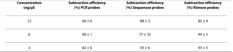
Three different capture probes for human genomic DNA (hgDNA) were synthesized by isothermal amplification ("Klenow probes"), strand displacement ("Sequenase probes") and polymerase chain reaction ("PCR probes"). Gel electrophoresis of the probes shows a length variation from ~100 300 bp for the Klenow probes to ~200 -600 bp for the Sequenase and ~300 - 800 bp for PCR probes (Figure 1). To compare the subtraction efficiency, the probes were covalently immobilized on magnetic microbeads and subtraction was performed using a pre-viously developed single step protocol [See additional file 1: Supplemental Figure S1]. The supernatant containing the remnant DNA was precipitated and analyzed with qPCR, and the subtraction efficiency for each probe type was compared (Table 1). The results indicated that the Klenow probes showed the best subtraction efficiency
with less experimental variation, while the Sequenase probes showed similar subtraction efficiency with higher variations. The PCR probes showed the lowest subtrac-tion efficiency in comparison to the Klenow and Seque-nase probes. The results also revealed a general trend that reduction in the amount of probe enhanced the subtrac-tion efficiency. Addisubtrac-tional experiments performed at higher probe densities (up to 30 ng/μl) using various buf-fer systems indicated that the subtraction efficiency decreases as the amount of probe increases (data not shown).
Given the low subtraction efficiency of PCR probes, further characterizations were undertaken with only the Klenow and Sequenase probes. Further experiments con-firmed the observation that, reduction in the amount of probe enhanced the subtraction efficiency with Klenow probes which exhibited less experimental variations (Fig-ure 2).
These results indicated that the probe length plays a major role in the subtraction efficiency and that the shorter probes provided better subtraction efficiency. This is probably due to the fact that longer probes form secondary structures (hairpin or loop) which are thought to negatively affect the hybridization efficiency in solu-tion and solid phase even with oligonucleotides as short as 60 nt [14,15]. They also represent a more complex sce-nario since bending induced by electrostatic interactions and intermolecular hybridization can occur and affect hybridization [20]. Our results also indicate that a lower input concentration (3 ng/μl) of shorter probes (~100-200 bp) can be utilized to subtract up to 1000 ng of hgDNA. Larger spacing between the probes reduces steric interference and facilitates diffusion of the targets which is further enhanced by the gradual decrease in temperature and the intermittent mixing. These results contradict the observations by Chan et al. who deter-mined that the rate of hybridization per unit area was equally efficient, regardless of the probe size, if there was proper spacing and as long as the target was between 100 to 160 bp [11]. However, these simulations did not
con-Table 1: Subtraction efficiencies of human genomic DNA using three different types of probes
Concentration (ng/μl)
Subtraction efficiency (%) PCR probes
Subtraction efficiency (%) Sequenase probes
Subtraction efficiency (%) Klenow probes
12 66 ± 6 88 ± 3 85 ± 4
6 80 ± 1 77 ± 25 94 ± 5
3 82 ± 6 93 ± 6 95 ± 5
[image:3.595.56.541.613.721.2]sider secondary structures or targets of variable molecu-lar sizes.
Interestingly, the Sequenase probes contain probes sizes similar to the Klenow probes (100 to 200 bp) (Figure 1), however, the capture efficiency was lower and more variable. This may reflect the fact that longer probes rep-resent the majority within the Sequenase generated probes. In this case the hybridization would be
domi-nated by these longer fragments which exhibit an inher-ently lower subtraction efficiency than do shorter probes. Even if short probes are present, diffusion of the targets to these sites might be hindered by secondary structure or steric interference. In addition to length, the sequence of the probes is another parameter to consider since it influences the structure of the probes [14,15,21]. How-ever, we have not performed sequencing analysis and we cannot discuss in further detail this particular parameter.
Conclusions
In conclusion, we have investigated the effect of probe length, conformation and amount on the solid phase cap-ture efficiency of human genomic DNA. In contrast with the published literature, our results indicate that, probes as short as 200 bp can capture between 500-1000 ng of human genomic DNA (100 and 5000 bp). Longer probes (600-1000 bp) exhibited lower subtraction efficiency (~10% difference) with greater variability. Secondary structure and steric interference might be responsible for these differences. In all cases, a lower amount of immobi-lized probe on the magnetic beads correlated with an enhanced performance. The results presented here are of relevance for the design of efficient solid phases for the selective capture of genomic targets.
List of Abbreviations
[image:4.595.62.539.94.316.2]NA: nucleic acids; SDS: sodium dodecyl sulfate; SSC: standard sodium citrate; PCR: Polymerase chain reaction; TMAC: tetramethylammonium chloride.
Figure 1 Capture probe characterization. (A) Representative image of different capture probes. Lane 1, molecular weight marker; lane 2, PCR probes; lane 3, probes synthesized by strand displacement; lane 4, probes generated by isothermal amplification. The products were run on a 1.2% TAE agarose gel and visualized by ethidium bromide staining. (B) Histogram showing the distribution of the sizes of the capture probes. The corre-sponding molecular weight marker sizes are indicated in the x-axis.
1 2 3 4
650 bp
100 bp 1 Kbp
300 bp 2 Kbp
A)
100 1k
2k 650
1.20
1.00
0.80
0.60
0.40
0.20
0.00
N
o
rm
alized Intensity
(a.u)
Ladder Size (bp)
Klenow Probes PCR Probes
Sequenase Probes
300
B)
Figure 2 Comparison of subtraction efficiency between the
Kle-now and the Sequenase probes. The Klenow (solid black) and the
Se-quenase (solid grey) probes were further evaluated for their subtraction efficiency. The Y-axis shows % subtraction, and the x-axis indicates the input probes concentration. These experiments com-prised at least 5 subtractions and the qPCR was performed in triplicate. Error bars correspond to the standard deviation of the mean.
100 90 80 70 60 50 40 30 20 10
% Subtracted
[image:4.595.56.290.494.658.2]Additional material
Competing interests
There are two pending patent applications, one for preparation and function-alized of magnetic beads and one for genomic DNA subtraction protocol, that are related to this article. Both MJA, and BL are listed as inventors of these two patent applications.
Authors' contributions
MJA participated in the concept development, characterization and fabrica-tion the solid phase, and drafting the manuscript. NL performed the probes synthesis and subtraction experiments. BL participated in the concept devel-opment, performing quantitative real time PCR analysis, and drafting the man-uscript.
Acknowledgements
This study was sponsored by the Defense Threat Reduction Agency. We thank Dr. Nazir Barekzi and Mr. Paul Charles for critically reviewing the manuscript. We also thank Dr. Thomas O'Shaugnessy for his assistance in generating the cap-ture probe histograms. The views expressed here are those of the authors and none are to be construed as those of the Defense Threat Reduction Agency, the U. S. Navy, the U. S. Department of Defense, or the U. S. Government at large.
Author Details
1US Naval Research Laboratory, Center for Bio/Molecular Science &
Engineering, 4555 Overlook Avenue, S W, Washington, DC, 20375, USA and
2NOVA Research Inc, 1900 Elkin St, Suite 230, Alexandria, VA, 22308, USA
References
1. Millar DS, Withey SJ, Tizard MLV, Ford JG, Hermon-Taylor J: Solid phase hybridization capture of low-abundance target DNA sequences: Applications to the polymerase chain reaction detection of
Mycobacterium Paratuberculosis and Mycobacterium avium subsp.
silvaticum. Anal Biochem 1995, 226:325-330.
2. Mangiapan G, Vokurka M, Schouls L, Cadranel J, Lecossier D, Van Embden J, Hance AJ: Sequence capture-PCR improves detection of
mycobacterial DNA in clinical specimens. J Clin Microbiol 1996,
34:1209-1215.
3. Pradel N, Leroy-Setrin S, Joly B, Livrelli V: Genomic subtraction to identify and characterize sequences of shiga toxin producing Escherichia coli
O91:H21. Appl Environ Microbiol 2002, 68:2316-2325.
4. Boni J, Shah C, Flepp M, Luthy R, Schupbach J: Detection of low copy numbers of HIV-1 proviral DNA in patient PBMCs by a high-input, sequence capture PCR (Mega-PCR). J Med Virol 2004, 72:1-9. 5. Day PJR, Flora PS, Fox JE, Walker MR: Immobilization of polynucleotides
on magnetic particles. Factors influencing hybridization efficiency.
Biochem J 1991, 278:735-740.
6. Stevens PW, Henry MR, Kelso DM: DNA hybridization on microparticles: determining capture-probe density and equilibrium dissociation constants. Nucleic Acids Res 1999, 27:1719-1727.
7. Peterson AW, Heaton RJ, Georgiadis RM: The effect of surface probe density on DNA hybridization. Nucleic Acids Res 2001, 29:5163-5168. 8. Zeng J, Almadidy A, Watterson J, Krull UJ: Interfacial hybridization
kinetics of oligonucleotides immobilized on fused silica surfaces. Sens Actuators B 2003, 90:68-75.
9. Yeung SW, Hsing IM: Manipulation and extraction of genomic DNA form cell lysate by functionalized magnetic particles for lab on a chip applications. Biosens Bioelectron 2006, 21:989-997.
10. Parham NJ, Picard FJ, Peytavi R, Gagnon M, Seyrig G, Gagne PA, Boissinot M, Bergeron MG: Specific magnetic bead-based capture of genomic DNA from clinical samples: application to the detection of group B
Streptococci in vaginal/anal swabs. Clin Chem 2007, 53:1570-1576.
11. Chan V, Graves DJ, McKenzie SE: The biophysics of DNA hybridization with immobilized oligonucleotide probes. Biophys J 1995,
69:2243-2255.
12. Okahata Y, Kawase M, Niikura K, Ohtake F, Furusawa H, Ebara Y: Kinetic Measurements of DNA Hybridization on an Oligonucleotide-Immobilized 27-MHz Quartz Crystal Microbalance. Anal Chem 1998,
70:1288-1296.
13. Hagan MF, Chakraborty AK: Hybridization dynamics of surface immobilized DNA. J Chem Phys 2004, 120:4958-4968. 14. Chien FD, Liu JS, Su HJ, Kao LA, Chiou CF, Chen WY, Chen SJ: An
investigation into the influence of secondary structures on DNA hybridization using surface Plasmon resonance biosensing. Chem Phys Lett 2004, 397:429-434.
15. Gao Y, Wolf LK, Georgiadis RM: Secondary structure effects on DNA hybridization kinetics: a solution versus surface comparison. Nucleic Acids Res 2006, 34:3370-3377.
16. Zammateo N, Alexandre I, Ernest I, Le L, Brancart F, Remacle J:
Comparison between microwell and bead supports for the detection of human cytomegalovirus amplicons by sandwich hybridization. Anal Biochem 1997, 253:180-189.
17. Archer MJ, Lin B, Wang Z, Stenger DA: Magnetic bead-based solid phase for selective extraction of genomic DNA. Anal Biochem 2006,
355:285-297.
18. Archer MJ, Lin B, Stenger DA: Selective solid phase for capture of genomic nucleic acids in complex mixtures in a single step. 2007. U.S. Patent Application 11/751,096
19. Archer MJ, Stenger DA, Lin B: Development and characterization of a solid phase for single step enrichment of pathogenic targets. The Open Analytical Chemistry Journal 2008, 2:47-54.
20. Fenley MO, Olson WK, Tobias I, Manning GS: Electrostatic effects in short superhelical DNA. Biophys Chem 1994, 50:255-271.
21. Matsumoto A, Olson WK: Sequence-dependent motions of DNA: A normal mode analysis at the base pair level. Biophys J 2002, 83:22-41.
doi: 10.1186/1756-0500-3-109
Cite this article as: Archer et al., Effect of probe characteristics on the sub-tractive hybridization efficiency of human genomic DNA BMC Research Notes
2010, 3:109
Additional file 1 Supplemental material to the methods. Details on the magnetic bead bases solid support preparation, hybridization capture assays (Figure S1) and primer sequences and PCR conditions (Table S1).
Received: 18 December 2009 Accepted: 20 April 2010 Published: 20 April 2010
This article is available from: http://www.biomedcentral.com/1756-0500/3/109 © 2010 Archer et al; licensee BioMed Central Ltd.
This is an Open Access article distributed under the terms of the Creative Commons Attribution License (http://creativecommons.org/licenses/by/2.0), which permits unrestricted use, distribution, and reproduction in any medium, provided the original work is properly cited.