R E S E A R C H A R T I C L E
Open Access
Efficacy of sodium butyrate adjunct therapy
in shigellosis: a randomized, double-blind,
placebo-controlled clinical trial
Rubhana Raqib
1,5*, Protim Sarker
1,2, Akhirunnesa Mily
1, Nur Haque Alam
1, Abu Saleh Mohammed Arifuzzaman
1,
Rokeya Sultana Rekha
1,2, Jan Andersson
3, Gudmundur H Gudmundsson
4, Alejandro Cravioto
1and Birgitta Agerberth
2Abstract
Background:Treatment of shigellosis in rabbits with butyrate reduces clinical severity and counteracts the downregulation of cathelicidin (CAP-18) in the large intestinal epithelia. Here, we aimed to evaluate whether butyrate can be used as an adjunct to antibiotics in the treatment of shigellosis in patients.
Methods:A randomized, double-blind, placebo-controlled, parallel-group designed clinical trial was conducted. Eighty adult patients with shigellosis were randomized to either the Intervention group (butyrate, n = 40) or the Placebo group (normal saline, n = 40). The Intervention group was given an enema containing sodium butyrate (80 mM), twice daily for 3 days, while the Placebo group received the same dose of normal saline. The primary endpoint of the trial was to assess the efficacy of butyrate in improving clinical, endoscopic and histological features of shigellosis. The secondary endpoint was to study the effect of butyrate on the induction of antimicrobial peptides in the rectum. Clinical outcomes were assessed and concentrations of antimicrobial peptides (LL-37, human beta defensin1 [HBD-1] and human beta defensin 3 [HBD-3]) and pro-inflammatory cytokines (interleukin-1β [IL-1β] and interleukin-8 [IL-8]) were measured in the stool. Sigmoidoscopic and histopathological analyses, and immunostaining of LL-37 in the rectal mucosa were performed in a subgroup of patients.
Results:Compared with placebo, butyrate therapy led to the early reduction of macrophages, pus cells, IL-8 and IL-1β in the stool and improvement in rectal histopathology. Butyrate treatment induced LL-37 expression in the rectal epithelia. Stool concentration of LL-37 remained significantly higher in the Intervention group on days 4 and 7.
Conclusion:Adjunct therapy with butyrate during shigellosis led to early reduction of inflammation and enhanced LL-37 expression in the rectal epithelia with prolonged release of LL-37 in the stool.
Trial Registration:ClinicalTrials.gov, NCT00800930.
Keywords:Short chain fatty acids, Butyrate, Shigellosis, Innate immunity, Antimicrobial peptides, Cathelicidin, LL-37, Inflammation, Pro-inflammatory cytokines, Rectal mucosa
Background
Shigellosis continues to be a major health burden with an annual incidence rate of 125 million cases in Asia, al-though the fatality rate has decreased substantially over
the last two decades [1]. Invasion of the colonic mucosa by Shigella leads to inflammation-mediated destruction of the mucosal barrier. The resultant manifestations are the passage of bloody mucoid loose stools, abdominal cramps, rectal tenesmus and fever [2]. The management of shigellosis depends on antibiotics, but the emergence of antibiotic-resistant strains is limiting their effective-ness. Moreover, it is often the case that antibiotic treat-ment cannot resolve the chronic inflammatory response * Correspondence:rubhana@icddrb.org
1
International Centre for Diarrheal Disease Research, Dhaka, Bangladesh 5Nutritional Biochemistry Laboratory, Laboratory Sciences Division, International Centre for Diarrheal Disease Research, Bangladesh (icddr,b), Mohakhali, Dhaka 1212, Bangladesh
Full list of author information is available at the end of the article
of shigellosis [3,4]. Furthermore, the use of antibiotics can have systemic side-effects and disturb the balance of normal flora, exacerbating the disease or making the host prone to other opportunistic pathogens [5]. There-fore, new drugs need to be developed that may act alone or as adjunct to antibiotic therapy.
Antimicrobial peptides (AMPs) are front-line compo-nents of innate immunity in multicellular organisms [6]. These peptides constitute an antimicrobial arsenal against a wide range of pathogens at the host-microbe interfaces. Our group and others have previously shown thatShigellaspp downregulates the expression of human cathelicidin LL-37 and beta defensins in colonic epithe-lial cells, one of several mechanisms employed by this pathogen to evade host defenses [7,8].
Short chain fatty acids (SCFAs), primarily acetate, propi-onate and butyrate are bacterial fermentation products of undigested dietary carbohydrates in the colon. SCFAs, principally butyrate, supply energy and exert various effects on colonocytes, influencing colonic health [9]. Rab-baniet al.first demonstrated that a mixture of SCFAs can improve the clinicopathologic and bacteriologic features of experimental shigellosis in rabbits [10]. Later, we showed that oral butyrate treatment of rabbits leads to clinical recovery and reduced Shigella count in the stool [11]. In addition, Shigella-mediated downregulation of cathelicidin CAP-18 in the large intestinal epithelia of rab-bits was counteracted by butyrate treatment [11]. In a ran-domized clinical trial, ingestion of green banana, which induces luminal SCFA production, reduced clinical sever-ity of childhood shigellosis [12]. However, to our know-ledge, no study has been carried out to evaluate the efficacy of butyrate therapy on recovery following shigello-sis in humans. Therefore, in this study, we aimed to assess the potential of butyrate as an adjunct to antibiotic therapy in adult shigellosis in terms of clinical, endo-scopic and histopathological recovery. The effect of butyrate on the expression of antimicrobial peptides and pro-inflammatory cytokines was also investigated.
Methods
This study is reported according to CONSORT (Consoli-dated Standards of Reporting Trials) guidelines.
Study design
A double-blind, placebo-controlled, parallel-group designed, equally randomized (1:1) clinical trial was con-ducted in Bangladesh from January 2005 to January 2009. The Trial Registration number is ClinicalTrials.gov, NCT00800930.
Ethics statement
The trial was conducted in accordance with the declar-ation of Helsinki. The study protocol was approved by the
Ethical Review Committee of the International Centre for Diarrheal Disease Research, Bangladesh (icddr,b) and by the Directorate General of Drug Administration (DGDA) of the Government of the People’s Republic of Bangladesh. Written informed consent was obtained from all eligible participants before participation.
Intervention compound
Sodium butyrate was purchased from Merck Schuchardt OHG, Hohenbrun, Germany and the analysis for pharmaceutical grade was performed by Apoteket Pro-duction Laboratory, Stockholm, Sweden.
Selection of patients, study settings and locations
Adult (18–55 years) patients of both sexes having occult blood and mucus in their stool and with a history of 0– 4 days of diarrhea were selected as presumptive cases of shigellosis in the outpatient clinic of the Dhaka Hospital and the Matlab Hospital of icddr,b. The Dhaka Hospital serves the metropolitan district of Dhaka and its sur-rounding areas, while the Matlab Hospital provides health care in a rural setting.
Clinical management
Following selection, standard clinical history was taken and physical examinations were performed. All patients were given pivmecillinam (400 mg, every 8 hours for 5 days) as empirical therapy. If required, oral or intraven-ous rehydration was given to patients during hospitalization. The patients were kept in the study ward for 4 days to administer enema and were released on the 5th day. If the diarrhea did not subside by 5thday, they were kept in the study ward for additional days until diar-rhea resolved. Standard treatment was maintained, even if the patients were no longer in the study. All patients received the usual hospital food three times a day.
Inclusion and exclusion criteria for enrollment
Patients aged 18–55 years with 0–4 days duration of diarrhea and with culture-confirmed Shigella spp (all Shigella spp.) in their stool were eligible for the study, if they did not meet any exclusion criteria. The exclusion criteria were: (1) treatment with antimicrobial agents be-fore attending the icddr,b hospital; and (2) presence of clinical symptoms of other concomitant infections such as chronic respiratory infections, other concomitant gastrointestinal infections.
Outcome measures
improvement. The levels of pro-inflammatory cytokines, interleukin-8 (IL-8) and interleukin-1β (IL-1β) were assessed in the stool of all patients to support endo-scopic and histopathological features of rectal inflamma-tion, which were analyzed in a subgroup of patients.
The secondary endpoint was the induction of en-dogenous AMPs in the rectum through butyrate treat-ment. The release of LL-37, human beta defensin 1 (HBD-1) and human beta defensin 3 (HBD-3) in the stool, and the expression of LL-37 in the rectal mucosa were evaluated.
Sample size calculation
Sample size estimation was based on the assumption of a 30% clinical improvement in the Intervention group over the Placebo group at a 5% significance level with 80% statistical power. The estimated sample size was 38 in each group. Accounting for 5% loss to follow-up or dropouts, the sample size was finalized at 40 cases per group.
Randomization, allocation concealing, blinding, implementation and intervention
Patients were randomized with a 1:1 allocation ratio using a simple randomization procedure (computer generated list of random numbers prepared by an independent per-son not involved in the study) to either the Intervention group (butyrate, n = 40) or the Placebo group (normal sa-line, n = 40). An independent pharmacist dispensed either sodium butyrate or placebo into bottles and consecutively numbered the bottles for each patient according to the randomization list. Enrollment of patients to the study ward was carried out by the nursing staff on duty. The as-signment of patients to the Intervention or Placebo group was concealed to both the investigators and patients by enclosing the assignment card that contained the code number for each patient in a sealed envelope. The enve-lope was opened only at the time of allocating patients to the study by the responsible physician. After enrollment, the Intervention group received 80 mM sodium butyrate isotonic enema, every 12 hours for 72 hours. Sodium bu-tyrate was dissolved in water and osmolarity of the enema solution was adjusted to 295 mOsm/L with sterile non-pyrogenic normal saline. The Placebo group received the same dose of normal saline enema (308 mOsm/L). The enema solution was administered over 7–8 minutes after which the patients were kept in a supine position for an-other 60 minutes. If the patients could not retain the enema for 30 minutes due to defecation, they were given a 2nddose of enema.
Clinical investigations and follow-up visits
Patients were observed for clinical outcomes for 4 con-secutive days starting from the enrollment day (day 1).
Each clinical parameter was given grading scores: dur-ation of diarrhea: 1 = 1-2 days, 2 = 3 days, 3 = 4 days; stool frequency: 1 = 1-3 times, 2 = 4-7 times, 3 ≥8 times; stool output (g): 1 = 0-76, 2 = 77-190, 3 = 191-382, 4 ≥383; stool consistency: 1 = formed, 2 = soft, 3 = watery; body temperature (°C): 1 = 36.1-37.4, 2≥37.5; for vomit-ing, tenesmus, dehydration, abdominal pain, anorexia and mucus in stool: 1 = absent, 2 = present. Scores for each parameter were combined to set an overall clinical score. Frequency of red blood cells (RBC), pus cells and macrophages in the stool was observed by routine microscopic examinations (RME). The grading for RBC was: 1 = 0, 2 = 1-10, 3 = 11-20, 4 = 21-50 and 5 >50; for pus cells: 1 = 0-10, 2 = 11-20, 3 = 21-50 and 4 >50; for macrophages: 1 = 0, 2 = 1-5, 3 = 6-10 and 4>10. If diar-rhea subsided, patients were released from the hospital on 5th day and asked to return for a follow up visit on day 7.
Sigmoidoscopic (Olympus Tokyo, Japan) examination was performed in patients, enrolled only in Dhaka Hos-pital (Intervention group, n = 15 and Placebo group, n = 11) on day 1 and day 7 to monitor inflammation in the rectal mucosa. Inflammation was graded as mild, moderate or severe based on the modified Baron score [13]. One patient in the Placebo group declined to undergo sigmoidoscopic examination on day 7. However, follow-up stools were collected from that patient. There-fore, that patient was excluded from the sigmoidoscopic and subsequent histologic and immunohistochemical analyses. Due to lack of endoscopic facilities in the Matlab Hospital, patients in Matlab (n = 49) did not undergo sigmoidoscopic examination. The patients who underwent sigmoidoscopy are referred hereafter as a subgroup.
Specimen collection and processing
Stool specimens were collected from each patient on the day of enrollment (day 1), on each of the following 3 days and on day 7. Stool samples were diluted 10-fold with 60% acetonitrile in 1% aqueous trifluoroacetic acid (TFA) and extracted overnight at 4°C. The extracts were centrifuged and supernatants were passed through a 0.45 μm filter, aliqouted, lyophilized and stored at −20°C.
Blood was collected on days 1 and 4 for toxicity assessments.
Bacterial count in stool
Bacterial load in the stool was quantified by plating ser-ial dilutions of stool onto MacConkey agar plates with colonies being counted following overnight incubation at 37°C. The results were expressed as colony forming units (CFU) per gram of stool.
Enzyme linked immunosorbent assay (ELISA)
LL-37, HBD-1, HBD-3, IL-8 and IL-1β were measured in stool extracts by sandwich ELISA. Lyophilized stool extracts were dissolved in Tris-buffer saline (TBS) for LL-37 or in appropriate dilution buffer as recommended by the manufacturer for HBD-1, HBD-3 (Alpha diagnos-tic, Texas, USA), IL-8 and IL-1β(BD Biosciences Phar-mingen, California, USA).
For LL-37 concentration, an in-house method was used. Microtiter plates (96-well, black; Nunc, Roskidle, Denmark) were coated with monoclonal anti-LL-37 (produced in our laboratory). After blocking nonspecific sites, standards (synthetic LL-37 peptide [Innovagen AB, Lund, Sweden]) and stool samples were added in dupli-cate and incubated overnight at 4°C. Plates were then incubated with biotinylated polyclonal LL-37 (Innovagen AB) and streptavidin-alkaline phosphatase (Millipore, California, USA) for 2 hours each at room temperature. Finally, substrate 4-methylumbelliferyl phosphate (4-MUP) (Invitrogen, Leiden, The Netherlands) was added to produce a fluorescent end product. The fluorescent intensity was measured by Infinite 200 spectrophotom-eter (Tecan, Männedorf, Switzerland) at an excitation wavelength of 360 nm and an emission wavelength of 450 nm. The LL-37 concentration in stool extracts was then extrapolated from the standard curve.
ELISA for HBD-1, HBD-3, IL-8 and IL-1βwas carried out according to manufacturer’s instructions.
Histology and immunohistochemistry
Paraffin embedded rectal biopsy sections were deparaffi-nized, stained with hematoxylin and eosin and examined by a pathologist. Histological grading of inflammation as mild, moderate or severe was done according to the cri-teria described earlier [14].
For immunohistochemical detection of LL-37, deparaf-finized sections were microwave-treated in retrieval buf-fer (Dako, Glostrup, Denmark) and endogenous peroxidase activity was quenched by hydrogen per-oxide (H2O2).The sections were then incubated overnight with
rabbit polyclonal LL-37 antibody (3 μg/ml) (Innovagen AB), followed by sequential incubation with biotinylated goat anti-rabbit IgG (Dako) and avidin-biotin horserad-ish peroxidase complex (Dako), each for 1 hour. The
color reaction (brown) was developed by adding diami-nobenzidine (Dako) as the substrate for peroxidase en-zyme. To control for specific staining, synthetic LL-37 peptide was incubated overnight at 4°C at a 20-fold higher concentration with the LL-37 antibody, and the mixture was used for immunostaining.
Image analyses
In situimmunohistochemical staining of LL-37 was ana-lyzed by the image analysis system Quantimate Q550 (Leica, Wetzlar, Germany) according to Cunnane G et al.[15]. The epithelial and non-epithelial areas of rec-tal mucosa were separately assessed for the quantifica-tion of LL-37 staining in each tissue secquantifica-tion at 400x magnification and the results were given as ACIA (Acquired Computerized Image Analysis) score. Since there were erosions of surface epithelium (SE) at mul-tiple locations, ACIA scores of LL-37 in SE were expressed in terms of ACIA score per unit SE length.
Biosafety evaluation of treatment
For biosafety evaluation, the serum levels of biomarkers for kidney (urea and creatinine) or liver (alanine transaminase andγ-glutamyl transferase) toxicity were assessed.
Statistical analyses
Statistical analyses were performed using the statistical software packages SigmaStat (version 3.1; Systat Soft-ware Inc., Point Richmond, CA, USA) and SPSS for Windows (release 17; SPSS Inc, Chicago, Illinois, USA). Data were expressed as number of patients, n (% of patients) for categorical variables, and as mean with standard deviation or median with 25–75 percentiles for quantitative variables. Changes of categorical variables over time in two groups were compared using Chi-square test. Quantitative data were transformed (e.g. nat-ural log or log) when not normally distributed, and ana-lyses were performed on the transformed variables. Two-way repeated measures ANOVA was performed to determine significant interaction between butyrate and placebo therapy on different days, and when interaction was significant, the Holm-Sidak post-hoc comparison procedure was used to compare the effects of butyrate therapy on outcome measures. The overall significance level was set atP<0.05.
distribution for the Intervention group overlaps almost completely with the score distribution for the Placebo group with 0% to 14.7% of non-overlap (small). An ES
between 0.6 and 0.8 indicates that the mean of the Inter-vention group is between 73 and 79 percentiles (large) with 38.2% to 47.4% of non-overlap, respectively. Patients with shigellosis assessed
for eligibility (n=94)
Excluded (n=14): Declined to participate (n=2) Lacking inclusion criteria (n=7) Meeting exclusion criteria (n=5)
Randomized (n=80)
Dhaka hospital (n=31) Matlab hospital (n=49)
t
n
e
mll
or
n
E
Allocated to butyrate intervention (n=16)
Received allocated intervention (n=16)
Allocated to placebo intervention (n=15)
Received allocated intervention (n=15)
Allocated to butyrate intervention (n=24)
Received allocated intervention (n=24)
Allocated to placebo intervention (n=25)
Received allocated intervention (n=25)
Lost to follow-up (n=1) (Declined to
continue)
Lost to follow-up (n=3) (Declined to
continue)
Lost to follow-up (n=0)
Lost to follow-up (n=0)
Allocation
Follow-up
Analyzed (n=15)
Endoscopic & histological analysis
(n=15)
Analyzed (n=12)
Endoscopic & histological analysis
(n=11) (1 patient declined to give biopsy on day-7)
Analyzed (n=24)
Endoscopic & histological analysis
(n=0) (Endoscopy facility
absent in Matlab hospital)
Analyzed (n=25)
Endoscopic & histological analysis
(n=0) (Endoscopy facility
absent in Matlab hospital)
si
s
yl
a
n
[image:5.595.56.541.88.669.2]A
Results
Demography
Figure 1 shows the flow of patients from screening, en-rollment, allocation to follow-up and analysis. Forty two male and 34 female patients with a mean age of 34.5 years and average body weight of 45.4 kg were ana-lyzed in the study from January 2005 to January 2009. There were no significant differences in the baseline characteristics and the clinical features between butyrate and placebo treated patients (Table 1).
Effect of butyrate treatment on clinical and microbiological outcomes in shigellosis
There were no significant differences in the clinical re-covery between the Intervention and Placebo groups of patients in terms of disease scores (p = 0.73) (Data not
shown). However, routine microscopic examination of stool samples revealed significant reduction in pus cells (p = 0.009) and macrophages (p = 0.043) by day 2 in the Intervention group compared with the Placebo group (Table 2). Since all patients received antibiotic treatment, Shigellabacterial count in the stool samples disappeared within 48 hours in all patients (data not shown) and hence, the effects of butyrate therapy on bacterial counts could not be evaluated.
Butyrate treatment reduces inflammation of rectal mucosa in shigellosis
Sigmoidoscopic examination showed that all 15 patients in the Intervention group had inflammation in the rectal mucosa, either mild (n = 7), moderate (n = 5) or severe (n = 3) on day 1. In the Placebo group, all 11 patients had either mild (n = 6), moderate (n = 4) or severe (n = 1) inflammation on day 1. Rectal inflammation was healed/ reduced on day 7 in 11 patients (73.3%) in the Interven-tion group in contrast to 6 patients (54.5%) in the Pla-cebo group (Table 3). Notably, in the Intervention group, inflammation was completely healed in 9 patients (60%) by day 7, who had presented either severe (n = 3), moderate (n = 2) or mild (n = 4) inflammation on day 1 (Table 3). In the Placebo group, 4 patients (36%) includ-ing 1 with moderate and 3 with mild inflammation on day 1 showed no inflammation by day 7 (Table 3).
[image:6.595.56.290.111.308.2]Histological analysis revealed that 14 patients in the Intervention group had inflammation in the rectal mucosa on day 1; either mild (n = 7), moderate (n = 4) or severe (n = 3) (Figure 2). Ten patients in the Placebo group had either mild (n = 5), moderate (n = 3) or severe (n = 2) in-flammation. One patient in each group had no inflamma-tion on day 1 and was thus excluded from analysis. On day 7, 13 patients (92.8%) in the Intervention group had improved histological features (Figure 2) of inflammation in contrast to 5 patients (50%) in the Placebo group (Table 3). The number of patients with improved histology on day 7 was significantly higher in the Intervention group Table 1 Demographic data ofShigella-infected patients
on the day of enrollmenta
Features Study patients (N = 76) p
valueb Intervention
group
Placebo group
n = 39 n = 37
Age, yrs 34.72 ± 13.80 34.97 ± 11.20 0.93
Sex, Male: Female 23:16 19:18 0.66
Body weight, kg 46.04 ± 6.08 45.03 ± 6.96 0.5
Duration of diarrhea, days 3.41 ± 1.46 3.21 ± 0.71 0.97
Frequency of defecation, times/d
4.41 ± 4 5.06 ± 4 0.32
Stool output, g 124.71 ±148.35 83.42 ± 77.69 0.21
Fever, n (%) 18 (46.1%) 16 (43.2%) 0.82
Tenesmus, n (%) 25 (64.1%) 26 (70.3%) 0.63
Abdominal cramping, n (%) 32 (82%) 31 (83.8%) 1.0
Blood in stool, n (%) 17 (43.6%) 17 (45.9%) 1.0
a
Data are given as mean ± standard deviation, proportion of patients or number of patients (% of total patients in each group).
b
[image:6.595.66.540.592.702.2]Student-t test or Chi-square test was used to compare between the groups for quantitative or categorical data, respectively. Significance: p≤0.05.
Table 2 Comparison of stool microscopic outcomes between Intervention and Placebo groups of patients with shigellosisa
No. of patient improved n (%) compared to day 1 Odds ratiob
(95% CI)
p valueb
Microscopic features in stool Intervention groupn = 39 Placebo groupn = 37
RBC at day-2 29 (74.4) 22 (59.5) 1.85 (0.7-4.9) 0.22
RBC at day-3 32 (82)) 31 (83.8) 0.88 (0.3-2.9) 1.0
Pus cell at day-2 30 (76.9) 17 (45.9) 3.72 (1.4-10) 0.009
Pus cell at day-3 32 (82) 30 (81.1) 1.1 (0.3-3.4) 1.0
Macrophage at day-2 32 (82) 22 (59.5) 3.12 (1.1-8.9) 0.043
Macrophage at day-3 38 (97.4) 34 (91.9) 3.3 (0.3-33.8) 0.35
Abbreviations:RBCRed blood cell,CIConfidence interval.
a
Data are given as number of patients (% of total patients in each group).
b
than that in the Placebo group (p = 0.035) (Table 3). In the Intervention group, histology was normal (Figure 2) by day 7 in 11 patients (78.6%) (1 patient with severe, 3 patients with moderate and 7 patients with mild inflam-mation on day 1) (Table 3). In the Placebo group, only 1 patient (10%) had completely healed rectal mucosa on day 7, which was significantly lower compared with the Inter-vention group (p = 0.005) (Table 3). Notably, 1 patient with moderate inflammation on day 1 got worse with severe histology on day 7 in the Placebo group. These data sug-gested the contribution of butyrate treatment in the re-duction of rectal inflammation.
Effect of butyrate treatment on release of pro-inflammatory cytokines in stool
Concentration of IL-8 in the stool declined significantly over time (from day 1 to days 4 and 7) (p≤0.001) in both
Placebo and Intervention groups. However, the decline in the Intervention group was significantly higher compared with that found in the Placebo group (p = 0.048) (Figure 3A). Similarly, IL-1βlevel in the stool reduced in both groups over time, and the reduction was higher in the Intervention group compared with the Placebo group, although the difference was not significant (p = 0.078) (Figure 3B). In terms of the decrease of IL-8 level in the stool on day 4, the effect size was 0.18 (small) for the Intervention group compared with the Placebo group.
Effect of butyrate treatment on LL-37 expression in rectal mucosa
[image:7.595.58.539.99.194.2]There was a significant increase in the expression of LL-37 in the surface epithelium (SE) of the rectum from day 1 to day 7 in the Intervention group compared with the Pla-cebo group (p = 0.04) (Figures 2 and 4). No significant Table 3 Comparison of rectal inflammation between Intervention and Placebo groups of patients with shigellosisa
Assessment of Inflammation on day 7
Number of patients improved n (%) compared to day 1
Odds ratiob
(95%CI) p
valueb Number of patients healed n (%)compared to day 1 Oddsratiob
(95%CI) p valueb
Intervention group Placebo group Intervention group Placebo group
By sigmoidoscopy 11 (73.3) 6 (54.5) 2.29 0.324 9 (60) 4 (36) 2.63 0.239
n = 15 n = 11 (0.4-11.9) n = 15 n = 11 (0.5-13.1)
By histology 13 (92.8) 5 (50) 13 0.035 11 (78.6) 1(10) 33 0.005
n = 14c n = 10c (1.2-140.7) n = 14c n = 10c (2.9-374.3)
Abbreviation:CIConfidence interval.
a
Data are given as number of patients (% of total patients in each group).
b
Chi-square test was used to compare between groups. Significance: p≤0.05.
c
One patient in the Intervention and one in the Placebo group did not have histological feature of inflammation on day 1 and thus excluded from the analysis.
Figure 2Rectal tissues from adult patients with shigellosis representing histology (A-C) and LL-37 immunostaining (D-F). (A)SE was completely eroded at the onset of disease (arrow heads). Huge appearance of blood (deep pink) due to hemorrhage was also evident in LP.(B)
[image:7.595.59.538.460.655.2]changes over time were observed between the two groups in the expression of LL-37 in the lamina propria (LP) (p = 0.14) (Figure 4). The effect size for increased epithelial LL-37 expression on day 7 was small (0.21) for the Inter-vention group compared with the Placebo group.
Effect of butyrate treatment on release of antimicrobial peptides in stool
A significant decrease in LL-37 level in the stool from day 1 to days 4 and 7 was observed (p<0.001) for both Placebo and Intervention groups. However, LL-37
concentration on day 4 and day 7 in the Intervention group remained significantly higher than those in the Placebo group (p <0.001) (Figure 5). There was no sig-nificant difference between the two groups in terms of the levels of HBD-1 and HBD-3 throughout the study period (data not shown). For elevated concentration of LL-37 in the stool on days 4 and 7, the effect size was 0.96 (large) and 1.53 (large), respectively for the Inter-vention group compared with the Placebo group.
Biosafety of topical butyrate treatment
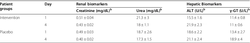
[image:8.595.57.291.88.408.2]The serum levels of urea, alanine transaminase and γ -glutamyl transferase were within the normal range in both Intervention and Placebo groups of patients (Table 4). The levels of creatinine was slightly below the normal range. However, there was no difference in the creatinine levels between the Intervention and the Pla-cebo groups of patients (Table 4).
Figure 3Levels of Pro-inflammatory cytokines in the stool of
Shigella-infected patients, treated with butyrate or placebo.
[image:8.595.306.539.88.257.2]Stool specimens were collected on indicated time points from Intervention (n = 39) and Placebo (n = 37) groups of patients. Concentrations of(A)IL-8 and(B)IL-1βin stool extracts were measured by enzyme linked immunosorbent assay (ELISA). Data are represented as mean ± SEM. Two-way repeated measure ANOVA was performed to determine significant interaction between butyrate and placebo therapy on different days, and when interaction was significant the Holm-Sidak post hoc comparison procedure was used to compare the effects of butyrate therapy on outcome measures. Significance: p≤0.05. Levels of IL-8 and IL-1βdiminished significantly from day 1 to days 4 and 7 (≤0.001) in both groups. Diminution of IL-8 in the Intervention group was significantly higher than the Placebo group (p = 0.048). Attenuation of IL-1βin the Intervention group was higher but not significant compared with the Placebo group (p = 0.078). IL-8: interleukin-8; IL-1β: interleukin-1β.
Discussion
The results of this study show that adjunct therapy with sodium butyrate enema leads to an early decline in fre-quency of inflammatory cells and concentrations of pro-inflammatory cytokines in the stool, but has no obvious effect on clinical recovery from shigellosis. In a subgroup ofShigella-infected patients, butyrate treatment resulted in early improvement of rectal inflammation compared with placebo-treated patients. Furthermore, the expres-sion of cathelicidin LL-37 in the rectal epithelium was significantly enhanced following butyrate treatment.
In our earlier study in a rabbit model of shigellosis, butyrate treatment resulted in a marked improvement in clinical outcomes and early reduction of Shigella count in the stool [11]. We did not observe similar effects in
this study. It is worth mentioning that while rabbits were given butyrate treatment without any antibiotics, patients were given butyrate in addition to antibiotics. Therefore, it is not unexpected that the clinical features and Shigella count in the stool subsided in all study patients simultaneously.
The potential anti-inflammatory effects of butyrate have long been documented. Preventive and therapeutic effects of butyrate on inflammation were evaluated in animal models of colitis [16-18]. In patients with ulcera-tive colitis (UC) or Crohn’s disease (CD), oral/topical ad-ministration of butyrate (or SCFA mixture), or stimulation of luminal butyrate production by feeding dietary fibers, have been shown to reduce clinical and in-flammatory index in several studies (reviewed in [19,20]). In experimental shigellosis in rabbits, the in-flammatory parameters were also reduced with colonic infusion of an SCFA mixture [10], or by oral administra-tion of butyrate [11]. No adverse effects of butyrate treatment were reported in these studies. Here, we have shown that local luminal administration of butyrate can significantly improve the histological features of inflam-mation in the rectum in a subgroup of patients. Butyrate treatment also resulted in an early decline in pus cells, macrophages and pro-inflammatory cytokines in the stool. Hence, butyrate treatment can resolve the persist-ence of inflammation, which often occurs after clinical recovery from shigellosis following antibiotic treatment [3,21]. Furthermore, patients did not exhibit untoward effects of butyrate treatment as assessed by the levels of renal and hepatic biomarkers.
[image:9.595.57.291.89.244.2]The anti-inflammatory effect of butyrate is mediated primarily through the inhibition of nuclear factor κB (NF-κB) activation in the large intestinal mucosa [22-24]. Dysregulated activation of NF-κB in animal models of colitis, and in patients with UC or CD, was reported to be inhibited by butyrate, which correlated with decreased inflammation [25,26]. The anti-inflammatory activity of butyrate might also be due to the activation of peroxisome proliferator-activated receptor-γ (PPARγ), a ligand-activated transcription factor in colonic epithelial cells [27,28], and the inhibition of interferon-γ signaling
Figure 5LL-37 level in the stool ofShigella-infected patients, treated with butyrate or placebo.Stool specimens were collected on indicated time points from Intervention (n = 39) or Placebo (n = 37) groups of patients. Level of LL-37 in stool extracts was measured by enzyme linked immunosorbent assay (ELISA). Data are represented as mean ± SEM. Two-way repeated measure ANOVA was performed to determine significant interaction between butyrate and placebo therapy on different days, and when interaction was significant the Holm-Sidak post hoc comparison procedure was used to compare the effects of butyrate therapy on outcome measures. Significance: p≤0.05. Concentration of LL-37 decreased significantly from day 1 to days 4 and 7 (<0.001) in both groups. The decrease was significantly higher in the Placebo group than that in the Intervention group (p<0.001).
Table 4 Levels of hepatic and renal biomarkers in serum ofShigella-infected patients at different timea
Patient groups
Day Renal biomarkers Hepatic Biomarkers
Creatinine (mg/dL)b Urea (mg/dL)b ALT (U/L)b γ-GT (U/L)b
Intervention 1 0.51 ± 0.04 21.3 ± 3 15.5 ± 1.6 11.4 ± 0.8
4 0.43 ± 0.02 18 ± 1.1 21.9 ± 2.3 11 ± 0.6
Placebo 1 0.49 ± 0.03 18.7 ± 2.6 18.6 ± 2.2 13.4 ± 2.7
4 0.40 ± 0.02 17.3 ± 1.5 21.1 ± 2.4 18.9 ± 4
Abbreviation:ALTAlanine transaminase,γ−GTGamma glutamyl transferase.
a
Data are given as mean ± standard error of mean.
b
[image:9.595.57.539.615.699.2][29]. Moreover, involvement of the G-protein coupled receptor (GPCR) in the anti-inflammatory effect of SCFA has been demonstrated recently in animal models of colitis, arthritis and asthma [30].
Treatment with butyrate showed increased expression of LL-37 in the rectal epithelia compared with placebo treatment, although there was a general increase in its ex-pression in both groups after one week. This was in ac-cordance with our previous finding in experimental shigellosis, where butyrate treatment counteracted the downregulation of rabbit cathelicidin in the rectal epithelia [11]. However, the effect size in patients was small, which could be due to the fact that butyrate was given as an enema in patients in contrast to oral therapy in the rabbit model. Patients may not have completely retained the bu-tyrate after infusion because of repeated defecation, which would affect the absorption of butyrate in serum. Recently, we have shown that the systemic dissemination of butyrate is necessary to induce cathelicidin expression in epithelial cells [31]. In parallel to epithelial expression, the release of LL-37 peptide in the stool also remained significantly higher on days 4 and 7 in the Intervention group. Since there was significant reduction of inflammatory cells in stool from the Intervention group, the prolonged secretion of LL-37 in the stool may have originated from the healed epithelium of the large intestine and may play a role in bactericidal activities.
The current study has a number of limitations. Since both groups of patients were given antibiotics, it was not possible to evaluate whether butyrate treatment enhanced shigellacidal activity in the stool. Butyrate was given as an enema instead of oral therapy as in rabbits since the bad smell of butyrate makes it unsuitable for oral therapy in humans. Repeated use of enemas is troublesome, espe-cially with regards to patient compliance and the need for hospital facilities. Oral administration of enteric coated tablets containing butyrate can be a better alternative, which was proved to be effective previously in patients with ulcerative colitis [32]. Induction of luminal butyrate via ingestion of fermentable fiber supplementation, which has been successfully used in clinical trials for ulcerative colitis (reviewed in [20]) and childhood shigellosis [12], may also be suitable for healing inflammation in shigello-sis. In addition, consumption of probiotics has recently been suggested as an interesting approach to reduce intes-tinal inflammation through the upregulation of luminal levels of butyrate and butyrate-producing commensal bac-teria, and the lowering of cecal pH [33]. In fact, these and additional metabolic shifts in T-bet−/−Rag2−/− mice were shown to improve colitis scores by creating an unfavorable environment for the colitogenicEnterobacteriaceae. How-ever, whether these therapies would also be suitable for the induction of antimicrobial peptides in the epithelium remains to be seen.
Conclusion
The current study demonstrates that adjunct therapy with butyrate enema during shigellosis promotes healing of the rectal mucosa and reduces luminal content of inflamma-tory cells and pro-inflammainflamma-tory cytokines. Butyrate treat-ment also resulted in enhanced expression of LL-37 in the rectal epithelia and prolonged secretion of LL-37 in the stool. However, efficacy of butyrate in clinical recovery from shigellosis was not evident. Recently, we have shown that sodium 4-phenylbutyrate (PB), a derivative of butyrate without the foul smell, provides similar treatment efficacy as butyrate when given orally to rabbits with experimental shigellosis [31]. Since PB is already an approved drug for treating urea cycle disorder, it holds much promise as a therapeutic alternative for human shigellosis.
Abbreviations
CONSORT: Consolidated Standards of Reporting Trials; HBD-1: Human beta defensin 1; HBD-3: Human beta defensin 3; IL-1β: Interleukin-1β; IL-8: Interleukin-8; AMP: Antimicrobial peptide; SCFA: Short chain fatty acid; RBC: Red blood cell; RME: Routine microscopic examination;
TFA: Trifluoroacetic acid; CFU: Colony forming unit; ELISA: Enzyme linked immunosorbent assay; TBS: Tris-buffer saline; 4-MUP: 4-methylumbelliferyl phosphate; H2O2: Hydrogen per-oxide; ACIA: Acquired Computerized Image Analysis; SE: Surface epithelium; LP: Lamina propria; ES: Effect size; UC: Ulcerative colitis; CD: Crohn’s disease; NF-κB: Nuclear factorκB; PPARγ: Peroxisome proliferator-activated receptor-γ; GPCR: G-protein coupled receptor; CI: Confidence interval.
Competing interest
The authors declare that they have no competing interest.
Authors’contributions
RR, BA, GHG, NHA and JA conceived and designed the trial. NHA performed sigmoidoscopy and collected biopsies. ASMA were responsible for data acquisition and specimen collection. PS, AM and RSR performed the laboratory experiments. RR, PS, AM and RSR carried out the statistical analysis. RR and BA supplied reagents/materials/analysis tools. RR, PS and BA drafted the manuscript. JA, GHG and AC revised the manuscript. All authors approved the final version of the manuscript before submission.
Acknowledgement
The work was supported by“the Swedish Agency for Research Cooperation with Developing Countries (Sida/SAREC Agreement support; grant 384)”; “The Swedish Research Council”;“Swedish Cancer Society”;“The Swedish Strategic Foundation (SSF)”;“Thorsten and Ragnar Söderberg´s Foundations”; “The Swedish Institute”;“Karolinska Institutet”;“icddr,b”;“The Icelandic Centre for Research (RANNIS)”and“University of Iceland research fund”. Icddr,b acknowledges with gratitude the commitment of“Sida/SAREC”to the Centre's research efforts. Icddr,b also gratefully acknowledges the following donors, which provide unrestricted support to the Centre's research efforts: “Australian Agency for International Development (AusAID)”;“Government of the People’s Republic of Bangladesh”;“Canadian International Development Agency (CIDA)”;“Embassy of the Kingdom of the Netherlands (EKN)”; “Swedish International Development Cooperation Agency (Sida)”;“Swiss Agency for Development and Cooperation (SDC)”and“Department for International Development, UK (DFID)”. We acknowledge the patients who generously gave consent to participate in this clinical trial. We thank all individuals who helped us with randomization, blinding, concealing and implementation of interventions and for collection of specimens.
Author details
1International Centre for Diarrheal Disease Research, Dhaka, Bangladesh. 2
Biochemistry Laboratory, Laboratory Sciences Division, International Centre for Diarrheal Disease Research, Bangladesh (icddr,b), Mohakhali, Dhaka 1212, Bangladesh.
Received: 15 December 2011 Accepted: 2 May 2012 Published: 10 May 2012
References
1. Bardhan P, Faruque AS, Naheed A, Sack DA:Decrease in shigellosis-related deaths withoutShigellaspp.-specific interventions, Asia.Emerg Infect Dis
2010,16(11):1718–1723.
2. Sansonetti PJ:Microbes and microbial toxins: paradigms for microbial-mucosal interactions III. Shigellosis: from symptoms to molecular pathogenesis.Am J Physiol Gastrointest Liver Physiol2001,280(3):G319–G323. 3. Raqib R, Lindberg AA, Wretlind B, Bardhan PK, Andersson U, Andersson J:
Persistence of local cytokine production in shigellosis in acute and convalescent stages.Infect Immun1995,63(1):289–296.
4. Raqib R, Moly PK, Sarker P, Qadri F, Alam NH, Mathan M, Andersson J: Persistence of mucosal mast cells and eosinophils inShigella-infected children.Infect Immun2003,71(5):2684–2692.
5. Niyogi SK:Shigellosis.J Microbiol2005,43(2):133–143.
6. Zasloff M:Antimicrobial peptides of multicellular organisms.Nature2002, 415(6870):389–395.
7. Islam D, Bandholtz L, Nilsson J, Wigzell H, Christensson B, Agerberth B, Gudmundsson G:Downregulation of bactericidal peptides in enteric infections: a novel immune escape mechanism with bacterial DNA as a potential regulator.Nat Med2001,7(2):180–185.
8. Sperandio B, Regnault B, Guo J, Zhang Z, Stanley SL Jr, Sansonetti PJ, Pedron T:VirulentShigella flexnerisubverts the host innate immune response through manipulation of antimicrobial peptide gene expression.J Exp Med2008,205(5):1121–1132.
9. Mortensen PB, Clausen MR:Short-chain fatty acids in the human colon: relation to gastrointestinal health and disease.Scand J Gastroenterol Suppl
1996,216:132–148.
10. Rabbani GH, Albert MJ, Hamidur Rahman AS, Moyenul Isalm M, Nasirul Islam KM, Alam K:Short-chain fatty acids improve clinical, pathologic, and microbiologic features of experimental shigellosis.J Infect Dis1999,179 (2):390–397.
11. Raqib R, Sarker P, Bergman P, Ara G, Lindh M, Sack DA, Nasirul Islam KM, Gudmundsson GH, Andersson J, Agerberth B:Improved outcome in shigellosis associated with butyrate induction of an endogenous peptide antibiotic.Proc Natl Acad Sci U S A2006,103(24):9178–9183.
12. Rabbani GH, Ahmed S, Hossain I, Islam R, Marni F, Akhtar M, Majid N: Green banana reduces clinical severity of childhood shigellosis: a double-blind, randomized, controlled clinical trial.Pediatr Infect Dis J
2009,28(5):420–425.
13. Jakobovits SL, Travis SP:Management of acute severe colitis.Br Med Bull
2005,75–76:131–144.
14. Raqib R, Reinholt FP, Bardhan PK, Karnell A, Lindberg AA:
Immunopathological patterns in the rectal mucosa of patients with shigellosis: expression of HLA-DR antigens and T-lymphocyte subsets.
APMIS1994,102(5):371–380.
15. Cunnane G, Bjork L, Ulfgren AK, Lindblad S, FitzGerald O, Bresnihan B, Andersson U:Quantitative analysis of synovial membrane inflammation: a comparison between automated and conventional microscopic measurements.Ann Rheum Dis1999,58(8):493–499.
16. Andoh A, Bamba T, Sasaki M:Physiological and anti-inflammatory roles of dietary fiber and butyrate in intestinal functions.JPEN J Parenter Enteral Nutr1999,23(5 Suppl):S70–S73.
17. Butzner JD, Parmar R, Bell CJ, Dalal V:Butyrate enema therapy stimulates mucosal repair in experimental colitis in the rat.Gut1996,38(4):568–573. 18. Song M, Xia B, Li J:Effects of topical treatment of sodium butyrate and
5-aminosalicylic acid on expression of trefoil factor 3, interleukin 1β, and nuclear factorκB in trinitrobenzene sulphonic acid induced colitis in rats.Postgrad Med J2006,82(964):130–135.
19. Canani RB, Costanzo MD, Leone L, Pedata M, Meli R, Calignano A:Potential beneficial effects of butyrate in intestinal and extraintestinal diseases.
World J Gastroenterol2011,17(12):1519–1528.
20. Hamer HM, Jonkers D, Venema K, Vanhoutvin S, Troost FJ, Brummer RJ: Review article: the role of butyrate on colonic function.Aliment Pharmacol Ther2008,27(2):104–119.
21. Raqib R, Wretlind B, Andersson J, Lindberg AA:Cytokine secretion in acute shigellosis is correlated to disease activity and directed more to stool than to plasma.J Infect Dis1995,171(2):376–384.
22. Andoh A, Fujiyama Y, Hata K, Araki Y, Takaya H, Shimada M, Bamba T: Counter-regulatory effect of sodium butyrate on tumour necrosis factor-alpha (TNF-α)-induced complement C3 and factor B biosynthesis in human intestinal epithelial cells.Clin Exp Immunol1999,118(1):23–29. 23. Inan MS, Rasoulpour RJ, Yin L, Hubbard AK, Rosenberg DW, Giardina C:The
luminal short-chain fatty acid butyrate modulates NF-κB activity in a human colonic epithelial cell line.Gastroenterology2000,118(4):724–734. 24. Place RF, Noonan EJ, Giardina C:HDAC inhibition prevents NF-κB
activation by suppressing proteasome activity: down-regulation of proteasome subunit expression stabilizes IκBα.Biochem Pharmacol2005, 70(3):394–406.
25. Luhrs H, Gerke T, Muller JG, Melcher R, Schauber J, Boxberge F, Scheppach W, Menzel T:Butyrate inhibits NF-κB activation in lamina propria macrophages of patients with ulcerative colitis.Scand J Gastroenterol
2002,37(4):458–466.
26. Segain JP, Raingeard de la Bletiere D, Bourreille A, Leray V, Gervois N, Rosales C, Ferrier L, Bonnet C, Blottiere HM, Galmiche JP:Butyrate inhibits inflammatory responses through NFκB inhibition: implications for Crohn's disease.Gut2000,47(3):397–403.
27. Kinoshita M, Suzuki Y, Saito Y:Butyrate reduces colonic paracellular permeability by enhancing PPARγactivation.Biochem Biophys Res Commun2002,293(2):827–831.
28. Schwab M, Reynders V, Loitsch S, Steinhilber D, Stein J, Schroder O: Involvement of different nuclear hormone receptors in butyrate-mediated inhibition of inducible NFκB signalling.Mol Immunol2007,44(15):3625–3632. 29. Klampfer L, Huang J, Sasazuki T, Shirasawa S, Augenlicht L:Inhibition of
interferonγsignaling by the short chain fatty acid butyrate.Mol Cancer Res2003,1(11):855–862.
30. Maslowski KM, Vieira AT, Ng A, Kranich J, Sierro F, Yu D, Schilter HC, Rolph MS, Mackay F, Artis D,et al:Regulation of inflammatory responses by gut microbiota and chemoattractant receptor GPR43.Nature2009,461 (7268):1282–1286.
31. Sarker P, Ahmed S, Tiash S, Rekha RS, Stromberg R, Andersson J, Bergman P, Gudmundsson GH, Agerberth B, Raqib R:Phenylbutyrate counteractsShigella mediated downregulation of cathelicidin in rabbit lung and intestinal epithelia: a potential therapeutic strategy.PLoS One2011,6(6):e20637. 32. Vernia P, Monteleone G, Grandinetti G, Villotti G, Di Giulio E, Frieri G,
Marcheggiano A, Pallone F, Caprilli R, Torsoli A:Combined oral sodium butyrate and mesalazine treatment compared to oral mesalazine alone in ulcerative colitis: randomized, double-blind, placebo-controlled pilot study.Dig Dis Sci2000,45(5):976–981.
33. Veiga P, Gallini CA, Beal C, Michaud M, Delaney ML, DuBois A, Khlebnikov A, van Hylckama Vlieg JE, Punit S, Glickman JN,et al:Bifidobacterium animalis subsp.lactisfermented milk product reduces inflammation by altering a niche for colitogenic microbes.Proc Natl Acad Sci U S A2010,107 (42):18132–18137.
doi:10.1186/1471-2334-12-111
Cite this article as:Raqibet al.:Efficacy of sodium butyrate adjunct 758 therapy 759 in shigellosis: a randomized, double-blind, placebo-controlled
clinical 760 trial.BMC Infectious Diseases201212:111.
Submit your next manuscript to BioMed Central and take full advantage of:
• Convenient online submission
• Thorough peer review
• No space constraints or color figure charges
• Immediate publication on acceptance
• Inclusion in PubMed, CAS, Scopus and Google Scholar
• Research which is freely available for redistribution