R E S E A R C H
Open Access
Investigation of tick-borne bacteria (
Rickettsia
spp.
, Anaplasma
spp.
, Ehrlichia
spp. and
Borrelia
spp.) in ticks collected from Andean tapirs, cattle
and vegetation from a protected area in Ecuador
Cristina Pesquera, Aránzazu Portillo, Ana M Palomar and José A Oteo
*Abstract
Background:Ixodid ticks play an important role in the transmission and ecology of infectious diseases. Information about the circulation of tick-borne bacteria in ticks is lacking in Ecuador. Our aims were to investigate the tick species that parasitize Andean tapirs and cattle, and those present in the vegetation from the buffer zone of the Antisana Ecological Reserve and Cayambe-Coca National Park (Ecuador), and to investigate the presence of tick-borne bacteria.
Methods:Tick species were identified based on morphologic and genetic criteria. Detection of tick-borne bacteria belonging toRickettsia, Anaplasma, EhrlichiaandBorreliagenera was performed by PCRs.
Results:Our ticks included 91Amblyomma multipunctum, 4Amblyommaspp., 60Rhipicephalus microplus, 5Ixodes
spp. and 1Ixodes boliviensis. A potentialCandidatusRickettsia species closest toRickettsia monacensisandRickettsia tamurae(designatedRickettsiasp. 12G1) was detected in 3R. microplus(3/57, 5.3%). In addition,Anaplasmaspp., assigned at least toAnaplasma phagocytophilum(or closely related genotypes) andAnaplasma marginale, were found in 2A. multipunctum(2/87, 2.3%) and 13R. microplus(13/57, 22.8%).
Conclusions:This is the first description ofRickettsiasp. in ticks from Ecuador, and the analyses of sequences suggest the presence of a potential novelRickettsiaspecies. Ecuadorian ticks from Andear tapirs, cattle and vegetation belonging toAmblyommaandRhipicephalusgenera were infected withAnaplasmataceae. Ehrlichiaspp. andBorrelia burgdorferisensu lato were not found in any ticks.
Keywords:Ticks,Amblyomma multipunctum,Amblyomma scalpturatum,Amblyomma sp.,Rhipicephalus microplus,
Ixodes lasallei,Ixodes boliviensis,Ixodes sp.,Rickettsia,Anaplasma,Ehrlichia,Borrelia, Ecuador
Background
Hard ticks (Ixodidae) are arthropods that suck blood from their vertebrate hosts and play an important role in the transmission and ecology of infectious diseases [1]. At least 30 ixodid tick species belonging to Amblyomma, Dermacentor, Haemaphysalis, Ixodes and Rhipicephalus genera have been documented in Ecuador [2]. These genera are recognized vectors of pathogenic bacteria
with medical and veterinary relevance in neotropical regions [3].
In South America, information about the occurrence of tick-borne bacteria in wild mammals, which are fre-quently exposed to tick-bites, is limited [4,5]. Moreover, several severe and economically important diseases of livestock in tropical regions are caused by tick-borne path-ogens (i.e. bovine anaplasmosis caused by Anaplasma marginale) that can also infect wildlife species [6].
In Ecuador, the Andean tapir (Tapirus pinchaque) is listed as endangered species. Cattle introduction into the Andean tapir refuges (i.e. Cayambe-Coca Ecological Reserve) is ne-gatively affecting tapir populations due to loss of habitat. In * Correspondence:jaoteo@riojasalud.es
Departamento de Enfermedades Infecciosas, Hospital San Pedro- Centro de Investigación Biomédica de La Rioja (CIBIR), C/ Piqueras 98, 26006 - Logroño, La Rioja, Spain
this environment, pathogens of domestic animals may threaten health of wild animals and vice versa [7].
It is known thatAmblyomma scalpturatum, Amblyom-ma latepunctatum, AmblyomAmblyom-ma multipunctum and Am-blyomma ovale tick species infest the Andean tapir in Ecuador [7]. All but A. multipunctum have been found biting humans in South America, and harboring tick-borne microorganisms [8-11]. The knowledge of bacteria transmitted by ticks (potential vectors and reservoirs of microorganisms) in a given area is useful for assessing the risk of infection in humans and animals. Therefore, the aims of our study were: 1- To investigate which tick species parasitize the Andean tapirs and cattle, and those present in the vegetation from the buffer zone of the Antisana Ecological Reserve and Cayambe-Coca National Park in Ecuador, and 2.- To detect and to identify tick-borne bacteria belonging toRickettsiaspp., Anaplasma spp., Ehrlichia spp. and Borrelia spp. gen-era in the collected tick specimens.
Methods
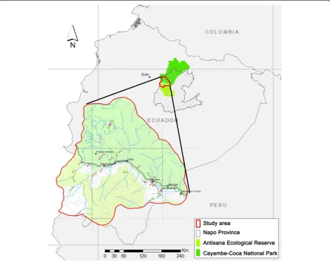
From May to October 2011 and during February 2012, an investigation was conducted in the buffer zone of the Antisana Ecological Reserve and Cayambe-Coca National Park, Napo Province, Ecuador (Figure 1). This area is located in the basin of the Papallacta River, where‘The Andean tapir conservation project’was developing.
Ticks were removed from 6 Andean tapirs, cattle [13 cows (Bos taurus) from 4 farms] and vegetation (10 tran-sects of 2-Km long that were toured twice). Arthropods were kept in tubes with ethanol recording the host/sam-pling and date, and sent to the Center of Rickettsioses and Arthropod-Borne Diseases, located at the Center of Biomedical Research from La Rioja (CIBIR), Logroño (Spain) for further analysis.
[image:2.595.59.538.338.715.2]The species were identified based on morphologic cri-teria following taxonomic keys from the Neotropical re-gion [3,12,13]. DNA was individually extracted using DNeasy Blood & Tissue kit (Qiagen, Hilden, Germany).
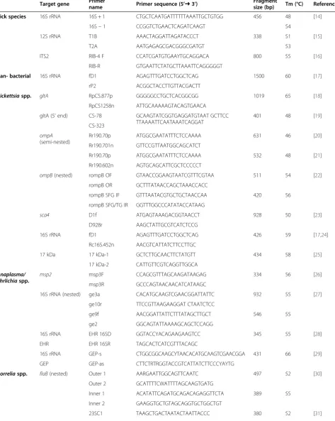
Table 1 PCR primer pairs used in this study
Target gene Primername Primer sequence (5’➜3’) Fragmentsize (bp) Tm (°C) Reference
Tick species 16S rRNA 16S + 1 CTGCTCAATGATTTTTTAAATTGCTGTGG 456 48 [14]
16S–1 CCGGTCTGAACTCAGATCAAGT 54
12S rRNA T1B AAACTAGGATTAGATACCCT 338 51 [15]
T2A AATGAGAGCGACGGGCGATGT 53
ITS2 RIB-4 F CCATCGATGTGAAYTGCAGGACA 800 55 [16]
RIB-R GTGAATTCTATGCTTAAATTCAGGGGGT
Pan- bacterial 16S rRNA fD1 AGAGTTTGATCCTGGCTCAG 1500 60 [17]
rP2 ACGGCTACCTTGTTACGACTT
Rickettsiaspp. gltA RpCS.877p GGGGGCCTGCTCACGGCGG 1019 65 [18]
RpCS1258n ATTGCAAAAAGTACAGTGAACA
gltA (5’end) CS-78 GCAAGTATCGGTGAGGATGTAAT GCTTCC
TTAAAATTCAATAAATCAGGAT
401 48 [19]
CS-323 ompA
(semi-nested)
Rr190.70p ATGGCGAATATTTCTCCAAAA 631 46 [20]
Rr190.701n GTTCCGTTAATGGCAGCATCT
Rr190.70p ATGGCGAATATTTCTCCAAAA 532 48 [21]
Rr190.602n AGTGCAGCATTCGCTCCCCCT
ompB(nested) rompB OF GTAACCGGAAGTAATCGTTTCGTAA 511 54 [22]
rompB OR GCTTTATAACCAGCTAAACCACC
rompB SFG IF GTTTAATACGTGCTGCTAACCAA 420 56
rompB SFG/TG IR GGTTTGGCCCATATACCATAAG
sca4 D1f ATGAGTAAAGACGGTAACCT 928 50 [23]
D928r AAGCTATTGCGTCATCTCCG
16S rRNA fD1 AGAGTTTGATCCTGGCTCAG 426 59 [17,24]
Rc16S.452n AACGTCATTATCTTCCTTGC
17 kDa 17 kDa-1 GCTCTTGCAACTTCTATGTT 434 58 [25]
17 kDa-2 CATTGTTCGTCAGGTTGGCA
Anaplasma/ Ehrlichiaspp.
msp2 msp3F CCAGCGTTTAGCAAGATAAGAG 334 56 [26]
msp3R GCCCAGTAACAACATCATAAGC
16S rRNA (nested) ge3a CACATGCAAGTCGAACGGATTATTC 932 55 [27]
ge10r TTCCGTTAAGAAGGAT CTAATCTCC
ge9f AACGGATTATTCTTTATAGCTTGCT 546 55
ge2 GGCAGTATTAAAAGCAGCTCCAGG
16S rRNA EHR 16SD GGTACCYACAGAAGAAGTCC 345 55 [28]
EHR EHR 16SR TAGCACTCATCGTTTACAGC
16S rRNA GEP-s CTGGCGGCAAGCYTAACACATGCAAGTCGAACGGA 431 66 [29]
GEP GEP-as CTTCTRTRGGTACCGTCATTATCTTCCCYAYTG
Borreliaspp. flaB(nested) Outer 1 AARGAATTGGCAGTTCAATC 497 52 [30]
Outer 2 GCATTTTCWATTTTAGCAAGTGATG
Inner 1 ACATATTCAGATGCAGACAGAGGTTCTA 389 55
Inner 2 GAAGGTGCTGTAGCAGGTGCTGGCTGT
Each tick specimen was screened by PCR for both iden-tification of tick species and detection of bacteria includ-ing Rickettsia spp., Anaplasma spp., Ehrlichia spp. and Borrelia burgdorferi sensu lato (s. l.). Tick species were confirmed by PCR targeting the tick mitochondrial 16S ribosomal RNA (rRNA) [14]. PCR assays for the tick mi-tochondrial 12S rRNA gene and the tick nuclear 5.8S-28S rRNA intergenic transcribed spacer 2 (ITS2) were also performed for selected samples [15,16]. For the screening of tick-borne bacteria, at least two fragment genes of each genus were tested by PCR assays. The molecular bio-markers selected to identify ticks are among the ones most widely used for the phylogenetics of ticks, being suitable to distinguish between closely related species. Biomarkers for the detection of microorganisms were selected based on our own expertise and according to previously reported usefulness and sensitivity. Target genes, specific primers and PCR conditions are listed in Table 1. Two negative controls, one of them containing water instead of template DNA and the other with tem-plate DNA but without primers, as well as positive con-trols of Rickettsia slovaca strain S14ab DNA (obtained from Vero cells inoculated in our facility with a Derma-centor marginatustick from La Rioja, and known to be infected with R. slovaca), Anaplasma phagocytophilum strain Webster DNA kindly provided by Dr. Raoult (Unité de Recherche sur les Maladies Infectieuses et Tropicales Emergentes, France) and Dr. Dumler (The Johns Hopkins Hospital, USA), orBorrelia burgdorferisensu stricto DNA kindly provided by Dr. Fingerle (German National Refer-ence Centre for Borrelia, Germany) were included in all PCR assays. PCR products were sequenced in both direc-tions. Sequences were compared with those available in the NCBI database using BLAST.
Results
Identification of ticks
A total of 161 ticks (75 removed from Andean tapirs, 66 from cattle and 20 collected over vegetation) were in-cluded in the study. Ten specimens (one of each stage and gender in case of adult ticks) were deposited in the Museum of Zoology of Pontificia Universidad Católica from Ecuador.
Morphologically, 84 specimens (12 nymphs, 47 male and 25 female ticks) corresponded toA. multipunctum, 4 specimens toA. scalpturatumand 7 were classified as
Amblyommaspp. For all but 4 specimens, the mitochon-drial 16S rRNA sequences (409 bp) were identical to the 16S rRNA gene from A. multipunctum(GenBank acces-sion no. KC677673), or differed by 0.2-1.7% (1–7 bp) when compared to this species. No 12S rRNA sequence from A. multipunctumwas available in GenBank. There-fore, ours (from a specimen whose 16S rRNA sequence was identical to A. multipunctum KC677673) was de-posited in GenBank under no. KM077433. It differed in sequence by 10% when compared to those available, and showed the highest identity (90%) with the 12S rRNA gene from Amblyomma sp. (GenBank accession no. AY342251). For the 4 tick specimens morphologic-ally classified as A. scalpturatum, sequences of the 16S rRNA showed maximum identity (90%; 370/410 bp) withA. multipunctum, whereas 12S rRNA and ITS2 se-quences were closest to Amblyomma varium (90.6% identity; 309/341 bp and 93.6% identity; 836/893 bp, re-spectively). Obtained sequences showed lower percent-ages of identity when compared to those from A. scalpturatum: 87% for 12S rRNA (GenBank accession no. AY342276), and 90% for ITS2 (GenBank accession no. AY619574). Therefore, these 4 ticks were classified as Amblyomma spp. and these three fragment genes were deposited in GenBank under nos. KM077434-6.
A total of 60 specimens were morphologically classified as Rhipicephalus microplus (formerly, Boophilus micro-plus) (6 nymphs, 16 male and 38 female ticks). In all these cases, the 16S rRNA sequences were identical to the 16S rRNA gene from R. microplus (GenBank accession no. EU918187).
According to morphological features, 5 female ticks were classified asIxodes lasallei. The 16S rRNA sequen-ces did not match with those fromI. lasallei (GenBank accession no. AF549850) but were closest to this tick spe-cies (90% identity). Due to this discrepancy, they were classified asIxodesspp. and deposited in GenBank under no. KM077438.
Lastly, one specimen morphologically corresponded to Ixodes boliviensis. The 16S rRNA sequences showed the highest identity (94%) with the 16S rRNA gene from Ixodessp. (GenBank accession no. KF702351). It was de-posited in GenBank since no sequences forI. boliviensis were available (KM077437).
[image:4.595.61.548.102.145.2]According to morphological and genetic classifications, our ticks included 91 A. multipunctum, 4 Amblyomma
Table 1 PCR primer pairs used in this study(Continued)
5S-23S intergenic spacer (nested)
23SN1 ACCATAGACTCTTATTACTTTGAC
5SCB GAGAGTAGGTTATTGCCAGGG 226 55
23SN2 ACCATAGACTCTTATTACTTTGACCA
spp., 60R. microplus, 5Ixodesspp. and 1Ixodes boliviesis (Table 2).
Detection and identification of tick-borne bacteria Tick-borne bacteria were tested for 151/161 specimens, excluding those deposited in the museum.
The presence of rickettsiae was screened by PCR assays targeting 2 fragments of the gltA rickettsial gene (1019 and 401 pb, respectively). Positive amplicons were ob-tained for 3R. microplus(2 male and 1 female specimens) removed from 2 cows from different farms. There were no differences in the sequences ofgltAfor amplicons derived from the DNA of the 3 rickettsial-infected R. microplus, and showed maximum identities (99.7% -99.2%) withgltA gene from Rickettsia monacensis and Rickettsia tamurae as validated species (Table 3).
Subsequently, fragments of ompA (532 bp), ompB (420 bp), sca4 (928 bp), 16S rRNA gene (426 bp and 1500 bp, respectively), and 17 kDa-antigen gene (334 bp) were amplified to classify the Rickettsia at the species level.
The sequences of ompA (also identical each other) were closest toR. tamurae(95.9% identity) andR. mona-censis(95.7% identity) (Table 3).
For ompB, the DNA sequences of the 3 rickettsiae-positive R. microplus were identical to each other and showed 99.2% identity with R. monacensis and 97.1% identity withR. tamurae(Table 3).
Unfortunately, no amplicons were obtained in PCR as-says targetingsca4 gene. Attempts to sequence the rick-ettsial 16S rRNA and pan-bacterial 16S rRNA amplicons for the 3 R. microplus were inconclusive for Rickettsia.
Curiously, A. marginale was amplified in 1 out of these 3 specimens using pan-bacterial 16S rRNA primers (see below). In addition, the sequences of 17 kDa antigen gene did not match with those available in GenBank.
In 2005, Raoult et al. established the criteria for the taxonomic classification of potential newRickettsia spe-cies [32]. They proposed the ‘Candidatus’ status for a bacterium not established in pure culture that did not exhibit more than one of the following percentages of nucleotide identity: >99.8, >99.9, >98.8, >99.2, and >99.3 for rrs (16S rRNA), gltA, ompA, ompB, and sca4, re-spectively, with a validated Rickettsiaspecies. According to our results, only amplicons for thegltA, ompA,ompB and 17KDa were obtained. Therefore, based on the rec-ommended nomenclature [32], aCandidatusstatus could not be assigned to this microorganism. We designated this bacterium asRickettsiasp. 12G1.
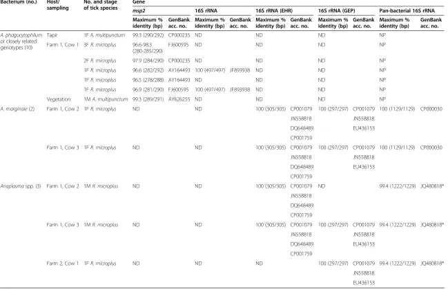
The presence of Anaplasma spp. was detected in 15 out of 151 samples, including 2A. multipunctumand 13 R. microplus. On the one hand, the partial sequences of msp2 and 16S rRNA gene from Anaplasma spp. for A. multipunctum (a female tick from Andean tapir and a male tick from vegetation) and 8R. microplus(all female ticks from one cow) were, when available, closest (96.6-100% identity) to A. phagocytophilum (Table 4). On the other hand, the 16S rRNA sequences (EHR and GEP re-gions) for 5R. microplus(3 female and 2 male specimens) removed from 3 cows in two farms, were respectively identical each other, and matched (100% identity) with more than oneAnaplasmaspecies (assigned toA. margin-ale, Anaplasma ovis, A. phagocytophilumandAnaplasma centrale) for both PCR targets. Maximum identity with
Table 3 Maximum identities of rickettsial sequences detected in 3Rhipicephalus microplusfrom Ecuador with validated Rickettsiaspecies
Gene sequence % identity withRickettsiaspp. (bp)
R. monacensis GenBank no. R. tamurae GenBank no.
gltA[KF831358] 99.5 (625/628) DQ100163 99.2 (623/628) AF394896
gltA(5’end) [KF831359] 99.7 (349/350) DQ100163 99.4 (348/350) AF394896
ompA[KF831361] 95.7 (444/464) DQ100169 95.9 (445/464) DQ103259
ompB[KF831360] 99.2 (379/382) EF380356 97.1 (371/382) DQ113910
[image:5.595.58.540.642.724.2]bp: base pairs; [ ]: GenBank accession number generated in this study; GenBank no.: GenBank accession number;R.:Rickettsia. Table 2 Ticks included in this study
Host/sampling Tick species Nymph Male Female Total number
Andean tapir Amblyomma multipunctum 2 45 24 71
Amblyommaspp. 4* 4
Cattle Rhipicephalus microplus 6* 16* 38* 60
Ixodesspp. 5* 5
Ixodes boliviensis 1* 1
Vegetation A. multipunctum 10* 9** 1* 20
msp2 16S rRNA 16S rRNA (EHR) 16S rRNA (GEP) Pan-bacterial 16S rRNA Maximum %
identity (bp)
GenBank acc. no.
Maximum % identity (bp)
GenBank acc. no.
Maximum % identity (bp)
GenBank acc. no.
Maximum % identity (bp)
GenBank acc. no.
Maximum % identity (bp)
GenBank acc. no.
A. phagocytophilum or closely related genotypes (10)
Tapir 1FA. multipunctum 99.3 (290/292) CP000235 ND ND ND NP
Farm 1, Cow 1 3FR. microplus 96.6-98.3 (280-285/290)
FJ600595 ND ND ND NP
2FR. microplus 97.9 (284/290) CP000235 ND ND ND NP
1FR. microplus 96.6 (282/292) AY164493 100 (497/497) JF893938 ND ND NP
1FR. microplus 96.5 (278/288) AY164493 ND ND ND NP
1FR. microplus 96.9 (281/290) FJ600595 100 (497/497) JF893938 ND ND NP
Vegetation 1MA. multipunctum 99.3 (289/291) AY626255 ND ND ND NP
A. marginale(2) Farm 1, Cow 2 1FR. microplus ND ND 100 (305/305) CP001079 100 (297/297) CP001079 100 (1129/1129) CP000030
JN558818 JN558818
DQ648489 EU436153
CP001759
Farm 1, Cow 3 1FR. microplus ND ND 100 (305/305) CP001079 100 (297/297) CP001079 100 (1129/1129) CP000030
JN558818 JN558818
DQ648489 EU436153
CP001759
Anaplasmaspp. (3) Farm 1, Cow 2 1MR. microplus ND ND 100 (305/305) CP001079 ND 99.4 (1222/1229) JQ480818*
JN558818
DQ648489
CP001759
Farm 1, Cow 3 1MR. microplus ND ND 100 (305/305) CP001079 100 (297/297) CP001079 99.4 (1222/1229) JQ480818*
JN558818 JN558818
DQ648489 EU436153
CP001759
Farm 2, Cow 1 1FR. microplus ND ND ND 100 (297/297) CP001079 99.4 (1222/1229) JQ480818*
JN558818
EU436153
*Coxiellaendosymbiont ofRhipicephalus turanicusisolate DGGE.
A. phagocytophilum:Anaplasma phagocytophilum;A. multipunctum:Amblyomma multipunctum;A. marginale:Anaplasma marginale;R.:Rhipicephalus; M: male; F: Female; ND: Not detected; NP: Not performed; CP001079-CP000030:Anaplasma marginalesequences from GenBank; JN558818:Anaplasma ovissequence from GenBank; DQ648489-EU436153:Anaplasma phagocytophilumsequences from GenBank; CP001759: Anaplasma centralesequence from GenBank.
al.
Parasit
es
&
Vectors
(2015) 8:46
Page
6
of
[image:6.794.61.710.80.499.2]validated species of Ehrlichia genus did not exceed 95% with any of these 16S rRNA target genes (Table 4). Since these fragment genes were highly conserved for these spe-cies, in an attempt to identify the Anaplasma species, DNA extracts of these 5 samples were used as templates of pan-bacterial 16S rRNA PCR assays. The sequences corresponding to 2 out of 5R. micropluswere identical to each other and homologous (100% identity) toA. margin-ale. In these 2 cases, percentages of identity were 99.6, 99.5 and 97.2% when compared toA. ovis,A. centraleand human pathogenic A. phagocytophilum, respectively. Se-quencing results for the 3 remaining ticks matched (99.4% identity) with a Coxiella endosymbiont of Rhipicephalus turanicus(GenBank accession no. J480818) (Table 4).
Table 5 summarizes the detection rates for Rickettsia spp. and Anaplasma spp. Ehrlichia species were not amplified in any of the 151 ticks analyzed in this study. Lastly, B. burgdorferi s.l. was not detected in any ticks when flaBgene and 5S-23S rRNA intergenic spacer re-gion were tested by PCR.
Co-infections
Out of 18 positive ticks, one of them (5.6%) was found co-infected with 2 bacteria. The co-infection detected was A. marginalewithRickettsiasp. 12G1 in oneR. microplus tick collected from a cow.
GenBank accession numbers
Sequences obtained in this study have been deposited in the GenBank database under the following acces-sion numbers: KM077433-8 (identification of ticks) and KF831358-62 (rickettsial genes).
Discussion
A total of 161 ticks (nymphs or adult specimens) re-moved from Andean tapirs, cattle and vegetation, and belonging to Amblyomma, Rhipicephalus and Ixodes genera, was included in the present study. These tick ge-nera had been previously reported to occur in Ecuador
[3,33,34]. Based on morphological and genetic criteria, arthropods were classified as 91 A. multipunctum, 4 Amblyommaspp., 60 R. microplus, 5Ixodes spp. and 1 I. boliviesis. On the one hand, A. multipunctum was collected from vegetation and found attached to An-dean tapirs. This tick species was originally described from a Tapirus sp. in North America, and it has been reported in Venezuela, Colombia and Ecuador [35,36]. Partial sequences of the mitochondrial 16S rRNA gene of A. multipunctumspecimens from Ecuador had been previously generated [37]. Our group has completed this molecular description with sequences of the 12S rRNA fragment gene (GenBank accession no. KM077433). On the other hand, R. microplus and Ixodes spp. were re-moved from cows, as well as one specimen ofI. boliviensis that was genetically characterized herein using mitochon-drial 16S rRNA gene as PCR target (GenBank accession no. KM077437).R. microplus, known as the cattle tick, is widely distributed in cattle from tropical regions [3]. This is the first description ofI. boliviensisin Ecuador, although it has been found in cattle from Costa Rica [38].
As far as we know, this is the first report where ticks from Ecuador were evaluated for the presence ofRickettsia spp., Anaplasmaspp., Ehrlichiaspp. andBorreliaspp.
[image:7.595.58.540.593.716.2]The circulation of a potential Candidatus Rickettsia species (designated Rickettsia sp. 12G1) in R. microplus ticks removed from cattle in Ecuador is reported. Ac-cording to our data, this novel Rickettsiawas closest to R. monacensis and R. tamurae, as validated species. R. monacensishas been so far reported fromIxodes ricinus, and R. tamurae from Amblyomma testudinarium [39]. The human pathogenic role of R. monacensis was first reported in Spain [18], and one case ofR. tamurae infec-tion has been detected in Japan [40]. Nevertheless, no evidence of human pathogenicity is presented herein for Rickettsiasp. 12G1, and there is no evidence to suggest that thisRickettsiais transmissible to humans. Other new genotypes with unknown pathogenicity that also belong to the same lineage ofR. tamuraeandR. monacensis, such as
Table 5 Detection rates forRickettsiaspp. andAnaplasmaspp.
Host/sampling Tick species Detection rate% (number of infected ticks/number of total ticks)
Rickettsiaspp. A. phagocytophilum A. marginale Anaplasmaspp.
Andean tapir A. multipunctum 0 1.4 (1/71) 0 0
Amblyommaspp. 0 0 0 0
Cattle R. microplus 5.3 (3/57) 14.0 (8/57) 3.5 (2/57) 5.3 (3/57)
Ixodesspp. 0 0 0 0
I. boliviensis 0 0 0 0
Vegetation A. multipunctum 0 6.3 (1/16) 0 0
Total 2 (3/151) 6.6 (10/151) 1.3 (2/151) 2 (3/151)
Rickettsiasp. strain Colombianensi orRickettsiasp. strain IbR/CRC, have been documented in R. microplus or I. boliviensisfrom the New World [41,42].
In our study,A. phagocytophilumor closely related ge-notypes have been detected in ticks removed from An-dean tapirs, cows and vegetation. It is known that the high intraspecific variability observed in the msp2 gene of A. phagocytophilum promotes the adaptation of the bacterium to different hosts and could justify its distri-bution in various environments [43]. As expected, the msp2sequences obtained in this study (corresponding to 10 ticks) showed high genetic variability. Whereas the 16S rRNA sequences matched, when available (n = 2), with A. phagocytophilumpathogenic for humans (GenBank ac-cession no. CP000235), msp2sequences for 5 specimens (1A. multipunctumfrom an Andean tapir and 4R. micro-plus from cows) demonstrated relatedness with human pathogenic A. phagocytophilum but differed by 0.7-3.4% [44,45]. In addition, msp2 sequences obtained from 4 R. micropluswere closest (96.6-98.3% identity) toA. phagocy-tophilum from Japanese Ixodes persulcatus [46]. Lastly, themsp2sequence for 1A. multipunctumfrom vegetation was 99% identical to oneA. phagocytophilumstrain from rodents in Florida (also highly similar to human patho-genic reference strain) [47].
As far as we know, the occurrence ofA. phagocytophi-lumor closely related genotypes had not been previously detected neither in Ecuador nor in ticks removed from tapirs. Nevertheless, A. phagocytophilum or closely re-latedAnaplasma spp. have been found in blood samples from domestic (dogs and cats) and wild animals (deer) in Brazil [48-50]. This is the first evidence ofA. phagocy-tophilum inR. microplus in the New World. Neverthe-less, this bacterium had been previously found in R. microplusfrom China [51].
Based on the sequencing results of the 16S rRNA gene, 2 R. microplus specimens removed from cows tested positive for A. marginale and 3 harbored Anaplasma spp. (assigned to A. marginale, A. ovis, A. phagocytophi-lumandA. centrale).
A. marginale, which is transmitted by R. microplus, has a worldwide occurrence and is considered as one of the most prevalent pathogens causing cattle morbidity and mortality in subtropical and tropical countries, in-cluding Latin America [52,53]. Our study evidences the first molecular detection ofA. marginaleinR. microplus from Ecuador. This bacterium had been previously de-tected in Ecuadorian blood samples from cattle by PCR [54] and also inR. microplusticks in Philipinnes [55].
Moreover, no evidence ofEhrlichiaspp. orB. burgdorferi s.l.-infected ticks has been found in Ecuador. Nevertheless, in South American countries, new members of the Ehrli-chia genus and the B. burgdorferis.l. complex have been recently described in Brazil, Uruguay and Chile [56-59].
Conclusions
In summary, this is the first description ofRickettsiasp. in ticks from Ecuador, and the analyses of sequences suggest the presence of a potential novelRickettsiaspecies. The complete characterization and distribution of the novelRickettsiasp. 12G1, as well as its possible pathogenic role for animals and humans, needs to be determined.
Our data also showed that ticks from Andean tapirs, cattle and vegetation in Ecuador (Amblyomma and Rhi-picephalus) were naturally infected with Anaplasmata-ceae and that co-infection (A. marginale and Rickettsia sp.) occurred.
Competing interests
The authors declare they have no competing interests.
Authors’contributions
Designed the study: JAO, AP. Collected and identified ticks: CP, AMP. Processed samples and analyzed sequences: CP, AMP. Analyzed the data: AP, AMP, JAO. Wrote the paper: AP, AMP, JAO. All authors read and approved the final version of the manuscript.
Acknowledgements
This study operated under the following permit from the government of Ecuador: N°020-IC-FAU/FLO-DPN/MA, as well as appropriate export permits, as required.
We are grateful to the veterinary team from‘The Andean tapir conservation project’(EcoCiencia Foundation), and local farmers for their help with ticks collection.
We are also grateful to José M. Venzal (University of the Republic, Uruguay) and Valeria C. Onofrio (Instituto Butantan, Brazil) for their help with taxonomic identification of ticks.
We appreciate the support from Red Iberoamericana de Investigación y Control de Enfermedades Rickettsiales (RIICER, N° 210RT0403), Programa Iberoamericano de Ciencia y Tecnologías para el Desarrollo (CYTED). This study was presented in part in the I Congreso Latinoamericano de Tapires y II Congreso Ecuatoriano de Mastozoología, Puyo, Pastaza (Ecuador), May 8–11, 2013 (oral communication I-CLT 048), and in the IV Congreso Latinoamericano de Enfermedades Rickettsiales, San José (Costa Rica), July 22–24, 2013 (abstracts A-17 and B-13).
Received: 12 July 2014 Accepted: 15 January 2015
References
1. Sonenshine DE, Lane RS, Nicholson WL. Ticks (Ixodida). In: Mullen G, Durden L, editors. Medical and Veterinary Entomology. New York: Academic Press; 2002. p. 517–58.
2. Guglielmone AA, Estrada-Peña A, Keirans JE, Robbins RG. Ticks (Acari: Ixodida) of the Neotropical Zoogeographic Region. Houten: Atalanta; 2003. 3. Barros-Battesti D, Arzua M, Bechara GH. Carrapatos de Importância
Medico-Veterinaria da Região Neotropical: Um Guia Ilustrado para Identificação de Espécies. Sao Pablo: Vox/ICTTD-3/ Butantan; 2006. 4. Spolidorio MG, Andreoli GS, Martins TF, Brandão PE, Labruna MB. Rickettsial
infection in ticks collected from road-killed wild animals in Rio de Janeiro, Brazil. J Med Entomol. 2012;49:1510–4.
5. André MR, Dumler JS, Scorpio DG, Teixeira RH, Allegretti SM, Machado RZ. Molecular detection of tick-borne bacterial agents in Brazilian and exotic captive carnivores. Ticks Tick Borne Dis. 2012;3:247–53.
6. Barbosa da Silva J, Vinhote WM, Oliveira CM, André MR, Machado RZ, DaFonseca AH, et al. Molecular and serological prevalence ofAnaplasma marginalein water buffaloes in northern Brazil. Ticks Tick Borne Dis. 2014;5:100–4.
7. Diaz AG, Castellanos A, Piñeda C, Downer C, Lizcano DJ, Constantino E, et al.Tapirus pinchaque. In: IUCN 2014. IUCN Red List of Threatened Species. Version 2014.1. 2008. http://www.iucnredlist.org/details/21473/0.
the State of Rondônia, Western Amazon, Brazil. J Med Entomol. 2004;41:1073–81.
9. Guglielmone AA, Beati L, Barros-Battesti DM, Labruna MB, Nava S, Venzal JM, et al. Ticks (Ixodidae) on humans in South America. Exp Appl Acarol. 2006;40:83–100.
10. Szabó MP, Pinter A, Labruna MB. Ecology, biology and distribution of spotted-fever tick vectors in Brazil. Front Cell Infect Microbiol. 2013;3:27. 11. Londoño AF, Díaz FJ, Valbuena G, Gazi M, Labruna MB, Hidalgo M, et al. Infection ofAmblyomma ovalebyRickettsiasp. strain Atlantic rainforest, Colombia. Ticks Tick Borne Dis. 2014;5:672–5.
12. Fairchild GB, Kohls GM, Tipton VJ. The ticks of Panama (Acarina: Ixodoidea). In: Wenzel WR, Tipton VJ, editors. Ectoparasites of Panama. Chicago: Field Museum of Natural History; 1966. p. 167–219.
13. Jones EK, Clifford CM, Keirans JE, Kohls GM. The ticks of Venezuela (Acarina: Ixodoidea) with a key to the species ofAmblyommain the western hemisphere. Brigham Young University Science Bulletin, Biological Series. 1972;17:1–40.
14. Black WC, Piesman J. Phylogeny of hard- and soft-tick taxa (Acari: Ixodida) based on mitochondrial 16S rDNA sequences. Proc Natl Acad Sci U S A. 1994;91:10034–8.
15. Beati L, Keirans JE. Analysis of the systematic relationships among ticks of the generaRhipicephalusandBoophilus(Acari: Ixodidae) based on mitochondrial 12S ribosomal DNA gene sequences and morphological characters. J Parasitol. 2001;87:32–48.
16. Zahler M, Gothe R, Rinder H. Genetic evidence against a morphologically suggestive conspecificity ofDermacentor reticulatusandDermacentor marginatus(Acari: Ixodidae). Int J Parasitol. 1995;25:1413–9. 17. Weisburg WG, Barns SM, Pelletier DA, Lane DJ. 16S ribosomal DNA
amplification for phylogenetic study. J Bacteriol. 1991;173:697–703. 18. Jado I, Oteo JA, Aldámiz M, Gil H, Escudero R, Ibarra V, et al.Rickettsia
monacensisand human disease, Spain. Emerg Infect Dis. 2007;13:1405–7. 19. Labruna MB, Whitworth T, Horta MC, Bouyer DH, McBride JW, Pinter A, et al.
Rickettsiaspecies infectingAmblyomma cooperiticks from an area in the state of São Paulo, Brazil, where Brazilian spotted fever is endemic. J Clin Microbiol. 2004;42:90–8.
20. Roux V, Fournier PE, Raoult D. Differentiation of spotted fever group rickettsiae by sequencing and analysis of restriction fragment length polymorphism of PCR-amplified DNA of the gene encoding the protein rOmpA. J Clin Microbiol. 1996;34:2058–65.
21. Regnery RL, Spruill CL, Plikaytis BD. Genotypic identification of rickettsiae and estimation of intraspecies sequence divergence for portions of two rickettsial genes. J Bacteriol. 1991;173:1576–89.
22. Choi YJ, Lee SH, Park KH, Koh YS, Lee KH, Baik HS, et al. Evaluation of PCR-based assay for diagnosis of spotted fever group rickettsiosis in human serum samples. Clin Vaccine Immunol. 2005;12:759–63.
23. Sekeyova Z, Roux V, Raoult D. Phylogeny ofRickettsiaspp. inferred by comparing sequences of‘gene D’, which encodes an intracytoplasmic protein. Int J Syst Evol Microbiol. 2001;51:1353–60.
24. Márquez FJ, Muniain MA, Soriguer RC, Izquierdo G, Rodríguez-Baño J, Borobio MV. Genotypic identification of an undescribed spotted fever group rickettsia inIxodes ricinusfrom southwestern Spain. Am J Trop Med Hyg. 1998;58:570–7.
25. Oliveira RP, Galvão MA, Mafra CL, Chamone CB, Calic SB, Silva SU, et al. Rickettsia felisinCtenocephalidesspp. fleas, Brazil. Emerg Infect Dis. 2002;8:317–9.
26. Zeidner NS, Burkot TR, Massung R, Nicholson WL, Dolan MC, Rutherford JS, et al. Transmission of the agent of human granulocytic ehrlichiosis byIxodes spinipalpisticks: evidence of an enzootic cycle of dual infection withBorrelia burgdorferiin Northern Colorado. J Infect Dis. 2000;182:616–9.
27. Massung R, Slater K, Owens JH, Nicholson WL, Mather TN, Solberg VB, et al. Nested PCR assay for detection of granulocytic ehrlichiae. J Clin Microbiol. 1998;36:1090–5.
28. Inokuma H, Raoult D, Brouqui P. Detection ofEhrlichia platysDNA in brown dog ticks (Rhipicephalus sanguineus) in Okinawa Island, Japan. J Clin Microbiol. 2000;38:4219–21.
29. Eddlestone SM, Diniz PP, Neer TM, Gaunt SD, Corstvet R, Cho D, et al. Doxycycline clearance of experimentally induced chronicEhrlichia canis infection in dogs. J Vet Intern Med. 2007;21:1237–42.
30. Clark K, Hendricks A, Burge D. Molecular identification and analysis of Borrelia burgdorferisensu lato in lizards in the southeastern United States. Appl Environ Microbiol. 2005;71:2616–25.
31. Rijpkema SG, Molkenboer MJ, Schouls LM, Jongejan F, Schellekens JF. Simultaneous detection and genotyping of three genomic groups of Borrelia burgdorferisensu lato in DutchIxodes ricinusticks by
characterization of the amplified intergenic spacer region between 5S and 23S rRNA genes. J Clin Microbiol. 1995;33:3091–5.
32. Raoult D, Fournier PE, Eremeeva M, Graves S, Kelly PJ, Oteo JA, et al. Naming of Rickettsiae and rickettsial diseases. Ann N Y Acad Sci. 2005;1063:1–12.
33. Labruna MB, Guglienmone AA. Ticks of New World Tapirs. Tapir Conservation. 2009;18:21–8.
34. Vásquez CL, Muro JJ, Clavijo JJ. Garrapatas del géneroIxodesLatreille, 1795 y Rhipicephalus(Boophilus) Koch, 1844 (Acari: Ixodidae) presentes en la colección de Zoología Agrícola, Decanato de Agronomía, UCLA, Lara, Venezuela. Entomotropica. 2011;26:89–97.
35. Neumann LG. Révision de la famille des Ixodidés. Mémoires de la Société de Zoologie de France. 1899;12:211–7.
36. Voltzit OV. A review of NeotropicalAmblyommaspecies (Acari: Ixodidae). Acarina. 2007;15:3–134.
37. Labruna MB, Martins TF, Nunes PH, Costa FB, Portero F, Venzal JM. New records ofAmblyomma multipunctumandAmblyomma naponensefrom Ecuador, with description ofA. multipunctumnymph. J Parasitol. 2013;99:973–7.
38. Alvarez V, Bonilla R, Chacón I. Relative abundance ofAmblyommaspp. (Acari: Ixodidae) in bovines (Bos taurusandB. indicus) from Costa Rica. Rev Biol Trop. 2003;51:435–43.
39. Parola P, Paddock CD, Socolovschi C, Labruna MB, Mediannikov O, Kernif T, et al. Update on tick-borne rickettsioses around the world: a geographic approach. Clin Microbiol Rev. 2013;26:657–702.
40. Imaoka K, Kaneko S, Tabara K, Kusatake K, Morita E. The first human case of Rickettsia tamuraeinfection in Japan. Case Rep Dermatol. 2011;3:68–73. 41. Miranda J, Portillo A, Oteo JA, Mattar S.Rickettsiasp. strain Colombianensi
(Rickettsiales: Rickettsiaceae): A New Proposed Rickettsia Detected in Amblyomma dissimile(Acari: Ixodidae) from iguanas and free-living larvae ticks from vegetation. J Med Entomol. 2012;49:960–5.
42. Troyo A, Moreira-Soto A, Carranza M, Calderón-Arguedas O, Hun L, Taylor L. Detection of an undescribedRickettsiasp. inIxodes boliviensisfrom Costa Rica. Ticks Tick Borne Dis. 2014;5:672–5.
43. Rymaszewska A. Variability within themsp2gene in populations of Anaplasma phagocythopilum. Folia Biol (Praha). 2010;56:269–75. 44. Barbet AF, Meeus PF, Bélanger M, Bowie MV, Yi J, Lundgren AM, et al.
Expression of multiple outer membrane protein sequence variants from a single genomic locus ofAnaplasma phagocytophilum. Infect Immun. 2003;71:1706–18.
45. Lin M, Kikuchi T, Brewer HM, Norbeck AD, Rikihisa Y. Global proteomic analysis of two tick-borne emerging zoonotic agents:Anaplasma phagocytophilumandEhrlichia chaffeensis. Front Microbiol. 2011;2:24. 46. Wuritu, Ozawa Y, Gaowa, Kawamori F, Masuda T, Masuzawa T, et al.
Structural analysis of ap44/msp2expression site ofAnaplasma phagocytophilumin naturally infected ticks in Japan. J Med Microbiol. 2009;58:1638–44.
47. Clark KL.Anaplasma phagocytophilumin small mammals and ticks in northeast Florida. J Vector Ecol. 2012;37:262–8.
48. Santos HA, Pires MS, Vilela JA, Santos TM, Faccini JL, Baldani CD, et al. Detection ofAnaplasma phagocytophilumin Brazilian dogs by real-time polymerase chain reaction. J Vet Diagn Invest. 2011;23:770–4.
49. Sacchi AB, Duarte JM, André MR, Machado RZ. Prevalence and molecular characterization ofAnaplasmataceaeagents in free-ranging Brazilian marsh deer (Blastocerus dichotomus). Comp Immunol Microbiol Infect Dis. 2012;35:325–34.
50. André MR, Baccarim Denardi NC, de Sousa KC M, Gonçalves LR, Henrique PC, Grosse Rossi Ontivero CR, et al. Arthropod-borne pathogens circulating in free-roaming domestic cats in a zoo environment in Brazil. Ticks Tick Borne Dis 2014. 2014;5:545–51.
51. Zhang L, Liu H, Xu B, Lu Q, Li L, Chang L, et al.Anaplasma phagocytophilum infection in domestic animals in ten provinces/cities of China. Am J Trop Med Hyg. 2012;87:185–9.
52. Vidotto O, Barbosa CS, Andrade GM, Machado RZ, Da Rocha MA, Silva SS. Evaluation of a frozen trivalent attenuated vaccine against babesiosis and anaplasmosis in Brazil. Ann N Y Acad Sci. 1998;849:420–3.
54. Soto KK. Determinación de la prevalencia de anaplasmosis en el ganado bovino faenado en la empresa metropolitana de Rastro de Quito (EMRQ) mediante la aplicación de las técnicas de diagnóstico: microscopía de frotis sanguíneos, reacción en cadena de la polimerasa (PCR) y ensayo inmunoenzimático competitivo (cELISA). In: PhD thesis. Sangolquí: Escuela Politécnica del Ejército; 2010.
55. Ybañez AP, Sivakumar T, Ybañez RH, Ratilla JC, Perez ZO, Gabotero SR, et al. First molecular characterization ofAnaplasma marginalein cattle and Rhipicephalus (Boophilus) microplusticks in Cebu, Philippines. J Vet Med Sci. 2013;75:27–36.
56. Cabezas-Cruz A, Vancová M, Zweygarth E, Ribeiro MF, Grubhoffer L, Passos LM. Ultrastructure ofEhrlichia mineirensis, a new member of the Ehrlichiagenus. Vet Microbiol. 2013;167:455–8.
57. Zweygarth E, Schöl H, Lis K, Cabezas-Cruz A, Thiel C, Silaghi C, et al. In vitro culture of a novel genotype ofEhrlichiasp. from Brazil. Transbound Emerg Dis. 2013;60:86–92.
58. Barbieri AM, Venzal JM, Marcili A, Almeida AP, Gonzalez EM, Labruna MB. Borrelia burgdorferisensu lato infecting ticks of theIxodes ricinuscomplex in Uruguay: first report for the Southern Hemisphere. Vector Borne Zoonotic Dis. 2013;13:147–53.
59. Ivanova LB, Tomova A, González-Acuña D, Murúa R, Moreno CX, Hernández C, et al.Borrelia chilensis, a new member of theBorrelia burgdorferisensu lato complex that extends the range of this genospecies in the Southern Hemisphere. Environ Microbiol. 2014;16:1069–80.
Submit your next manuscript to BioMed Central and take full advantage of:
• Convenient online submission
• Thorough peer review
• No space constraints or color figure charges
• Immediate publication on acceptance
• Inclusion in PubMed, CAS, Scopus and Google Scholar
• Research which is freely available for redistribution