RESEARCH
Prediction of hepatic necroinflammatory
activity in patients with chronic hepatitis B
by a simple noninvasive model
Fei‑Fei Shen
1, Yan Wang
1†, Yi‑Fei Wang
1†, Rui‑Dan Zheng
2, Jian‑Chun Xian
3, Jun‑Ping Shi
4, Ying Qu
1,
Yu‑Wei Dong
1, Ming‑Yi Xu
1*and Lun‑Gen Lu
1*Abstract
Background: A model was constructed using clinical and serum variables to discriminate between chronic hepatitis B (CHB) patients with and without significant necroinflammatory activity (score 4–18 vs. score 0–3).
Methods: Consecutive CHB patients who underwent liver biopsy were divided into two sequential groups: a training group (n = 401) and a validation group (n = 401). Multivariate analysis identified alanine aminotransferase, γ‑glutamyltransferase, prothrombin time and albumin as independent predictors of necroinflammatory activity.
Results: The area under the receiver operating characteristic curve was 0.826 for the training group and 0.847 for the validation group. Using a cut‑off score of H ≤ 0.375, significant necroinflammatory activity (score 4–18) was excluded with high accuracy [78.2% negative predictive value (NPV), 72% positive predictive value (PPV), and 90.8% sensitivity] in 238 (59.4%) of 401 patients in the training group and with the same certainty (88.1% NPV, 61.2% PPV, and 95.1% sensitivity) among 204 (50.9%) of 401 patients in the validation group. Similarly, applying a cut‑off score of H > 0.720, significant necroinflammatory activity was correctly identified with high accuracy (90.8% PPV, 57.7% NPV, and 92.0% specificity) in 150 (37.4%) of 401 patients in the training group and with the same certainty (91.8% PPV, 64.6% NPV, and 95.4% specificity) in 188 (46.9%) of 401 patients in the validation group.
Conclusions: A predictive model based on easily accessible variables identified CHB patients with and without sig‑ nificant necroinflammatory activity with a high degree of accuracy. This model may decrease the need for liver biopsy for necroinflammatory activity grading in 72.1% of CHB patients.
Keywords: Chronic hepatitis B, Hepatic necroinflammatory activity, Noninvasive, Prediction
© The Author(s) 2018. This article is distributed under the terms of the Creative Commons Attribution 4.0 International License (http://creat iveco mmons .org/licen ses/by/4.0/), which permits unrestricted use, distribution, and reproduction in any medium, provided you give appropriate credit to the original author(s) and the source, provide a link to the Creative Commons license, and indicate if changes were made. The Creative Commons Public Domain Dedication waiver (http://creat iveco mmons .org/ publi cdoma in/zero/1.0/) applies to the data made available in this article, unless otherwise stated.
Background
Hepatitis B virus (HBV) infection remains a worldwide public health problem with high morbidity and mortal-ity. Approximately 2 billion people have been infected with HBV, and approximately 240 million people are chronic hepatitis B surface antigen (HBsAg) carriers [1, 2]. Approximately 7.2% of people in China have been infected with HBV [3]. The number of HBV-related deaths caused by liver failure, liver cirrhosis and/or
hepatocellular carcinoma increased by 33% between 1990 and 2003. In 2013, there were > 686,000 HBV-related deaths worldwide [4]. The incidence of HBV-related liver cirrhosis and HCC is 30 and 60% worldwide and 45 and 80% in China, respectively [5, 6]. The annual rate of decompensated cirrhosis is approximately 3–5%, and the 5-year cumulative incidence is approximately 16% in patients with chronic hepatitis B (CHB) [7]. The 5-year mortality rate of CHB with compensated and decompen-sated cirrhosis is 14–20% and 70–86%, respectively.
For many years, liver biopsy has been the gold stand-ard for diagnosing steatosis, fibrosis, and necroinflam-matory activity in liver tissues. Because of its invasive nature, both patients and doctors may prefer to avoid a
Open Access
*Correspondence: xumingyi2014@163.com; lungenlu1965@163.com
†Yan Wang and Yi‑Fei Wang contributed equally to this work
1 Department of Gastroenterology, Shanghai General Hospital, Shanghai
liver biopsy procedure. In addition, the procedure has some disadvantages, for example, intra- and interob-server inconsistencies may arise, dynamic observation and follow-up are difficult, and the size of the specimens obtained is sometimes inadequate. For these reasons, the gold standard of liver biopsy can result in misdiagnosis of cirrhosis in 10–30% of patients [8]. Therefore, a conveni-ent and reliable noninvasive diagnostic index or method for evaluating necroinflammatory activity and fibrosis is needed to replace liver biopsy. Optimal methods for the diagnosis of liver disease should be noninvasive, low cost, and easy to reproduce and should have a high sensitivity, specificity and accuracy.
Several diagnostic models, such as the classic ami-notransferase to platelet ratio index (APRI) (aspar-tate aminotransferase [AST] × 100/platelet count) [9], the Forns index [10], the Fibrotest [11], the Shang-hai liver fibrosis group (SLFG) model [12], Ho’s model (α2-macroglobulin, vitamin D binding protein, and apoli-poprotein A1) [13] and the S index [γ-glutamyltransferase (GGT), platelet count, and albumin (ALB)] [14], have been used to diagnose liver fibrosis with high accuracy, sensitivity and specificity. However, few noninvasive diagnostic models have been established for the study of chronic hepatitis necroinflammatory activity. The ActiT-est diagnosis model was ActiT-established for chronic hepatitis C (CHC) [15]. Although CHB and CHC are both of viral origin, there are significant differences between the two in etiology, natural history, histopathology and treatment. We assessed the medical histories, physical examinations and blood test results in CHB patients and constructed and validated a model and scoring system by combin-ing the Knodell histologic activity index (HAI) score [16] with routine laboratory tests to distinguish patients with and without significant necroinflammatory activity. This model may render liver biopsy unnecessary in a consider-able proportion of CHB patients.
Methods Patients
Between July 2006 and December 2012, 978 consecutive patients with HBV infections from five hospitals (Shang-hai First People’s Hospital, Shang(Shang-hai Jiaotong University School of Medicine; Zhengxing Hospital, Zhangzhou, Fujian Province; Taizhou People’s Hospital, Jiangsu ince; Hangzhou Second People’s Hospital, Zhejiang Prov-ince, and Zhoushan People’s Hospital, Zhejiang Province) in China were recruited for this study. The inclusion cri-teria were CHB patients aged 18–65 years who were pos-itive for serum HBsAg and/or HBV DNA for 6 months or more before enrollment. The exclusion criteria were decompensated cirrhosis; coinfection with HIV or hepa-titis C virus (HCV); having received antiviral treatment;
taking immunoregulation drugs such as cytotoxic agents and hormones; using drugs, such as traditional Chinese medicines, capable of reducing serum liver enzyme activ-ity and bilirubin levels; alcohol consumption > 30 g/day; autoimmune disease or antinuclear antibody titers higher than 1:160; and other chronic liver disease. The study was approved by the Ethics Committee of Shanghai First Peo-ple’s Hospital, Shanghai Jiao Tong University School of Medicine. Informed consent to participate in the study was obtained from each patient.
Serum markers
Blood samples were obtained from all patients on the day before liver biopsy. Serum markers were measured in either fresh blood or frozen serum samples stored at − 40 °C. Hematological (Sysmex XE-2100, Sysmex Corporation, Japan) or common biochemical (Hitachi 7600-020 Analyzer, Hitachi, Japan; Wako Diagnostics reagents, Wako Pure Chemical Industries Ltd, Japan) and coagulation function (MC-2000 blood coagulation analyzer, Meichuang Company, Germany) tests were performed using standard methodologies. The reference value was 5–50 IU/L for alanine aminotransferase (ALT) (IFCC, 37 °C), 15–60 IU/L for GGT, 40–55 g/L for ALB, and 9–13 s for prothrombin time (PT). Hepatitis virus markers (Abbott ARCHITECT i2000 SR system, Abbott Laboratories, Abbott Park, IL, USA) including HBsAg, hepatitis B surface antibody (HBsAb), hepatitis B early antigen (HBeAg), hepatitis B early antibody (HBeAb), hepatitis B core antibody (HBcAb), and anti-HCV were measured with Clinical Laboratory Improvement Amendment (CLIA) systems. HBV DNA concentrations were measured using the COBAS TaqMan assay (Roche Molecular Systems, Branchburg, NJ, USA), which has a lower limit of quantification of 100 copies/mL. All mark-ers described above were measured by the Department of Laboratory Medicine, Shanghai First People’s Hospital, Shanghai Jiaotong University School of Medicine, Shang-hai, China.
Liver biopsy
9–12: moderate; and score 13–18: severe), while a score of 0–3 indicated no significant necroinflammatory activ-ity [2]. Liver necroinflammatory activactiv-ity was staged on a scale of 0–4: G0–1 (HAI 0–3) = nonspecific reactive hep-atitis, CLH, or CPH; G2 (HAI 4–8) = severe CLH, CPH or mild CAH; G3 (HAI 9–12) = moderate CAH; G4 (HAI 13–18) = severe CAH with bridging necrosis. All sec-tions were blindly and independently assessed by three pathologists, and the observed results were processed by the Kappa concordance test. The inter- and intraob-server agreements were excellent (P < 0.01). When the three pathologists did not agree, the specimens were re-examined to analyze discrepancies, and a consensus was reached.
Statistical analysis
Statistical analysis was performed using SPSS V20.0 (SAS Institute Inc., Cary, NC, USA) software. The patient char-acteristics are expressed as the median (25th–75th per-centile), and categorical data are expressed as a number (percentage). Univariate analysis (Student’s t test, Mann– Whitney U test or χ2 test) was carried out to identify var-iables that were significantly different between patients with and without significant necroinflammatory activ-ity. Categorical variables were analyzed using the χ2 test, while continuous variables were assessed with an inde-pendent samples t test or the Mann–Whitney U test as appropriate. Correlation was evaluated by Spearman’s rank correlation coefficient. The predictive variables were selected by a stepwise forward analysis [likelihood ratio (LR), enter P < 0.05, remove P > 0.10] from the sig-nificant variable from the univariate analysis (P < 0.05). The model was constructed using the results of the mul-tivariate logistic regression analysis. The diagnostic value of the model was assessed by calculating the area under the receiver operating characteristic curve (AUROC). The diagnostic accuracy was calculated using sensitivity (SEN, specificity (SPE), positive predictive value (PPV), negative predictive value (NPV), and LR.
Result
Patient characteristics
As described above, a total of 978 patients were recruited for this study. According to the inclusion and exclusion criteria, 176 patients were excluded because of prior interferon and/or nucleoside/nucleotide therapy, con-comitant liver disease, inadequate liver tissues for fibro-sis staging and necroinflammatory activity grading, or incomplete data. Eventually, a total of 802 patients were enrolled in the study and separated into two groups, the training cohort and the validation cohort (Fig. 1). Con-secutive patients who were biopsied between July 2006 and October 2009 comprised the training group. Patients
who were biopsied between November 2009 and Decem-ber 2012 comprised the validation group. The character-istics of the patients at the time of liver biopsy are shown in Table 1. There were no significant differences (P > 0.05) between the training group and the validation group in any of the variables. There was also no significant dif-ference between the training cohort and the valida-tion cohort in the degree of hepatic necroinflammatory activity.
Predictors of necroinflammatory activity
The training cohort was divided into patients without significant necroinflammatory activity (scores of 0–3) and patients with significant necroinflammatory activity (scores of 4–18) based on HAI scores. Variables that were determined to be associated with the presence of signifi-cant necroinflammatory activity (scores of 4–18) in the training cohort (401 patients) by univariate analysis are shown in Table 2.
Variables including weight, TC, ALT, AST, GGT, ALP, TB, DB, ALB, AFP and PT were identified as predic-tors of necroinflammatory activity by univariate analysis (Table 2).
Establishment and evaluation of the predictive model The indexes associated with significant necroinflam-matory activity in the training group were screened to establish the predictive model, which was constructed by stepwise forward logistic regression analysis (Table 3). The formula acquired was G = Exp (1.214 + 0.006 × ALT (IU/L) + 0.008 × GGT (IU/L) + 0.324 × PT (s)
− 0.143 × ALB (g/L)). The formula was converted as fol-lows: H index = G/(G + 1).
Correlations of hepatic necroinflammatory activity with biopsy and the H index calculated by the model we constructed were evaluated by Spearman’s rank cor-relation coefficient. The corcor-relation coefficient r was significant (r = 0.625, P < 0.01), whereas the correlation coefficients of hepatic necroinflammatory activity with biopsy and serum biomarkers (ALT, GGT, PT and ALB) were 0.291, 0.525, 0.360 and − 0.525, respectively. The correlations of the abovementioned values (H index, ALT, GGT, PT and ALB) and hepatic necroinflammatory activity with biopsy are shown in box plots (Fig. 2).
Cut‑off H index values
We selected low (0.375) and high (0.720) cut-off values according to the Youden index (Youden index = sensi-tivity + specificity − 1) to identify the absence and pres-ence of significant necroinflammatory activity (Fig. 3). The diagnostic accuracy was analyzed. The SEN, SPE, PPV, NPV, positive LR (LR+) and negative LR (LR−) are shown in Table 4. In this training cohort, at an H index cut-off of 0.375, 216 of 300 (72%) patients without significant necroinflammatory activity were identified correctly. Patients with significant necroinflamma-tory activity could be diagnosed with a high NPV of 78%, as 79 of 101 (78.2%) patients were correctly ruled out. Therefore, more than half of the patients could be
excluded from undergoing liver biopsy. At an H index cut-off of 0.720, 150 of 260 (57.7%) patients with necro-inflammatory activity in the liver biopsy were correctly identified. Patients without significant necroinflam-matory activity could be diagnosed with a high PPV of 91%.
Assessment of the predictive model in the validation cohort
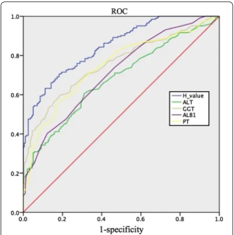
In predicting significant necroinflammatory activity in the validation cohort, the AUROC was 0.847 for the H index (Fig. 4) and was higher than that of any single variable. Using the cut-off values of 0.375 and 0.720, the presence of significant necroinflammatory activity
[image:4.595.61.539.82.526.2]was predicted with high sensitivity (95%) and high specificity (95.4%) in the validation cohort (Table 4). In the validation cohort, at an H index cut-off of 0.375, 194 of 317 (61.2%) patients without significant necroinflammatory activity were identified correctly. Patients with significant necroinflammatory activity could be diagnosed with a high NPV of 88%, as 74 of 84 (88.1%) patients were correctly ruled out. There-fore, almost half of the patients could be excluded from undergoing liver biopsy. At an H index cut-off of 0.720, 188 of 291 (64.6%) patients with necroinflammatory activity in the liver biopsy were correctly identified. Patients without significant necroinflammatory activ-ity could be diagnosed with a high PPV of 92%. Only 9 of 110 (8.1%) patients without significant necroinflam-matory activity were classified incorrectly.
Discussion
[image:5.595.62.537.114.425.2]Globally, an estimated 240 million individuals have CHB; the prevalence varies geographically and is high-est in Africa and Asia [2]. In China, a national survey of HBV seroepidemiology has already shown a decrease in the general prevalence of HBsAg, from 9.75% in 1992 to 7.18% in 2006, and a decrease in children less than 5 years of age, from 9.67% in 1992 to 0.96% in 2006 [5]. Death from cirrhosis and hepatocellular carcinoma (HCC) has been estimated at 310,000 and 340,000 per year, respectively [16]. The goal of HBV therapy is to pre-vent liver-related morbidity and mortality. Patients in the immune-active phases of infection (HBeAg-positive or HBeAg-negative) display elevated ALT, histological evidence of liver injury (significant necroinflammatory activity and/or fibrosis), and elevated HBV DNA levels, Table 1 Baseline characteristics of the 802 patients with chronic hepatitis B at the time of liver biopsy: comparison between the training set and the validation set
The patient characteristics are expressed as the median (25th–75th percentile), and categorical data are expressed as a number (percentage). There were no significant differences between the training group and the validation group in any of the variable
RBC red blood cell, WBC white blood cell, PLT platelets, HB haemoglobin, TG triglyceride, TC total cholesterol, ALT alanine aminotransferase, AST aspartate
aminotransferase, GGT γ-glutamyl transpeptidase, ALP alkaline phosphatase, TB total bilirubin, DB indirect bilirubin, ALB albumin, AFP α-fetal protein, PT prothrombin time
Variables The training cohort (n = 401) The validation cohort (n = 401) All patients (n = 802)
Age (years) 33 (26.5–41) 36 (29–42) 34 (27–41)
Sex (male n, %) 322 (80.2%) 318 (79.3%) 640 (79.8%)
Weight (kg) 62 (56–68) 61 (56–66) 61 (56–67)
RBC (1012/L) 4.8 (4.3–5.5) 4.6 (4.3–5) 4.7 (4.3–5.3)
WBC (109/L) 5.13 ± 1.23 5.24 ± 1.41 5.19 ± 1.32
PLT (109/L) 144 (110–193) 138 (111–176) 140 (110–86.25)
HB (g/L) 140 (130–153) 139 (125–150.5) 139 (128–152)
TG (mmol/L) 1.2 (0.9–1.5) 1.2 (0.9–1.4) 1.2 (0.9–1.4)
TC (mmol/L) 4.2 (3.6–4.9) 4.2 (3.6–4.65) 4.2 (3.6–4.7)
ALT (IU/L) 75 (50–142.5) 62 (42–101.5) 68 (45–121)
AST (IU/L) 56 (37.5–101.5) 50 (34–75) 51 (35–86)
GGT (IU/L) 54 (29–111) 56 (35–82) 54 (32–93)
ALP (IU/L) 79 (64.5–108) 79 (64–100) 78 (63.75–104.25)
TB (μmol/L) 14.5 (10.95–20.2) 15.3 (12.3–19.85) 15 (11.5–20.125)
DB (μmol/L) 5 (3.5–8.3) 5 (3.9–6.95) 5 (3.6–7.4)
ALB (g/L) 41 (37.85–46) 41 (38–44) 42 (38–45.1)
AFP (ng/mL) 3.8 (2.6–7.25) 4.2 (3.05–5.9) 3.9 (2.8–6.7)
PT (s) 12.5 (11.75–13.4) 12.3 (11.7–13.2) 12.3 (11.7–13.2)
HBVDNA (log10 copies/mL) 5.69 (3.34–6.92) 6.37 (3.67–7.52) 5.84 (3.41–7.08)
Grading of liver necroinflammatory activity
G0–1:HAI (0–3) (n, %) 163 (40.6%) 170 (42.4%) 333 (41.5%)
G2:HAI (4–8) (n, %) 119 (29.7%) 116 (28.9%) 235 (29.3%)
G3:HAI (9–12) (n, %) 91 (22.7%) 88 (21.9%) 179 (22.3%)
with a greater risk of progressive liver disease and its associated complications [17]. Significant necroinflam-matory activity and liver tissue fibrosis are risk factors for these complications and are indications for therapeutic intervention.
The American Association for the Study of Liver Dis-eases (AASLD) [16], European Association for the Study of the Liver (EASL) [18], and Asian Pacific Association for the Study of the Liver (APASL) [19, 20] guidelines for treatment of CHB patients specify that the presence of moderate to severe necroinflammatory activity in liver biopsy specimens is an indication for antiviral therapy. At present, no single noninvasive indicator can be used to accurately diagnose and assess pathological changes in CHB patients. Due to the severity of liver disease, the degree of fibrosis and necroinflammatory activity should be determined to ensure that patients receive effective antiviral treatment.
In recent years, many studies have aimed to identify an optimal model for the noninvasive diagnosis of liver fibrosis, and many combined indexes for diagnosis have been developed. However, the noninvasive assessment of liver necroinflammatory activity in CHB patients is less well studied. One representative model is the ActiTest diagnostic model, which includes alpha 2-macroglobu-lin, apolipoprotein A1, haptoglobin, GGT, and ALT [15]. However, the ActiTest model was established using data from CHC patients. Whether ActiTest is useful for Chi-nese CHB patients requires further study. Hence, it is necessary to establish a simple, low-cost model for the noninvasive diagnosis of liver necroinflammatory activity in CHB patients.
A simple diagnostic model distinguishing CHB patients with significant necroinflammatory activity from those without significant necroinflammatory activity was established in this study based on commonly used, rou-tine clinical tests. We developed the index using factors that were independently associated with liver necroin-flammatory activity obtained by routine blood, serum biochemistry, coagulation function and virology tests. The training cohort was divided into two groups, namely, patients with or without significant necroinflammatory activity, based on HAI scores. We eventually selected ALT, GGT, PT and ALB as the most valuable diagnostic indexes and established the H value of a simple scoring system for predicting the absence or presence of signifi-cant necroinflammatory activity.
Table 2 Variables associated with the presence
of significant necroinflammatory activity (score 4–18) in the training group (401 patients) by univariate analyses cohort
RBC red blood cell, WBC white blood cell, PLT platelets, HB haemoglobin, TG triglyceride, TC total cholesterol, ALT alanine aminotransferase, AST aspartate aminotransferase, GGT γ-glutamyl transpeptidase, ALP alkaline phosphatase, TB total bilirubin, DB indirect bilirubin, ALB albumin, AFP α-fetal protein, PT prothrombin time
a Values are comparisons between the training and validation set using an
independent samples t test, except nonparametric test
b Values are comparisons between the training and validation set using an
independent samples t test, except x2 test
Variables No significant inflammation (n = 163)
Significant inflammation (n = 238)
P value
Age (year) 32 (25–40) 34 (27–41.25) 0.114 Weight (kg) 64 (58–69) 61 (56–66) 0.013 Male (n) 128 (78.5%) 194 (81.5%) 0.523b
RBC (1012/L) 4.8 (4.2–5.4) 4.8 (4.3–5.5) 0.119
WBC (109/L) 5 (4.5–5.625) 4.95 (4.3–5.5) 0.056
PLT (109/L) 147 (110–193) 141.5 (109–192) 0.78
HB (g/L) 141 (131–154) 139.5 (129–152.25) 0.533 TG (mmol/L) 1.2 (0.8–1.5) 1.2 (0.9–1.5) 0.793 TC (mmol/L) 4.3 (3.9–5) 4.1 (3.5–4.7) 0.005 ALT (IU/L) 64 (42–85) 95.5 (55–209.75) < 0.001a
AST (IU/L) 46 (30–67) 72 (42–139) < 0.001a
GGT (IU/L) 32 (20–54) 108.57 ± 94.57 < 0.001a
ALP (IU/L) 72 (60–88) 87.5 (70–117) < 0.001a
TB (μmol/L) 12.9 (10.4–17.6) 15.9 (11.4–24.45) < 0.001a
DB (μmol/L) 4.4 (3.2–6) 5.85 (3.9–9.7) < 0.001a
ALB (g/L) 45 (41–48.2) 39 (36–43.725) < 0.001 AFP (ng/mL) 3 (2.3–4.5) 4.75 (3.1–11) < 0.001a
PT (s) 12.1 (11.5–12.8) 12.8 (12–13.725) < 0.001a
HBVDNA (log10
copies/mL) 5.43 (3–6.64) 5.8 (4.08–6.98) 0.063
a
Table 3 Predicotors of significant necroinflammatory activity according to stepwise forward logistic regression analysis
ALT alanine aminotransferase, GGT γ-glutamyl transpeptidase, ALB albumin, PT prothrombin time
Parameter B S.E Wals df Sig. Exp (B) (95% CI)
ALT 0.006 0.002 9.994 1 0.002 1.004 (1.002, 1.009)
GGT 0.008 0.002 11.859 1 0.001 1.008 (1.004, 1.013)
ALB − 0.143 0.024 36.274 1 0.000 0.867 (0.827, 0.908)
PT 0.324 0.108 8.988 1 0.003 1.383 (1.119, 1.710)
[image:6.595.57.291.135.412.2] [image:6.595.59.540.632.714.2]The H value predicted significant necroinflammatory activity in the training group when the AUROC value was at least 0.826 (95% CI 0.786–0.866). With an H value cut-off of 0.560 (the max Youden index point), the sensitiv-ity, specificity and diagnosis accuracy were 83.3, 67.0 and 75.6%, independently. These results show that this model has a high accuracy for evaluating significant liver necro-inflammation. In this study, we constructed and vali-dated a model and scoring system to distinguish patients with and without significant necroinflammatory activ-ity. Meanwhile, our aim for this model is to render liver biopsy unnecessary in a considerable proportion of CHB patients. In recent years, many studies have aimed to identify an optimal model for the noninvasive diagnosis of liver fibrosis. However, the noninvasive assessment of liver necroinflammatory activity in CHB patients is less well studied. Therefore, we further improved the model by referring to the research methods adopted by other researchers to diagnose liver fibrosis [12, 21]. We selected low (0.375) and high (0.720) cut-off values according to the Youden index, sensitivity and specificity to identify the absence and presence of significant necroinflam-matory activity. At an H index cut-off of 0.375, patients without significant necroinflammatory activity were cor-rectly identified by a high NPV. The H value cut-off of 0.375 was used as an NPV, the sensitivity of diagnosing of liver necroinflammatory activity approached 90.8%. At the same time, patients with significant necroinflamma-tory activity were diagnosed correctly at an H index cut-off of 0.720 due to the high PPV. The H value cut-cut-off was
Fig. 2 The correlation of values and hepatic necroinflammatory activity with biopsy. The correlations of values (a ALT, b GGT, c PT, d ALB, e H value) and hepatic necroinflammatory activity with biopsy are shown in box plots. The top and bottom of each box represents the 25th and 75th centile intervals. The line through the box in the median
[image:7.595.58.540.88.298.2] [image:7.595.57.292.401.627.2]Table 4 D iagnostic ac cur ac
y of the H v
alue in the tr aining and v alida tion c ohor ts
In the v
alida
tion c
ohor
t, using a cut
-off sc
or
e of H
≤ 0.375, sig nifican t necr oinflamma tor y ac tivit y w as e
xcluded with high ac
cur
ac
y (88.1% NPV and 95.1% sensitivit
y). Similar
ly
, applying a cut
-off sc
or
e of H > 0.720,
sig nifican t necr oinflamma tor y ac tivit y w as c or rec tly iden
tified with high ac
cur
ac
y (91.8% PPV and 95.4% specificit
y) Italic v alues indica te sig nifican t positiv e v
alues in the v
alida tion c ohor t SEN sensitivit y, SPE specificit y, PPV positiv e pr edic tiv e v alue , NPV nega tiv e pr edic tiv e v alue , LR + positiv
e likelihood r
atio , LR − nega tiv
e likelihood r
atio Cut off G0–1 ≥ G2 SEN SPE PPV N PV LR + LR − Popula tion in volv ed (%) In terpr eta tion Training cohor t (n = 401) Lo w cut off 0.908 0.485 0.720 0.782 1.761 0.191 59.4
Absence of necr
oinflammat
or
y ac
tivit
y (78% cer
taint y) < 0.375 216 22 > 0.375 84 79 H igh cut off 0.538 0.920 0.908 0.577 6.743 0.502 40.6 Pr
esence of necr
oinflammat
or
y ac
tivit
y (91% cer
taint y) < 0.720 128 110 > 0.720 13 150 Validation cohor t (n = 401) Lo w cut off 0.951 0.376 0.612 0.881 1.523 0.130 50.9
Absence of necr
oinflammat
or
y ac
tivit
y (88% cer
taint y) < 0.375 194 10 > 0.375 123 74 H igh cut off 0.495 0.954 0.918 0.646 10.83 0.529 49.1 Pr
esence of necr
oinflammat
or
y ac
tivit
y (92% cer
[image:8.595.210.388.109.727.2]used as a PPV, the specificity of determining the absence of significant necroinflammatory activity reached 92%. With a combination of low and high cutoff values, the model would provide a more accurate diagnosis. In addi-tion, with more CHB patients, this model would render liver biopsy unnecessary.
The indexes used to establish the model are commonly used clinical tests. ALT is the simplest and most com-monly used enzyme for assessing hepatic parenchymal cell injury. In this study, the AUROC of ALT in assessing hepatic necroinflammatory activity in CHB patients was 0.669. Increased serum ALT is an independent risk factor for liver necroinflammatory activity [22]. The serum ALT level in CHB patients should be checked regularly [23]. The use of ALT alone to assess necroinflammatory activ-ity in hepatitis B is not ideal [24], and the ALT level was normal over the long term in 37% of CHB patients with significant liver necroinflammatory activity. Thus, ALT should be combined with other indicators. GGT reflects the degree of liver necroinflammatory activity with a high sensitivity but low specificity. The AUROC value of GGT was 0.771, the highest of all the individual indicators. Elevated GGT in CHB and CHC patients is often associ-ated with bile duct injury [25]. GGT is used in many non-invasive diagnostic models, including the Forns index, the Fibrotest, and the S index. In this study, GGT was found to be an important index with high accuracy in our
predictive model. The Child–Pugh classification, which includes ALB and PT, is useful for evaluating liver func-tion reserve and the degree of liver cirrhosis. ALB and PT often decrease with increased liver necroinflammatory activity, indicating that these parameters might reflect the severity of necroinflammatory activity and therefore have greater significance. According to guidelines for the pre-vention and treatment of chronic hepatitis B published in 2017 by EASL, antiviral treatment should be considered for patients whose liver histology reveals a Knodell HAI
≥ 4 or inflammatory necrosis stage ≥ G2; therefore, the H value is useful for distinguishing between the absence and presence of significant necroinflammatory activity.
Myers et al. assessed 209 CHB patients using the ActiTest model and found that the diagnostic accu-racy remained high. The AUROC was 0.82 ± 0.04 [26], but the model, which includes alpha 2-macroglobu-lin and haptoglobin, can only be used in a few medi-cal institutions and laboratories. The model established in our study is superior to the ActiTest model not only because of the diagnostic accuracy but also due to the lower cost and ease of access. In addition, the param-eters in our model exhibit high reproducibility in the clinic. The number of samples included this study is larger than those used in other studies. Similar to other noninvasive diagnostic models, the H value could reduce the requirement of liver biopsy.
Chen et al. assessed 200 CHB patients with cirrhosis using a model constructed by six variables (AST, TBIL, TBA, PT, APRI and serum HBV-DNA) and found that the diagnostic accuracy of this model was high. The AUROC was 0.859 [27], but this model can only be applied to patients with cirrhosis. Thus, whether this model is use-ful for CHB patients without cirrhosis requires further study. Some advantages of our study include the larger cohort, its applicability to all CHB patients, and the lower cost and ease of access of our model.
However, we should acknowledge that the H value, like other models, has some deficiencies and requires further improvement. First, although it can distinguish somewhat between patients with or without significant necroinflammatory activity, the H value might not be able to accurately grade the necroinflammatory activ-ity or provide prognostic information for CHB patients. Second, even though definite values have been set for the diagnostic model, the diagnostic accuracy is not high enough to predict significant necroinflammatory activity in all cases. As it cannot correctly diagnose liver necroin-flammatory activity in all CHB patients, the model would need to be combined with liver biopsy or other diagnos-tic tests. Finally, more indicators need to be screened and more cases need to be assessed to verify the diagnostic performance of the model.
[image:9.595.56.292.87.324.2]Conclusions
The model established in this study is especially useful for evaluating necroinflammatory activity and monitor-ing therapy outcomes since few patients are willmonitor-ing to have repeated liver biopsies. The H index has excellent diagnostic value for hepatic necroinflammatory activity and appears to be a good noninvasive panel for assess-ing liver inflammatory activity.
Abbreviations
HBV: hepatitis B virus; HCV: hepatitis C virus; CHB: chronic hepatitis B; CHC: chronic hepatitis C; HCC: hepatocellular carcinoma; HBsAg: hepatitis B surface antigen; HAI: histologic activity index; APRI: aminotransferase to platelet ratio index; HE: hematoxylin and eosin; RBC: red blood cell; WBC: white blood cell; PLT: platelets; HB: haemoglobin; TG: triglyceride; TC: total cholesterol; ALT: alanine aminotransferase; AST: aspartate aminotransferase; GGT : γ‑glutamyl transpeptidase; ALP: alkaline phosphatase; TB: total bilirubin; DB: indirect bili‑ rubin; ALB: albumin; AFP: α‑fetal protein; PT: prothrombin time; SEN: sensitivity; SPE: specificity; PPV: positive predictive values; NPV: negative predictive values; LR+: positive likelihood ratios; LR−: negative likelihood ratios; AUROC: the area under the receiver operating characteristic curve.
Authors’ contributions
Acquisition of data: FFS, RDZ, JCX, JPS, YW, YFW; analysis and interpretation of data: FFS, YW, YFW; drafting of the manuscript: FFS, YW, YFW; critical revision of the manuscript for important intellectual content: LGL, MYX; statistical analysis: FFS, YW, YFW; administrative, technical, or material support: YQ, YWD; study concept and design and study supervision: LGL, MYX; obtained funding: LGL. All authors read and approved the final manuscript.
Author details
1 Department of Gastroenterology, Shanghai General Hospital, Shanghai Jiao
Tong University School of Medicine, Shanghai, China. 2 Research and Therapy
Center for Liver Diseases, Zhengxing Hospital, Zhangzhou, Fujian Province,
China. 3 Department of Infectious Disease, Taizhou People’s Hospital, Taizhou,
Jiangsu Province, China. 4 Research and Therapy Center for Liver Diseases,
Hangzhou Second People’s Hospital, Hangzhou, Zhejiang Province, China.
Acknowledgements Not applicable.
Competing interests
The authors declare that they have no competing interests.
Availability of data and materials
The datasets used and analyzed during the current study are available from the corresponding author on reasonable request.
Consent for publication Not applicable.
Ethics approval and consent to participate
The study was approved by the Ethics Committee of Shanghai First People’s Hospital, Shanghai Jiao Tong University School of Medicine. Informed consent to participate in the study was obtained from each patient.
Funding
This study was funded by the National Key Technologies Research and Devel‑ opment Program of China during the 11th and 12th Five Year Plan Period (2008ZX1002‑006, 2012ZX10002007‑001‑040 and 2013ZX10002004‑002‑003), Science and Technology Commission of Shanghai Municipality (10411955300 and 09XD1403200), Shanghai Municipal Health Bureau (XBR2011012).
Publisher’s Note
Springer Nature remains neutral with regard to jurisdictional claims in pub‑ lished maps and institutional affiliations.
Received: 17 January 2018 Accepted: 6 June 2018
References
1. Schweitzer A, Horn J, Mikolajczyk RT, et al. Estimations of worldwide prevalence of chronic hepatitis B virus infection: a systematic review of data published between 1965 and 2013. Lancet. 2015;386:1546–55. 2. Ott JJ, Stevens GA, Groeger J, et al. Global epidemiology of hepatitis B
virus infection: new estimates of age‑specific HBsAg seroprevalence and endemicity. Vaccine. 2012;30:2212–9.
3. Luo Z, Li L, Ruan B. Impact of the implementation of a vaccination strategy on hepatitis B virus infections in China over a 20‑year period. Int J Infect Dis. 2012;16:e82–8.
4. Stanaway JD, Flaxman AD, Naghavi M, et al. The global burden of viral hepatitis from 1990 to 2013: findings from the global burden disease study 2013. Lancet. 2016;388:1081–8.
5. Lozano R, Naghavi M, Foreman K, et al. Global and regional mortal‑ ity from 235 causes of death for 20 age groups in 1990 and 2010: a systematic analysis for the Global Burden of Disease Study 2010. Lancet. 2012;380:2095–128.
6. Wang FS, Fan JG, Zhang Z, et al. The global burden of liver disease: the major impact of China. Hepatology. 2014;60:2099–108.
7. Peng CY, Chien RN, Liaw YF. Hepatitis B virus‑related decompensated liver cirrhosis: benefits of antiviral therapy. J Hepatol. 2012;57:442–50. 8. Afdhal NH, Nunes D. Evaluation of liver fibrosis: a concise review. Am J
Gastroenterol. 2004;99:1160–74.
9. Wai CT, Greenson JK, Fontana RJ, et al. A simple noninvasive index can predict both significant fibrosis and cirrhosis in patients with chronic hepatitis C. Hepatology. 2003;38:518–26.
10. Forns X, Ampurdanes S, Llover JM, et al. Identification of chronic hepatitis C patients without hepatic fibrosis by a simple predictive model. Hepa‑ tology. 2002;36:986–92.
11. Imbert‑Bismut F, Ratziu V, Pieroni L, et al. Biochemical markers of liver fibrosis in patients with hepatitis C virus infection: a prospective study. Lancet. 2001;357:1069–75.
12. Zeng MD, Lu LG, Mao YM, et al. Prediction of significant fibrosis in HBeAg‑positive patients with chronic hepatitis B by a noninvasive model. Hepatology. 2005;42:1437–45.
13. Ho AS, Cheng CC, Lee SC, et al. Novel biomarkers predict liver fibrosis in hepatitis C patients: alpha 2 macroglobulin, vitamin D binding protein and apolipoprotein A1. J Biomed Sci. 2010;17:58.
14. Zhou K, Gao CF, Zhao YP, et al. Simpler score of routine laboratory tests predicts liver fibrosis in HBeAg‑positive patients with chronic hepatitis B. J Gastroenterol Hepatol. 2010;25:1569–77.
15. Poynard T, Deckmyn O, Munteanu M, et al. Awareness of the severity of liver disease reexamined using software combined biomarkers of liver fibrosis and necroinflammatory activity. BMJ Open. 2015;5:e010017. 16. Terrault NA, Bzowej NH, Chang KM, Hwang JP, Jonas MM, Murad MH.
AASLD guidelines for treatment of chronic hepatitis B. Hepatology. 2016;63:261–83.
17. Rüeger S, Bochud PY, Dufour JF, et al. Impact of common risk factors of fibrosis progression in chronic hepatitis C. Gut. 2015;64:1605–15. 18. European Association for the Study of the Liver. EASL 2017 Clinical
Practice Guidelines on the management of hepatitis B virus infection. J Hepatol. 2017;67:370–98.
19. Sarin SK, Kumar M, Lau GK, et al. Asian‑Pacific clinical practice guide‑ lines on the management of hepatitis B: a 2015 update. Hepatol Int. 2016;10(1):1–98.
20. Yapali S, Talaat N, Lok AS. Management of hepatitis B: our practice and how it relates to the guidelines. Clin Gastroenterol Hepatol. 2014;12:16–26.
21. Feng L, Sun K, Zhang J, et al. A novel non‑invasive index using AFP and APTT is associated with liver fibrosis in patients with chronic hepatitis B infection: a retrospective cohort study. BMJ Open. 2015;5(9):e008032. 22. Kobayashi Y, Kawaguchi Y, Mizuta T, Fujimoto K, et al. Metabolic factors
•fast, convenient online submission
•
thorough peer review by experienced researchers in your field
• rapid publication on acceptance
• support for research data, including large and complex data types
•
gold Open Access which fosters wider collaboration and increased citations maximum visibility for your research: over 100M website views per year
•
At BMC, research is always in progress.
Learn more biomedcentral.com/submissions
Ready to submit your research? Choose BMC and benefit from: 23. Gish RG, Given BD, Lai CL, et al. Chronic hepatitis B: virology, natural his‑
tory, current management and a glimpse at future opportunities. Antivir Res. 2015;121:47–58.
24. Michelle L, Benjamin JH, Imad N, et al. The clinical significance of persistently normal ALT in chronic hepatitis B infection. Hepatology. 2007;47:760–7.
25. Loomba R, Rao F, Zhang L, et al. Genetic covariance between γ‑glutamyl transpeptidase and fatty liver risk factors: role of β2‑adrenergic receptor genetic variation in twins. Gastroenterology. 2010;139:836–45.
26. Myers RP, Tainturier MH, Ratziu V, et al. Prediction of liver histological lesions with biochemical markers in patients with chronic hepatitis B. J Hepatol. 2003;39:222–30.