REVIEW
Roles of airway smooth muscle
dysfunction in chronic obstructive pulmonary
disease
Furong Yan
1, Hongzhi Gao
1, Hong Zhao
1, Madhav Bhatia
2and Yiming Zeng
3*Abstract
The airway smooth muscle (ASM) plays an indispensable role in airway structure and function. Dysfunction in ASM plays a central role in the pathogenesis of chronic obstructive pulmonary disease (COPD) and contributes to altera-tions of contractility, inflammatory response, immunoreaction, phenotype, quantity, and size of airways. ASM makes a key contribution in COPD by various mechanisms including altered contractility and relaxation induce by [Ca2+]
i, cell proliferation and hypertrophy, production and modulation of extracellular cytokines, and release of pro-and-anti-inflammatory mediators. Multiple dysfunctions of ASM contribute to modulating airway responses to stimuli, remod-eling, and fibrosis, as well as influence the compliance of lungs. The present review highlights regulatory roles of multiple factors in the development of ASM dysfunction in COPD, aims to understand the regulatory mechanism by which ASM dysfunctions are initiated, and explores the clinical significance of ASM on alterations of airway structure and function in COPD and development of novel therapeutic strategies for COPD.
Keywords: COPD, ASM dysfunction, Proliferation, Phenotype shift
© The Author(s) 2018. This article is distributed under the terms of the Creative Commons Attribution 4.0 International License (http://creat iveco mmons .org/licen ses/by/4.0/), which permits unrestricted use, distribution, and reproduction in any medium, provided you give appropriate credit to the original author(s) and the source, provide a link to the Creative Commons license, and indicate if changes were made. The Creative Commons Public Domain Dedication waiver (http://creat iveco mmons .org/ publi cdoma in/zero/1.0/) applies to the data made available in this article, unless otherwise stated.
Background
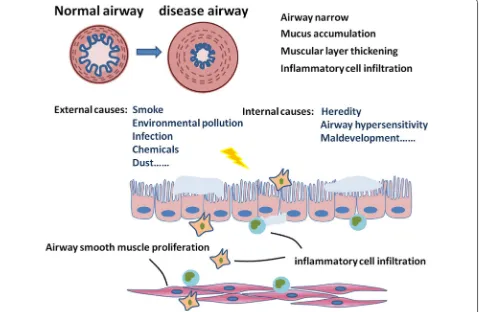
Chronic obstructive pulmonary disease (COPD) is a major cause of morbidity and mortality of patients with lung diseases, characterized by persistent airflow obstruc-tion, with an enhanced inflammatory response in lungs and airways. COPD was the third leading cause of death in China with more than 0.9 million deaths in 2013 [1]. Overwhelming majority of patients had varying degrees of airway remodeling and narrowing. The number of air-way smooth muscle (ASM) of COPD patients with the GOLD standard in grade 3 and 4 increased by nearly 50% [2], which was negatively correlated with lung function in COPD patients. A variety of cellular mediators and path-ways contribute to the pathogenesis of COPD, evoking a large number of airway and lung dysfunctions [3]. Ciga-rette smoking is the major cause for COPD, while the
pollution exposure such as biomass cooking, heating, and exhaust gas, are also very important factors [4]. In addi-tion, environmental alterations, genetic abnormalities, abnormal lung development and accelerated aging also contribute to the development of COPD [3]. The limita-tion of airflow as the principal feature of COPD is pro-gressive and not completely reversible [5] and is caused by airway remodeling, loss of small airways, and emphy-sema [6]. Of those, the airway remodeling and inflam-mation are considered as the major factors resulting in irreversible airflow limitation [2]. The airway remodeling in COPD includes mucosal hyperproduction, epithelial hyperplasia and metaplasia, increased basement mem-brane thickness, and connective tissue over-deposition, as well as increased mass of ASM (Fig. 1).
Smooth muscle cells are a crucial component of air-way for the contractile function and contributions to the production of inflammatory factors, proteases and growth factors [7]. The altered contractile func-tion and mass of ASM leads to airway inflammafunc-tion, hyperresponsiveness, and remodeling [8], which are
Open Access
*Correspondence: zeng_yi_ming@126.com
3 Department of Pulmonary and Critical Care Medicine, Respiratory Medicine Center of Fujian Province, Second Affiliated Hospital of Fujian Medical University, Quanzhou, Fujian, China
Page 2 of 9 Yan et al. J Transl Med (2018) 16:262
key defining features of COPD. Recent studies demon-strated that changes in other ASM functions like oxi-dant/antioxidant imbalance, inflammatory secretion and metabolic disorder also contribute significantly to COPD pathophysiology. For example, the nicotinamide adenine dinucleotide phosphate oxidase 4 (NOX4) expression was up-regulated in ASM during COPD, and strongly related with smoking [9]. ASM in COPD showed imbalance and accumulation in some glyco-lytic products like lactate, glutamine, fatty acids and amino acids [10]. Moreover, ASM also increases the release of inflammatory mediators to play an impor-tant role in many aspects of COPD pathogenesis. The present review aims to briefly overview how the abnor-mal contractility of ASM contributes to the airway remodeling in COPD, ASM-dominated airway inflam-mation occurs, ASM is involved in the development of local and systemic immune response to challenges, and the ASM mass and phenotype changes during COPD. Altered ASM plays an important role in the
pathogenesis of COPD and contributes to the severity of the disease.
ASM contractility
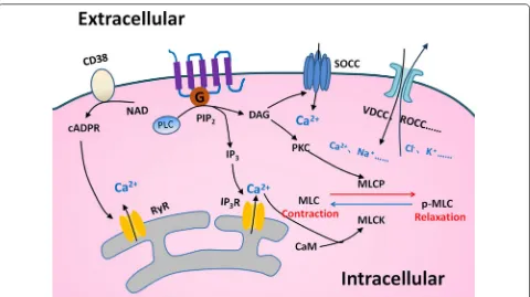
Airway smooth muscle plays a vital role in the regulation of bronchomotor tone and in the control of the airway caliber, by which COPD patients may have an exagger-ated progression of ASM contractility. A variety of regu-latory mechanisms are involved in the ASM contraction, leading to the occurrence of airway hyperresponsiveness. Key mechanisms include G-protein coupled receptor-based pathways [11] such as Gq and Gi-dependent sign-aling, nonselective cation channels especially transient receptor potential channels [12], and store-operated calcium channel [13] (Fig. 2). ASM maintains a balance between the airway hyper-reactivity and bronchodilation when it response to stimuli [14]. The abnormal regulation of contraction and relaxation in ASM leads to the devel-opment of disease. For example, the ASM contractile activity altered in lung fibrotic processes, leading to the
[image:2.595.61.540.90.402.2]abnormality of the mechanical properties of airway and contributing to the pathogenesis of COPD.
A number of factors are involved in the dysregulated mechanism of ASM tone in COPD, e.g. ion channels reside in ASM cell plasma membrane, including voltage-gated channels, receptor- and store-depended channels which are consisted of a variety of transient receptor potential (TRP) channels, stretch-activated channels, and Ca2+-dependent K+ channels [15]. Of these, TRPV4,
the subtype of TRP channels plays an important role in the development of COPD-related airway activities. The bronchial epithelial cells serve as the primary receptor to be initially stimulated by external challenges and as the barrier to separate ASM from the air. When ASM was exposed to hypotonic airway space liquid and gas, the airway epithelial cell barrier dysfunction occurred in patients with COPD. The activation of TRPV4 in ASM can trigger the ASM contraction, damaged epi-thelial cells induce the loss of ASM constrictive capabil-ity for releasing NO, and the airway can be consistently contracted in COPD patients under hypotonic stimula-tion [16]. TRPA1, another subtype of TRP channels are
a determinant of susceptibility in the development of COPD, mobilize Ca2+ influx in ASM upon activation, and
regulate airway contraction via release of neuropeptides, e.g. calcitonin related polypeptide alpha and substance P [17]. G protein-coupled receptors especially Gq-cou-pled pathways which mainly affects the contractility of ASM could be used as a major drug targets for COPD through elevating Ca2+ levels by activating the
phospho-lipase C-inositol 1,4,5-trisphosphate pathway in ASM. Conversely, Gs-coupled pathway is important for airway dilation, particularly through β2-adrenoceptor action
enhancing cAMP [11, 18, 19]. Recent studies suggests that the interactions between taste 2 receptor member 1-bronchoconstrictor and G protein-coupled receptors may contribute to airway contractility and its agonist such as caffeine can influence the actin polymerization of ASM in COPD [20]. Local levels of thromboxane 2 are elevated in the airway of patients with COPD [21]. It indi-cates that endogenous bronchoconstrictors can be one of the major reasons responsible for the high intension of ASM and the airway hypercontraction in COPD through the coupling between the thromboxane-prostanoid
Fig. 2 Mechanisms of contraction in ASM. Many regulatory mechanisms in ASM to control the contraction and relaxation are well recognized. The ectoenzyme CD38 could produce the second messenger cyclic ADP ribose (cADPR), which causes Ca2+ release through ryanodine receptor (RyR)
channels from sarcoplasmic reticulum (SR). The G-protein coupled receptor (GPCR)-based pathway activates phospholipase C (PLC) and breaks up phosphatidyl diphosphate inositol 2 (PIP2) into inositol trisphosphate (IP3) and diacylglycerol (DAG). Intracellular Ca2+ binds to intracellular
calmodulin (CAM) to alter the phosphorylation status of myosin light chain (MLC) and regulates ASM function. Depletion of Ca2+ in SR calcium
stores induces Ca2+ influx through store-operated Ca2+ channel (SOCC). There are other mechanisms that take part in regulating the intracellular
[image:3.595.59.539.85.354.2]Page 4 of 9 Yan et al. J Transl Med (2018) 16:262
receptor with Gq-coupled pathway and through the PLC/ IP3/Ca2+ pathway.
ASM‑dominated inflammatory responses
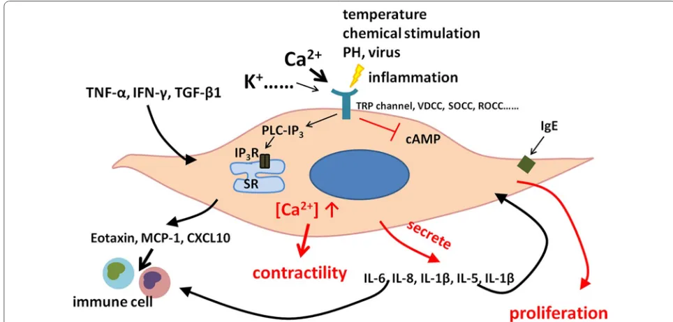
The over-contraction of ASM induced through the acti-vation of multi-kinases also plays a key role in the devel-opment of chronic inflammation and airway remodeling in patients with COPD [18]. When the airway is exposed to viruses, bacteria, or other pathogens, a number of airway and lung cells are provoked and over-reacted to resulting in the development of the airway inflammation in COPD. ASM cells are activated by inflammatory sig-nals from airway epithelial cells, leukocytes, and others, and then induce the secondary inflammation by produc-ing inflammatory mediators. A recently study has shown that ASM cells can act as producers of pro-and anti-inflammatory mediators (e.g. cytokines, chemokines, growth factors) to regulate the local immune environ-ment and influence proliferation, migration, and apopto-sis of other resident cells [22].
Airway smooth muscle can also act as a source of extra-cellular matrix proteins, leading to structural changes and remodeling of the airway and alveoli. ASM cells pro-duce monocyte chemoattractant protein-1, C-X-C motif chemokine ligand 10, interleukin (IL)-6, IL-8, IL-1β and eotaxin [23–25], through the selective expression of most Toll-like receptors isoforms. In human ASM, the active or inactivated virus could cause the release of IL-5 and IL-1β from human ASM cells, probably through inter-cellular adhesion molecule-1, since such production is prevented by treatment with the antibody against inter-cellular adhesion molecule-1 [26]. IL-1β and tumor necrosis factor-alpha (TNFα) can modulate inflamma-tory responses during the exacerbations of COPD.
In addition, the abnormality of the mitochondrial func-tion occurs within ASM cells from patients with COPD, evidenced by the excessive production of reactive oxy-gen species. The production of inflammatory responses and mitochondrial dysfunction are the part of cell self-defenses and the secondary inducer of tissue inflamma-tion and injury to activate and drive the innate immune system in the pathogenesis of COPD [27]. β-catenin as a cellular homeostasis regulator can control the cell divi-sion and differentiation and take part in the inflammatory processes of chronic airway disease, primarily through the co-action with nuclear factor-κB in human ASM and with the nuclear cofactors CREB-binding protein (CBP) and its homologue p300 [28].
ASM‑associated immunoreactions
The tissue forming cells, especially of ASM, play an important role in the immune reaction during the devel-opment of COPD by recognizing environmental factors
through immune globulin receptors or/and through non-immune systems [29]. ASM may amplify or dampen responses to pathogens by releasing mediators, inter-acting with recruiting immune cells, and increasing the responsiveness of ASM to stimuli. The immune-reg-ulatory capacity of ASM also includes its response to cytokines such as IL-1β, TNF-α and IFN-γ. During such process, ASM can initiate the over-expression of cell adhesion and co-stimulatory molecules, which attract the recruitment of multiple immune cells into airways and modulate responses to irritants. ASM-produced cytokines can induce the hyperplasia and modulate immune cell function. ASM-origin TNF-α stimulates eosinophilia and neutrophilia in the airway, results in the maturation and differentiation of structural tissues, and leads to the over-expression of cell adhesion molecules and T cell activation [30]. On the other hand, exter-nal TNF-α can promote roles of ASM in the infiltration and adhesion of activated immune cells and interaction with and adhesion to inflammatory cells [31–33]. ASM can also communicate with airway epithelial cells which can be activated to produce the inflammatory media-tors, resulting to the secondary systemic inflammation [34]. ASM can activate immune cells like T lymphocytes and dendritic cells (DCs) by producing thymic stromal lymphopoietin (TSLP), and promote the maturation of T cell phenotype shift [35, 36]. TSLP as a proinflamma-tory cytokines has been linked to chronic airway dis-eases. Recent studies have shown that ASM are capable in expressing TSLP in vitro and in vivo, and the enhanced production of it, in turn, creates an inflammatory micro-environment to activating local inflammatory response and aggravating airway remodeling, which is associated with COPD. TSLP-activated DCs in vitro play an impor-tant role in promoting the differentiation of Th17 cells with the central memory T cell phenotype [37]. They also induce a unique Th2 cell phenotype that may produces a large amounts of TNF, but little or no IL-10, which differ-ent with normal Th2 cells [38].
Change of ASM remodeling
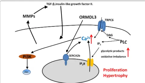
of COPD. The damaged energy balance and accumula-tion of the glycolytic products, e.g. lactate, glutamine, fatty acids, and amino acids, increased biosynthesis and redox imbalance in ASM of patients with COPD, and supported ASM proliferation and survival [10]. The ciga-rette smoke extract (CSE) is the most dangerous environ-mental factor that promotes the proliferation of ASM in COPD, and it happens in connection with up-regulated expression of calreticulin and down-regulated expression of C/EBPα [39]. Moreover, CSE enhances proliferation of ASM by up-regulation of PCNA and Cyclin E [40]. Calcium influx in ASM by α7 nAChR-PI3K/Akt-TRPC6 pathway contributes to the proliferation process of ASM primarily associated with cigarette smoke [41]. Hypertro-phy of ASM is also an important mechanism of airway remodeling. The elevated expression of NOX4 in ASM of small airway may increased the volume of ASM [42]. Up-regulation in orosomucoid-like 3 (ORMDL3) expres-sion can stimulate ASM proliferation, hypertrophy and contractility through enhancing Ca2+ flux induced by
increased sarcoplasmic reticulum Ca2+ ATPase 2b
(SER-CA2b) expression [43]. The ASM remodeling has become an intractable problem in COPD, because of its irrevers-ible changes and multiple mechanism regulation.
Phenotype shift
The ASM phenotype shift is characterized by revers-ible switching between contractile and proliferative phenotype, of which such shift altered obviously in COPD. It has been demonstrated that ASM cells switch from a contractile to a proliferative phenotype are usu-ally accompanied with reduced KCa1.1 channels and
enhanced KCa3.1 channels. Up-regulated KCa3.1 channels
caused the increased expression of contractile pheno-typic marker proteins and induced ASM migration and proliferation. Blockade of KCa3.1 channels is considered
as a therapeutic target in COPD [44]. Long non-coding RNAs (lncRNAs) are associated with ASM phenotype in COPD. The expression of some lncRNAs in healthy ASM cells was increased after stimulation with prolifera-tion inducer [45]. Muscarinic receptors are activated to enhance functional effects of TGF-β1 in ASM, under-pin ASM remodeling in COPD [46]. Smoking and local inflammation could directly lead to ASM proliferation through transformation from contractile ASM pheno-type into proliferative phenopheno-type in airway remodeling in COPD, which depends on phosphorylation of ERK 1/2 and p38 MAP kinase and downstream mitogenic signal-ing [47] (Fig. 4).
[image:5.595.57.536.86.360.2]Page 6 of 9 Yan et al. J Transl Med (2018) 16:262
Regulation of TRP channels in the function of ASM
Adjustment in intracellular calcium concentration ([Ca2+]
i) homeostasis directly affects ASM multiple
func-tions. Several members of TRP channels is calcium per-meable cation channels and are important for calcium signal transduction in many cell types of respiratory pathophysiology. The TRP channels superfamily can be divided into seven subfamilies based on sequence homol-ogy: TRPA (ANKTM1), TRPC (canonical), TRPM (mel-astatin), TRPML (mucolipidins), TRPN (NompC), TRPV (vanil) and TRPP (polycystin) [48]. Transient receptor potential canonical (TRPC) channels belong to the TRP superfamily and most of them expressed in ASM [49, 50]. They may mediated changes in [Ca2+]
i and induced ASM
abnormal contraction, proliferation, hypertrophy, as well as secretion of inflammatory mediators [41, 51]. Calcium influx in ASM stimulating by cigarette smoke activates α7 nAChR-PI3K/Akt-TRPC6 pathway and contributes to the proliferation process of ASM [41]. Deletion of TRPC6 has both elevated in airway resistance and arterial pres-sure. TRPC3 involved in both TNF-α and acetylcholine induced Ca2+ influx in ASM [51] and overexpression of it
may contributes to hyperresponsiveness and remodeling in ASM [52]. TRPV4 as an osmolarity sensor is expressed in ASM and may induced Ca2+ influx when stimulated
by hypotonic stimulation [53]. In recent years, TRPA1 has also been found to be highly expressed on the mem-brane of ASM and can be activated by temperature and
exogenous irritants, leading to airway inflammation and high reactivity [54]. TRP channels were widely involved in the physiological and pathological changes of various respiratory systems, and have become a new target for the treatment of COPD.
ASM in the development of novel therapeutic approaches for COPD
[image:6.595.54.536.88.319.2]channels is used to treat COPD by suppressing ASM phenotype shift to proliferative type [45]. Oxidative stress is crucial in the pathogenesis of COPD, but current treat-ment do not specifically target oxidative stress. Imbal-ance of oxidative stress responses in ASM have been shown in patients with COPD and contributed to airway inflammatory reaction and influenced ASM functions, probably through mitochondrial dysfunction. Targeting treatment with the mitochondria-targeted antioxidant leads to a reduction of the increased the secretion func-tions and reduce proliferation of ASM from patients with COPD [27]. Inhibition of NOX4 expression was mark-edly slowing COPD progress by modulating ROS pro-duction in ASM [9]. Sul-121 is a novel compound with anti-oxidative capacity and can effectively inhibit airway inflammation and airway hyperresponsiveness in COPD models by reducing intracellular reactive oxygen spe-cies production in ASM [55]. Leptin receptor increased expressed on ASM of COPD and has be found inhibited ASM proliferation, migration and eotaxin production through stimulating ASM to secrete prostaglandin E2 [56]. Retinoic acid can inhibit ASM migration by block-ing the PI3K/Akt pathway [57]. There are huge research prospects on the treatment of COPD. The main thera-pies for ASM to treat COPD have been summarised in Table 1.
Conclusion
Alterations of ASM morphology and function are con-sidered as the determinant of the airway function and contribute to the severity of COPD. Different factors
produced by ASM can induce inflammation, prolif-eration, apoptosis, and differentiation of ASM per se and also epithelial cells and immune cells. ASM can serve as a receptor cell to be stimulated and activated by systemic inflammation to develop hypertrophy and hyperplasia in COPD. More importantly, ASM can act as an initiator of secondary inflammation to activate other cells. Thus, ASM plays critical and irreplace-able roles in the development of airway inflammation and remodeling during COPD. Current researches on COPD often using stimulant likes cigarette, lipopoly-saccharide and protease either individually or jointly to construct models. A low dose of rhinovirus infection in patient with COPD would reproduce the features of COPD exacerbations and be know as a human model of COPD exacerbations [58]. Although these models have provided great help for clinical and scientific research on COPD, there are still some weakness on them for ASM dysfunction researches in COPD because of the complex mechanism. Due to limited understanding of mechanism and lack of appropriate models, the impor-tance of ASM in COPD has not been given sufficient attention. To elucidate the etiology and improve treat-ment of COPD, further research is needed. As we find out more about the ASM dysfunction in COPD, we will find more options for specific therapeutic method to improve the clinical symptoms and the quality of life in patients with COPD.
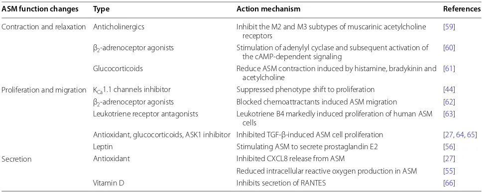
Table 1 Therapeutic approaches targeted on ASM
ASM function changes Type Action mechanism References
Contraction and relaxation Anticholinergics Inhibit the M2 and M3 subtypes of muscarinic acetylcholine
receptors [59]
β2-adrenoceptor agonists Stimulation of adenylyl cyclase and subsequent activation of
the cAMP-dependent signaling [60] Glucocorticoids Reduce ASM contraction induced by histamine, bradykinin and
acetylcholine [61]
Proliferation and migration KCa1.1 channels inhibitor Suppressed phenotype shift to proliferation [44]
β2-adrenoceptor agonists Blocked chemoattractants induced ASM migration [62]
Leukotriene receptor antagonists Leukotriene B4 markedly induced proliferation of human ASM
cells [63]
[image:7.595.55.548.496.689.2]Page 8 of 9 Yan et al. J Transl Med (2018) 16:262
Abbreviations
ASM: airway smooth muscle; COPD: chronic obstructive pulmonary disease; [Ca2+]i: intracellular free calcium concentration; CSE: cigarette smoke extract;
EBP-α: enhancer-binding protein-α; IFN-γ: interferon-γ; IL: interleukin; IP3: inositol tris-phosphate; MMPs: matrix metalloproteinases; NOX4: nicotinamide adenine dinucleotide phosphate oxidase 4; PLC: phospholipase C; TGF-β: transforming growth factor-β; TNF-α: tumor necrosis factor-α; TRP: transient receptor potential.
Authors’ contributions
All authors contributed to the writing of this review. All authors read and approved the final manuscript.
Author details
1 Center for Molecular Diagnosis and Therapy, Second Affiliated Hospital of Fujian Medical University, Quanzhou, Fujian, China. 2 Department of Pathol-ogy and Biomedical Science, University of Otago, Christchurch, New Zealand. 3 Department of Pulmonary and Critical Care Medicine, Respiratory Medicine Center of Fujian Province, Second Affiliated Hospital of Fujian Medical Univer-sity, Quanzhou, Fujian, China.
Acknowledgements
Not applicable.
Competing interests
The authors declare that they have no competing interests.
Availability of data and materials
Not applicable.
Consent for publication
Not applicable.
Ethics approval and consent to participate
Not applicable.
Funding
This work was supported by The National Key Research and Development Program of China (2016YFC1304000; 2016YFC1304003).
Publisher’s Note
Springer Nature remains neutral with regard to jurisdictional claims in pub-lished maps and institutional affiliations.
Received: 4 May 2018 Accepted: 16 September 2018
References
1. Zhou M, Wang H, Zhu J, Chen W, Wang L, Liu S, Li Y, Wang L, Liu Y, Yin P, et al. Cause-specific mortality for 240 causes in China during 1990–2013: a systematic subnational analysis for the Global Burden of Disease Study 2013. Lancet. 2016;387:251–72.
2. Hogg JC, Chu F, Utokaparch S, Woods R, Elliott WM, Buzatu L, Cherniack RM, Rogers RM, Sciurba FC, Coxson HO, Pare PD. The nature of small-airway obstruction in chronic obstructive pulmonary disease. N Engl J Med. 2004;350:2645–53.
3. Vogelmeier CF, Criner GJ, Martinez FJ, Anzueto A, Barnes PJ, Bourbeau J, Celli BR, Chen R, Decramer M, Fabbri LM, et al. Erratum to “Global strategy for the diagnosis, management, and prevention of chronic obstructive lung disease 2017 report: gOLD executive summary” [Arch Bronconeu-mol. 2017;53:128-49]. Arch BronconeuBronconeu-mol. 2017;53:411–2.
4. Gan WQ, FitzGerald JM, Carlsten C, Sadatsafavi M, Brauer M. Associations of ambient air pollution with chronic obstructive pulmonary disease hospitalization and mortality. Am J Respir Crit Care Med. 2013;187:721–7. 5. Woodruff PG, Barr RG, Bleecker E, Christenson SA, Couper D, Curtis JL,
Gouskova NA, Hansel NN, Hoffman EA, Kanner RE, et al. Clinical signifi-cance of symptoms in smokers with preserved pulmonary function. N Engl J Med. 2016;374:1811–21.
6. Chen L, Ge Q, Tjin G, Alkhouri H, Deng L, Brandsma CA, Adcock I, Timens W, Postma D, Burgess JK, et al. Effects of cigarette smoke extract on human airway smooth muscle cells in COPD. Eur Respir J. 2014;44:634–46. 7. Howarth PH, Knox AJ, Amrani Y, Tliba O, Panettieri RA Jr, Johnson M.
Synthetic responses in airway smooth muscle. J Allergy Clin Immunol. 2004;114:S32–50.
8. Hirota N, Martin JG. Mechanisms of airway remodeling. Chest. 2013;144:1026–32.
9. Hollins F, Sutcliffe A, Gomez E, Berair R, Russell R, Szyndralewiez C, Saun-ders R, Brightling C. Airway smooth muscle NOX4 is upregulated and modulates ROS generation in COPD. Respir Res. 2016;17:84.
10. Michaeloudes C, Kuo CH, Haji G, Finch DK, Halayko AJ, Kirkham P, Chung KF, Adcock IM. Copdmap: metabolic re-patterning in COPD airway smooth muscle cells. Eur Respir J. 2017. https ://doi.org/10.1183/13993 003.00202 -2017.
11. Billington CK, Penn RB. Signaling and regulation of G protein-coupled receptors in airway smooth muscle. Respir Res. 2003;4:2.
12. Gosling M, Poll C, Li S. TRP channels in airway smooth muscle as therapeutic targets. Naunyn Schmiedebergs Arch Pharmacol. 2005;371:277–84.
13. Ay B, Prakash YS, Pabelick CM, Sieck GC. Store-operated Ca2+ entry in porcine airway smooth muscle. Am J Physiol Lung Cell Mol Physiol. 2004;286:L909–17.
14. Prakash YS. Airway smooth muscle in airway reactivity and remod-eling: what have we learned? Am J Physiol Lung Cell Mol Physiol. 2013;305:L912–33.
15. Perez-Zoghbi JF, Karner C, Ito S, Shepherd M, Alrashdan Y, Sanderson MJ. Ion channel regulation of intracellular calcium and airway smooth muscle function. Pulm Pharmacol Ther. 2009;22:388–97.
16. Zhu G, Investigators I, Gulsvik A, Bakke P, Ghatta S, Anderson W, Lomas DA, Silverman EK, Pillai SG. Association of TRPV4 gene polymor-phisms with chronic obstructive pulmonary disease. Hum Mol Genet. 2009;18:2053–62.
17. Jha A, Sharma P, Anaparti V, Ryu MH, Halayko AJ. A role for transient receptor potential ankyrin 1 cation channel (TRPA1) in airway hyper-responsiveness? Can J Physiol Pharmacol. 2015;93:171–6.
18. Penn RB, Bond RA, Walker JK. GPCRs and arrestins in airways: implications for asthma. Handb Exp Pharmacol. 2014;219:387–403.
19. Pera T, Penn RB. Crosstalk between beta-2-adrenoceptor and mus-carinic acetylcholine receptors in the airway. Curr Opin Pharmacol. 2014;16:72–81.
20. Tazzeo T, Bates G, Roman HN, Lauzon AM, Khasnis MD, Eto M, Janssen LJ. Caffeine relaxes smooth muscle through actin depolymerization. Am J Physiol Lung Cell Mol Physiol. 2012;303:L334–42.
21. Rolin S, Masereel B, Dogne JM. Prostanoids as pharmacological targets in COPD and asthma. Eur J Pharmacol. 2006;533:89–100.
22. Barnes PJ. Kinases as novel therapeutic targets in asthma and chronic obstructive pulmonary disease. Pharmacol Rev. 2016;68:788–815. 23. Sukkar MB, Xie S, Khorasani NM, Kon OM, Stanbridge R, Issa R, Chung KF.
Toll-like receptor 2, 3, and 4 expression and function in human airway smooth muscle. J Allergy Clin Immunol. 2006;118:641–8.
24. Chiou YL, Lin CY. Der p2 activates airway smooth muscle cells in a TLR2/ MyD88-dependent manner to induce an inflammatory response. J Cell Physiol. 2009;220:311–8.
25. Cooper PR, Lamb R, Day ND, Branigan PJ, Kajekar R, San Mateo L, Hornby PJ, Panettieri RA Jr. TLR3 activation stimulates cytokine secretion without altering agonist-induced human small airway contraction or relaxation. Am J Physiol Lung Cell Mol Physiol. 2009;297:L530–7.
26. Grunstein MM, Hakonarson H, Whelan R, Yu Z, Grunstein JS, Chuang S. Rhinovirus elicits proasthmatic changes in airway responsiveness inde-pendently of viral infection. J Allergy Clin Immunol. 2001;108:997–1004. 27. Wiegman CH, Michaeloudes C, Haji G, Narang P, Clarke CJ, Russell KE, Bao
W, Pavlidis S, Barnes PJ, Kanerva J, et al. Oxidative stress-induced mito-chondrial dysfunction drives inflammation and airway smooth muscle remodeling in patients with chronic obstructive pulmonary disease. J Allergy Clin Immunol. 2015;136:769–80.
28. Koopmans T, Eilers R, Menzen M, Halayko A, Gosens R. Beta-catenin directs nuclear factor-kappaB p65 output via CREB-binding protein/p300 in human airway smooth muscle. Front Immunol. 2017;8:1086.
30. Matera MG, Calzetta L, Cazzola M. TNF-alpha inhibitors in asthma and COPD: we must not throw the baby out with the bath water. Pulm Phar-macol Ther. 2010;23:121–8.
31. Hughes JM, Arthur CA, Baracho S, Carlin SM, Hawker KM, Johnson PR, Armour CL. Human eosinophil-airway smooth muscle cell interactions. Mediators Inflamm. 2000;9:93–9.
32. Lee CW, Lin CC, Luo SF, Lee HC, Lee IT, Aird WC, Hwang TL, Yang CM. Tumor necrosis factor-alpha enhances neutrophil adhesiveness: induc-tion of vascular cell adhesion molecule-1 via activainduc-tion of Akt and CaM kinase II and modifications of histone acetyltransferase and histone deacetylase 4 in human tracheal smooth muscle cells. Mol Pharmacol. 2008;73:1454–64.
33. Lee CW, Lin WN, Lin CC, Luo SF, Wang JS, Pouyssegur J, Yang CM. Tran-scriptional regulation of VCAM-1 expression by tumor necrosis factor-alpha in human tracheal smooth muscle cells: involvement of MAPKs, NF-kappaB, p300, and histone acetylation. J Cell Physiol. 2006;207:174–86. 34. Damera G, Zhao H, Wang M, Smith M, Kirby C, Jester WF, Lawson JA,
Panettieri RA Jr. Ozone modulates IL-6 secretion in human airway epithelial and smooth muscle cells. Am J Physiol Lung Cell Mol Physiol. 2009;296:L674–83.
35. Redhu NS, Saleh A, Halayko AJ, Ali AS, Gounni AS. Essential role of NF-kappaB and AP-1 transcription factors in TNF-alpha-induced TSLP expression in human airway smooth muscle cells. Am J Physiol Lung Cell Mol Physiol. 2011;300:L479–85.
36. Shan L, Redhu NS, Saleh A, Halayko AJ, Chakir J, Gounni AS. Thymic stromal lymphopoietin receptor-mediated IL-6 and CC/CXC chemokines expression in human airway smooth muscle cells: role of MAPKs (ERK1/2, p38, and JNK) and STAT3 pathways. J Immunol. 2010;184:7134–43. 37. Tanaka J, Watanabe N, Kido M, Saga K, Akamatsu T, Nishio A, Chiba T.
Human TSLP and TLR3 ligands promote differentiation of Th17 cells with a central memory phenotype under Th2-polarizing conditions. Clin Exp Allergy. 2009;39:89–100.
38. Liu YJ, Soumelis V, Watanabe N, Ito T, Wang YH, Malefyt Rde W, Omori M, Zhou B, Ziegler SF. TSLP: an epithelial cell cytokine that regulates T cell differentiation by conditioning dendritic cell maturation. Annu Rev Immunol. 2007;25:193–219.
39. Guan P, Cai W, Yu H, Wu Z, Li W, Wu J, Chen J, Feng G. Cigarette smoke extract promotes proliferation of airway smooth muscle cells through suppressing C/EBP-alpha expression. Exp Ther Med. 2017;13:1408–14. 40. Wylam ME, Sathish V, VanOosten SK, Freeman M, Burkholder D,
Thomp-son MA, Pabelick CM, Prakash YS. Mechanisms of cigarette smoke effects on human airway smooth muscle. PLoS ONE. 2015;10:e0128778. 41. Hong W, Peng G, Hao B, Liao B, Zhao Z, Zhou Y, Peng F, Ye X, Huang L,
Zheng M, et al. Nicotine-induced airway smooth muscle cell proliferation involves TRPC6-dependent calcium influx via alpha7 nAChR. Cell Physiol Biochem. 2017;43:986–1002.
42. Liu X, Hao B, Ma A, He J, Liu X, Chen J. The expression of NOX4 in smooth muscles of small airway correlates with the disease severity of COPD. Biomed Res Int. 2016;2016:2891810.
43. Chen J, Miller M, Unno H, Rosenthal P, Sanderson MJ, Broide DH. Oroso-mucoid-like 3 (ORMDL3) upregulates airway smooth muscle proliferation, contraction, and Ca(2+) oscillations in asthma. J Allergy Clin Immunol. 2018;142(207–218):e206.
44. Yu ZH, Wang YX, Song Y, Lu HZ, Hou LN, Cui YY, Chen HZ. Up-regulation of KCa3.1 promotes human airway smooth muscle cell phenotypic modula-tion. Pharmacol Res. 2013;77:30–8.
45. Perry MM, Tsitsiou E, Austin PJ, Lindsay MA, Gibeon DS, Adcock IM, Chung KF. Role of non-coding RNAs in maintaining primary airway smooth muscle cells. Respir Res. 2014;15:58.
46. Oenema TA, Smit M, Smedinga L, Racke K, Halayko AJ, Meurs H, Gosens R. Muscarinic receptor stimulation augments TGF-beta1-induced contrac-tile protein expression by airway smooth muscle cells. Am J Physiol Lung Cell Mol Physiol. 2012;303:L589–97.
47. Pera T, Gosens R, Lesterhuis AH, Sami R, van der Toorn M, Zaagsma J, Meurs H. Cigarette smoke and lipopolysaccharide induce a proliferative airway smooth muscle phenotype. Respir Res. 2010;11:48.
48. Nilius B, Owsianik G. The transient receptor potential family of ion chan-nels. Genome Biol. 2011;12:218.
49. Zhang X, Zhao Z, Ma L, Guo Y, Li X, Zhao L, Tian C, Tang X, Cheng D, Chen Z, Zhang L. The effects of transient receptor potential channel (TRPC) on airway smooth muscle cell isolated from asthma model mice. J Cell Biochem. 2018;119:6033–44.
50. Corteling RL, Li S, Giddings J, Westwick J, Poll C, Hall IP. Expression of transient receptor potential C6 and related transient receptor potential family members in human airway smooth muscle and lung tissue. Am J Respir Cell Mol Biol. 2004;30:145–54.
51. White TA, Xue A, Chini EN, Thompson M, Sieck GC, Wylam ME. Role of transient receptor potential C3 in TNF-alpha-enhanced calcium influx in human airway myocytes. Am J Respir Cell Mol Biol. 2006;35:243–51. 52. Xu BM, Zhang JH, Wang JL, Xiao JH. TRPC3 overexpression and
interven-tion in airway smooth muscle of ovalbumin-induced hyperresponsive-ness and remodeling. Cell Biol Int. 2018;42:1021–9.
53. Jia Y, Wang X, Varty L, Rizzo CA, Yang R, Correll CC, Phelps PT, Egan RW, Hey JA. Functional TRPV4 channels are expressed in human airway smooth muscle cells. Am J Physiol Lung Cell Mol Physiol. 2004;287:L272–8.
54. Nassini R, Pedretti P, Moretto N, Fusi C, Carnini C, Facchinetti F, Viscomi AR, Pisano AR, Stokesberry S, Brunmark C, et al. Transient receptor potential ankyrin 1 channel localized to neuronal airway cells promotes non-neurogenic inflammation. PLoS ONE. 2012;7:e42454.
55. Han B, Poppinga WJ, Zuo H, Zuidhof AB, Bos IS, Smit M, Vogelaar P, Kren-ning G, HenKren-ning RH, Maarsingh H, et al. The novel compound Sul-121 inhibits airway inflammation and hyperresponsiveness in experimental models of chronic obstructive pulmonary disease. Sci Rep. 2016;6:26928. 56. Nair P, Radford K, Fanat A, Janssen LJ, Peters-Golden M, Cox PG. The
effects of leptin on airway smooth muscle responses. Am J Respir Cell Mol Biol. 2008;39:475–81.
57. Day RM, Lee YH, Park AM, Suzuki YJ. Retinoic acid inhibits airway smooth muscle cell migration. Am J Respir Cell Mol Biol. 2006;34:695–703. 58. Mallia P, Message SD, Gielen V, Contoli M, Gray K, Kebadze T, Aniscenko
J, Laza-Stanca V, Edwards MR, Slater L, et al. Experimental rhinovirus infection as a human model of chronic obstructive pulmonary disease exacerbation. Am J Respir Crit Care Med. 2011;183:734–42.
59. Hansel TT, Barnes PJ. Tiotropium bromide: a novel once-daily anticho-linergic bronchodilator for the treatment of COPD. Drugs Today (Barc). 2002;38:585–600.
60. Giembycz MA, Newton R. Beyond the dogma: novel beta2-adrenoceptor signalling in the airways. Eur Respir J. 2006;27:1286–306.
61. Sun HW, Miao CY, Liu L, Zhou J, Su DF, Wang YX, Jiang CL. Rapid inhibi-tory effect of glucocorticoids on airway smooth muscle contractions in guinea pigs. Steroids. 2006;71:154–9.
62. Carlin SM, Roth M, Black JL. Urokinase potentiates PDGF-induced chemo-taxis of human airway smooth muscle cells. Am J Physiol Lung Cell Mol Physiol. 2003;284:L1020–6.
63. Watanabe S, Yamasaki A, Hashimoto K, Shigeoka Y, Chikumi H, Hasegawa Y, Sumikawa T, Takata M, Okazaki R, Watanabe M, et al. Expression of func-tional leukotriene B4 receptors on human airway smooth muscle cells. J Allergy Clin Immunol. 2009;124(59–65):e51–3.
64. Xie S, Sukkar MB, Issa R, Khorasani NM, Chung KF. Mechanisms of induc-tion of airway smooth muscle hyperplasia by transforming growth factor-beta. Am J Physiol Lung Cell Mol Physiol. 2007;293:L245–53.
65. Eapen MS, Kota A, Vindin H, McAlinden KD, Xenaki D, Oliver BG, Desh-pande DA, Sohal SS, Sharma P. Apoptosis signal-regulating kinase 1 (ASK1) inhibition attenuates human airway smooth muscle growth and migration in chronic obstructive pulmonary disease (COPD). Clin Sci (Lond). 2018. https ://doi.org/10.1042/CS201 80398 .