Open Access
Research
T cell responses against tumor associated antigens and prognosis in
colorectal cancer patients
Dirk Nagorsen*
1, Carmen Scheibenbogen
1, Anne Letsch
1,
Christoph-Thomas Germer
2, Heinz-Johannes Buhr
2, Susanna Hegewisch-Becker
3,
Licia Rivoltini
4, Eckhard Thiel
1and Ulrich Keilholz
1Address: 1Medical Department III, Hematology, Oncology, and Transfusion Medicine, Charité University Medicine Berlin, Campus Benjamin
Franklin, Berlin, Germany, 2Department of Surgery, Charité University Medicine Berlin, Campus Benjamin Franklin, Berlin, Germany, 3Department of Oncology and Hematology, University Clinic Eppendorf, Hamburg, Germany and 4Istituto Nazionale Tumori, Milan, Italy
Email: Dirk Nagorsen* - dirk.nagorsen@charite.de; Carmen Scheibenbogen - carmen.scheibenbogen@charite.de; Anne Letsch - anne.letsch@charite.de; Christoph-Thomas Germer - germer@klinikum-nuernberg.de; Heinz-Johannes Buhr - heinz.buhr@charite.de; Susanna Hegewisch-Becker - s.hegewisch-becker@uke.uni-hamburg.de;
Licia Rivoltini - rivoltini@istitutotumori.mi.it; Eckhard Thiel - eckhard.thiel@charite.de; Ulrich Keilholz - ulrich.keilholz@charite.de * Corresponding author
T cell responseantigencolorectal cancersurvivalprognosis
Abstract
Introduction: Spontaneous T cell responses against specific tumor-associated antigens (TAA) are frequently detected in peripheral blood of tumor patients of various histiotypes. However, little is known about whether these circulating, spontaneously occurring, TAA-reactive T cells influence the clinical course of disease.
Methods: Fifty-four HLA-A2 positive colorectal cancer patients had been analyzed for the presence of T cell responses against epitopes derived from the TAA Ep-CAM, her-2/neu, and CEA either by ELISPOT assay or by intracellular cytokine staining. Then, Kaplan-Meier survival analysis was performed comparing T-cell-responders and T-cell-non-responders. For comparison, a group of T-cell-non-responders was compiled stringently matched to T-cell-responders based on clinical criteria and also analyzed for survival.
Results: Sixteen out of 54 patients had a detectable T cell response against at least one of the three tested TAA. Two out of 21 patients (9.5%) with limited stage of disease (UICC I and II) and 14 out of 33 patients (42.4%) with advanced disease (UICC III and IV) were T cell response positive. Comparing all T-cell-responders (n = 16) and all T-cell-non-responders (n = 38), no survival difference was found. In an attempt to reduce the influence of confounding clinical factors, we then compared 16 responders and 16 non-responders in a matched group survival analysis; and again no survival difference was found (p = 0.7).
Conclusion: In summary, we found no evidence that spontaneous peripheral T cell responses against HLA-A2-binding epitopes of CEA, her-2/neu and Ep-CAM are a strong prognostic factor for survival.
Published: 19 January 2005
Journal of Translational Medicine 2005, 3:3 doi:10.1186/1479-5876-3-3
Received: 22 November 2004 Accepted: 19 January 2005
This article is available from: http://www.translational-medicine.com/content/3/1/3
© 2005 Nagorsen et al; licensee BioMed Central Ltd.
Introduction
The importance of the immune system in containing tumor growth is supported by animal studies and various observations in humans [1,2]. These include increased prevalence of certain tumors following immunosuppres-sion as well as the demonstration, that the presence of int-ralesional T cells is correlated with improved clinical outcome in various solid tumors [1,3-6]. In particular in CRC, the presence of CD8+ T cells within the tumor microenvironment was significantly associated with a bet-ter survival in several studies [3,7-9]. However, the anti-gen-specificity of these cells was not determined. T cell responses against specific tumor-associated antigens (TAA) are frequently detected in the peripheral blood of tumor patients [reviewed in [10]] of various histiotypes including colorectal cancer [11], melanoma [12,13], acute myeloid leukemia [14], breast cancer [15], neuroblastoma [16], and head and neck cancer [17]. Data from selected single patients suggest a favorable clinical course in patients with peripheral, spontaneous TAA-directed T cells [18,19]. However, this type of analysis does not allow firm conclusions. The only study comparing clinical out-come of patients with presence of antigen-specific immune responses including natural as well as vaccine-induced antibodies against melanoma antigen GM2 showed an improved survival in favor of immune responders [20]. TAA-directed T cell responses can reliably be induced using various vaccination approaches [reviewed in [21]]. Several recent reports have found a cor-relation between induction of a TAA-directed T cell response by vaccination and clinical response [22-25]. Preliminary data also suggest a possibly favorable clinical effect of vaccine-induced T cells in adjuvant vaccination [26-29]. Taken together, these data lead to the question, whether the presence of spontaneous TAA-specific T cells might be a positive prognostic factor. So far, however, no study has systematically compared survival data of patients with and without presence of a spontaneous TAA-directed T cell response.
In previous studies, we have demonstrated spontaneous T cell responses against the TAA CEA, Ep-CAM, or her-2/neu in peripheral blood of approximately 25% of colorectal cancer patients [11,30]. These cells were identified in
functional T cell assays by antigen-induced IFNγ
produc-tion. More detailed analyses in some samples revealed a
CD3+ CD8+ IFNγ+ CD69+ CD45RA+ phenotype [11],
indicative of an effector T cell subset that is able to directly mediate tumor cell lysis [19]. Spontaneous TAA-specific T cells with the potential of effector cells should, theoreti-cally, be capable of destroying tumor cells and thereby lead to elimination of residual disease or prevent tumor progression. To investigate whether a peripheral, sponta-neous T cell response has an effect on the clinical outcome of tumor patients, we analyzed survival data of CRC
patients with a TAA-directed T cell response and com-pared these data with the clinical course of CRC patients without detectable T cell response.
Patients, materials, and methods
Patient selection and T cell assaysAfter institutional review board approval and informed consent, peripheral blood mononuclear cells from 132 patients with CRC in all stages of disease had been pro-spectively collected and frozen for T cell analysis. All anal-yses have been performed in compliance with the Helsinki Declaration. Fifty-four patients were tested posi-tive for HLA-A2 and were subsequently analyzed for the presence of T cell responses against the HLA-A*0201 pre-sented T cell epitopes Ep-CAM p263–271 [31], her-2/neu p654–662 [32,33], and CEA p571–579 [34] either by ELISPOT assay or by intracellular cytokine staining. HLA analysis, ELISPOT, and intracellular cytokine staining were performed as previously described [11,30]. Positive responses were defined as previously described [11,30].
Survival analysis
First, we performed a Kaplan-Meier survival analysis com-paring all T-cell-responders and all T-cell-non-responders. Additionally, a two-sided log rank test was used to test sta-tistical significance. Then, in an attempt to reduce the influence of external factors, we compiled a patient group from non-responders matching them to responders according to the following criteria: UICC stage of disease, gender, presence of clinically detectable tumor at time of blood draw, duration of disease until blood draw, age at first diagnosis, and previous therapy. Survival in both groups was compared using Kaplan-Meier analysis and by a two-sided log rank test. A level of p < 0.05 was consid-ered significant. SPSS (11.5) software was used.
Results
Patients, T cell response, survival of responders and non-responders
Fifty-four HLA-A2 positive CRC patients who had been analyzed for T cell responses were included in this study [11,30] and retrospectively analyzed for survival. The overall survival rate was 66.7% at a median of 27.5 months follow-up after blood draw for T cell analysis. In 16 out of 54 patients a total number of 26 T cell responses
(between 10 and 1110 specific T cell per 106 PBMC)
data for all patients; and found that among non-respond-ers only 50% (19 out of 38) had clinical stage III or IV dis-ease while 87.5% (14 out of 16) responding patients had stage III or IV disease. Thus, the data on survival are possi-bly based on a strong stage-related bias. Therefore, we sub-sequently used an approach matching non-responders to responders.
Survival in matched patient groups
Sixteen patients from the above group of 38 non-respond-ers were matched with 16 respondnon-respond-ers to obtain two groups comparable for potentially confounding clinical factors, in particular stage of disease (see table 1). At the time of the survival analysis 13 of the total of 32 (40.6%) patients had died: seven T-cell-responders (n = 1 stage III,
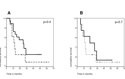
[image:3.612.107.513.148.405.2]Kaplan-Meier survival analyses of colorectal cancer patients based on their T cell response state
Figure 1
Kaplan-Meier survival analyses of colorectal cancer patients based on their T cell response state. Dashed lines refer to T-cell-responders, full lines refer to T-cell-non-T-cell-responders, crosses mark censored cases. Time point 0 refers to the time of blood draw for T cell analysis. A. Two groups of CRC patients (total n = 54) were analyzed for survival after T cell analysis. One group had a spontaneous T cell response against the tumor associated antigens CEA, Ep-CAM, or her-2/neu (n = 16, dashed line). The other group had no T cell response against these antigens (n = 38, full line). No survival difference between the groups was found (log rank, p = 0.4). B. Two matched groups of CRC patients (total n = 32) were analyzed for survival after T cell analysis. One group had a spontaneous T cell response against tumor antigens CEA, Ep-CAM, or her-2/neu (n = 16, dashed line). The other group had no T cell response against these antigens and was selected by having similar clinical characteristics for stage, gender, presence of clinically detectable tumor at time of blood draw, duration of disease, age, and prior therapy (n = 16, full line). No survival difference was found (log rank, p = 0.7).
Time in months
70 60 50 40 30 20 10 0
cu
m
u
la
ti
ve
s
u
rv
iv
a
l
1,0
,9
,8
,7
,6
,5
Time in months
70 60 50 40 30 20 10 0
cu
m
u
la
ti
ve
s
u
rv
iv
a
l
1,0
,9
,8
,7
,6
,5
A
B
n = 6 stage IV) and six T-cell-non-responders (n = 1 stage III, n = 5 stage IV). All deaths were CRC-related. Median time to death among T-cell-responders was 11 months, among T-cell-non-responders 14.5 months. The calcu-lated mean survival time after blood draw for patients without T cell response was 37.0 months (± 4.8 SEM) with a 95% confidence interval 27.5–46.5. Mean survival of T-cell-responders was 40.2 months (± 6.5 SEM) with 95% confidence interval of 27.5–52.9 (Fig. 1B). In a two-sided log rank test, survival did not show a statistically signifi-cant difference between responders and non-responders (p = 0.7). Of note, one to two years after blood draw, patients without T cell response had an up to 20% higher survival rate (approx. 80% vs. approx 60%). These results
were, however, not significant. In a two-sided test with β
= 0.2, a survival difference of 70% could have been
con-sidered significant at a level of α = 0.05 in a population of
this size.
Discussion
In the present study, we analyzed the clinical course of colorectal cancer patients with or without T cells reactive against HLA-A2-binding epitopes of Ep-Cam, her-2/neu, and CEA. No survival difference between T-cell-respond-ers and T-cell-non-respondT-cell-respond-ers was found. This result has to be interpreted cautiously due to the small number of HLA-A2+ patients responding to the above antigens. Obviously, these small numbers cause a high beta error. Thus, this study is only a first indication that the tested spontaneous T cell responses are not important prognos-tic factors for survival.
The second limitation of our study is that the known rep-ertoire of TAA as potentially important T cell targets in
CRC grows every year; and our T cell analysis included only a fraction of potential epitopes. Various other CRC-associated antigens, such as MUC1 or p53, and additional MHC class I antigenic epitopes of CEA and her2-neu have been described [summarized in [35]]. T cell responses against antigens in addition to the ones tested here could potentially play a role in immune surveillance of CRC.
Immune surveillance is understood as a complex process in which T cells and tumor cells influence each other in several ways ["immunoediting", [1]]. There are various potential factors related to tumor cells as well as T cells which may explain a lack of survival effect by TAA-specific T cells. The frequency of TAA-specific T cells detected in most patients was quite low in the range of 10 to 100 T
cells per 106 PBMC. These numbers may be too low to
control tumor growth especially in patients with a higher tumor burden. Furthermore, a general T cell dysfunction including anergic T cells and T cells with downregulated CD3-zeta chains has been described in CRC patients [36,37]. It is possible that the specific T cells detected in the present study are functionally unable to destroy tumor cells. This assumption, however, is not supported by our previous finding that TAA-specific T cells have an effector potential as analyzed in selected patients [11]. Since we have analyzed peripheral blood, we do not know if the cir-culating T cells have the potential to migrate to the tumor site or compartments where CRC cells frequently migrate to including lymph node, liver and bone marrow.
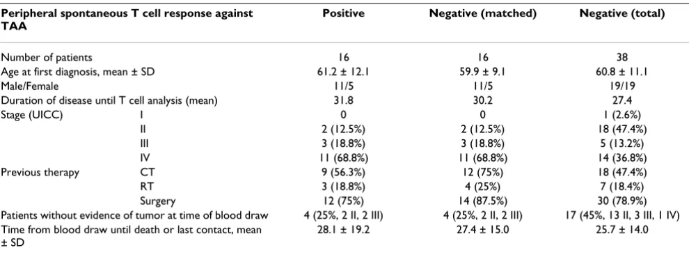
[image:4.612.58.553.99.283.2]Furthermore, tumor cells may not be recognizable by TAA-specific T cells. It has been shown that CRC cell lines secrete immunosuppressive cytokines and that develop-ment of T cell responses is impeded due to low HLA Table 1: Patient characteristics
Peripheral spontaneous T cell response against TAA
Positive Negative (matched) Negative (total)
Number of patients 16 16 38
Age at first diagnosis, mean ± SD 61.2 ± 12.1 59.9 ± 9.1 60.8 ± 11.1
Male/Female 11/5 11/5 19/19
Duration of disease until T cell analysis (mean) 31.8 30.2 27.4
Stage (UICC) I 0 0 1 (2.6%)
II 2 (12.5%) 2 (12.5%) 18 (47.4%)
III 3 (18.8%) 3 (18.8%) 5 (13.2%)
IV 11 (68.8%) 11 (68.8%) 14 (36.8%)
Previous therapy CT 9 (56.3%) 12 (75%) 18 (47.4%)
RT 3 (18.8%) 4 (25%) 7 (18.4%)
Surgery 12 (75%) 14 (87.5%) 30 (78.9%)
Patients without evidence of tumor at time of blood draw 4 (25%, 2 II, 2 III) 4 (25%, 2 II, 2 III) 17 (45%, 13 II, 3 III, 1 IV) Time from blood draw until death or last contact, mean
± SD
28.1 ± 19.2 27.4 ± 15.0 25.7 ± 14.0
expression and lack of intercellular adhesion molecule-1 (ICAM-1) and HLA-DR [38,39]. This is especially relevant considering the fact that TAA-specific T cell responses in peripheral blood are more frequently detectable in advanced stages of CRC [11,40], as well as other tumors [40,41]. These data led to the hypothesis that metastasiz-ing of tumor cells to lymph nodes is a prerequisite for the development of circulating T cell responses [11,42]. Fur-thermore, the presence of TAA-directed T cell responses may have selected immune escape tumor variants. A broad variety of tumor escape mechanisms, such as anti-gen loss or loss of HLA expression, is described in various clinical conditions [43]. It is possible that we encounter similar mechanisms in the present study since malignant cells had grown in vivo during the presence of specific T cell responses. Finally, the role of suppressor and regula-tory T cells in this specific context is unknown.
Taken together, no evidence was found that peripheral, spontaneous T cell responses against HLA-A*0201-bind-ing epitopes of CEA, Ep-CAM, or her-2/neu influence sur-vival of CRC patients. Since the low patient number limits the conclusion, further studies should investigate more patients, more detailed function and migratory pattern of spontaneous T cell responses as well as the genetic profile of the tumor; and consider a broader antigen and epitope repertoire. These studies could have implications for vac-cination therapy as we learn more about why immune surveillance may fail to control tumors and if the presence of a natural T cell response may impact on the efficacy of a vaccine.
Abbreviations
CEA carcinoembryonic antigen, CRC, colorectal
carci-noma; ELISPOT, enzyme-linked immunospot; IFNγ,
Interferon-γ; HLA, human leukocyte antigen; PBMC,
peripheral blood mononuclear cells, TAA, tumor associ-ated antigen.
Competing interests
The author(s) declare that they have no competing interests.
Acknowledgments
We thank Dr. Dr. W. Hopfenmüller and Prof. Dr. P. Martus, both at the Institute for Medical Informatics, Biometry and Epidemiology, Charité Uni-versity Medicine Berlin, Germany, for statistical advice. Furthermore, we thank Orfea Zehm for excellent documentation and Sandra Bauer for excellent technical assistance.
References
1. Dunn GP, Bruce AT, Ikeda H, Old LJ, Schreiber RD: Cancer immu-noediting: from immunosurveillance to tumor escape.Nat Immunol 2002, 3:991-8.
2. Boon T, van Baren N: Immunosurveillance against cancer and immunotherapy – synergy or antagonism?N Engl J Med 2003,
348:252-4.
3. Naito Y, Saito K, Shiiba K, Ohuchi A, Saigenji K, Nagura H, Ohtani H:
CD8+ T cells infiltrated within cancer cell nests as a prognos-tic factor in human colorectal cancer. Cancer Res 1998,
58:3491-3494.
4. Zhang L, Conejo-Garcia JR, Katsaros D, Gimotty PA, Massobrio M, Regnani G, Makrigiannakis A, Gray H, Schlienger K, Liebman MN, Rubin SC, Coukos G: Intratumoral T cells, recurrence, and sur-vival in epithelial ovarian cancer. N Engl J Med 2003,
348:203-213.
5. Mihm M, Clemente C, Cascinelli N: Tumor infiltrating lym-phocytes in lymph node melanoma metastases: a his-topathologic prognostic indicator and an expression of local immune response.Lab Investig 1996, 74:43-47.
6. Schumacher K, Haensch W, Roefzaad C, Schlag PM: Prognostic sig-nificance of activated CD8(+) T cell infiltrations within esophageal carcinomas.Cancer Res 2001, 61:3932-3936. 7. Ropponen KM, Eskelinen MJ, Lipponen PK, Alhava E, Kosma VM:
Prognostic value of tumour-infiltrating lymphocytes (TILs) in colorectal cancer.J Pathol 1997, 182:318-24.
8. Oberg A, Samii S, Stenling R, Lindmark G: Different occurrence of CD8+, CD45R0+, and CD68+ immune cells in regional lymph node metastases from colorectal cancer as potential prog-nostic predictors.Int J Colorectal Dis 2002, 17:25-9.
9. Funada Y, Noguchi T, Kikuchi R, Takeno S, Uchida Y, Gabbert HE:
Prognostic significance of CD8+ T cell and macrophage per-itumoral infiltration in colorectal cancer. Oncol Rep 2003,
10:309-13.
10. Nagorsen D, Scheibenbogen C, Marincola FM, Letsch A, Keilholz U:
Natural T cell immunity against cancer.Clin Cancer Res 2003,
9:4296-303.
11. Nagorsen D, Keilholz U, Rivoltini L, Schmittel A, Letsch A, Asemissen AM, Berger G, Buhr HJ, Thiel E, Scheibenbogen C: Natural T cell response against MHC class I epitopes of epithelial cell adhe-sion molecule, her-2/neu, and carcinoembryonic antigen in patients with colorectal cancer.Cancer Res 2000, 60:4850-4854. 12. Pittet MJ, Valmori D, Dunbar PR, Speiser DE, Lienard D, Lejeune F, Fleischhauer K, Cerundolo V, Cerottini JC, Romero P: High fre-quencies of naive Melan-A/MART-1-specific CD8(+) T cells in a large proportion of human histocompatibility leukocyte antigen (HLA)-A2 individuals.J Exp Med 1999, 190:705-715. 13. Lee PP, Yee C, Savage PA, Fong L, Brockstedt D, Weber JS, Johnson
D, Swetter S, Thompson J, Greenberg PD, Roederer M, Davis MM:
Characterization of circulating T cells specific for tumor-associated antigens in melanoma patients. Nat Med 1999,
5:677-685.
14. Scheibenbogen C, Letsch A, Thiel E, Schmittel A, Mailaender V, Baer-wolf S, Nagorsen D, Keilholz U: CD8 T-cell responses to Wilms tumor gene product WT1 and proteinase 3 in patients with acute myeloid leukemia.Blood 2002, 100:2132-2137.
15. Feuerer M, Beckhove P, Bai L, Solomayer EF, Bastert G, Diel IJ, Pedain C, Oberniedermayr M, Schirrmacher V, Umansky V: Therapy of human tumors in NOD/SCID mice with patient-derived reactivated memory T cells from bone marrow.Nat Med 2001, 7:452-458.
16. Rodolfo M, Luksch R, Stockert E, Chen YT, Collini P, Ranzani T, Lom-bardo C, Dalerba P, Rivoltini L, Arienti F, Fossati-Bellani F, Old LJ, Parmiani G, Castelli C: Antigen-specific immunity in neuroblas-toma patients: antibody and T-cell recognition of NY-ESO-1 tumor antigen.Cancer Res 2003, 63:6948-55.
17. Albers A, Ferris R, Whiteside T, DeLeo A: Immune Responses to P53 in Patients with Cancer: Elevated Frequencies of Tetramer+ P53 Peptide-Specific T Cells and Regulatory CD4+CD25+ Cells at Tumor Sites Compared to the Periph-eral Circulation.J Immunother 2003, 26(6):. Abstract
18. Karanikas V, Colau D, Baurain JF, Chiari R, Thonnard J, Gutierrez-Roelens I, Goffinet C, Van Schaftingen EV, Weynants P, Boon T, Coulie PG: High frequency of cytolytic T lymphocytes directed against a tumor-specific mutated antigen detecta-ble with HLA tetramers in the blood of a lung carcinoma patient with long survival.Cancer Res 2001, 61:3718-3724. 19. Valmori D, Scheibenbogen C, Dutoit V, Nagorsen D, Asemissen AM,
exert-Publish with BioMed Central and every scientist can read your work free of charge "BioMed Central will be the most significant development for disseminating the results of biomedical researc h in our lifetime."
Sir Paul Nurse, Cancer Research UK
Your research papers will be:
available free of charge to the entire biomedical community
peer reviewed and published immediately upon acceptance
cited in PubMed and archived on PubMed Central
yours — you keep the copyright
Submit your manuscript here:
http://www.biomedcentral.com/info/publishing_adv.asp
BioMedcentral
ing ex vivo tumor-specific cytolytic activity.Cancer Res 2002,
62:1743-1750.
20. Livingston PO, Wong GY, Adluri S, Tao Y, Padavan M, Parente R, Hanlon C, Calves MJ, Helling F, Ritter G, Oetten HF, Lloyd JO:
Improved survival in stage III melanoma patients with GM2 antibodies: a randomized trial of adjuvant vaccination with GM2 ganglioside.J Clin Oncol 1994, 12:1036-44.
21. Scheibenbogen C, Letsch A, Schmittel A, Asemissen AM, Thiel E, Keil-holz U: Rational peptide-based tumour vaccine development and T cell monitoring.Seminars in Cancer biology 2003, 13:423-429. 22. Banchereau J, Palucka AK, Dhodapkar M, Burkeholder S, Taquet N, Rolland A, Taquet S, Coquery S, Wittkowski KM, Bhardwaj N, Pineiro L, Steinman R, Fay J: Immune and clinical responses in patients with metastatic melanoma to CD34(+) progenitor-derived dendritic cell vaccine.Cancer Res 2001, 61:6451-6458.
23. Fong L, Hou Y, Rivas A, Benike C, Yuen A, Fisher GA, Davis MM, Eng-leman EG: Altered peptide ligand vaccination with Flt3 ligand expanded dendritic cells for tumor immunotherapy.Proc Natl Acad Sci U S A 2001, 98:8809-14.
24. Coulie PG, Karanikas V, Colau D, Lurquin C, Landry C, Marchand M, Dorval T, Brichard V, Boon T: A monoclonal cytolytic T-lym-phocyte response observed in a melanoma patient vacci-nated with a tumor-specific antigenic peptide encoded by gene MAGE-3.Proc Natl Acad Sci U S A 2001, 98:10290-5. 25. Belli F, Testori A, Rivoltini L, Maio M, Andreola G, Sertoli MR, Gallino
G, Piris A, Cattelan A, Lazzari I, Carrabba M, Scita G, Santantonio C, Pilla L, Tragni G, Lombardo C, Arienti F, Marchiano A, Queirolo P, Bertolini F, Cova A, Lamaj E, Ascani L, Camerini R, Corsi M, Cascinelli N, Lewis JJ, Srivastava P, Parmiani G: Vaccination of metastatic melanoma patients with autologous tumor-derived heat shock protein gp96-peptide complexes: clinical and immuno-logic findings.J Clin Oncol 2002, 20:4169-4180.
26. Wang F, Bade E, Kuniyoshi C, Spears L, Jeffery G, Marty V, Groshen S, Weber J: Phase I trial of a MART-1 peptide vaccine with incomplete Freund's adjuvant for resected high-risk melanoma.Clin Cancer Res 1999, 5:2756-65.
27. Slingluff CL Jr, Yamshchikov G, Neese P, Galavotti H, Eastham S, Engelhard VH, Kittlesen D, Deacon D, Hibbitts S, Grosh WW, Pet-roni G, Cohen R, Wiernasz C, Patterson JW, Conway BP, Ross WG:
Phase I trial of a melanoma vaccine with gp100(280–288) peptide and tetanus helper peptide in adjuvant: immuno-logic and clinical outcomes.Clin Cancer Res 2001, 7:3012-24. 28. Lee P, Wang F, Kuniyoshi J, Rubio V, Stuges T, Groshen S, Gee C, Lau
R, Jeffery G, Margolin K, Marty V, Weber J: Effects of interleukin-12 on the immune response to a multipeptide vaccine for resected metastatic melanoma.J Clin Oncol 2001, 19:3836-47. 29. Weber J, Sondak VK, Scotland R, Phillip R, Wang F, Rubio V, Stuge
TB, Groshen SG, Gee C, Jeffery GG, Sian S, Lee PP: Granulocyte-macrophage-colony-stimulating factor added to a multipep-tide vaccine for resected Stage II melanoma.Cancer 2003,
97:186-200.
30. Nagorsen D, Scheibenbogen C, Schaller G, Leigh B, Schmittel A, Let-sch A, Thiel E, Keilholz U: Differences in T cell immunity towards tumor associated antigens between colorectal can-cer and breast cancan-cer.Int J Cancer 2003, 105:221-225.
31. Ras E, Burg SH, van der, Zegveld ST, Brandt RMP, Kuppen PJK, Offringa R, Warnarr SO, Velde CJH, van de, Melief CJ: Identification of potential HLA-A 0201 restricted CTL epitopes derived from the epithelial cell adhesion molecule (Ep-CAM) and the carcinoembryonic antigen (CEA). Human Immunology 1997,
53:81-89.
32. Peoples GE, Goedegebuure PS, Smith R, Linehan DC, Yoshino I, Eber-lein TJ: Breast and ovarian cancer-specific cytotoxic T lym-phocytes recognize the same HER-2/neu-derived peptide.
Proc Natl Acad Sci U S A 1995, 92:432-436.
33. Peiper M, Goedegebuure PS, Linehan DC, Ganguly E, Douville CC, Eberlein TJ: The Her-2/neu-derived peptide p654–662 is a tumor-associated antigen in human pancreatic cancer rec-ognized by cytotoxic T lymphocytes. Eur J Immunol 1997,
27:1115-1123.
34. Tsang KY, Zaremba S, Nieroda CA, Zhu MZ, Hamilton JM, Schlom J:
Generation of human cytotoxic T cells specific for human carcinoembryonic antigen epitopes from patients immu-nized with recombinant vaccinia-CEA vaccine.J Natl Cancer Inst 1995, 87:982-990.
35. Titu LV, Monson JR, Greenman J: The role of CD8(+) T cells in immune responses to colorectal cancer. Cancer Immunol Immunother 2002, 51:235-47.
36. Pellegrini P, Berghella AM, Del Beato T, Cicia S, Adorno D, Casciani CU: Disregulation in TH1 and TH2 subsets of CD4+ T cells in peripheral blood of colorectal cancer patients and nvolve-ment in cancer establishnvolve-ment and progression.Cancer Immunol Immunother 1996, 42:1-8.
37. Yoong KF, Adams DH: Interleukin 2 restores CD3-ζ chain expression but fails to generate tumour-specific lytic activity in tumour-infiltrating lymphocytes derived from human colorectal hepatic metastases.Br J Cancer 1998, 77:1072-1081. 38. Luo JS, Kammerer R, Schultze H, von Kleist S: Modulations of the
effector function and cytokine production of human lym-phocytes by secreted factors derived from colorectal-carci-noma cells.Int J Cancer 1997, 72:142-148.
39. Lindauer M, Rudy W, Guckel B, Doeberitz MV, Meuer SC, Moebius U: Gene transfer of costimulatory molecules into a human colorectal cancer cell line: requirement of CD54, CD80 and class II MHC expression for enhanced immunogenicity. Immu-nology 1998, 93:390-397.
40. Harashima N, Tanaka K, Sasatomi T, Shimizu K, Miyagi Y, Yamada A, Tamura M, Yamana H, Itoh K, Shichijo S: Recognition of the Lck tyrosine kinase as a tumor antigen by cytotoxic T lym-phocytes of cancer patients with distant metastases.Eur J Immunol 2001, 31:323-32.
41. Mortarini R, Piris A, Maurichi A, Molla A, Bersani I, Bono A, Bartoli C, Santinami M, Lombardo C, Ravagnani F, Cascinelli N, Parmiani G, Ani-chini A: Lack of terminally differentiated tumor-specific CD8+ T cells at tumor site in spite of antitumor immunity to self-antigens in human metastatic melanoma.Cancer Res 2003,
63:2535-45.
42. Parmiani G, Sensi M, Castelli C, Rivoltini L, Anichini A: T-cell response to unique and shared antigens and vaccination of cancer patients.Cancer Immun 2002, 2:6.