R E S E A R C H
Open Access
A three-gene signature as potential predictive
biomarker for irinotecan sensitivity in gastric
cancer
Jie Shen
1, Jia Wei
1, Hao Wang
2, Guofeng Yue
3, Lixia Yu
1, Yang Yang
1, Li Xie
1, Zhengyun Zou
1, Xiaoping Qian
1,
Yitao Ding
2, Wenxian Guan
2*and Baorui Liu
1*Abstract
Objective:Personalized chemotherapy based on molecular biomarkers can maximize anticancer efficiency. We aim to investigate predictive biomarkers capable of predicting response to irinotecan-based treatment in gastric cancer. Methods:We examined gene expression of APTX, BRCA1, ERCC1, ISG15, Topo1 and methylation of SULF2 in formalin-fixed paraffin-embedded gastric cancer tissues from 175 patients and evaluated the association between gene expression levels or methylation status andin vitrosensitivity to irinotecan. We used multiple linear regression analysis to develop a gene-expression model to predict irinotecan sensitivity in gastric cancer and validated this modelin vitroandvivo.
Results:Gene expression levels of APTX, BRCA1 and ERCC1 were significantly lower in irinotecan-sensitive gastric cancer samples than those irinotecan-resistant samples (P< 0.001 for all genes), while ISG15 (P= 0.047) and Topo1 (P= 0.002) were significantly higher. Based on those genes, a three-gene signature were established, which was calculated as follows: Index =0.488 - 0.020× expression level of APTX + 0.015× expression level of Topo1 - 0.011 × expression level of BRCA1. The three-gene signature was significantly associated with irinotecan sensitivity (rho = 0.71, P< 0.001). The sensitivity and specificity for the prediction of irinotecan sensitivity based on the three-gene signature reached 73% and 86%, respectively. In another independent testing set, the irinotecan inhibition rates in gastric samples with sensitive-signature were much higher than those with resistant-signature (65% vs. 22%,P< 0.001). Irinotecan therapy with 20 mg/kg per week to immunodeficient mice carrying xenografts with sensitive-signature dramatically arrested the growth of tumors (P< 0.001), but had no effect on mice carrying xenografts with resistant-signature.
Conclusions:The three-gene signature established herein is a potential predictive biomarker for irinotecan sensitivity in gastric cancer.
Keywords:Personalized chemotherapy, Irinotecan, Gastric cancer, HDRA, Immunodeficient mice
* Correspondence: guanwenxiannj@163.com; baoruiliu@nju.edu.cn 2Department of General Surgery, Drum Tower Hospital, Medical School of Nanjing University, Clinical Cancer Institute of Nanjing University, 321 Zhongshan Rd, Nanjing 210008, China
1The Comprehensive Cancer Centre of Drum Tower Hospital, Medical School of Nanjing University, Clinical Cancer Institute of Nanjing University, 321 Zhongshan Rd, Nanjing 210008, China
Full list of author information is available at the end of the article
Introduction
Gastric cancer remains one of the leading causes of can-cer death worldwide [1,2]. There is no “gold standard” chemotherapy for advanced gastric cancer up to now. In recent years, new generation chemotherapy agents, such as docetaxel, oxaliplatin, irinotecan, capecitabine and S-1 have been studied in phase III studies [3]. However, the median survival remained below one year [2], and the response rate was only approximately 30%-50% [4]. Irinotecan (CPT-11) is a semisynthetic derivative of camptothecin (CPT) which interferes with DNA replica-tion and cell division through its potent interacreplica-tion with the enzyme topoisomerase I (Topo1). Both irinotecan and CPT belong to Topo1 inhibitors. Irinotecan is mainly used in colorectal cancer and also frequently used in the treatment of gastric cancer, showing response rates vary-ing from 14% to 23% as svary-ingle agent and approximately 50% in combination [5].
A number of molecular biomarkers capable of pre-dicting the probability of response to chemotherapeutic agents have been investigated over the last decades [6]. Our previous studies have identified breast cancer suscep-tibility gene 1 (BRCA1) as potential predictive biomarker for cisplatin and docetaxel sensitivity [7,8], thymidylate synthase (TS) for 5-FU and raltitrexed [9,10], and excision repair cross-complementing 1 (ERCC1) for platinum [11]. However, there has been limited progress in the identifica-tion of biomarkers capable of predicting response to irinotecan-based treatment in gastric cancer. Topo1 regu-lates DNA supercoiling during replication through the way of causing single-strand breaks and religation [12]. Irinotecan and its active metabolite SN-38 induce DNA damage by stabilizing a transient covalent complex be-tween DNA and Topo1, which then results in DNA strand breaks, replication arrest, and apoptosis [13]. High tumor levels of Topo1 protein have recently been reported to identify a subgroup of metastatic colorectal cancer pa-tients with good response to irinotecan [14]. Repair of irinotecan-associated and Topo1-mediated DNA damage requires removal of the stalled Topo1 and resolution of the associated DNA break. During this process, a variety of repair proteins, including aprataxin (APTX), BRCA1 and ERCC1, are involved, some of which may have clinical potential as predictive biomarkers [15].
Besides of the candidate biomarkers mentioned above, recent studies suggested that the methylation of the heparan sulfate 6-O-endosulfatase (SULF2) promoter was associated with sensitivity to Topo1 inhibitors in Non-Small-Cell Lung Cancer (NSCLC) [16]. INF-inducible regulator of ubiquitination (ISG15) could block the ubiquitin/26S proteasomal pathway leading to accu-mulation of CPT-induced DNA damage which resulted in an increased apoptosis [17]. SULF2 methylation
(SULF2M) and high ISG15 expressing NSCLC cell lines showed 134-fold sensitivity to CPT than SULF2 unmethylation (SULF2U) and low ISG15 expressing cell lines [16].
Based on the above evidences, we hypothesized that APTX, BRCA1, ERCC1, ISG15, SULF2, and Topo1 or their combination might play important roles in pre-dicting irinotecan sensitivity in gastric cancer. In the current study, we investigated each gene as predictive biomarker by itself, and then established algorithm combining those genes together to more accurately pre-dict irinotecan sensitivity. We also validated the model in another independent set of gastric cancer samples and two cohorts of immunodeficient mice models. The aim of this study was to identify a clinically useful clas-sification signature that could predict the irinotecan sensitivity in gastric cancer.
Materials and methods Patient samples
All specimens and relevant clinical data were obtained from the department of oncology and general surgery, Drum Tower Hospital Affiliated to Medical School of Nanjing University during the period from August 2010 to June 2012. The specimens include 175 freshly-removed gastric tumors, which were randomly classified as either training set (n = 100) or testing set (n = 75) by using computer-generated random numbers. Each tumor tissue was divided into two parts once it removed in the surgery: (1) one part was kept in 4°C Hanks’balanced salt solution with 1% penicillin/streptomycin and detected chemo-sensitivity in vitro by histoculture drug response assay (HDRA); (2) the rest part was left in formalin and made into formalin-fixed paraffin-embedded (FFPE) tumor blocks for pathological observation and gene detection. Diagnosis of patients with gastric tumor was confirmed by histopathology. Clinical and histopathological data, in-cluding age, sex, histology, tumor site, stage, histological grade and lymph node metastasis were all collected. Clin-ical characteristics of the patients were summarized in Table 1. Informed consent was obtained from all patients and the protocols for this study were approved by the Hu-man Research Protective Committee of Drum Tower Hospital Affiliated to Medical School of Nanjing University. All animal experiments were performed in accordance with the Chinese Coordinating Committee on Cancer Research Regulations for the Welfare of Animals and the Animal Protection Law.
HDRA
in diameter, which were then placed on prepared colla-gen (Health Design, Rochester, NY) surfaces in 24-well microplates. There were 8 parallel culture wells for irinotecan sensitivity testing and 8 parallel culture wells for control. After incubation for 7 days at 37°C (in a humidified atmosphere containing 95% air−5% CO2) in
the presence of drugs dissolved with RPMI 1640 medium containing 20% fetal calf serum, 100 μl type I collagenase (0.1 mg/ml, Sigma) and MTT (5 mg/ml, Sigma) were added to each culture well and incubated for another 16 hours. Concentration of irinotecan was 20 μg/ml according to its peak plasma concentration (ppc) in patients [19]. After extraction with dimethyl sulfoxide (DMSO, Sigma), absorbance of the solution in each well was read at 540 nm. Absorbance per gram of cultured tumor tissue was calculated from the mean absorbance of tissue from 8 parallel culture wells, and the tumor-tissue weight was determined before culture.
The inhibition rate was calculated by using the following formula:
Inhibition rate ð Þ ¼% ð1T=CÞ 100%
T is the mean absorbance of treated tumor/Weight C is the mean absorbance of control tumor/Weight
mRNA expression level detection Total RNA extraction from FFPE tissue
Six 7-μm sections were prepared from FFPE tumor blocks that contained at least 80% tumor cells. After hematoxylin-eosin staining, the cancerous parts were microdissected and transferred into a microcentrifuge tube. RNA was isolated in accordance with a proprietary procedure (European patent number EP1945764-B1). Briefly, paraffin was removed by xylene, and micro-dissected cancerous parts were lysed in a proteinase K-containing buffer at 60°C for 16 h. RNA was purified by phenol and chloroform extractions followed by precipi-tation with isopropanol in the presence of sodium acetate at−20°C. The RNA pellet was washed in 70% ethanol and resuspended in 53 μl of RNase-free water followed by treatment with DNase I (Life Technologies).
QPCR assessment of gene expression
M-MLV Reverse Transcriptase Kit (Invitrogen) was applied to generate cDNA for Quantitative polymerase chain reac-tion (qPCR) to detect theβ-actin (ACTB), APTX, BRCA1, ERCC1, ISG15 and Topo1. Each batch of reaction included a positive control from commercial human lung and liver RNA (Stratagene, La Jolla, CA, USA) as calibrators and negative controls without RNA and reverse transcriptase. Total RNA 1μg was used for each RT reaction. Template cDNA was amplified with specific primers and probes for ACTB, APTX, BRCA1, ERCC1, ISG15 and Topo1 using Taqman Universal Master Mix (Applied Biosystems, Foster City, CA). The Assay IDs (Applied Biosystems, Foster City, CA) for the primers and probes were as fol-lows: Hs99999903_m1 (ACTB), Hs00214452_m1 (APTX), Hs00157415_m1 (ERCC1), Hs00192713_m1 (ISG15), Hs00243257_m1 (Topo1), BRCA1 (NM_007294):
for-ward 5’ GGCTATCCTCTCAGAGTGACATTTTA 3’,
reverse 5’ GCTTTATCAGGTTATGTTGCATGGT 3’,
and probe 6FAM −5’CCACTCAGCAGAGGG 3’MGB.
[image:3.595.55.290.102.499.2]QPCR was performed to quantify gene expression using the ABI Prism 7900HT Sequence Detection System (Applied Biosystems). The PCR conditions were 50°C for 2 min, 95°C for 15 min, followed by 40 cycles at 95°C for 15 s and 60°C for 1 min. Relative gene expres-sion quantifications were calculated according to the comparative Ct method using ACTB as an endogenous control, based on our previous experience comparing different housekeeping genes [7,20], and commercial Table 1 Patient characteristics
Characteristic Training set (N = 100)
Independent testing set (N = 75)
In total (N = 175)
Age, y median (range)
63 (29–83) 63 (29–83) 63 (29–83)
≥63 51 (51%) 40 (53%) 91 (52%)
< 63 49 (49%) 35 (47%) 84 (48%)
Sex
Male 74 (74%) 57 (76%) 131 (75%)
Female 26 (26%) 18 (24%) 44 (25%)
Tumor Site
Distal stomach
34 (34%) 28 (37%) 62 (35%)
Proximal stomach
41 (41%) 29 (39%) 70 (40%)
Whole stomach
25 (25%) 18 (24%) 43 (25%)
Stage
I 13 (13%) 7 (9%) 20 (11%)
II 20 (20%) 21 (28%) 41 (23%)
III 65 (65%) 45 (60%) 110 (63%)
IV 2 (2%) 2 (3%) 4 (3%)
Histological grade
2 20 (20%) 16 (21%) 36 (21%)
3 47 (47%) 34 (46%) 81 (46%)
Mixed 1–2 3 (3%) 3 (4%) 6 (3%)
Mixed 2–3 30 (30%) 22 (29%) 52 (30%)
Lymph node metastasis
No 21 (21%) 19 (25%) 40 (23%)
human lung and liver RNAs (Stratagene, La Jolla, CA, USA) as calibrators, which enables us to compare gene expression levels between different patients. Final re-sults were determined by the formula mRNA expression level = 2-(dCt sample-dCt calibrator)(dCt = Ctgene- CtACTB) [7,21]
and were analyzed with the Stratagene analysis software.
DNA methylation detection
DNA extraction and modification Three 7-μm sec-tions were prepared from primary tumor blocks that contained at least 80% tumor cells. After hematoxylin-eosin staining, the cancerous parts were microdissected and transferred into a microcentrifuge tube. DNA was isolated routinely and then was chemically modified by sodium bisulphite to convert all unmethylated cytosines to uracils while leaving methylcytosines unaltered [16]. Then they were stored at−20°C for further analysis.
Methylation-specific polymerase chain reaction (MSP)
MSP was performed to determine the methylation of SULF2 using the ABI Prism 7300HT Sequence Detec-tion System (Applied Biosystems). Each PCR reacDetec-tion contained genomic DNA 2 μl, SYBR Green PCR Mix (TaKaRa, Japan) 10 μl, water 7.7 μl, and primers 0.15μl (10 μmol/ l). The PCR conditions were 95°C for 10 min, followed by 45 cycles at 59°C for 30 s, 72°C for 30 s and 95°C for 30 s. Primers for SULF2 methylated PCR (TaKaRa, Japan) were as follows: forward 5’ TAAGT
GTTTTTTTTATAGCGGC 3’, reverse 5’TACCGTAAT
TTCCGCTATC 3’. Primers for SULF2 unmethylated PCR (TaKaRa, Japan) were as follows: forward 5’ GTTTA
TAAGTGTTTTTTTATAGTGGT3’, reverse 5’TACCATA
ATTTCCACTATCCCT 3’. Each batch of reaction in-cluded a positive control from Methyltransferase (M.SssI)-treated human genomic DNA (fully methylated), a negative control from DNA samples which has been confirmed unmethylated and another negative control without DNA. All tests were performed in duplicate.
The establishment and validation of the gene-expression model for irinotecan sensitivity prediction
We adopted multiple linear regression analysis to establish the optimized gene-expression model based on the training set of 100 gastric cancers [22]. According to the results of stepwise regression (entry:α= 0.10, remove:α= 0.15), model consisted of APTX, Topo1 and BRCA1 is the optimized one. We assigned each patient an index according to the lin-ear combination of the expression level of the mRNA weighted by the regression coefficient from the training samples. The index of the gene-expression model was calcu-lated as follows: Index =0.488 - 0.020× expression level of APTX + 0.015× expression level of Topo1 - 0.011 × expres-sion level of BRCA1. This model was later validated in
another independent testing set of 75 patients with gastric cancer. In the testing set, patients were ranked according to their gene signature index and divided into sensitive-signature and resistant-sensitive-signature groups by using the me-dian index as the cutoff point. The irinotecan sensitivity of these two groups were tested by HDRA and compared with each other.
In vivo validation of the gene-expression model for irinotecan sensitivity prediction
To establish immunodeficient mice models with patient-derived gastric cancer xenografts, each freshly-removed surgical tumor tissue was cut into pieces of 3 × 3 × 3 mm3, which were transplanted within 30 min to 12 athymic immunodeficient mice, termed a “cohort” [23]. In each cohort, when the tumor grew to a size of 50–100 mm3, mice with xenografts were randomized to treatment with irinotecan 20 mg/kg/w, ip (n = 6) or no treatment as the control (n = 6). Individual tumor volumes (V) were calculated by the formula“V = (length × width × width)/2” and compared to the values at the start of treatment to obtain the relative tumor volume. Mice were observed every other day for tumor growth.
In order to evaluate the consistent inhibition of irinotecan in the sensitive-signature mice, three weeks after first administration, all the tumors were separated from the first generation mice and passaged to second generation mice. In the second generation, no drug was administrated. Mice were observed every day for another two weeks.
Statistical analysis
The Mann–Whitney U-test and the Kruskal-Wallis test were used to test the association between mRNA expres-sion levels and clinical characteristics, and the association between irinotecan sensitivity and patients’ clinicopatho-logical parameters. The Spearman’s rank method was used to assess the correlation of the mRNA expression levels between different genes as well as the correlation be-tween mRNA levels and in vitro irinotecan sensitivity.
The Mann–Whitney U-test was used to compare
Results
Patient characteristics
Characteristics of all patients are shown in Table 1. In the 175 patients, the majority of patients were males (75%), and the histology of every sample was adenocar-cinoma. In 62 (35%) patients, the tumor was located in the distal stomach, in 70 (40%) in the proximal stomach, and in 43 (25%) in the whole stomach. One hundred and ten (63%) patients had stage III disease. Lymph node metastasis was present in 135 (77%) patients.
Gene expression levels
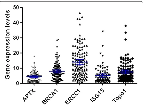
The mRNA expression levels of APTX, BRCA1, ERCC1, ISG15 and Topo1 were detected in all tumors, with me-dian gene expression level relative to housekeeping ACTB of 4.32 for APTX (range 0.26–17.99, 95% confi-dence interval (CI): 3.78–5.19), 7.91 for BRCA1 (range 0.37–29.04, 95% CI: 7.07–9.51), 14.03 for ERCC1 (range 0.33–46.30, 95% CI: 13.01–17.07), 5.07 for ISG15 (range 0.04–34.24, 95% CI: 3.94–6.21), and 7.51 for Topo1 (range 1.07–37.81, 95% CI: 6.38–9.17) (Figure 1). A significant association was observed between APTX mRNA expression levels and histological grade (P= 0.03), and ISG15 mRNA and gender (P= 0.017). No other asso-ciation between clinical characteristics and tumor mRNA levels was found (Table 2). However, a strong correlation was observed between the mRNA expression levels of APTX and BRCA1 (rho = 0.53, P< 0.001), APTX and ERCC1 (rho = 0.73, P< 0.001), BRCA1 and ERCC1 (rho = 0.48,P< 0.001) in tumor.
The relationship between gene expression and chemosensitivity to irinotecan
In the training set, the irinotecan sensitivity was suc-cessfully tested in all tumors, with median inhibition rate of 43.3% (range 2%–89%, CI: 39%–47%). There was no significant association between irinotecan sensitivity and clinical characteristics (Table 3), including age (P =0.51), sex (P =0.77), tumor site (P =0.64), stage (P =0.41), histological grade (P =0.48) and lymph node metastasis (P =0.47). However, mRNA levels of APTX (rho =−0.48, P< 0.001), BRCA1 (rho =−0.49, P< 0.001), ERCC1 (rho =−0.42, P< 0.001), ISG15 (rho = 0.34, P= 0.001), and Topo1 (rho = 0.43,P< 0.001) showed a correlation to irinotecan sensitivity. Patients were ranked according to their irinotecan inhibition rates and divided into irinotecan-sensitive and irinotecan-resistant groups by using the median inhibition rate as the cutoff point [19]. Gene expression levels of APTX (P< 0.001), BRCA1 (P< 0.001) and ERCC1 (P< 0.001) were significantly lower in irinotecan-sensitive patients than in irinotecan-resistant patients, while ISG15 (P= 0.047) and Topo1 (P= 0.002) were significantly higher (Figure 2A-E). ROC curves were generated to calculate the sensitivity and specificity of the
each gene in predicting irinotecan sensitivity (Figure 2G-J). The areas under the ROC curve (AUC), sensitivity and specificity for the prediction of irinotecan sensitivity based on APTX, BRCA1, ERCC1, ISG15 and Topo1 mRNA levels were listed in Table 4.
The relationship between SULF2 methylation and sensitivity to irinotecan
In the training set, the methylation status of SULF2 was successfully detected in all patients, with thirty-three (28%) carrying SULF2M and eighty-four (72%) carrying SULF2U. There was no significant association between SULF2 methylation status and clinical characteristics. The irinotecan inhibition rates were 49.8% (range 2%– 89%, 95% CI: 41%–59%) for SULF2M group, and 40.2% for SULF2U group (range 2%–84%, 95% CI: 34%–46%, P= 0.08).
The establishment of the gene-expression model and its association with sensitivity to irinotecan
[image:5.595.306.538.88.259.2]Based on the expression level of five genes and status of SULF2 methylation, we constructed a signature by mul-tiple linear regression analysis as mentioned in the methods. The index of the three-gene signature ranged from 0.08 to 0.99 with mean value of 0.43 ± 0.16. There was a significant correlation between the index and irinotecan sensitivity (rho = 0.71, P< 0.001) (Figure 3A). ROC curve was generated to calculate the sensitivity and specificity of the three-gene signature in predicting irinotecan sensitivity (Figure 3B). The AUC was 0.828 (95% CI: 0.755–0.901, P< 0.001). With the threshold value of 0.43, the sensitivity and specificity for the pre-diction of irinotecan sensitivity based on the three-gene signature reached 73% and 86%, respectively.
Figure 1Gene expression levels of APTX, BRCA1, ERCC1, ISG15 and Topo1 in 175 patients analyzed by quantitative RT-PCR.
Validation of the three-gene signature in the independent testing set
To determine if the three-gene signature could signifi-cantly distinguish the sensitive and resistant group to irinotecan, we applied it to an independent testing set in-cluding 75 gastric adenocarcinomas. All the patients’ char-acteristics were listed in Table 1. In the testing set, the irinotecan sensitivity was successfully tested in all tumors, with median inhibition rate of 44.0% (range 2%–89%, CI: 37%–51%). There was no significant association between irinotecan sensitivity and clinical characteristics (Table 3), including age (P =0.54), sex (P =0.81), tumor site (P = 0.54), stage (P =0.61), histological grade (P =0.52) and lymph node metastasis (P =0.79). The mRNA expression levels of APTX, BRCA1, ERCC1, ISG15 and Topo1 were detected in all tumors and the gene signature index was calculated as mentioned above. We divided those patients into two groups according to their gene signature index (sensitive-signature group: index > 0.43, n = 37; resistant-signature group: index≤0.43, n = 38). The irinotecan in-hibition rates in gastric samples with sensitive-signature were much higher than those with resistant-signature
(65% vs. 22%,P< 0.001) (Table 3, Figure 3C). There was a significant correlation between the index and irinotecan sensitivity (rho = 0.79, P< 0.001). ROC curve was gener-ated to calculate the sensitivity and specificity of the three-gene signature in predicting irinotecan sensitivity in this set of patients. The AUC was 0.939 (95% CI: 0.882– 0.996, P< 0.001). With the threshold value of 0.43, the sensitivity and specificity for the prediction of irinotecan sensitivity based on the three-gene signature reached 84% and 94%, respectively.
In vivovalidation of the three-gene signature
[image:6.595.60.537.100.452.2]Twenty cohorts of immunodeficient mice (12 mice per cohort, 240 mice in total) with human-derived xenografts were successfully established from the 75 surgical speci-mens of the independent testing set. Based on the mRNA expression of APTX, BRCA1 and Topo1, one cohort of mice (n = 12) carrying surgical tumors with sensitive-signature (Index = 0.95, gene expression level: APTX = 1.01, BRCA1 = 2.78 and Topo1 = 33.96) and another cohort (n = 12) with resistant-signature (Index = 0.28, gene expres-sion level: APTX = 8.33, BRCA1 = 6.87 and Topo1 = 2.09) Table 2 The association of the gene expressions and pathological characteristics
Characteristic No. of
Patients
APTX mRNA BRCA1 mRNA ERCC1 mRNA ISG15 mRNA Topo1 mRNA
mean ± SD mean ± SD mean ± SD mean ± SD mean ± SD
Age, y
≥63 91 (52%) 4.81 ± 3.78 8.61 ± 5.73 15.28 ± 10.01 4.48 ± 3.69 8.03 ± 7.30 < 63 84 (48%) 4.09 ± 3.76 7.90 ± 5.95 14.75 ± 9.28 5.79 ± 6.90 7.47 ± 5.81
Sex
Male 131 (75%) 4.77 ± 3.33 8.41 ± 5.57 15.74 ± 10.19 5.54 ± 5.62 7.95 ± 7.17
Female 44 (25%) 3.58 ± 3.29 7.92 ± 6.67 12.77 ± 7.30 3.53 ± 4.27* 7.21 ± 4.62
Tumor Site
Distal stomach 62 (35%) 4.79 ± 2.87 8.97 ± 6.52 16.12 ± 10.36 7.05 ± 7.75 8.09 ± 5.93
Proximal stomach 70 (40%) 4.16 ± 3.10 8.33 ± 5.72 13.97 ± 9.09 3.61 ± 2.64 7.40 ± 6.63
Whole stomach 43 (25%) 4.60 ± 4.44 7.12 ± 4.74 15.31 ± 9.70 4.61 ± 3.43 7.06 ± 7.94
Stage
I 20 (11%) 4.25 ± 4.47 5.91 ± 3.40 13.40 ± 7.07 1.89 ± 0.83 5.89 ± 4.03
II 41 (23%) 4.19 ± 3.01 8.58 ± 5.09 14.70 ± 9.56 6.76 ± 7.96 8.91 ± 6.35
III 110 (63%) 4.66 ± 3.38 8.19 ± 5.75 15.56 ± 10.16 4.67 ± 4.01 7.70 ± 7.07
IV 4 (3%) 4.20 ± 4.61 15.88 ± 17.90 11.01 ± 5.00 7.25 ± 6.33 4.03 ± 1.63
Histological grade
2 36 (21%) 5.05 ± 3.54* 10.97 ± 7.33 15.78 ± 9.52 4.86 ± 4.75 8.08 ± 6.61
3 81 (46%) 3.92 ± 3.55 7.81 ± 5.16 14.30 ± 9.64 3.68 ± 2.75 7.18 ± 6.08
Mixed 1–2 6 (3%) 3.63 ± 4.51 7.17 ± 2.13 15.17 ± 12.40 8.00 ± 8.94 8.42 ± 6.02
Mixed 2–3 52 (30%) 4.55 ± 2.53 7.06 ± 5.27 15.65 ± 9.96 7.16 ± 7.74 8.44 ± 7.18
Lymph node metastasis
No 40 (23%) 4.58 ± 3.39 8.70 ± 5.08 14.68 ± 7.31 5.97 ± 8.17 7.48 ± 4.47
Yes 135 (77%) 4.46 ± 3.35 8.16 ± 6.05 15.16 ± 10.33 4.78 ± 4.12 7.87 ± 7.23
were chosen forin vivovalidation of the three-gene signa-ture. There were 12 mice in each cohort (six for irinotecan administration and six for control). Irinotecan therapy with 20 mg/kg per week to immunodeficient mice carrying xenografts with sensitive-signature was well tolerated and dramatically arrested the growth of tumors (P< 0.001, Figure 4A), but there was no effect for the same treatment on mice carrying xenografts with resistant-signature (P= 0.83, Figure 4B).
Three weeks after first irinotecan administration, all the tumors were separated from the first generation mice and passaged to the second generation. There were in total three groups in the second generation: mice
carrying xenografts with sensitive-signature and having been treated with irinotecan (named“sensitive-signature mice” for short, n = 6), mice carrying xenografts with resistant-signature and having been treated with irinotecan (named “resistant-signature mice” for short, n = 6) and the controls (no irinotecan administration be-fore, n = 6). The subsequent tumor size in sensitive-signature mice kept exhibiting a reduced tumor size compared with the tumor size in resistant-signature and control groups (sensitive-signature mice vs. control: P< 0.001; resistant-signature mice vs. control: P= 0.10, Figure 4C).
Discussion
The screening and validation of molecular biomarkers capable of predicting response to different chemothera-peutic agents constitutes a significant step towards per-sonalized treatment for cancer patients. In the field of prognostic biomarkers, advances in genome-wide se-quencing and microarray analysis have allowed the iden-tification of molecular signatures that can promote more precise classification and prognostication of human can-cers [24-26]. It was reported that a five-gene signature was closely associated with relapse-free and overall sur-vival among patients with NSCLC, and a 54-gene signa-ture could predict the risk of recurrence in NSCLC [25]. A 21-gene signature also has been demonstrated to pre-dict the risk of distant recurrence in postmenopausal patients with breast cancer treated with anastrozole or tamoxifen [26]. However, up to now, in the field of chemosensitivity predictive biomarkers, there is still no such gene signature for personalizing chemotherapy in gastric cancer.
In the present study, we firstly investigated the value of APTX, BRCA1, ERCC1, ISG15, SULF2 and Topo1 as predictive biomarkers to irinotecan, respectively. We found that although those genes had correlation with irinotecan sensitivity, their function in irinotecan sensi-tivity prediction was limited and their combination might improve efficiency. Therefore, we established a three-gene signature by multiple linear regression ana-lysis and demonstrated the promising value of this signa-ture in distinguishing two subgroups of advanced gastric cancer patients that widely differed in their sensitivity to irinotecan treatment. Moreover, validation was carried out in another independent testing set and two cohorts of immunodeficient mice models with patient-derived gastric cancer xenografts. It was showed that samples with sensitive-signature were significantly more sensitive to irinotecan than those with resistant-signature. Immu-nodeficient mice model with human-derived xenograft also showed that mice with sensitive-signature (high three-gene signature index) could benefit from irinotecan therapy dramatically and were delayed in tumor growth in Table 3 Association between irinotecan sensitivity and
clinical characteristics Characteristic Irinotecan
inhibition rate
Irinotecan inhibition rate
mean (95% CI) mean (95% CI)
Training set (N = 100)
Independent testing set (N = 75)
Age, y median (range)
≥63 42% (36–47%) 44% (31–57%) < 63 45% (39–51%) 44% (34–54%)
Sex
Male 43% (38–47%) 42% (33–51%)
Female 45% (36–55%) 50% (31–68%)
Tumor Site
Distal stomach 42% (35–49%) 44% (28–60%)
Proximal stomach
44% (38–51%) 47% (35–58%)
Whole stomach 43% (35–51%) 37% (16–59%)
Stage
I 34% (21–47%) 36% (3–70%)
II 49% (40–57%) 50% (31–68%)
III 43% (38–48%) 44% (34–54%)
IV 33% 34%
Histological grade
2 41% (33–49%) 44% (21–68%)
3 42% (36–48%) 39% (27–51%)
Mixed 1–2 54% 58%
Mixed 2–3 46% (38–54%) 49% (35–62%)
Lymph node metastasis
No 42% (33–51%) 46% (27–65%)
Yes 44% (39–48%) 44% (34–53%)
Signature index
> 0.43 57% (52–63%) ** 65% (61–70%) **
≤0.43 31% (27–36%) 22% (17–28%)
the second generation; whereas mice with resistant-signature (low three-gene resistant-signature index) had no re-sponse to irinotecan and their tumor kept growing in both first and second generation. Taken together, these findings indicate that this three-gene signature is closely associated with irinotecan sensitivity among patients with gastric cancer.
The chemosensitivity assay we adopted in the present study included HDRA forin vitro testing and immuno-deficient mice models with patient-derived gastric cancer xenografts for in vivo validation. HDRA has been dem-onstrated by varieties of studies as a useful predictor for chemosensitivity at different cancerous sites, including gastrointestinal cancer [18]. It has been reported in gas-tric cancer[18], esophageal cancer [19], breast cancer [27], oral squamous cell carcinomas [28] and head and neck cancer [29] that efficacy rate for an individual agent using HDRA assayin vitro has a considerable good cor-relation with clinical response rate to each agent. The value of patient-derived tumor xenograft model has been investigated and evaluated in various studies, including retrospective and prospective clinical studies [23,30-33]. Similar to the original tumor sample in histological and gene status, the response of xenograft models could pre-dict the efficiency of chemotherapeutic agents in more
[image:8.595.61.539.89.241.2]than 90% patients [23,30]. Good correlations between ef-ficacy rate for an individual agent using such model and clinical response rate to each agent have been well demon-strated [31]. A patient with advanced and gemcitabine-resistant pancreatic cancer resulted in long-lasting tumor response after the efficient treatment guided by the per-sonalized xenograft model generated from the patient’s freshly-removed tumor [32]. In another pilot clinical study, patients with advanced cancer were treated with 17 selected regimens on the basis of personalized tumor grafts. Consequently, durable partial remissions were ob-served in 15 cases [33]. These results supported the notion of patient-derived tumor xenograft models as a powerful platform for chemosensitivity evaluation. In present study, we established different cohorts of immunodeficient mice models with patient-derived gastric cancer xenografts, and demonstrated that tumor growth were significantly suppressed in the cohort with sensitive-signature (low APTX and BRCA1, but high Topo1 mRNA expression level, Index = 0.95) when treated with irinotecan, but had no differences compared with cohort with resistant-signature (high APTX and BRCA1, but low Topo1 mRNA expression level, Index = 0.28). The results of the second generation tumor showed that irinotecan might have anti-cancer efficiency on stem-like cells and therefore the
Figure 2Gene expression levels of APTX (P< 0.001), BRCA1 (P< 0.001) and ERCC1 (P< 0.001) were significantly lower in irinotecan-sensitive patients than in irinotecan-resistant patients, while ISG15 (P= 0.047) and Topo1 (P= 0.002) were significantly higher.Box plots showed the mRNA expression levels of APTX (A), BRCA1 (B), ERCC1 (C), ISG15 (D) and Topo1 (E) in irinotecan-sensitive and irinotecan-resistant groups, respectively (n = 100). The lines inside the boxes denoted the medians. The whiskers of box plots: Min to Max. Graphs of ROC curve showed the AUCs of APTX (F), BRCA1 (G), ERCC1 (H), ISG15 (I) and Topo1 (J) for predicting irinotecan sensitivity. Sensitivity (Y-axis) was plotted against false-positive fraction (1 - specificity).
Table 4 The sensitivity and specificity of five gene expression levels for the prediction of irinotecan sensitivity
Genes Chemotheraputic agents Sensitivity Specificity AUC (95% CI) P
APTX Irinotecan 73% 74% 0.758 (0.669-0.847) <0.001
BRCA1 Irinotecan 91% 58% 0.760 (0.673-0.846) < 0.001
ERCC1 Irinotecan 64% 79% 0.726 (0.632-0.821) <0.001
ISG15 Irinotecan 59% 73% 0.617 (0.494-0.740) 0.047
[image:8.595.56.537.649.733.2]tumor growth was delayed in the second generation of the sensitive-signature group.
We have to admit that HDRA and mice model might still not be representative of the behavior of the patient’s tumors because of the cancer heterozygote and patients’ characteristics, such as age, gender, tumor size and loca-tion. There may unavoidably be an imperfect relationship between tumor response and survival because of treatment associated adverse events. In order to avoid tumor hetero-geneity, we designed 8 parallel culture wells for irinotecan sensitivity testing and 8 parallel culture wells for control from different parts of one patient’s tumor sample. The mice models we have established were derived from 75 pa-tients with different clinicopathological parameters. We will follow up those patients to confirm that whether this in vivoandin vitroinhibition would have consequences for the therapy as well as patients’outcome.
Gene-expression signature usually established by the use of microarrays and later validated by qPCR [24]. However, in clinical practice, microarrays usually involv-ing a large number of genes in the analysis are limited by complicated methods, lack of reproducibility, the need for fresh-frozen tissues and further indepen-dent validation of the results [34]. RT-PCR comprising a smaller number of genes may be more clinically useful, allowing for reproducible and accurate quantification of results for small amounts of RNA obtained from FFPE specimens [24,34]. In the current study, we firstly selected six candidate predictive biomarkers for irinotecan based on widely literature review and previous investigation, and then RT-PCR were performed for gene detection and fur-ther analysis. The method we adopted to detect mRNA expression levels in FFPE specimens is feasible for routine gene expression analysis in daily clinical practice.
According to the results of stepwise regression, the predictive model we established consists of three genes (APTX, BRCA1 and Topo1) finally. mRNA levels of APTX and BRCA1 are both negatively correlated with irinotecan sensitivity, while Topo1 level is positively cor-related with irinotecan sensitivity. Irinotecan, as a kind of Topo1 inhibitors, can stabilize of Topo1-DNA com-plex that upon collision with the replication fork causes double-strand DNA breaks, cell cycle arrest and death [12]. Therefore, the direct molecular target Topo1 was regarded as the best-characterized biomarker capable of predicting response to irinotecan [14]. A clinical study in metastatic colorectal cancer has reported that higher protein levels of Topo1 were correlated longer overall survival (17.4 months vs. 14.7 months,P= 0.005) and bet-ter response to irinotecan significantly [14]. Staying with the same line of the previous study, the current study demonstrated that both singly or combined in the three-gene signature, tumors with higher mRNA levels of Topo1 were more sensitive to irinotecan in gastric cancer.
[image:9.595.60.539.90.240.2]Irinotecan treatment results in the accumulation of DNA strand breaks in tumor cells, and APTX, BRCA1 and ERCC1 have been shown to have important roles in the repair of DNA single- and double-strand breaks [15]. Validation in a panel of 30 colorectal cancer cell lines, the levels of APTX were significantly associated with CPT sensitivity (P= 0.004) [35]. It also reported that APTX as a predictive biomarker was capable of identifying a subset of advanced colorectal cancer patients with high probability of response to irinotecan-based treatment. Patients with low levels of APTX had improved progression-free (9.2 vs. 5.5 months, P= 0.03) and overall survival (36.7 vs. 19 -months, P= 0.008) [13]. Both BRCA1 and ERCC1 play central roles in nucleotide excision repair in DNA damage
response pathways. BRCA1 has been identified as differ-ential modulators of sensitivity to cisplatin and docetaxel [7]. BRCA1 was also been reported to be related with the sensitivity of Topo1 poison in a study of mice model with mammary tumors [36]. ERCC1 is part of the ERCC1– ERCC4 (XPF) heterodimeric structure-specific endonucle-ase, and has been implicated in platinum resistance. Recently, ERCC1 was also demonstrated by a cell line study to be involved in repair of CPT-induced DNA dam-age and had potential value in predicting CPT sensitivity [37]. As a supplement to the previous studies, the current study further demonstrated that higher APTX, BRCA1 and ERCC1 mRNA expression levels suggested lower
[image:10.595.61.539.87.478.2]likelihood of response to irinotecan-based chemotherapy in gastric cancer. The three-gene signature with APTX and BRCA1 could predict sensitivity to irinotecan more precisely. This may result from the reason that DNA dam-age caused by irinotecan would be repaired more effi-ciently when APTX, BRCA1 and ERCC1 expression in high levels, and therefore, these samples would have a poor response to this form of treatment. Moreover, the re-gression coefficient for APTX was higher than for Topo1, which might indicate that the response to irinotecan in gastric tumors could highly dependent on DNA repair mechanisms. The specific mechanisms remain to be fur-ther studied and elucidated.
SULF2 promotes growth and metastasis of solid tumors. It has been demonstrated that promoter CpG island methylation of SULF2 is highly prevalent in resected lung adenocarcinomas and is significantly associated with bet-ter survival [38]. ISG15 inbet-terferes with the ubiquitin/26S proteasome pathway and increase the sensitivity to Topo1 inhibitors by leading to accumulation of CPT-induced DNA damage and resulting in an increased level of apop-tosis [17]. In NSCLC, silencing SULF2 through methyla-tion could result the significant increase of ISG15 mRNA expression levels and increase sensitivity to Topo1 inhibi-tors in vitro[16]. In the present study, based on freshly-removed gastric tumors, ISG15 was demonstrated to correlate with irinotecan sensitivity positively. Samples with higher mRNA expression levels of ISG15 were more sensitive to irinotecan. Our study also showed that SULF2M group might have a higher likelihood of benefit from irinotecan-based treatment than SULF2U group. Further validation is warranted.
Conclusion
In conclusion, the establishment of this three-gene sig-nature as a new model predicting the sensitivity to irinotecan treatment constitutes a new step towards the goal of individualized treatment for gastric cancer pa-tients. Our results suggest that a patient with a tumor that has high levels of the three-gene signature index would be an ideal candidate to receive single or com-bined treatment with irinotecan. These findings are pre-liminary and suggestive at this point, and this three-gene signature needs to be validated before being used in rou-tine daily clinical practice. A clinical trial is currently be-ing designed in order to validate the role of customizbe-ing treatment based on this three-gene signature.
Abbreviations
FFPE:Formalin-fixed paraffin-embedded; HDRA: Histoculture drug response assay; APTX: Aprataxin; BRCA1: Breast cancer susceptibility gene 1; ERCC1: Excision repair cross-complementing 1; ISG15: INF-inducible regulator of ubiquitination; Topo1: Topoisomerase I; SULF2: Heparan sulfate 6-O-endosulfatase.
Competing interest
We declare that we have no conflicts of interest.
Authors’contributions
JS, JW, WXG and BRL designed the research and wrote the paper. JS, HW, GFY and LXY performed the research. YY, LX and ZZ analyzed data. JW, XPQ and YTD edited paper. All authors read and approved the final manuscript.
Acknowledgments
This work was funded by grants from the National Natural Science Foundation of China (Grant No. 81172094), Natural Science Foundation of Jiangsu Province (Grant No. bk2011095) and Top Six Talents Project of Jiangsu Province (Grant No. 2011ws005).
Author details
1The Comprehensive Cancer Centre of Drum Tower Hospital, Medical School of Nanjing University, Clinical Cancer Institute of Nanjing University, 321 Zhongshan Rd, Nanjing 210008, China.2Department of General Surgery,
Drum Tower Hospital, Medical School of Nanjing University, Clinical Cancer Institute of Nanjing University, 321 Zhongshan Rd, Nanjing 210008, China. 3Nanjing University of Traditional Chinese Medicine, 138 Xianlin Rd, Nanjing 210029, China.
Received: 15 January 2013 Accepted: 8 March 2013 Published: 22 March 2013
References
1. Jemal A, Bray F, Center MM, Ferlay J, Ward E, Forman D:Global cancer statistics.CA Cancer J Clin2011,61:69–90.
2. Wagner AD, Grothe W, Haerting J, Kleber G, Grothey A, Fleig WE: Chemotherapy in advanced gastric cancer: a systematic review and meta-analysis based on aggregate data.J Clin Oncol2006,24:2903–2909. 3. Kubota T:New chemotherapy strategies for gastric cancer.In Vivo2008,
22:273–278.
4. Wesolowski R, Lee C, Kim R:Is there a role for second-line chemotherapy in advanced gastric cancer?Lancet Oncol2009,10:903–912.
5. Farhat FS:A general review of the role of irinotecan (CPT11) in the treatment of gastric cancer.Med Oncol2007,24:137–146.
6. Schilsky RL:Personalized medicine in oncology: the future is now.Nat Rev Drug Discov2010,9:363–366.
7. Wei J, Costa C, Ding Y, Zou Z, Yu L, Sanchez JJ, Qian X, Chen H, Gimenez-Capitan A, Meng F,et al:mRNA expression of BRCA1, PIAS1, and PIAS4 and survival after second-line docetaxel in advanced gastric cancer. J Natl Cancer Inst2011,103:1552–1556.
8. Wang L, Wei J, Qian X, Yin H, Zhao Y, Yu L, Wang T, Liu B:ERCC1 and BRCA1 mRNA expression levels in metastatic malignant effusions is associated with chemosensitivity to cisplatin and/or docetaxel.BMC Cancer2008,8:97.
9. Shen J, Wang H, Wei J, Yu L, Xie L, Qian X, Zou Z, Liu B, Guan W: Thymidylate synthase mRNA levels in plasma and tumor as potential predictive biomarkers for raltitrexed sensitivity in gastric cancer.Int J Cancer2012,131:E938–E945.
10. Wang L, Liu B, Wang T, Ding Y, Qian X, Zhao Y:Detection of cell-free ERCC1 and thymidylate synthase (TS) mRNA in malignant effusions and its association with anticancer drug sensitivity.Anticancer Res2008, 28:1085–1091.
11. Wei J, Zou Z, Qian X, Ding Y, Xie L, Sanchez JJ, Zhao Y, Feng J, Ling Y, Liu Y, et al:ERCC1 mRNA levels and survival of advanced gastric cancer patients treated with a modified FOLFOX regimen.Br J Cancer2008, 98:1398–1402.
12. Xu Y, Villalona-Calero MA:Irinotecan: mechanisms of tumor resistance and novel strategies for modulating its activity.Ann Oncol2002, 13:1841–1851.
13. Dopeso H, Mateo-Lozano S, Elez E, Landolfi S, Ramos Pascual FJ, Hernandez-Losa J, Mazzolini R, Rodrigues P, Bazzocco S, Carreras MJ,et al:Aprataxin tumor levels predict response of colorectal cancer patients to irinotecan-based treatment.Clin Cancer Res2010,16:2375–2382.
14. Braun MS, Richman SD, Quirke P, Daly C, Adlard JW, Elliott F, Barrett JH, Selby P, Meade AM, Stephens RJ,et al:Predictive biomarkers of chemotherapy efficacy in colorectal cancer: results from the UK MRC FOCUS trial.J Clin Oncol2008,26:2690–2698.
15. Gilbert DC, Chalmers AJ, El-Khamisy SF:Topoisomerase I inhibition in colorectal cancer: biomarkers and therapeutic targets.Br J Cancer2012, 106:18–24.
16. Tessema M, Yingling CM, Thomas CL, Klinge DM, Bernauer AM, Liu Y, Dacic S, Siegfried JM, Dahlberg SE, Schiller JH, Belinsky SA:SULF2 methylation is prognostic for lung cancer survival and increases sensitivity to topoisomerase-I inhibitors via induction of ISG15.Oncogene2012,31:4107–4116.
17. Desai SD, Haas AL, Wood LM, Tsai YC, Pestka S, Rubin EH, Saleem A, Nur EKA, Liu LF:Elevated expression of ISG15 in tumor cells interferes with the ubiquitin/26S proteasome pathway.Cancer Res2006,66:921–928. 18. Furukawa T, Kubota T, Hoffman RM:Clinical applications of the
histoculture drug response assay.Clin Cancer Res1995,1:305–311. 19. Fujita Y, Hiramatsu M, Kawai M, Nishimura H, Miyamoto A, Tanigawa N:
Histoculture drug response assay predicts the postoperative prognosis of patients with esophageal cancer.Oncol Rep2009,21:499–505. 20. Margeli M, Cirauqui B, Castella E, Tapia G, Costa C, Gimenez-Capitan A,
prognostic value of BRCA1 mRNA expression levels following neoadjuvant chemotherapy in breast cancer.PLoS One2010,5:e9499. 21. Livak KJ, Schmittgen TD:Analysis of relative gene expression data using
real-time quantitative PCR and the 2(−delta delta C(T)) method.Methods 2001,25:402–408.
22. Hu Z, Chen X, Zhao Y, Tian T, Jin G, Shu Y, Chen Y, Xu L, Zen K, Zhang C, Shen H:Serum microRNA signatures identified in a genome-wide serum microRNA expression profiling predict survival of non-small-cell lung cancer.J Clin Oncol2010,28:1721–1726.
23. Fichtner I, Rolff J, Soong R, Hoffmann J, Hammer S, Sommer A, Becker M, Merk J:Establishment of patient-derived non-small cell lung cancer xenografts as models for the identification of predictive biomarkers.Clin Cancer Res2008,14:6456–6468.
24. Chen HY, Yu SL, Chen CH, Chang GC, Chen CY, Yuan A, Cheng CL, Wang CH, Terng HJ, Kao SF,et al:A five-gene signature and clinical outcome in non-small-cell lung cancer.N Engl J Med2007,356:11–20.
25. Larsen JE, Pavey SJ, Passmore LH, Bowman RV, Hayward NK, Fong KM:Gene expression signature predicts recurrence in lung adenocarcinoma.Clin Cancer Res2007,13:2946–2954.
26. Dowsett M, Cuzick J, Wale C, Forbes J, Mallon EA, Salter J, Quinn E, Dunbier A, Baum M, Buzdar A,et al:Prediction of risk of distant recurrence using the 21-gene recurrence score in node-negative and node-positive postmenopausal patients with breast cancer treated with anastrozole or tamoxifen: a TransATAC study.J Clin Oncol2010,28:1829–1834. 27. Tanino H, Oura S, Hoffman RM, Kubota T, Furukawa T, Arimoto J, Yoshimasu
T, Hirai I, Bessho T, Suzuma T,et al:Acquisition of multidrug resistance in recurrent breast cancer demonstrated by the histoculture drug response assay.Anticancer Res2001,21:4083–4086.
28. Ariyoshi Y, Shimahara M, Tanigawa N:Study on chemosensitivity of oral squamous cell carcinomas by histoculture drug response assay.Oral Oncol2003,39:701–707.
29. Hasegawa Y, Goto M, Hanai N, Ijichi K, Adachi M, Terada A, Hyodo I, Ogawa T, Furukawa T:Evaluation of optimal drug concentration in histoculture drug response assay in association with clinical efficacy for head and neck cancer.Oral Oncol2007,43:749–756.
30. Sausville EA, Burger AM:Contributions of human tumor xenografts to anticancer drug development.Cancer Res2006,66:3351–3354. discussion 3354. 31. Talmadge JE, Singh RK, Fidler IJ, Raz A:Murine models to evaluate novel
and conventional therapeutic strategies for cancer.Am J Pathol2007, 170:793–804.
32. Villarroel MC, Rajeshkumar NV, Garrido-Laguna I, De Jesus-Acosta A, Jones S, Maitra A, Hruban RH, Eshleman JR, Klein A, Laheru D,et al:Personalizing cancer treatment in the age of global genomic analyses: PALB2 gene mutations and the response to DNA damaging agents in pancreatic cancer.Mol Cancer Ther2011,10:3–8.
33. Hidalgo M, Bruckheimer E, Rajeshkumar NV, Garrido-Laguna I, De Oliveira E, Rubio-Viqueira B, Strawn S, Wick MJ, Martell J, Sidransky D:A pilot clinical study of treatment guided by personalized tumorgrafts in patients with advanced cancer.Mol Cancer Ther2011,10:1311–1316.
34. Ramaswamy S:Translating cancer genomics into clinical oncology.N Engl J Med2004,350:1814–1816.
35. Mariadason JM, Arango D, Shi Q, Wilson AJ, Corner GA, Nicholas C, Aranes MJ, Lesser M, Schwartz EL, Augenlicht LH:Gene expression profiling-based prediction of response of colon carcinoma cells to 5-fluorouracil and camptothecin.Cancer Res2003,63:8791–8812.
36. Zander SA, Kersbergen A, van der Burg E, de Water N, van Tellingen O, Gunnarsdottir S, Jaspers JE, Pajic M, Nygren AO, Jonkers J,et al:Sensitivity and acquired resistance of BRCA1;p53-deficient mouse mammary tumors to the topoisomerase I inhibitor topotecan.Cancer Res2010,70:1700–1710. 37. Kirschner K, Melton DW:Multiple roles of the ERCC1-XPF endonuclease in
DNA repair and resistance to anticancer drugs.Anticancer Res2010, 30:3223–3232.
38. Tessema M, Yu YY, Stidley CA, Machida EO, Schuebel KE, Baylin SB, Belinsky SA:Concomitant promoter methylation of multiple genes in lung adenocarcinomas from current, former and never smokers. Carcinogenesis2009,30:1132–1138.
doi:10.1186/1479-5876-11-73
Cite this article as:Shenet al.:A three-gene signature as potential predictive biomarker for irinotecan sensitivity in gastric cancer.Journal of Translational Medicine201311:73.
Submit your next manuscript to BioMed Central and take full advantage of:
• Convenient online submission
• Thorough peer review
• No space constraints or color figure charges
• Immediate publication on acceptance
• Inclusion in PubMed, CAS, Scopus and Google Scholar
• Research which is freely available for redistribution