R E S E A R C H
Open Access
Epigenetic silencing of the 3p22 tumor
suppressor
DLEC1
by promoter CpG methylation
in non-Hodgkin and Hodgkin lymphomas
Zhaohui Wang
1,2†, Lili Li
1,2†, Xianwei Su
2, Zifen Gao
3, Gopesh Srivastava
4, Paul G Murray
5, Richard Ambinder
6and Qian Tao
1,2,6*Abstract
Background:Inactivaion of tumor suppressor genes (TSGs) by promoter CpG methylation frequently occurs in
tumorigenesis, even in the early stages, contributing to the initiation and progression of human cancers.Deleted in lung and esophageal cancer 1(DLEC1), located at the 3p22-21.3 TSG cluster, has been identified frequently silenced by promoter CpG methylation in multiple carcinomas, however, no study has been performed for lymphomas yet. Methods:We examined the expression ofDLEC1by semi-quantitative reverse transcription (RT)-PCR, and evaluated the promoter methylation ofDLEC1by methylation-specific PCR (MSP) and bisulfite genomic sequencing (BGS) in common lymphoma cell lines and tumors.
Results:Here we report thatDLEC1is readily expressed in normal lymphoid tissues including lymph nodes and PBMCs, but reduced or silenced in 70% (16/23) of non-Hodgkin and Hodgkin lymphoma cell lines, including 2/6 diffuse large B-cell (DLBCL), 1/2 peripheral T cell lymphomas, 5/5 Burkitt, 6/7 Hodgkin and 2/3 nasal killer (NK)/T-cell lymphoma cell lines. Promoter CpG methylation was frequently detected in 80% (20/25) of lymphoma cell lines and correlated withDLEC1downregulation/silencing. Pharmacologic demethylation reversedDLEC1expression in lymphoma cell lines along with concomitant promoter demethylation.DLEC1methylation was also frequently detected in 32 out of 58 (55%) different types of lymphoma tissues, but not in normal lymph nodes. Furthermore, DLEC1was specifically methylated in the sera of 3/13 (23%) Hodgkin lymphoma patients.
Conclusions:Thus, methylation-mediated silencing ofDLEC1plays an important role in multiple lymphomagenesis, and may serve as a non-invasive tumor marker for lymphoma diagnosis.
Keywords:DLEC1, CpG, Methylation, Tumor suppressor, Lymphoma
Introduction
Epigenetic silencing of tumor suppressor genes (TSGs) by promoter CpG methylation and histone modification has been widely recognized as one of the major causes of tumorigenesis including hematological malignancies [1,2]. Aberrant methylation of TSG is frequently
detected even in the early stage of tumorigenesis, sug-gesting its potential as tumor biomarker for early detec-tion and therapeutic targeting.
Deletions of the 3p22-21.3 region have been identified as one of the earliest molecular events in various malig-nancies [3], including naspharyngeal [4], head and neck [5], lung [6], gastric [7], breast [8], cervix [9] and renal [10] carcinomas, as well as lymphomas [11]. A growing
number of TSGs, including RASSF1A [12,13], BLU/
ZMYND10 [14,15], and CACNA2D2 [16], have been
identified in this region [17,18]. Frequent inactivation of several 3p21.3 genes as functional TSGs, such as RASSF1and BLU[14,19], by promoter CpG methylation had been identified associated with tumor initiation and * Correspondence:qtao@clo.cuhk.edu.hk
†Equal contributors 1
Shenzhen Institutes of Advanced Technology (SIAT), Chinese Academy of Sciences (CAS)-CUHK, Shenzhen, China
2
Cancer Epigenetics Laboratory, Department of Clinical Oncology, State Key Laboratory of Oncology in South China, Sir YK Pao Center for Cancer, The Chinese University of Hong Kong and CUHK Shenzhen Research Institute, Shatin, Hong Kong
Full list of author information is available at the end of the article
progression. For example, promoter methylation of RASSF1Ahas been shown related to poor prognosis and advanced tumor stage of certain tumor types [20-33]. Restoration of RASSF1A in cancer cell lines inhibited tumor cell growth and metastasis [34]. Thus, 3p22-21.3 is a critical TSG cluster in tumorigenesis [3,18].
Deleted in lung and esophageal cancer 1 (DLEC1), a 3p22 cluster genes, was first identified as a TSG in esophageal and lung cancers [35]. Downregulation of DLEC1by promoter methylation has been found in mul-tiple cancers, including nasopharyngeal [36,37], ovarian [38], lung [39], hepatocellular [40], gastric [41], renal [42], and breast carcinomas [43], suggesting its potential as a broad TSG [44,45]. Remarkably,DLEC1was methy-lated in breast cancer as well as pre-invasive lesions but rarely in normal breast tissues, indicating its potential as an epigenetic marker for early tumors [43].
In this study, we examined the expression and methy-lation status of DLEC1 in lymphoma cell lines and tis-sues, and evaluated its potential as a tumor marker for the early detection of hematologic tumors.
Methods
Cell lines and tumor samples
Non-Hodgkin and Hodgkin lymphoma cell lines studied included diffuse large B-cell lymphoma (DLBCL) cell lines (OCI-Ly1, Ly3, Ly7, Ly8, Ly18, SUDHL6); periph-eral T cell lymphoma (PTCL) cell lines (Ly13.2, Ly17); Burkitt lymphoma (BL) cell lines (AG876, BJAB, Namalwa, Rael, Raji); Hodgkin lymphoma (HL) cell lines (L428, L540, L591, L1236, KM-H2, HD-LM-2, HD-MY-Z) (DSMZ cell collection, Braunschweig, Germany); and nat-ural killer (NK)/T-cell lines (NL) (KHYG-1, SNK6, YT) [46-48]. Cell lines were maintained in RPMI1640 or
Dulbecco’s Modified Eagle’s Medium supplemented with 10% fetal calf serum (FBS) (Invitrogen, Paisley, Scotland) and 1% streptomycin/penicillin at 37°C in 5% CO2. HL cell
lines were treated with 5 μM of 5-aza-20-deoxycytidine (Aza) (Sigma) for 3 days, or further treated with 100 nmol/L trichostatin A (Cayman Chemical Co., Ann Arbor, MI, USA) for additional ~16 h as described previ-ously [15,49], and L428 and KM-H2 cell lines were treated with Aza for 6 days.
Normal peripheral blood mononuclear cells (PBMC), lymph node samples, different types of lymphoma sam-ples, as well as sera from healthy individuals and HL patients were collected as previously described, and the reliability and quality of all the studied samples in this study have been confirmed before [41,47,50]. The study was approved by Johns Hopkins Medicine Institutional Review Board. Normal adult tissue RNA samples were purchased commercially (Stratagene, La Jolla, CA, USA or Millipore-Chemicon, Billerica, MA, USA). DNA and RNA were extracted from lymphoma cell lines and pri-mary lymphomas using TRIzol reagent (Invitrogen) as previously described [51,52].
Semi-quantitative reverse transcription (RT)-PCR
RT-PCR was performed as previously described [40,41]. Primers used for RT-PCR are: DLEC1A: 50-ttcctccctcg cctactc,DLEC1B: 50-aaactcatccagccgctg; GAPDH33: 50-ga tgaccttgcccacagcct, GAPDH55: 50-atctctgccccctctgctga. RT-PCR was done with 32 cycles forDLEC1and 23 cycles for GAPDH.
Bisulfite treatment and promoter methylation analysis
[image:2.595.58.537.510.685.2]Bisulfite modification of DNA, methylation-specific PCR (MSP) and bisulfite genomic sequencing (BGS) were
performed as described [53,54]. MSP primers forDLEC1 were: DLEC1m1: 50-gtttcgtagttcggtttcgtc; DLEC1m2: 50-cgaaatatcttaaatacgcaacg; DLEC1u1: 50-tagttttgtagtttggt tttgtt; DLEC1u2: 50-acaaaatatcttaaatacacaaca. For BGS, bisulfite-treated DNA was amplified using primers DLEC1BGS1: 50-gaagatataaatgtttataatgatt; DLEC1BGS4:
[image:3.595.57.542.168.655.2]50-aactacaaccccaaatcctaa. ANKRD30Am1: 50-cggtagttgtta tttgtacgc; ANKRD30Am2: 50-tcctctctcaataaaatcgcg. MSP and BGS were performed for 40 cycles by using the AmpliTaq-Gold DNA polymerase (Applied Biosystems). All primer sets were previously tested for not amplifying any unbisulfited DNA. Amplified BGS products were
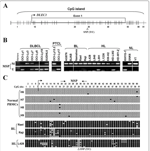
Figure 2Promoter CpG methylation ofDLEC1in lymphoma cell lines. A.Diagram shows theDLEC1CpG island, exon 1 (indicated with a rectangle), and CpG sites (short vertical lines). Numbers from 1 to 61 indicate the individual CpG sites.B.DLEC1methylation as measured by MSP in lymphoma cell lines. M: methylated; U: unmethylated; DLBCL: diffuse large B-cell lymphoma; BL: Burkitt lymphoma; HL: Hodgkin lymphoma; NL: nasal NK/T-cell lymphoma.C.High resolution mapping of the methylation status of theDLEC1promoter by BGS in normal PBMCs, and
cloned and 5 to 10 colonies were randomly chosen for sequencing.
Results
DLEC1was downregulated by promoter CpG methylation in lymphoma cell lines
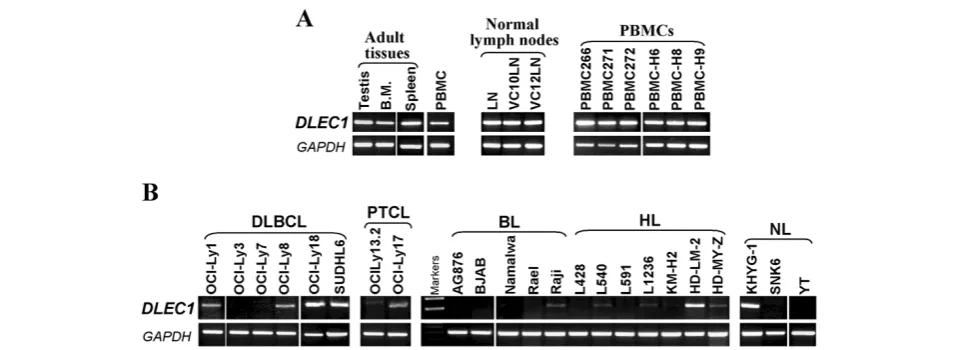
We first examined the expression of DLEC1 mRNA in 25 non-Hodgkin and Hodgkin lymphoma cell lines by
RT-PCR. DLEC1 was highly expressed in the normal
lymph node, PBMC samples, as well as human adult testis and bone marrow tissues (Figure 1A), but silenced or reduced in 33% (2/6) DLBCL, 50% (1/2) PTCL, 100% (5/5) BL, 86% (6/7) HL, and 67% (2/3) NL cell lines (Figure 1B), indicating that DLEC1 is a candidate TSG for lymphomas.
The promoter and exon 1 region ofDLEC1is a typical CpG island, with a T/C SNP site at CpG#29 (Figure 2A). MSP analysis showed thatDLEC1promoter methylation was frequently detected in 3/6 DLBCL, 1/2 PTCL, 5/5 BL, 7/7 (one weak) HL, and 2/3 NL cell lines (Figure 2B; Table 1), well correlated with its silencing or reduction. DLEC1methylation was further verified in detail by BGS analysis of 61 CpG sites within the CpG island. CpG sites ofDLEC1examined were heavily methylated in BL cell lines Rael, Raji, and in the HL cell line L428, but rarely in all normal PBMC samples, which confirmed the MSP data (Figure 2C). These results indicate that DLEC1 silencing by promoter methylation is a critical event in lymphomagenesis.
Silencing ofDLEC1could be reversed by pharmacologic demethylation
To further confirm whether promoter methylation med-iates the loss ofDLEC1expression in lymphoma, lymph-oma cell lines with methylated and reducedDLEC1were treated with the DNA methylation inhibitor, Aza, alone or combined with the HDAC inhibitor trichostatin A (TSA). After Aza treatment,DLEC1 expression was sig-nificantly induced in cell lines Rael, L428, L1236 and KM-H2. MSP analysis showed increased unmethylated alleles. Similar results were obtained using a combination treatment of Aza and TSA (Figure 3). Demethylation of the DLEC1 promoter was further confirmed by BGS. These results confirmed that promoter CpG methylation directly mediatesDLEC1silencing in lymphomas.
DLEC1is frequently methylated in primary lymphomas
We next investigated DLEC1 methylation in different types of primary lymphomas. Of 58 lymphoma tissues, DLEC1methylation was detected in 5/6 (83%) BL, 16/30 (53%, one weak) HL, 1/10 (10%) DLBCL, 6/8 (75%) NL, and 4/4 (100%, two weak) follicular lymphoma (FL) tumor samples, while no methylation was detected in normal lymph node samples (Figure 4A and B; Table 1).
Unmethylated bands were detected in all samples due to the inevitable inclusion of non-tumor cells in the analysis. Furthermore, the reduction or silencing ofDLEC1 expres-sion was observed in 6 out of 7 (86%) NL tumors as mea-sured by RT-PCR.
Furthermore as a pilot study, we examined DLEC1 methylation in serum samples of 13 HL patients with sera from 20 healthy individuals as controls.ANKRD30A is a normally methylated gene, thus used as an internal control for validating genomic DNA integrity of these samples, in addition to unmethylated DLEC1. Results showed that DLEC1 methylation was detected in 3/13 (23%) sera from HL patients (Figure 4D) but not in any normal sera.
Discussion
DLEC1 is located in the commonly deleted region 3p22-21.3 [3,18]. A cluster of TSGs, includingRASSF1A, BLU, and CACNA2D2, has been identified within this region. Silencing of RASSF1A, BLU, as well as DLEC1 by pro-moter CpG methylation had been extensively identified in multiple human cancers. Notably, promoter methylation of several 3p22-21.3 TSGs, such as RASSF1A and BLU, has also been identified in lymphomas [50,55], suggesting that TSGs in this region are frequently susceptible to epigenetic disruption during lymphoma pathogenesis.
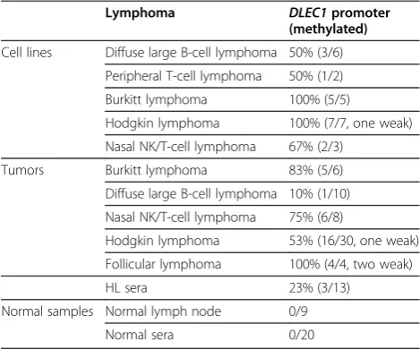
[image:4.595.304.539.111.306.2]Here, we report that DLEC1 is frequently silenced by promoter methylation both in lymphoma cell lines and primary lymphomas, but seldom in normal lymph nodes and PBMCs. Pharmacologic demethylation could reacti-vate DLEC1 expression, suggesting that epigenetic mechanism including DNA methylation and histone modification mediates DLEC1 transcriptional silencing. As primary tumor tissues usually contain infiltrating Table 1 Frequencies ofDLEC1methylation in various lymphoma tissues
Lymphoma DLEC1promoter
(methylated)
Cell lines Diffuse large B‑cell lymphoma 50% (3/6)
Peripheral T-cell lymphoma 50% (1/2)
Burkitt lymphoma 100% (5/5)
Hodgkin lymphoma 100% (7/7, one weak)
Nasal NK/T-cell lymphoma 67% (2/3)
Tumors Burkitt lymphoma 83% (5/6)
Diffuse large B‑cell lymphoma 10% (1/10)
Nasal NK/T‑cell lymphoma 75% (6/8)
Hodgkin lymphoma 53% (16/30, one weak)
Follicular lymphoma 100% (4/4, two weak)
HL sera 23% (3/13)
Normal samples Normal lymph node 0/9
non-malignant cells such as lymphocytes, unmethylated alleles were detected in all the tumor samples which also acting as an internal control for genomic DNA integrity.
DLEC1 unmethylated alleles and ANKRD30A
methy-lated alleles detected in different types of normal and
[image:5.595.59.538.88.294.2]lymphoma samples indicated the reliability of these sam-ples as shown before.DLEC1methylation showed a rela-tively low frequency in DLBCL samples, compared to other lymphoma types. The reason for this difference is not clear probably due to the special biologic features of Figure 3Pharmacologic demethylation restoredDLEC1expression in methylated/silenced lymphoma cell lines. A.Expression and methylation analyses ofDLEC1in HL cell lines with Aza treatment or combined with TSA (A+T). M: methylated; U: unmethylated.B.BGS analysis ofDLEC1methylation in lymphoma cell line KM-H2 before and after 5μM Aza treatment.
Figure 4DLEC1is frequently methylated in lymphoma tissues. A.MSP analysis ofDLEC1methylation in normal lymph node samples. Templates used for positive controls are: bisulfite treated DNA from HCT116 (+ve-control1) and HT-29 (+ve-control2) cell lines for methylation; SW480 (+ve-control1) and SNU16 (+ve-control2) cell lines for unmethylation.B.Representative analyses ofDLEC1methylation in primary lymphomas by MSP in primary lymphoma BL, DLBCL, HL and FL samples. BL: Burkitt lymphoma; DLBCL: diffuse large B-cell lymphoma; HL: Hodgkin lymphoma; NL: nasal NK/T-cell lymphoma; FL: follicular lymphoma.C.Representative analysis ofDLEC1expression in NL tumors.GAPDH
[image:5.595.58.539.432.655.2]DLBCL, which needs to be further confirmed by large sample size. Our results suggest that epigenetic silencing of DLEC1is important for lymphoma pathogenesis. Re-markably,DLEC1is specifically methylated in sera of HL patients, suggesting its potential as an epigenetic bio-marker for the non-invasive diagnosis of lymphomas.
Conclusions
This study identifies the frequent epigenetic inactivation of DLEC1 in various lymphomas and demonstrates its potential as a non-invasive tumor marker for the detec-tion of lymphomas. More molecular studies on the
tumor suppressive functions of DLEC1 in lymphoma
pathogenesis are needed.
Competing interests
The authors declare no conflict of interest.
Authors’contributions
ZW and LL analyzed data and drafted the manuscript. XS and ZW acquired data. ZG, GS, PGM and AR provided material and reviewed the manuscript. PGM contributed to the writing of the manuscript. QT conceived and supervised the study, analyzed data and finalized the manuscript. All authors read and approved the final manuscript.
Acknowledgments
This study was supported by National Natural Science Foundation of China (# 81071634 and 81172582), Shenzhen Science Fund for Distinguished Young Scholars (#JC201005270328A) the Leukaemia Lymphoma Research of the United Kingdom and the grants from The Chinese University of Hong Kong. We thank Dr. Riccardo Dalla-Favera for the DLBCL cell lines, Drs. Teresa Marafioti and (David Y Mason) for the L1236 cell line, Drs. Norio Shimizu for the SNK-6 lymphoma cell line.
Author details
1
Shenzhen Institutes of Advanced Technology (SIAT), Chinese Academy of Sciences (CAS)-CUHK, Shenzhen, China.2Cancer Epigenetics Laboratory,
Department of Clinical Oncology, State Key Laboratory of Oncology in South China, Sir YK Pao Center for Cancer, The Chinese University of Hong Kong and CUHK Shenzhen Research Institute, Shatin, Hong Kong.3Department of Pathology, Peking University Health Science Center, Beijing, China.
4
Department of Pathology, University of Hong Kong, Shatin, Hong Kong.
5Cancer Research UK Institute for Cancer Studies, University of Birmingham,
Birmingham, UK.6Johns Hopkins Singapore and Sidney Kimmel
Comprehensive Cancer Center, Johns Hopkins School of Medicine, Baltimore, MD, USA.
Received: 22 July 2012 Accepted: 4 October 2012 Published: 11 October 2012
References
1. Jones PA, Baylin SB:The epigenomics of cancer.Cell2007,128:683–692. 2. Baylin SB, Ohm JE:Epigenetic gene silencing in cancer - a mechanism for
early oncogenic pathway addiction?Nat Rev Cancer2006,6:107–116. 3. Hesson LB, Cooper WN, Latif F:Evaluation of the 3p21.3
tumour-suppressor gene cluster.Oncogene2007,26:7283–7301.
4. Cheng Y, Poulos NE, Lung ML, Hampton G, Ou B, Lerman MI, Stanbridge EJ:
Functional evidence for a nasopharyngeal carcinoma tumor suppressor gene that maps at chromosome 3p21.3.Proc Natl Acad Sci USA1998,
95:3042–3047.
5. Maestro R, Gasparotto D, Vukosavljevic T, Barzan L, Sulfaro S, Boiocchi M:
Three discrete regions of deletion at 3p in head and neck cancers.
Cancer Res1993,53:5775–5779.
6. Hibi K, Takahashi T, Yamakawa K, Ueda R, Sekido Y, Ariyoshi Y, Suyama M, Takagi H, Nakamura Y:Three distinct regions involved in 3p deletion in human lung cancer.Oncogene1992,7:445–449.
7. Pizzi S, Azzoni C, Bassi D, Bottarelli L, Milione M, Bordi C:Genetic alterations in poorly differentiated endocrine carcinomas of the gastrointestinal tract.Cancer2003,98:1273–1282.
8. Yang Q, Yoshimura G, Mori I, Sakurai T, Kakudo K:Chromosome 3p and breast cancer.J Hum Genet2002,47:453–459.
9. Acevedo CM, Henriquez M, Emmert-Buck MR, Chuaqui RF:Loss of heterozygosity on chromosome arms 3p and 6q in microdissected adenocarcinomas of the uterine cervix and adenocarcinoma in situ.
Cancer2002,94:793–802.
10. van den Berg A, Buys CH:Involvement of multiple loci on chromosome 3 in renal cell cancer development.Genes Chromosomes Cancer1997,
19:59–76.
11. Kimm LR, DeLeeuw RJ, Savage KJ, Rosenwald A, Campo E, Delabie J, Ott G, Muller-Hermelink H-K, Jaffe ES, Rimsza LM,et al:Frequent occurrence of deletions in primary mediastinal B-cell lymphoma.Genes Chromosomes Cancer2007,46:1090–1097.
12. Agathanggelou A, Cooper WN, Latif F:Role of the Ras-association domain family 1 tumor suppressor gene in human cancers.Cancer Res2005,
65:3497–3508.
13. Dammann R, Schagdarsurengin U, Seidel C, Strunnikova M, Rastetter M, Baier K, Pfeifer GP:The tumor suppressor RASSF1A in human carcinogenesis: an update.Histol Histopathol2005,20:645–663.
14. Agathanggelou A, Dallol A, Zochbauer-Muller S, Morrissey C, Honorio S, Hesson L, Martinsson T, Fong KM, Kuo MJ, Yuen PW,et al:Epigenetic inactivation of the candidate 3p21.3 suppressor gene BLU in human cancers.Oncogene2003,22:1580–1588.
15. Qiu GH, Tan LK, Loh KS, Lim CY, Srivastava G, Tsai ST, Tsao SW, Tao Q:
The candidate tumor suppressor gene BLU, located at the commonly deleted region 3p21.3, is an E2F-regulated, stress-responsive gene and inactivated by both epigenetic and genetic mechanisms in nasopharyngeal carcinoma.Oncogene2004,23:4793–4806.
16. Lerman MI, Minna JD:identification and evaluation of the resident candidate tumor suppressor genes. the international lung cancer chromosome 3p21.3 tumor suppressor gene consortium.Cancer Res
2000,60:6116–6133.
17. Ji L, Nishizaki M, Gao B, Burbee D, Kondo M, Kamibayashi C, Xu K, Yen N, Atkinson EN, Fang B,et al:Expression of several genes in the human chromosome 3p21.3 homozygous deletion region by an adenovirus vector results in tumor suppressor activities in vitro and in vivo.
Cancer Res2002,62:2715–2720.
18. Ji L, Minna JD, Roth JA:3p21.3 tumor suppressor cluster: prospects for translational applications.Future Oncol2005,1:79–92.
19. Hesson L, Bieche I, Krex D, Criniere E, Hoang-Xuan K, Maher ER, Latif F:
Frequent epigenetic inactivation of RASSF1A and BLU genes located within the critical 3p21.3 region in gliomas.Oncogene2004,23:2408–2419. 20. Wang Y, Yu Z, Wang T, Zhang J, Hong L, Chen L:Identification of
epigenetic aberrant promoter methylation of RASSF1A in serum DNA and its clinicopathological significance in lung cancer.Lung Cancer2007,
56:289–294.
21. Lai HC, Lin YW, Chang CC, Wang HC, Chu TW, Yu MH, Chu TY:
Hypermethylation of two consecutive tumor suppressor genes, BLU and RASSF1A, located at 3p21.3 in cervical neoplasias.Gynecol Oncol2007,
104:629–635.
22. Pan ZG, Kashuba VI, Liu XQ, Shao JY, Zhang RH, Jiang JH, Guo C, Zabarovsky E, Ernberg I, Zeng YX:High frequency somatic mutations in RASSF1A in nasopharyngeal carcinoma.Cancer Biol Ther2005,4:1116–1122. 23. Pizzi S, Azzoni C, Bottarelli L, Campanini N, D’Adda T, Pasquali C, Rossi G,
Rindi G, Bordi C:RASSF1A promoter methylation and 3p21.3 loss of heterozygosity are features of foregut, but not midgut and hindgut, malignant endocrine tumours.J Pathol2005,206:409–416. 24. Tomizawa Y, Iijima H, Nomoto T, Iwasaki Y, Otani Y, Tsuchiya S, Saito R,
Dobashi K, Nakajima T, Mori M:Clinicopathological significance of aberrant methylation of RARbeta2 at 3p24, RASSF1A at 3p21.3, and FHIT at 3p14.2 in patients with non-small cell lung cancer.Lung Cancer2004,
46:305–312.
25. Chow LS, Lo KW, Kwong J, To KF, Tsang KS, Lam CW, Dammann R, Huang DP:RASSF1A is a target tumor suppressor from 3p21.3 in
nasopharyngeal carcinoma.Int J Cancer2004,109:839–847.
26. Horiguchi K, Tomizawa Y, Tosaka M, Ishiuchi S, Kurihara H, Mori M, Saito N:
27. Cohen Y, Singer G, Lavie O, Dong SM, Beller U, Sidransky D:The RASSF1A tumor suppressor gene is commonly inactivated in adenocarcinoma of the uterine cervix.Clin Cancer Res2003,9:2981–2984.
28. Wagner KJ, Cooper WN, Grundy RG, Caldwell G, Jones C, Wadey RB, Morton D, Schofield PN, Reik W, Latif F, Maher ER:Frequent RASSF1A tumour suppressor gene promoter methylation in Wilms’tumour and colorectal cancer.Oncogene2002,21:7277–7282.
29. Liu L, Yoon JH, Dammann R, Pfeifer GP:Frequent hypermethylation of the RASSF1A gene in prostate cancer.Oncogene2002,21:6835–6840. 30. Hogg RP, Honorio S, Martinez A, Agathanggelou A, Dallol A, Fullwood P,
Weichselbaum R, Kuo MJ, Maher ER, Latif F:Frequent 3p allele loss and epigenetic inactivation of the RASSF1A tumour suppressor gene from region 3p21.3 in head and neck squamous cell carcinoma.Eur J Cancer
2002,38:1585–1592.
31. Dreijerink K, Braga E, Kuzmin I, Geil L, Duh FM, Angeloni D, Zbar B, Lerman MI, Stanbridge EJ, Minna JD,et al:The candidate tumor suppressor gene, RASSF1A, from human chromosome 3p21.3 is involved in kidney tumorigenesis.Proc Natl Acad Sci USA2001,98:7504–7509. 32. Lo KW, Kwong J, Hui AB, Chan SY, To KF, Chan AS, Chow LS, Teo PM,
Johnson PJ, Huang DP:High frequency of promoter hypermethylation of RASSF1A in nasopharyngeal carcinoma.Cancer Res2001,61:3877–3881. 33. Burbee DG, Forgacs E, Zochbauer-Muller S, Shivakumar L, Fong K, Gao B,
Randle D, Kondo M, Virmani A, Bader S,et al:Epigenetic inactivation of RASSF1A in lung and breast cancers and malignant phenotype suppression.J Natl Cancer Inst2001,93:691–699.
34. Chow LS, Lam CW, Chan SY, Tsao SW, To KF, Tong SF, Hung WK, Dammann R, Huang DP, Lo KW:Identification of RASSF1A modulated genes in nasopharyngeal carcinoma.Oncogene2006,25:310–316.
35. Daigo Y, Nishiwaki T, Kawasoe T, Tamari M, Tsuchiya E, Nakamura Y:
Molecular cloning of a candidate tumor suppressor gene, DLC1, from chromosome 3p21.3.Cancer Res1999,59:1966–1972.
36. Kwong J, Chow LS, Wong AY, Hung WK, Chung GT, To KF, Chan FL, Daigo Y, Nakamura Y, Huang DP, Lo KW:Epigenetic inactivation of the deleted in lung and esophageal cancer 1 gene in nasopharyngeal carcinoma.
Genes Chromosomes Cancer2007,46:171–180.
37. Ayadi W, Karray-Hakim H, Khabir A, Feki L, Charfi S, Boudawara T, Ghorbel A, Daoud J, Frikha M, Busson P, Hammami A:Aberrant methylation of p16, DLEC1, BLU and E-cadherin gene promoters in nasopharyngeal carcinoma biopsies from Tunisian patients.Anticancer Res2008,
28:2161–2167.
38. Kwong J, Lee JY, Wong KK, Zhou X, Wong DT, Lo KW, Welch WR, Berkowitz RS, Mok SC:Candidate tumor-suppressor gene DLEC1 is frequently downregulated by promoter hypermethylation and histone hypoacetylation in human epithelial ovarian cancer.Neoplasia2006,
8:268–278.
39. Seng TJ, Currey N, Cooper WA, Lee CS, Chan C, Horvath L, Sutherland RL, Kennedy C, McCaughan B, Kohonen-Corish MR:DLEC1 and MLH1 promoter methylation are associated with poor prognosis in non-small cell lung carcinoma.Br J Cancer2008,99:375–382.
40. Qiu GH, Salto-Tellez M, Ross JA, Yeo W, Cui Y, Wheelhouse N, Chen GG, Harrison D, Lai P, Tao Q, Hooi SC:The tumor suppressor gene DLEC1 is frequently silenced by DNA methylation in hepatocellular carcinoma and induces G1 arrest in cell cycle.J Hepatol2008,48:433–441. 41. Ying J, Poon FF, Yu J, Geng H, Wong AH, Qiu GH, Goh HK, Rha SY, Tian L,
Chan AT,et al:DLEC1 is a functional 3p22.3 tumour suppressor silenced by promoter CpG methylation in colon and gastric cancers.Br J Cancer
2009,100:663–669.
42. Zhang Q, Ying J, Li J, Fan Y, Poon FF, Ng KM, Tao Q, Jin J:Aberrant promoter methylation of DLEC1, a critical 3p22 tumor suppressor for renal cell carcinoma, is associated with more advanced tumor stage.
J Urol2010,184:731–737.
43. Park SY, Kwon HJ, Lee HE, Ryu HS, Kim SW, Kim JH, Kim IA, Jung N, Cho NY, Kang GH:Promoter CpG island hypermethylation during breast cancer progression.Virchows Arch2010,458:73–84.
44. Kang GH, Lee S, Cho NY, Gandamihardja T, Long TI, Weisenberger DJ, Campan M, Laird PW:DNA methylation profiles of gastric carcinoma characterized by quantitative DNA methylation analysis.Lab Invest2008,
88:161–170.
45. Zhang Y, Wang R, Song H, Huang G, Yi J, Zheng Y, Wang J, Chen L:
Methylation of multiple genes as a candidate biomarker in non-small cell lung cancer.Cancer Lett2011,303:21–28.
46. Ying J, Li H, Murray P, Gao Z, Chen YW, Wang Y, Lee KY, Chan AT, Ambinder RF, Srivastava G, Tao Q:Tumor-specific methylation of the 8p22 tumor suppressor gene DLC1 is an epigenetic biomarker for Hodgkin, nasal NK/T-cell and other types of lymphomas.Epigenetics2007,2:15–21. 47. Murray PG, Fan Y, Davies G, Ying J, Geng H, Ng KM, Li H, Gao Z, Wei W,
Bose S,et al:Epigenetic silencing of a proapoptotic cell adhesion molecule, the immunoglobulin superfamily member IGSF4, by promoter CpG methylation protects Hodgkin lymphoma cells from apoptosis.Am J Pathol2010,177:1480–1490.
48. Ying J, Li H, Chen YW, Srivastava G, Gao Z, Tao Q:WNT5A is epigenetically silenced in hematologic malignancies and inhibits leukemia cell growth as a tumor suppressor.Blood2007,110:4130–4132.
49. Ying J, Li H, Seng TJ, Langford C, Srivastava G, Tsao SW, Putti T, Murray P, Chan AT, Tao Q:Functional epigenetics identifies a protocadherin PCDH10 as a candidate tumor suppressor for nasopharyngeal, esophageal and multiple other carcinomas with frequent methylation.
Oncogene2006,25:1070–1080.
50. Murray PG, Qiu GH, Fu L, Waites ER, Srivastava G, Heys D, Agathanggelou A, Latif F, Grundy RG, Mann JR,et al:Frequent epigenetic inactivation of the RASSF1A tumor suppressor gene in Hodgkin’s lymphoma.Oncogene
2004,23:1326–1331.
51. Cheng Y, Geng H, Cheng SH, Liang P, Bai Y, Li J, Srivastava G, Ng MH, Fukagawa T, Wu X,et al:KRAB zinc finger protein ZNF382 is a proapoptotic tumor suppressor that represses multiple oncogenes and is commonly silenced in multiple carcinomas.Cancer Res2010,
70:6516–6526.
52. Jin H, Wang X, Ying J, Wong AH, Cui Y, Srivastava G, Shen ZY, Li EM, Zhang Q, Jin J,et al:Epigenetic silencing of a Ca(2+)-regulated Ras GTPase-activating protein RASAL defines a new mechanism of Ras activation in human cancers.Proc Natl Acad Sci USA2007,
104:12353–12358.
53. Tao Q, Robertson KD, Manns A, Hildesheim A, Ambinder RF:
The Epstein-Barr virus major latent promoter Qp is constitutively active, hypomethylated, and methylation sensitive.J Virol1998,72:7075–7083. 54. Tao Q, Swinnen LJ, Yang J, Srivastava G, Robertson KD, Ambinder RF:
Methylation status of the Epstein-Barr virus major latent promoter C in iatrogenic B cell lymphoproliferative disease. Application of PCR-based analysis.Am J Pathol1999,155:619–625.
55. Toujani S, Dessen P, Ithzar N, Danglot G, Richon C, Vassetzky Y, Robert T, Lazar V, Bosq J, Da Costa L,et al:High resolution genome-wide analysis of chromosomal alterations in Burkitt’s lymphoma.PLoS One2009,4:e7089.
doi:10.1186/1479-5876-10-209
Cite this article as:Wanget al.:Epigenetic silencing of the 3p22 tumor
suppressorDLEC1by promoter CpG methylation in non-Hodgkin and
Hodgkin lymphomas.Journal of Translational Medicine201210:209.
Submit your next manuscript to BioMed Central and take full advantage of:
• Convenient online submission
• Thorough peer review
• No space constraints or color figure charges
• Immediate publication on acceptance
• Inclusion in PubMed, CAS, Scopus and Google Scholar
• Research which is freely available for redistribution