This is a repository copy of
Aerial dissemination of Clostridium difficile spores
.
White Rose Research Online URL for this paper:
http://eprints.whiterose.ac.uk/7800/
Article:
Roberts, K., Smith, C.F., Snelling, A.M. et al. (4 more authors) (2008) Aerial dissemination
of Clostridium difficile spores. BMC Infectious Diseases, 8 (7). ISSN 1471-2334
https://doi.org/10.1186/1471-2334-8-7
eprints@whiterose.ac.uk https://eprints.whiterose.ac.uk/
Reuse
See Attached
Takedown
If you consider content in White Rose Research Online to be in breach of UK law, please notify us by
Open Access
Research article
Aerial Dissemination of
Clostridium difficile
spores
Katherine Roberts
1, Caroline F Smith
2, Anna M Snelling
3, Kevin G Kerr
2,4,
Kathleen R Banfield
2,4, P Andrew Sleigh
1and Clive B Beggs*
2Address: 1Pathogen Control Engineering Research Group, School of Civil Engineering, University of Leeds, Leeds, LS2 9JT, UK, 2School of
Engineering, Design and Technology, University of Bradford, Bradford, BD7 1DP, UK, 3Medical Biosciences, University of Bradford, Bradford, BD7
1DP, UK and 4Harrogate Health Care Trust, Harrogate District Hospital, Lancaster Park Road, Harrogate HG2 7SX, UK
Email: Katherine Roberts - ka_roberts2003@yahoo.co.uk; Caroline F Smith - c.f.smith3@bradford.ac.uk;
Anna M Snelling - a.m.snelling@bradford.ac.uk; Kevin G Kerr - kevin.kerr@hdft.nhs.uk; Kathleen R Banfield - kath.banfield@hdft.nhs.uk; P Andrew Sleigh - p.a.sleigh@leeds.ac.uk; Clive B Beggs* - c.b.beggs@bradford.ac.uk
* Corresponding author
Abstract
Background: Clostridium difficile-associated diarrhoea (CDAD) is a frequently occurring healthcare-associated infection, which is responsible for significant morbidity and mortality amongst elderly patients in healthcare facilities. Environmental contamination is known to play an important contributory role in the spread of CDAD and it is suspected that contamination might be occurring as a result of aerial dissemination of C. difficile spores. However previous studies have failed to isolate C. difficile from air in hospitals. In an attempt to clarify this issue we undertook a short controlled pilot study in an elderly care ward with the aim of culturing C. difficile from the air.
Methods: In a survey undertaken during February (two days) 2006 and March (two days) 2007, air samples were collected using a portable cyclone sampler and surface samples collected using contact plates in a UK hospital. Sampling took place in a six bedded elderly care bay (Study) during February 2006 and in March 2007 both the study bay and a four bedded orthopaedic bay (Control). Particulate material from the air was collected in Ringer's solution, alcohol shocked and plated out in triplicate onto Brazier's CCEY agar without egg yolk, but supplemented with 5 mg/L of lysozyme. After incubation, the identity of isolates was confirmed by standard techniques. Ribotyping and REP-PCR fingerprinting were used to further characterise isolates.
Results: On both days in February 2006, C. difficile was cultured from the air with 23 samples yielding the bacterium (mean counts 53 – 426 cfu/m3 of air). One representative isolate from each
of these was characterized further. Of the 23 isolates, 22 were ribotype 001 and were indistinguishable on REP-PCR typing. C. difficile was not cultured from the air or surfaces of either hospital bay during the two days in March 2007.
Conclusion: This pilot study produced clear evidence of sporadic aerial dissemination of spores of a clone of C. difficile, a finding which may help to explain why CDAD is so persistent within hospitals and difficult to eradicate. Although preliminary, the findings reinforce concerns that current C. difficile control measures may be inadequate and suggest that improved ward ventilation may help to reduce the spread of CDAD in healthcare facilities.
Published: 24 January 2008
BMC Infectious Diseases 2008, 8:7 doi:10.1186/1471-2334-8-7
Received: 25 September 2007 Accepted: 24 January 2008
This article is available from: http://www.biomedcentral.com/1471-2334/8/7 © 2008 Roberts et al; licensee BioMed Central Ltd.
BMC Infectious Diseases 2008, 8:7 http://www.biomedcentral.com/1471-2334/8/7
Background
Clostridium difficile-associated diarrhoea (CDAD) is a fre-quently occurring nosocomial infection, which is respon-sible for significant morbidity and mortality amongst elderly patients in healthcare facilities. In many countries the incidence of CDAD seems to be increasing and a toxin-hyperproducing strain (ribotype 027) is becoming more common [1,2], despite the fact that many hospitals have made stringent efforts to control the infection through isolation of infected patients, improved compli-ance with handwashing and decontamination of the ward environment. Several studies have suggested that environ-mental contamination, particularly of fomites, may play a role in the spread of CDAD [3-5]. Nosocomial outbreaks can occur in spatial clusters [6], suggesting that physical proximity to infected patients might be an important risk
factor for acquisition of C. difficile [7]. This has led to
sus-picion that environmental contamination might be
occur-ring as a result of aerial dissemination of C. difficile spores.
The latter can survive on inanimate surfaces for months, and inactivating them is problematic since they are rela-tively resistant to disinfectants. Indeed, sporulation can actually be enhanced by exposure of cells to some types of cleaning agent [8]. Processes such as bed making are known to liberate large numbers of bacteria-carrying par-ticles into the air [9-11] and it would appear a reasonable
assumption that aerial dissemination of C. difficile
vegeta-tive cells and spores occurs in the same manner. However,
previous studies have failed to isolate C. difficile from air
in hospitals [3,12-14]. In an attempt to clarify this issue we undertook a short pilot study in an elderly care ward and orthopaedic ward at a 400-bedded district general
hospital, with the aim of culturing C. difficile from the air.
Methods
The first phase of the study was undertaken on the 21st
(day 1) and 22nd February 2006 (day 2). Sampling took
place in a mechanically ventilated six-bedded bay (Figure 1a) on a 29 bed elderly care ward which had sporadic cases of CDAD (four confirmed cases in 2005, and a fur-ther five confirmed cases up to March 2007). Sampling
was repeated on this bay 12 months later on the 1st (day
3) and 2nd (day 4) of March 2007. A naturally ventilated
four-bedded bay (Figure 1b) on a 15 bed orthopaedic ward, in which no CDAD cases had been reported in the previous year, was sampled concurrently during this sec-ond phase.
Ward air was sampled using a portable cyclone air sam-pler (Burkhard C90M, Burkhard Ltd, UK) located, in the elderly care ward, outside the entrance to the patient's toi-let room (positioned on a metal trolley, 870 mm above floor level at approximately bed height) and, in the ortho-paedic ward, on the window sill. During each two-day period, air samples were taken at 30-minute intervals throughout the day. For each sampling, 250 L of air was drawn into the device over 15 min. At the end of each day
air samplers were cleaned with Virkon® disinfectant
Floor plan of the study bay (a), with six open bed spaces separated by curtains (dashed lines) and the control bay (b) which contained four open bed spaces also separated by curtains
Figure 1
Floor plan of the study bay (a), with six open bed spaces separated by curtains (dashed lines) and the control bay (b) which contained four open bed spaces also separated by curtains. The 'X' indicates where the air sampling equipment was positioned. T = toilet.
Sink
X
Window Sill
C
B D
Corridor
A Windows
X
A
B
C D
E F
Nurses
(DuPont) which is known to be active against both C. dif-ficile spores and vegetative cells. When in use, the devices were observed closely by the operator to ensure patients and staff did not touch them or interfere with their opera-tion in any way.
Particulate matter from the air was collected into 1 mL of sterile Ringer's solution in Eppendorf tubes. Specimens were alcohol shocked [15] by mixing with an equal vol-ume of absolute ethanol. The solution was then vortexed and held for 1 h at room temperature after which 0.1 mL aliquots were plated out in triplicate onto Brazier's CCEY agar (Lab M Ltd, Bury, UK) without egg yolk, but supple-mented with 5 mg/L of lysozyme (Sigma-Aldrich, Poole,
Dorset, UK) to optimise the recovery of C. difficile [16].
Any alcohol present was allowed to evaporate and the plates were then incubated at 37°C anaerobically for 48 h.
The identity of isolates with morphology typical of C.
dif-ficile was confirmed using Gram staining and a commer-cially available latex agglutination test (Oxoid Ltd, Basingstoke, UK) which detects toxin A. Mean colony counts per cubic metre of air were calculated.
Environmental samples were taken using Clostridium
diff-icile contact plates (E & O Laboratories, UK). The sites selected for testing were disinfected at the beginning of the day and after each sample was taken. In the study ward, samples were taken from the floor, top of the radia-tor, top of the ward door, a ventilation extract grille above bed C (see Figure 1a) and a ventilation extract grille above the nurses' station. On the control ward, samples were taken from the floor, the top of the ward door and the shelf adjacent to the hand-wash sink. Each site was screened at three set times during the day using two con-tact plates each time. Plates were incubated under anaero-bic conditions at 37°C for 48 h.
Ribotyping [17] and repetitive extragenic palindromic REP-PCR typing [18] were used to compare the
related-ness of selected isolates. C. difficile NCTC 11209 was used
as a control for the DNA extraction and purification
proc-ess. Reference strains of C. difficile ribotypes 001, 002,
010, 014, 027 and 106 were included as standards. Strains were cultured in Brain Heart Infusion Broth supple-mented with 0.5% yeast extract and 0.5 mg of haemin, incubated anaerobically at 37°C for 24 h. Cells from 1.5 mL of culture were pelleted by centrifugation. DNA was
extracted using the GenEluteTM Bacterial Genomic DNA
kit (Sigma-Aldrich).
PCR ribotyping was performed based on a method described previously [17] using primers 5'-CTGGGGT-GAAGTCGTAACAAGG-3' and
5'-GCGCCCTTTGTAGCTT-GACC-3'. A 25 μl reaction mix contained 1× Thermopol II
Buffer (New England Biolabs), 200 mM of each dNTP,
2.25 mM of MgSO4, 50 pmol of each primer
(Sigma-Genosys), 2.5 units of Taq polymerase (New England
Biolabs) and 2.5 μl of DNA template. The reaction mix
was made up to 25 μL using molecular grade water
(Sigma-Aldrich). Thirty-five cycles of amplification were carried out, consisting of 1 min at 94°C, 1 min at 55°C and 2 min at 72°C. Amplimers were resolved on a 2% agarose gel (Invitrogen). After ethidium bromide staining, banding patterns were compared visually.
REP primers REP1R-I, 5'-IIIICGICGICATCIGGC-3' and REP2-I, 5'-ICGICTTATCIGGCCTAC-3' were used in a PCR reaction based on a previously described method for typ-ing C. difficile [18] with minor modifications. A 25 μL reaction mix contained 1× Thermopol II Buffer (New
Eng-land Biolabs), 200 μM of each dNTP, 2.5 mM of MgSO4,
25pmol of each primer (Sigma-Genosys), 2 units Taq
polymerase (New England Biolabs), 100 μg/mL bovine
serum albumin (Promega) and DNA template (60 ng).
The reaction mix was made up to 25 μL using molecular
grade water (Sigma-Aldrich). Initial denaturation was 2 min at 95°C and 3 secs at 94°C. Thirty cycles of amplifi-cation were then carried out, consisting of 30 secs at 92°C, 1 min at 40°C and 2 min at 65°C. The final extension was for 15 min at 65°C. Amplimers were resolved on a 1.5% agarose gel. After ethidium bromide staining, banding patterns were compared visually.
The work described in this paper was originally under-taken as part of enhanced environmental surveillance as contained in Harrogate Health Care Trust's Infection Con-trol Annual Plan ratified by the Trust Board. However, when it became clear that the surveillance yielded results of wider interest which might merit publication, the Trust's Research Governance Committee approved a request to submit.
Results
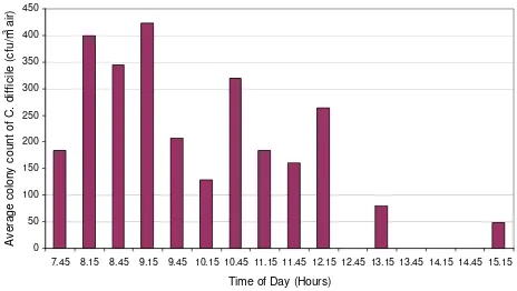
The results of the first phase of air sampling are presented in Figures 2 and 3. From these it can be seen that on both
days C. difficile was cultured from the air of the elderly care
bay, with counts of 53 – 426 cfu/m3 of air, indicating that
substantial numbers of C. difficile-spores were liberated
into the ward air throughout this period. The limit of
detection for the method was 27 cfu/m3 – this being the
count that would have been recorded if a single colony was observed amongst the triplicate agar plates (1 colony arising from 0.3 ml of the alcohol shock solution = 6.67 cfu from the 250 L of air sampled, which in turn
approxi-mates to 27 per m3).
It should be noted that no sample was collected at 09.45
on day 1. Twenty-three air samples in total yielded C.
BMC Infectious Diseases 2008, 8:7 http://www.biomedcentral.com/1471-2334/8/7
belonged to ribotype 001 (Figure 4). All of these had the same REP-PCR profile, but the pattern was different to that of the ribotype 001 reference strain (data not shown). The ribotype of the remaining isolate did not match any of the reference strains.
During the phase 1 survey, C. difficile was not isolated
from the surfaces sampled in the elderly care ward. During
the second phase of sampling, C. difficile was not cultured
from air samples or isolated from environmental sites in both the study and control bays.
Discussion
Earlier researchers, possibly because of sub-optimal recov-ery methods, such as the use of settle plates [3,12-14],
failed in attempts to culture C. difficile from the air of
hos-pital wards thus lending weight to the generally held opin-ion that airborne transmissopin-ion of the bacterium is
unimportant [12,19]. However, there is increasing evi-dence that airborne dissemination may play a role in the
spread of C. difficile within the clinical environment. For
example, air vents and high horizontal surfaces have been
noted to be contaminated with C. difficile, [5,20]
suggest-ing dissemination via the air. Furthermore, other studies
have found C. difficile on patients' bedding [12-14]. As
bed making is known to liberate large numbers of bacte-ria-carrying particles into the air [9-11], these
observa-tions would suggest that C. difficile may be disseminated
into the air by this route following these activities. We
were able to isolate C. difficile spores from the air on two
separate days, and to our best knowledge, this is the first report to suggest aerial dissemination of this bacterium within a hospital.
The cyclone air sampler was located outside the toilet and it is possible that many of the isolates recovered from the air originated from this area. Previous work [13,14] has found surfaces within bathrooms and toilets to be among the most contaminated areas within hospitals, which is
not surprising given that C. difficile colonises the colon.
However, during the two sampling days no one was observed using the toilet as many of the patients were bed-bound. Notwithstanding this, the patient in bed D (clos-est to the air sampler; Figure 1) did have diarrhoea during the sampling period and used a commode on several
occasions during day 2. A stool sample was negative for C.
difficile toxin A (at tha t time the laboratory did not test for toxin B production) but this does not rule out the possi-bility of asymptomatic gut carriage with the bacterium [21-24]. Indeed, asymptomatic carriers are recognised as a potential cause of environmental contamination [21,22].
PCR ribotype profiles of C. difficile isolates CAS 1 to 10 (lanes 1–10) obtained from air samples, Lane R = ribotype 001 ref-erence strain
Figure 4
PCR ribotype profiles of C. difficile isolates CAS 1 to 10 (lanes 1–10) obtained from air samples, Lane R = ribotype 001 ref-erence strain. Lane M = O'GeneRuler™ DNA ladder (10 kbp – 100 bp).
M 1 2 3 4 5 6 M 7 8 9 10 R M
[image:5.612.317.550.91.247.2]Mean C. difficile counts in the air of the study ward on day 1
Figure 2
Mean C. difficile counts in the air of the study ward on day 1. (NB. a value of zero denotes 'below the detection limit of 27 cfu/m3'; NDC denotes 'no data collection').
0 50 100 150 200 250 300 350 400 450
7.45 8.15 8.45 9.15 9.45 10.15 10.45 11.15 11.45 12.15 12.45 13.15 13.45 14.15 14.45 15.15
Time of Day (Hours)
A v erage c ol ony c ount of C . di ff ic ile ( c fu /m
3 ai
r)
NDC
Mean C. difficile counts in the air of the study ward on day 2 (NB. a value of zero denotes 'below the detection limit of 27cfu/m3'
Figure 3
Mean C. difficile counts in the air of the study ward on day 2 (NB. a value of zero denotes 'below the detection limit of 27cfu/m3'.
Figure 3 0 50 100 150 200 250 300 350 400 450
7.45 8.15 8.45 9.15 9.45 10.15 10.4511.15 11.45 12.15 12.45 13.15 13.45 14.15 14.45 15.15
Time of Day (Hours)
A v e rage c o lony c o u n t of C . di ff ic ile (c fu /m
3 ai
[image:5.612.61.293.539.670.2]Before the two sampling days in February 2006, the last time a patient with confirmed CDAD was on the ward was seven weeks earlier and the next case was four weeks later.
This shows that C. difficile can be isolated from the air in
the absence of a confirmed case/outbreak of CDAD.
The results of the ribo- and REP-PCR-typing indicate that all but one of the isolates found in the air were clonal and may have come from the same source. Earlier studies have
shown that some strains of C. difficile are more likely to
contaminate the local environment than others [5,25] and the 001 ribotype cultured in this study is currently the
most common strain of C. difficile in the UK.
Very small aerosol particles can remain airborne for long
periods, for example, a 2 μm diameter particle will take
4.2 hours to fall 2 m in a still room [26]. Spores of C.
dif-ficile are of this size range [27] and may thus become widely distributed around the clinical environment. Aer-ial dissemination of desiccation-tolerant microorganisms
such as Acinetobacter is also known to result in widespread
environmental contamination [28,29].
Our data give an interesting insight into the physical
nature of the C. difficile aerosolization and dispersal
within the ward. Aerosol particles can be removed from room air by two principal mechanisms, gravitational dep-osition and extraction via exhaust ventilation. The resi-dence time of true airborne particles in a well-ventilated
ward space is generally ≤ 30 min, depending on the
venti-lation rate. Therefore, in such a space most of the airborne particles will be purged from the air within 30 min of any aerosol liberation event. From Figures 2 and 3, it can be seen that, despite some significant fluctuations, large numbers of particles were found in the air at almost all of
the sampling points suggesting that numerous C. difficile
aerosolization events occurred throughout the sampling period and also that the cyclone sampler was located close
to the C. difficile dissemination source. It also indicates
that during this period the ventilation system was unable
to purge the ward air of C. difficile-bearing particles.
Con-tinuous observation of the device ensured that no inad-vertent direct contamination (e.g. from staff/patient hands or contact with other equipment) occurred during the sampling period.
Although the study reported here was only a short pilot
study, it produced evidence of aerial dissemination of C.
difficile, a phenomenon which may, at least in part, explain why CDAD is so persistent within hospitals and difficult to eradicate. It also demonstrates the transient nature of the airborne route of dissemination, since on both sampling days in phase one there were periods
where the spore count per m3 of air was below the limit of
detection. This has consequences for scheduling of air
sampling in future studies of aerial dissemination of C.
difficile.
It is surprising that no environmental specimens yielded C difficile, even on days when the bacterium was cultured from the air. This may be because we relied on 65 mm diameter contact plates. Sampling of larger areas using a
moistened swab may have resulted in a greater yield of C.
difficile [30]. Timing of sampling may also be important as sampling after ward cleaning may influence the likeli-hood of recovering the bacterium [30]. Furthermore, the inclusion of an agent, such as lysozyme which encourages germination of spores [16] or extending the time of incu-bation of cultures might have increased recovery. These issues will be addressed in future investigations.
Conclusion
The study produced clear evidence of sporadic aerial
dis-semination of spores of a clone of C. difficile, a finding
which may help to explain why CDAD is so persistent within hospitals and difficult to eradicate. As such, our report is timely because it coincides with concerns that
current C. difficile control measures are failing to halt the
spread of CDAD. Hopefully, it will encourage others to undertake aerobiological sampling in their own hospitals,
so that a proper evaluation of the extent to which C.
diffi-cile is being disseminated via the air can be made. If air-borne dissemination is a contributory factor to environmental contamination, then the use of negatively-pressurized isolation rooms and improved ward ventila-tion systems may help to reduce the spread of CDAD in healthcare facilities and these interventions warrant urgent evaluation.
Competing interests
The author(s) declare that they have no competing inter-ests.
Authors' contributions
AMS, KGK and CBB designed the study. KR and CFS were responsible for sample collection and laboratory analysis. KRB and KGK supervised the clinical aspects of the study. KGK and AMS supervised the microbiological sampling and analysis. PAS advised on the air sampling procedures and airborne particulate behaviour. CBB wrote the manu-script with major contributions from other authors. All authors read and approved the final manuscript.
Acknowledgements
This work has been sponsored by grants from the EPSRC (Ref. GR/S48462/ 01), Department of Health (Ref. B(02)09) and Arup. The authors wish to thank these organisations for their support. The authors also wish to thank Dr Ed J Kuijper, Céline Harmanus and Renate van den Berg from the Department of Medical Microbiology, National Reference Laboratory for
Publish with BioMed Central and every scientist can read your work free of charge
"BioMed Central will be the most significant development for disseminating the results of biomedical researc h in our lifetime."
Sir Paul Nurse, Cancer Research UK
Your research papers will be:
available free of charge to the entire biomedical community
peer reviewed and published immediately upon acceptance
cited in PubMed and archived on PubMed Central
yours — you keep the copyright
Submit your manuscript here:
http://www.biomedcentral.com/info/publishing_adv.asp
BioMedcentral
BMC Infectious Diseases 2008, 8:7 http://www.biomedcentral.com/1471-2334/8/7
References
1. Warny M, Pepin J, Fang A, Killgore G, Thompson A, Brazier J, Frost E, McDonald LC: Toxin production by an emerging strain of Clostridium difficile associated with outbreaks of severe dis-ease in North America and Europe. Lancet 2005,
366(9491):1079-1084.
2. HPA: Clostridium difficile: Findings and recommendations from a review of the epidemiology and a survey of Directors of Infection Prevention and Control in England. Health Pro-tection Agency; 2006.
3. Malamou-Ladas H, O'Farrell S, Nash JQ, Tabaqchali S: Isolation of Clostridium difficile from patients and the environment of hospital wards. J Clin Pathol 1983, 36(1):88-92.
4. Hota B: Contamination, disinfection, and cross-colonization: are hospital surfaces reservoirs for nosocomial infection? Clin Infect Dis 2004, 39(8):1182-1189.
5. Fawley WN, Wilcox MH: Molecular epidemiology of endemic Clostridium difficile infection. Epidemiol Infect 2001,
126(3):343-350.
6. Foulke GE, Silva J Jr.: Clostridium difficile in the intensive care unit: management problems and prevention issues. Crit Care Med 1989, 17(8):822-826.
7. Chang VT, Nelson K: The role of physical proximity in nosoco-mial diarrhea. Clin Infect Dis 2000, 31(3):717-722.
8. Wilcox MH, Fawley WN: Hospital disinfectants and spore for-mation by Clostridium difficile. Lancet 2000, 356(9238):1324. 9. Roberts K, Hathway A, Fletcher LA, Beggs CB, Elliott MW, Sleigh PA:
Bioaerosol production on a respiratory ward. Indoor and Built Environment 2006, 15:35-40.
10. Greene VW, Vesley D, Bond RG, Michaelsen GS: Microbiological contamination of hospital air. I. Quantitative studies. Appl Microbiol 1962, 10:561-566.
11. Greene VW, Vesley D, Bond RG, Michaelsen GS: Microbiological contamination of hospital air. II. Qualitative studies. Appl Microbiol 1962, 10:567-571.
12. Fekety R, Kim KH, Batts DH, Browne RA, Cudmore MA, Silva J Jr., Toshniwal R, Wilson KH: Studies on the epidemiology of antibi-otic-associated Clostridium difficile colitis. Am J Clin Nutr 1980,
33(11 Suppl):2527-2532.
13. Fekety R, Kim KH, Brown D, Batts DH, Cudmore M, Silva J Jr.: Epi-demiology of antibiotic-associated colitis; isolation of Clostridium difficile from the hospital environment. Am J Med
1981, 70(4):906-908.
14. Kim KH, Fekety R, Batts DH, Brown D, Cudmore M, Silva J Jr., Waters D: Isolation of Clostridium difficile from the environ-ment and contacts of patients with antibiotic-associated col-itis. J Infect Dis 1981, 143(1):42-50.
15. Borriello SP, Honour P: Simplified procedure for the routine isolation of Clostridium difficile from faeces. J Clin Pathol 1981,
34(10):1124-1127.
16. Wilcox MH, Fawley WN, Parnell P: Value of lysozyme agar incor-poration and alkaline thioglycollate exposure for the envi-ronmental recovery of Clostridium difficile. J Hosp Infect 2000,
44(1):65-69.
17. O'Neill GLO, Ogunsola FT, Brazier JS: Modification of a PCR Ribotyping Method for Application as a Routine Typing Scheme for Clostridium difficile. Anaerobe 1996, 2:205-209. 18. Rahmati A, Gal M, Northey G, Brazier JS: Subtyping of
Clostrid-ium difficile polymerase chain reaction (PCR) ribotype 001 by repetitive extragenic palindromic PCR genomic finger-printing. J Hosp Infect 2005, 60(1):56-60.
19. Kaatz GW, Gitlin SD, Schaberg DR, Wilson KH, Kauffman CA, Seo SM, Fekety R: Acquisition of Clostridium difficile from the hos-pital environment. Am J Epidemiol 1988, 127(6):1289-1294. 20. Fawley WN, Freeman J, Wilcox MH: Evidence to support the
existence of subgroups within the UK epidemic Clostridium difficile strain (PCR ribotype 1). J Hosp Infect 2003, 54(1):74-77. 21. Kato H, Kita H, Karasawa T, Maegawa T, Koino Y, Takakuwa H, Saikai T, Kobayashi K, Yamagishi T, Nakamura S: Colonisation and trans-mission of Clostridium difficile in healthy individuals exam-ined by PCR ribotyping and pulsed-field gel electrophoresis.
J Med Microbiol 2001, 50(8):720-727.
22. Delmee M: Laboratory diagnosis of Clostridium difficile dis-ease. Clin Microbiol Infect 2001, 7(8):411-416.
23. Clabots CR, Gerding SJ, Olson MM, Peterson LR, Gerding DN:
Detection of asymptomatic Clostridium difficile carriage by
an alcohol shock procedure. J Clin Microbiol 1989,
27(10):2386-2387.
24. Kyne L, Warny M, Qamar A, Kelly CP: Asymptomatic carriage of Clostridium difficile and serum levels of IgG antibody against toxin A. N Engl J Med 2000, 342(6):390-397.
25. Samore MH, Venkataraman L, DeGirolami PC, Arbeit RD, Karchmer AW: Clinical and molecular epidemiology of sporadic and clustered cases of nosocomial Clostridium difficile diarrhea.
Am J Med 1996, 100(1):32-40.
26. Beggs CB: The airborne transmission of infection in hospital buildings: Fact or fiction? Indoor and Built Environment 2003, 12(1-2):9-18.
27. Panessa-Warren BJ, Tortora GT, Warren JB: Exosporial mem-brane plasticity of Clostridium sporogenes and Clostridium difficile. Tissue Cell 1997, 29(4):449-461.
28. Allen KD, Green HT: Hospital outbreak of multi-resistant Aci-netobacter anitratus: an airborne mode of spread? J Hosp Infect 1987, 9(2):110-119.
29. Beggs CB, Kerr KG, Snelling AM, Sleigh PA: Acinetobacter spp. and the clinical environment. Indoor and Built Environment 2006,
15(1):19-24.
30. Wilcox MH, Fawley WN, Wigglesworth N, Parnell P, Verity P, Free-man J: Comparison of the effect of detergent versus hypochlo-rite cleaning on environmental contamination and incidence of Clostridium difficile infection. J Hosp Infect 2003,
54(2):109-114.
Pre-publication history
The pre-publication history for this paper can be accessed here: