Redefining the role of tumour necrosis factor in macrophage differentiation and effector function in bacterial and tumour defences
Xinying Li
Submitted in fulfilment of the requirements for the degree of Doctor of Philosophy
University of Tasmania
Declaration
"This thesis contains no material which has been accepted for a degree or diploma by the University or any other institution, except by way of background information and duly
acknowledged in the thesis, and to the best of my knowledge and belief no material previously published or written by another person except where due acknowledgement is
made in the text of the thesis, nor does the thesis contain any material that infringes copyright.”
Authority of Access
This thesis may be made available for loan and limited copying and communication in accordance with the Copyright Act 1968.
Statement of Ethical Conduct
“The research associated with this thesis abides by the international and Australian codes on human and animal experimentation, the guidelines by the Australian Government's Office of
the Gene Technology Regulator and the rulings of the Safety, Ethics and Institutional Biosafety Committees of the University.”
Publications
Part of the work contained in this thesis has been published or submitted for publication as detailed below:
Chapter 3 is based on the published paper
Li, X., Lyons, A. B., Woods, G. M., & Körner, H. (2017). The absence of TNF permits myeloid Arginase 1 expression in experimental L. monocytogenes infection. Immunobiology, 222(8), 913-917.
XL performed the laboratory analysis, prepared tables and figures, drafted and refined the manuscript.
Chapter 5 is based on the ‘to be submitted' paper
Li, X., Körner. H, Darby. J, Lyons, A.B. Woods, G.M. TNF signalling downregulates phagocytosis of tumour cells by activated macrophages, to be submitted.
XL performed the laboratory analysis, prepared tables and figures, drafted and refined the manuscript.
Conference presentations
Part of the work contained in this thesis has been presented at national and international conferences as detailed below:
Xinying Li, A. Bruce Lyons, Heinrich Korner and Greg Woods.2016. Murine Macrophage Phagocytosis of Devil Facial Tumour Disease Cells. ICI 2016 International Congress of Immunology. 2016, Melbourne, Poster presentation.
Table of contents
Acknowledgements……….………..6
Abstract ………....…....8
Chapter 1. Literature review……….10
Aims of thesis……….……….………37
Chapter 2. General Materials and Methods………39
Chapter 3. Lack of TNF leads to alternative activation in splenic macrophages during L. monocytogenes infection……….………61
Chapter 4. Role of TNF in alternative activation of peritoneal macrophages following L. monocytogenes infection………78
Chapter 5. Role of TNF in macrophage phagocytosis of L. monocytogenes and tumour cells………..93
Chapter 6. Final discussion………..……...108
References……….117
Appendix 1: Published articles ……….……….131
Acknowledgements
I would like to express my appreciation to my primary supervisor Heinrich Korner, for giving me the opportunity to study overseas and providing mentorship over the past 4 years. Thank you for allowing me to think independently which is essential in my life, and also for providing me with scientific guidance in the beginning of my academic life.
I am very grateful to my co-supervisor Gregory Woods, who provided me with great supervision. Thank you for the encouragement which always helped me to overcome difficulties in my research. Thank you for the assistance and advice throughout my project. I appreciate your wisdom, enthusiasm, understanding and pushing me farther than I thought I could go. Your comments on balance between lab work and academic writing helped me to overcome some bad moments in the beginning of my studies.
Thank you to my co-supervisor Bruce Lyons for all the mentorship, professional advice, and support on every aspect of my project. Thank you for all the quick feedback and comments, thank you for always being there when I needed support. I am very appreciative of all the support you provided to me.
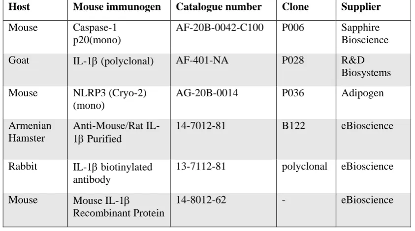
The inflammasome section in Chapter 3 came from the contribution of Kate Schroder and Jennifer Dou with the antibody staining in western blot analysis. Thank you Kate Schroder, the researcher of Institute for Molecular Bioscience in University of Queensland. Thank you for the insightful comments on the experimental design and data interpretation in the investigation of inflammasome. Thank you to Jennifer Dou, the research assistant in Kate Schroder’s group. Thank you very much for the laboratory performance of antibody staining of western blot.
Abstract
Chapter 1
Chapter 1: Literature review
1.1 Tumour necrosis factor ... 13
1.1.1 TNF in pathology... 13
1.1.2 TNF expression ... 13
1.1.3 TNF signal transduction ... 14
1.1.3.1 Activation of the NF-κB and MAPK pathways... 14
1.1.3.2 Induction of apoptosis and necrosis... 14
1.1.4 TNF and macrophages ... 15
1.2 Macrophages ... 16
1.2.1 Tissue resident macrophage origins ... 17
1.2.1.1 Splenic macrophages ... 18
1.2.1.2 Resident macrophages in peritoneum and central nervous system ... 18
1.2.2 Molecules related to monocyte development ... 20
1.2.3 Monocyte heterogeneity in mouse ... 21
1.2.4 Monocyte migration in infection ... 21
1.2.5 Macrophage polarization and functional phenotypes ... 22
1.2.5.1 Classically activated macrophages ... 22
1.2.5.2 Alternatively activated macrophages ... 24
1.2.5.3 Macrophage and cancer ... 25
1.2.6 Macrophage phagocytosis ... 26
1.2.7 The inflammasome ... 27
1.2.7.1 The NLRP3 inflammasome ... 28
1.2.7.2 The NLRC4 inflammasome ... 29
1.2.7.3 The AIM2 inflammasome ... 29
1.2.7.4 Inflammasome and human disease ... 30
1.3 The intracellular bacterium- Listeria monocytogenes ... 31
1.3.2 Macrophage responses during L. monocytogenes infection ... 32
1.3.3 Inflammasome activation in L. monocytogenes infection ... 33
1.4 Devil facial tumour disease ... 34
1.4.1 Pathology and origins of DFTD ... 34
1.4.2 The devil’s immune system ... 35
1.4.3 Low genetic diversity of devils ... 35
1.4.4 Immunosuppressive Cytokines ... 36
1.4.5 Macrophages and devils ... 37
1.5 Final remarks ... 37
Chapter 1: Literature review 1.1 Tumour necrosis factor
1.1.1 TNF in pathology
Tumour necrosis factor α (TNF) is an important inflammatory cytokine, which was first named in 1975 for its ability to induce tumour cell hemorrhagic necrosis in mice [5]. TNF has also been reported to play a role in a central endogenous mediator of endotoxic shock [5]. Additionally, as a pro-inflammatory cytokine, TNF is essential in initiation and resolution of inflammation. Overproduction of TNF is involved in many inflammatory diseases. Studies in TNF- or TNFR- deficient mice have indicated the essential role of TNF in host defence against bacterial and viral infections [5]. TNF is synthesised rapidly after infection and orchestrates pro-inflammatory cytokine production, which suggests its importance in the development of many inflammatory diseases including Rheumatoid arthritis (RA) and Crohn’s disease [6]. For instance, high concentrations of TNF have been found in the joints and synovial fluid from RA patients [7]. Thus, drugs targeting TNF have been developed for treatment of RA and Crohn’s disease. These include anti-TNF antibody (Remicade), and anti-TNF receptors Fc fusion protein (Etanercept, Lenercept) [8].
1.1.2 TNF expression
1.1.3 TNF signal transduction
TNF acts through binding with the receptors, TNF receptor 1 (TNFR1) and TNF receptor 2 TNFR2, in a trimeric manner [6]. TNFR1 and TNFR2 are expressed in most mammalian cells [17]. TNF binds to TNFR1 with higher affinity than TNFR2 [18], suggesting the predominant role of TNFR1 in TNF induced cellular responses. The trigger of TNF signaling pathway leads to various responses, including cell survival, apoptosis and necrosis.
1.1.3.1 Activation of the NF-κB and MAPK pathways
Following TNF binding, TNFR1 provides a docking site and interacts with the TNF receptor associated death domain (TRADD) [5]. TRADD then recruits TNFR-associated factors (TRAFs), which include TRAF2, TRAF5, receptor interacting protein-1 (RIP-1) and cellular inhibitor of apoptosis proteins 1 and 2 (cIAP-1 and 2), to form the TNF receptor signalling complex [5]. TRAF2 and TRAF5 double knockout cells demonstrated that TRAF2 and TRAF5 are required in TNF-induced NF-κB activation [19]. RIP-1 is a unique serine/threonine kinase with a death domain in the C-terminus [20]. The recruitment of RIP1 and TRAF2 are independent during the process of forming the signalling complex [21]. In addition, the lipid rafts provide the micro domain for the ubiquitylation of TNFR1 and RIP-1, as inhibition of the organization of lipid rafts impaired the NF-κB activation [22]. After formation of the TNF receptor signalling complex, RIP1 is polyubiquitinated by cIAP-1 and cIAP-2. This triggers activation of TGF-β activated kinase (TAK1) [22]. TAK1 then activates NF-κB which leads to NF-κB translocation into the cell nucleus, where it activates the transcription of genes such as cytokines, chemokines, growth factors and survival genes [23]. TAK1 activation also results in the activation of MAPK pathway which includes JNK and p38 [24]. The activation of MAPK pathway results in the activation of AP-1, it has similar activity to NF-κB in regulation of gene expression [5].
1.1.3.2 Induction of apoptosis and necrosis
RIP-1 interact with RIP-3 to form the RIP-1/RIP-3 necrosome. RIP-3 then phosphorylate phosphoglycerate mutase family member 5 (PGAM5), which dephosphorylates dynamin-related protein 1 (Drp1) and leads to necrosis [27]. Knockdown of PGAM5 or Drp1 impairs TNF induced necrosis [5]. In addition, the generation of reactive oxygen species (ROS) also plays a role in PGAM5/Drp1 dependent TNF activated necrosis [28].
Figure 1.1: Overview of TNF signaling pathway. TNF binds with TNFR1 and then TNFR1 recruits
TRADD. TRADD activates the formation of TNF receptor signalling complex, RIP-1 is subsequently
triggers TAK-1 activation and leads to the activation of NF-B and MAPK pathway. FADD interacts
with RIP-1 and leads to the activation of caspase-8, then triggers the induction of apoptosis.
RIP-1/RIP-3 necrosome formation leads to the phosphorylation of PGAM5S and subsequent necrosis. Adapted
from [6].
1.1.4 TNF and macrophages
pathophysiological processes, mechanisms of TNF activities in macrophage have been investigated.
TNFR1 deficient mice are susceptible to the infection by pathogens, including Leishmania major [30], Listeria. monocytogenes [31]. It indicates the requirement of TNF signaling in host defense against parasite and bacterium. TNF is known in the activation of macrophages. In combination with IFNγ, TNF activates macrophages of classically activated macrophage phenotypes with a high capacity for microbicidal or tumoricidal, secreting pro-inflammatory cytokines [6]. Typically, TLR ligands trigger macrophages in a MyD88-dependent pathway to produce TNF, which cooperates with IFNγ to activate macrophages in an autocrine manner [32]. Some TLR ligands can also trigger the production of IFN by regulating the IFN-regulatory factor 3 (IRF3) pathway [33]. The IFN replaces IFNγ and combines with TNF to induce the classically activated macrophages [33]. In addition to the role of promoting differentiation of classically activated macrophages, TNF has been shown to inhibit alternatively activated macrophage differentiation [34, 35]. In infection by L. major, macrophages from TNF-/- mice were indicated to exhibit the phenotype of AAM, with high expression of AAM markers, such as Arginase-1, CD206, Ym1 and Fizz1 [35]. TNF inhibits AAM markers expression in IL-4 induced macrophages [35]. The role of TNF in inhibiting AAM differentiation in other model needs to be investigated.
1.2 Macrophages
1.2.1 Tissue resident macrophage origins
Tissue resident macrophages have at least three lineages in mice, which arise at different developmental stages and persist to adulthood [38]. Macrophages are generated from the yolk sac in the embryo and colonise in the tissue where they persist in the adult [39]. The fetal liver is responsible for a second linage and gives rise to circulating monocytes during embryogenesis [40]. After birth, bone marrow is the main source of circulating monocytes and tissue resident macrophages [40]. Bone marrow derived monocytes originate from hematopoietic stem cells (HSC) with self-renewing ability [41]. After a series of differentiations, HSC become granulocyte and monocyte progenitors (GMP) [42]. GMPs then differentiate into monocyte and dendritic cell progenitors (MDP) [42]. These generate DCs and macrophages and lose the ability to differentiate into granulocytes [43]. Yolk sac and fetal liver are the main contributers of tissue resident macrophages in homeostasis. It is evident that adult microglial cells are generated from yolk sac and Langerhans cells in skin are derived from the yolk sac and fetal liver [37]. These yolk sac derived macrophages express high levels of the F4/80 marker (F4/80hi) in spleen, peritoneum, liver, skin and brain [44]. Under inflammatory conditions, bone marrow derived monocytes are recruited into inflammation sites and give rise to inflammatory dendritic cells (DC) and macrophages [41]. Due to the limitations of experimental approaches, the function of distinct source of macrophages in each tissue are unknown.
Figure 1.2: Tissue resident macrophage origins in mice. The tissue resident macrophages in
adults originate from three sources. Before birth, yolk sac generates progenitors and populates the
spleen and peritoneum. The second is fetal liver through progenitors generated from yolk sac. After
birth, bone marrow is the main origin of tissue resident macrophages. The circulating Ly6Chi and
1.2.1.1 Splenic macrophages
The spleen is a key organ for pathogen filtration, erythrocyte homeostasis and iron metabolism. It has all the major types of phagocytes such as monocytes, macrophages, and DCs [45]. These phagocytes are essential protectors in identifying cellular stress, pathogens, and phagocytosing dying cells [45]. The spleen is organized as red pulp, white pulp and marginal zones (MZ) [46]. Red pulp filters blood and recycles iron from old erythrocytes through phagocytosis by red pulp macrophages. White pulp has T cell and B cell follicles and protects the hosts against blood-borne pathogen infections and generates antigen specific immune responses [45]. The phenotype of red pulp macrophages is F4/80+CD206+CD11blo/-, and white pulp macrophages are identified by the pan-macrophage marker CD68 [47]. MZ surrounds the white pulp and promotes T cell responses by capturing antigens to white pulp [46]. MZ macrophages are characterized by the expression of MARCO (Macrophage receptor with collagenous structure), CD204 (Scavenger receptor A) and SINGR1 [47]. These MZ macrophages are efficient in trapping blood-borne antigens and pathogens. For example, SINGR1 recognizes polysaccharide antigens of Mycobacterium tuberculosis efficiently, then degrade these pathogens by phagocytosis [48].
1.2.1.2 Resident macrophages in peritoneum and central nervous system
The peritoneal cavity has the cell composition of B cells, T cells, NK cells, macrophages, eosinophils and DCs [1]. There are two subsets in peritoneal macrophages, large peritoneal macrophages (LPMs) and small peritoneal macrophages (SPMs) [1]. LPMs are F4/80highCD11b+ and SPMs are characterized as F4/80lowCD11b+ phenotype [49]. CD11b belongs to the integrin family and it forms the complement receptor 3 heterodimer together
Table 1.1 | Phenotypic profile of LPMs and SPMs[1]
Surface molecule LPMs SPMs
F4/80 +++ +
CD11b +++ +
MHC-II + ++
Ly6C - -
CD62L - ++
CD80 ++ +
with CD18 [50]. CD11b is expressed not only on macrophages, but also DCs, T and B cells [50]. It is required for monocyte adhesion with other immune cells, as the deficiency of CD11b leads to impaired myeloid cell recruitment in inflammation [51]. F4/80 has been used as a specific mouse macrophage marker and it is a glycoprotein that belongs to epidermal growth factor-transmembrane7 (TM7) family [1]. It is expressed in tissue resident macrophages (Kupffer cells, microglia, Langerhans cells), eosinophils and DCs, but not on fibroblasts or lymphocytes [51].
LPMs are the more abundant peritoneal macrophages in homeostasis [1]. LPMs and SPMs are present in the peritoneal cavity from several mouse strains, such as C57Bl/6, BALB/c, 129/S6, SJL/J, FVB/N [1]. These two peritoneal macrophage subsets have distinct morphologies and phenotypes. LPMs have abundant vacuoles in the cytoplasm, whereas SPMs exhibit dendrites with a polarized morphology in culture [52]. In addition, LPMs and SPMs express different levels of surface molecules (Table 1.1). LPMs and SPMs exhibit different phagocytic abilities, cytokines synthesis and nitric oxide production [49]. Phagocytosis analysis showed that SPMs appear to have higher efficiency in phagocytosis of zymosan and E. coli [49, 52]. SPMs and LPMs don't produce significant levels of inflammatory cytokines in steady-state conditions. SPMs secrete increased levels of pro-inflammatory cytokines such as TNF, IL-1β and IL-12 following stimulation with Staphylococcus epidermidis cell-free (SES) supernatant [53]. During stimulation with LPS in vitro, SPMs produce TNF, MIP-1α whereas LPMs secrete G-CSF, GM-CSF [53]. SPMs from Trypanoso macruzi, zymosan-inoculated mice produce high levels of NO in response to LPS in vitro [52]. In the LPS inoculated mice, SPMs produce larger amounts of NO than LPMs [49].
SPM [49]. LPMs undergo proliferation in homeostasis to maintain the number of F4/80hi peritoneal macrophages [1]. The proliferation ability of LPM is decreased in 12-week-old mice compared with newborn mice [56]. The transcription factor GATA-binding protein 6 (GATA6) is essential in LPM proliferation [55]. GATA6 is specifically expressed in LPMs and GATA-6 deficiency in macrophages leads to reduced number of LPMs in the peritoneal cavity [55]. Moreover, vitamin A induced retinoic acid induces the specific gene expression of GATA-6 in LPMs [55]. The self-proliferation of LPMs is a phenotype of AAMs which is related to tissue repair after resolution of the infection [57]. The macrophages in central nervous system, including microglia also have the ability to proliferate. Microglia are derived from yolk sac macrophages during early embryogenesis and express macrophage-associated markers, including CD11b and CD14 [58]. The self-proliferation of microglia is essential in infection, injury and irradiation throughout life. The transcription factor PU.1 and interferon regulatory factor 8 (IRF8) are implicated in microglia differentiation and development [59].
1.2.2 Molecules related to monocyte development
1.2.3 Monocyte heterogeneity in mouse
Monocytes are a population of innate immune cells which are highly heterogeneous in phenotype and function [41]. There are two subsets of monocytes in mice: Ly6Chi inflammatory and Ly6Clow non-inflammatory monocytes [68]. Ly6Chi inflammatory monocytes express high level of CC- chemokine receptor 2 (CCR2) and low level of CX3C- chemokine receptor 1(CX3CR1) [68]. The Ly6Clow non-inflammatory monocytes have low expression of CCR2 and high level of CX3CR1 [68]. The Ly6Chi monocytes and Ly6Clow monocytes exhibit distinct functions with Ly6Chi monocytes being pro-inflammatory and Ly6Clow monocytes having anti-inflammatory activities.
In bacterial (L. monocytogenes [69]) and protozoal (Leishmania major [70]) infections, Ly6Chi monocytes are recruited to the infected sites with a high capacity for producing TNF and inducible nitric oxide (iNOS). Conversely, Ly6Clow monocytes patrol blood vessels to scavenge dead cells, oxidized lipids, and pathogens in homeostasis [71]. To carry out these roles, Ly6Clow monocytes require integrin LFA-1 and chemokine receptor CX3CR1 [71]. During L. monocytogenes infection, Ly6Clow monocytes are recruited in the very early stage of infection and mediate early responses to bacterial infection [71]. In the spinal cord injury model, Ly6Clow monocytes are essential in wound recovery with high expression of IL-10 and arginase 1 [72]. Some studies have indicated that Ly6Chi monocytes are progenitors of Ly6Clow monocytes with decreasing expression of Ly6C [73]. Ly6Chi monocytes differentiate into Ly6Clow monocytes in homeostasis as demonstrated by a fate mapping study [40]. The M-CSFR antibody blockade results in an increased number of Ly6Chi monocytes and reduced number of Ly6Clow monocytes [74]. The decrease of Ly6Clow monocytes is balanced by the increase of Ly6Chi monocytes which suggested the requirement of M-CSF in the maturation of Ly6Chi to Ly6Clow monocytes [74].
1.2.4 Monocyte migration in infection
CCR2 and mediate the recruitment of Ly6Chi monocytes [79]. L. monocytogenes infection induces CCL2 and CCL7 production in the serum, liver, spleen and lung [79]. The enhanced CCL2, CCL7 expression in tissue results in the recruitment of inflammatory cells to infection sites [79]. The mechanism of CCL2 action in monocyte migration remains unclear. One idea is that the dimerization and glycosaminoglycan of CCL2 in specific tissues are required for gradient establishment and monocyte migration [75, 80]. The prevention of CCL2 dimerization or glycosaminoglycan leads to the impairment of monocyte recruitment. The recruitment of Ly6Clow monocytes is mediated through CX3CR1 [81]. Deficiency of CX3CR1 leads to diminished Ly6Clow monocyte recruitment [71]. The ligand of CX3CR1, CX3CL1 is upregulated in the marginal zone of the spleen in response to L. monocytogenes infection. It mediates early migration of Ly6Clow monocytes into spleen [81]. Moreover, CX3CR1 has some roles in Ly6Chi monocyte migration as CX3CR1 deficiency also leads to the reduced migration of Ly6Chi monocytes to the spleen following bacterial infection [81].
1.2.5 Macrophage polarization and functional phenotypes
Macrophages adopt several functional phenotypes in response to the tissue microenvironment in vivo. They recognize pathogens to initiate and direct T-cell dependent immune responses [38]. The activated T cells release cytokines such as IFN and IL-4 to further activate macrophages [38]. In response to stimulation, macrophages can be polarized into classically activated macrophages (CAM, M1 macrophages) and alternatively activated macrophages (AAM, M2 macrophages) [2]. Macrophages are polarized toward CAM in the activation of IFN alone or in concert with TNF or LPS. Conversely, Th2 cytokines such as IL-4 and IL-13 polarize macrophages toward to AAM phenotype [2]. CAMs and AAMs exhibit different functional phenotypes in terms of receptor expression, effector function and cytokine production as discussed later.
1.2.5.1 Classically activated macrophages
are inhibited by IL-4 [82]. The suppression of transcriptional activation of IFN and LPS by IL-4 requires ‘signal transducer and activator of transcription’ (STAT6), which inhibits the activation of STAT1 and NF-B [83]. NK cell production of IFNγ is required for the maturation of Ly6Chi monocytes following L. monocytogenes infection [84]. The NK cell secreted IFN is transient and the maintaining CAM activation needs adaptive immune responses [84]. TNF only activates macrophages in the presence of IFN, and the activated macrophages produce TNF and the new synthesised TNF activates macrophages with IFNγ in an autocrine manner [32]. IFN or TNF deficient mice are susceptible to L. monocytogenes infection, suggesting the requirement of both IFN and TNF in response to bacterial infection [85]. LPS combines with IFNγ and induces CAMs through toll like receptor 4 (TLR4) signalling [32]. Gene expression profiles of IFNγ in combination with LPS are different with the treatment of IFNγ, LPS alone [86]. IFNγ and LPS produce high levels of TNF in macrophages, as IFNγ enhances the transcription and mRNA stability of TNF [87]. These pro-inflammatory factors activate T cells and orchestrate a fully-activated pro-inflammatory response.
1.2.5.2 Alternatively activated macrophages
inflammatory cytokine expression (IFN, TNF) [115]. TGF-β induces collagen synthesis in fibroblasts and it is required in wound healing [116].
1.2.5.3 Macrophage and cancer
Macrophages play a key role in tumour cell migration, invasion and angiogenesis. High density of tumour-associated macrophages (TAMs) occurs in several human cancers, such as breast, bladder and prostate cancer [2]. This suggests that macrophage infiltration is essential in tumour progression [117]. The inflammatory cytokines (IFN, TNF) produced by macrophages sustain the chronic inflammation in the tumour initiation and invasion [118]. After tumour establishment, TAMs change their phenotype to promote tumour progression [2]. The invasion and proliferation ability of tumour cells are enhanced when culturing with macrophages as macrophage secreted substances (MMP-9, TNF, TGF-) stimulate tumour promotion [117]. They share the same characteristics with AAMs, with high expression of the surface markers (Arg1, Fizz1, Ym1) and release of the cytokines (IL-10, TGF-) [119]. Generation of new blood vessels in and around the tumour is required for tumour progression [2]. TAMs promote angiogenesis by producing angiogenesis-modulating enzymes such as MMP-9, MMP-2 and cyclo-oxygenase-2 (COX-2) [120]. In addition, tumour cells express high levels of integrin
Figure 1.3: Properties of polarized macrophages. Classically activated macrophages (CAM) are
activated by IFN and TNF, and LPS. Their phenotypes are characterized by the capacity for bacterial
killing and production of pro-inflammatory mediators (TNF, IL-1, iNOS). Conversly, alternatively
activated macrophages (AAM) are induced by IL-4/IL-13. They express high levels of surface
markers(Arg1, CD206, TGM2, Fizz1), and anti-inflamatory cytokines (IL-10, TGF-). Adapted from
[2].
Macrophage
CAM
Th1 cytokines IFNγ
LPS TNF
AAM Th2 cytokines
IL-4
IL-13
Arg1
IL-10 TGM2 Fizz1
CD206 Tissue repair
IL-1β TNF
Bacterial killing
iNOS
associated protein(CD47) to inhibit phagocytosis of TAMs [121]. Application of monoclonal antibody of CD47 enhances macrophage phagocytosis of bladder cancer cells in vitro [121]. Therefore, understanding the role of macrophages in tumour progression provides valuable ideas for anti-tumour therapy.
1.2.6 Macrophage phagocytosis
Phagocytosis is the main function of macrophages in defence against pathogens and contributes to innate and adaptive immune responses [122]. The process of phagocytosis includes receptor recognition, actin polymerization, phagosome maturation and degradation of pathogens [122]. Macrophages have ‘pattern recognition receptors’ (PRRs) to sense the ‘pathogen-associated molecular patterns’ (PAMPs) in microbial pathogens, including bacteria, parasites and virus [123]. Macrophage phagocytosis related PRRs include mannose receptor, complement receptors (CR) and the Fc family of receptors (FcR) [124], these mediate phagocytosis through binding non-opsonized targets or opsonic molecule coated targets [125].
The mannose receptor binds and internalizes non-opsonized bacterial and fungal pathogens [119] by recognizing mannose-rich glycoconjugates [126]. The mannose receptor mediated non-opsonized phagocytosis is important in the early immune response before antibodies are synthesized [122]. Opsonized phagocytosis with antibody or complement is more efficient than non-opsonized phagocytosis in eliminating pathogens [127]. The mannose receptor mediated phagocytosis leads to the production of reactive oxygen intermediates and pro-inflammatory cytokines, including TNF [127], IL-6 and IL-1β [128]. The members of the complement receptor family (CR1, CR3, CR4) recognize complement coated microbes [129]. CR1 is mainly involved in binding C3b, C3bi and C4b, while CR3 and CR4 bind C3bi specifically [122]. Unlike the mannose receptor mediated phagocytosis, CR-mediated phagocytosis does not release the pro-inflammatory cytokines or reactive oxygen [130].
and activates the tyrosine kinase Syk [132]. The impaired macrophage phagocytosis in Syk knockout mice indicates the essential role of Syk in phagocytosis [132]. The activation of Syk results in a series of downstream events such as rearrangement of actin and production of inflammatory mediators [122]. On the other hand, ITIMs mediate inhibition of phagocytosis which is important to avoid excessive phagocytosis. The phosphorylated ITIMs activate SH2 domain-containing inositol 5’-phosphatase (SHIP) and then dampen subsequent signalling in phagocytosis inhibition [132]. Some downstream kinases are activated to stimulate the actin polymerization and induce phagosome formation. These kinases include PI-3 kinase, the rho family of GTPases, and protein kinase C (PKC) [122]. The PI-3 kinase inhibitor wortmannin or Ly294002 impairs Fc-receptor mediated phagocytosis by preventing the closure of phagosomes [133]. Members of Rho family of GTPases regulate Fc-receptor mediated phagocytosis by regulating cell spread, cell membrane ruffling and formation of stress fibres [134].
After the recognition of FcRs, pathogens are formed into membrane wrapped phagosomes and phagocytosed by macrophages [125]. The phagosomes fuse with lysosomes to form the phagolysosome [125], which degrade the pathogens into small particles in the acidic environment [125]. The V-ATPase and GTPases (Rab5, Rab7) are associated with phagosome maturation [122]. V-ATPase establishes acidification by pumping protons to generate the acidification of phagolysosomes [135]. Rab5 induces membrane fusion of phagosome and endosomes in macrophages [136]. The overexpression of active Rab5 leads to increased killing ability of intracellular parasites [136]. Rab7 is required for late endosome formation as blocking of Rab7 leads to the impairment of phagolysosome formation and acidification [137]. The process of phagocytosis requires coordination of such events as receptor recognition, actin polarization, membrane trafficking, microbial killing and release of inflammatory cytokines, all of which regulate appropriate immune responses.
1.2.7 The inflammasome
helicases [139]. TLRs have 10 members in humans and 13 members in mice [140]. TLR2 binds a variety of bacterial products such as lipopeptide and peptidoglycan. TLR3 recognizes double-stranded RNA (dsRNA) and TLR4 recognizes the Gram-negative component LPS. The bacterial and viral CpG motifs are recognized by TLR9 [140]. NLRs have 23 members in human and over 34 members in mice [138]. They are composed of a N- terminal effector region caspase recruitment domain (CARD), C-terminal leucine-rich repeats (LRRs) and pyrin domain (PYD) acidic domain, a critical domain for sensing stimulus [141]. Some members of NLRs including NLRP1, NLRP3, NLRC4 are related to the formation of inflammasome, which is essential in host defense against infection [139].
1.2.7.1 The NLRP3 inflammasome
high extracellular K+ concentration abolishes NLRP3 inflammasome activation [147]. ATP induced ion channel purinergic ATP-gated P2X7 receptor (P2X7R) is required in NLRP3 inflammasome activation [148]. The activation of P2X7R leads to the collapse of normal ionic gradients, allowing for the release of intracellular K+ [148]. Moreover, the production of ROS has been suggested to be related to NLRP3 inflammasome activation. Inhibitors of ROS generation leads to the impaired IL-1β production, suggesting the requirement of ROS in inflammasome activation [149]. Mitochondria, but not NADPH oxidase, is the essential source of ROS required for inflammasome activation [150]. The inhibition of mitochondrial ROS scavenger Mito-TEMPO inhibits the activation of NLRP3 inflammasome [150]. The ligand for NLPR3 inflammasome might be from ROS released from the mitochondria, but requires further study. NLRP3 inflammasome senses mitochondrial dysfunction and initiates immune responses in the cellular stress.
1.2.7.2 The NLRC4 inflammasome
NLRC4 is another member in NLR family involved in inflammasome [151]. NLRC4 inflammasome is composed by NLRC4, ASC, caspase-1 and the BIR-domain-containing protein NAIP5 [152]. NLRC4 inflammasome recognizes flagellin from bacteria such as L. monocytogenes, Salmonella and Pseudomonas species [153]. Flagellin of bacteria binds NLRC4 and leads to the activation of NLRC4 inflammasome, thus results in the release of IL-1β [151]. The activation of NLRC4 inflammasome is inhibited when macrophages are infected with flagellin deficient Salmonella strains [154]. NLRC4 and NAIP5 appear to bind a similar region of flagellin in bacteria [155]. NLRC4 is required in NAIP5 induced caspase-1 activation, but NAIP5 is independent in the activation of caspase-1 by NLRC4 [155]. Interestingly, the flagellate free bacteria Shigella flexneri also activates NLRC4 inflammasome [156]. The activation of NLRC4 inflammasome can also be blocked by bacteria, such as Mycobacterium tuberculosis [157]. These data suggest the other molecules beside flagellin also can be recognized by NLRC4 inflammasome, which remains to be uncovered.
1.2.7.3 The AIM2 inflammasome
caspase-1 by recruiting the adaptor ASC [159]. Similar to other inflammasome, the activation of caspase-1 results in the maturation and secretion of IL-1 [159]. Ligands activate AIM2 inflammasome are cytosolic dsDNA from bacteria, virus or host. AIM2 inflammasome is the only inflammasome directly binds to the dsDNA that activates inflammasome formation [152]. As a sensor of cytoplasmic DNA, AIM2 inflammasome is an attractive target for treatment in dsDNA related autoimmune disease, including Systemic Lupus Erythematosis (SLE) [153]. AIM2 inflammasome triggers pyroptosis which is a caspase-1 dependent cell death [151]. Pyroptosis is often observed in cytosolic pathogen infection, and it is immunologically ‘silent’ [151]. In addition to the AIM2 inflammasome activation, the presence of cytosolic DNA is sensed by cGAS/STING [160]. Cyclic-GMP-AMP (cGAMP) synthase (cGAS) binds DNA and synthesizes cGAMP which subsequently binds and activates STING in endoplasmic reticulum membrane [161]. The activation of STING triggers the activation of transcription factor NF-B and IRF3 [160]. Cytosolic RNA is detected by RIG-I like receptors to produce cytokines such as IFN [161].
1.2.7.4 Inflammasome and human disease
inflammasome associated mechanisms will provide new approaches to autoimmune disease treatments.
1.3 The intracellular bacterium- Listeria monocytogenes
Listeria monocytogenes (L. monocytogenes) was first identified in 1926, as is a gram-positive bacterium which causes lethal disease in a rabbit colony [168]. L. monocytogenes is found in water, soil and uncooked meat and cheeses [169]. L. monocytogenes can cause serious infections in immunocompromised individuals, neonates and pregnant women [169]. Pregnant women develop chorioamnionitis and septic abortion with the infection of L. monocytogenes [170]. Gastroenteritis can be caused by the exposure to L. monocytogenes by ingesting incompletely cooked meats and unpasteurized dairy products [170]. Therefore, L. monocytogenes is a permanent pathogen in caring for pregnant women, neonates, and immunocompromised individuals. As L. monocytogenes is one of the best characterized and easily manipulated bacterial pathogens. It has been widely used in the research of interface between the mammalian immune system and a pathogenic microorganism [169].
1.3.1 Life cycle of L. monocytogenes in macrophages
After L. monocytogenes escape from the phagosome, the cell surface protein ActA is anchored to the membrane of mitochondria and modulates the movement of L. monocytogenes by rearranging the actin in the cytoplasm [178]. ActA is the product of gene actA and the actA mutant of L. monocytogenes leads to inhibition of intracellular movement [179]. ActA is a surface protein has 639 amino acids with a transmembrane motif at its carboxyl-terminal domain. The domain containing four proline-rich repeats triggers the Listeria actin-based motility [180]. The region of ActA (amino acids 31–263) is crucial in inducing bacterial movement, through activating Arp2/3, inducing actin polymerization and generating the array of actin filaments [181].
1.3.2 Macrophage responses during L. monocytogenes infection
Monocytes are essential phagocytes in host defence against L. monocytogenes infection. Ly6Chi monocytes, recruited from bone marrow, differentiate into TNFα and iNOS producing dendritic cells (TipDCs) [69]. The chemokine CCR2 is required for Ly6Chi monocyte recruitment into the infected sites [182]. CCR2 deficiency leads to reduced Ly6Chi monocyte infiltration to spleen and increased susceptibility to L. monocytogenes infection [182]. IFNγ produced by NK cells is important for maturation of Ly6Chi monocytes [84]. Ly6Clow monocytes are recruited in the early stages of L. monocytogenes infection. They transiently secrete TNF then display anti-inflammatory phenotypes with up-regulated expression of Fizz1
Figure 1.4: Macrophage phagocytosis of L. monocytogenes. L. monocytogenes is phagocytosed by
macrophages in membrane formed phagosome. L. monocytogenes then expresses LLO to lyse the
membrane and escape from the vacuole. In the cytoplasm, L. monocytogenes reproduces rapidly and
expresses ActA to rearrange the actin to move into the cytoplasm. Sequentially, the released L.
monocytogenes infects healthy neighbouring cells to start the new cycle of infection. Adapted from
[4].
Phagosome
Nucleus
Macrophage
L. monocytogenes
Actin
Neighbouring cell LLO
and CD206 [71]. The intracellular adaptive protein MyD88 is required for host defence against L. monocytogenes infection [183].
L. monocytogenes has a range of TLR ligands such as flagellin and peptidogylcan which can be recognized by macrophages [173]. TLRs are required for inflammatory cytokine production and host response to L. monocytogenes infection [184]. For instance, L. monocytogenes infected macrophages produce high amounts of TNF and IL-12 through the activation of the TLRs. Mice deficient in the TLR adaptor molecule MyD88 are susceptible to L. monocytogenes infection and are unable to produce TNF and IL-12 in response to TLR engagement [184]. Macrophages also produce chemokines such as CCL-2, and CCL-7 to recruit inflammatory monocytes into the infected tissue [184]. Activated macrophages are essential for sensing and eliminating L. monocytogenes infection. The activated macrophages are stimulated by IFN in combination with LPS or TNF. The high levels of nitric oxide produced by activated macrophages are bactericidal and effective in killing L. monocytogenes as iNOS deficient mice are highly susceptible to L. monocytogenes infection [185]. However, the enhanced production of NO is a double-edged sword in L. monocytogenes elimination [186]. L. monocytogenes takes advantage of the high NO concentration to promote their spread during infection. The iNOS inhibitor W1400 significantly reduces L. monocytogenes spread in the hosts [186]. Activated macrophages inhibit phagosome escape of L. monocytogenes through reactive oxygen intermediates (ROI) and reactive nitrogen intermediates (RNI) [97]. The inhibitors of ROI and RNI block L. monocytogenes vacuolar escape in macrophages [97]. The production of ROI and RNI are co-localized with L. monocytogenes, suggesting the direct microbial activities in the individual phagosomes of macrophages.
1.3.3 Inflammasome activation in L. monocytogenes infection
host responses to L. monocytogenes infection. In addition to the IL-1β release, L. monocytogenes infection also leads to caspase-1 dependent cell death [189]. NLRC4 inflammasome senses flagellin in L. monocytogenes and leads to caspase-1, IL-1β activation [190]. AIM2 inflammasome senses L. monocytogenes by recognizing DNA of L. monocytogenes in the cytosol [159]. The bacterial DNA is released after L. monocytogenes escape from phagolysosome [158]. Knockdown of AIM2 in macrophages leads to impaired caspase-1 activation, IL-1β release, and cell death after infection of L. monocytogenes [159]. 1.4 Devil facial tumour disease
The Tasmanian devil (Sarcophilus harissii) is the largest marsupial carnivore in the world, since the extinction of Tasmanian tiger (Thylacinus cyanocephalus) in 1936 [191]. Devils are nocturnal scavengers that feed on carrion and sometimes hunt for birds and small mammals [192]. They are medium sized and weigh between 4.5 kg and 13.0 kg [191]. The jaws and teeth of devils are extremely powerful which enable them to devour their prey [192]. This species is threatened by the fatal disease, devil facial tumour disease (DFTD). DFTD was first recorded in 1996 and has been the main factor responsible for the decline of devil population [192]. Consequently, devils are listed as endangered as a result of the DFTD epidemic. The extinction of the Tasmanian devil would be crucial in the ecosystem. It is risky to feral cats and potentially foxes, which would be the disastrous consequences for native species [193].
1.4.1 Pathology and origins of DFTD
microsatellite loci, and differed from their hosts [199]. In contrast with the female devil derived DFTD (DFT1), the second transmissible cancer, DFT2 was identified in 2015 [200]. DFT2 has distinct histological and genetic phenotypes from the DFT1. It carries a Y chromosome in comparison of the two X chromosomes, different alleles to its hosts at MHC loci and microsatellite [200]. As a transmissible allograft tumour, there are several possible explanations for the establishment of DFTD in the devil population: incompetent immune system of devils; low genetic especially at the MHC loci; or tumour escape from the host’s immune system.
1.4.2 The devil’s immune system
Tasmania devils are scavengers and are exposed to a wide range of bacteria and parasites due to their diet and biting behaviour. Little evidence shows that wild devils succumb to disease of bacteria and parasites. Similarly, research has demonstrated functional immune responses [201]. Thus, it is assumed that devils have a fully competent innate immune system which provides primary protection against these pathogens [196].
Studies on basic histological and immunology function assay suggest that devils have a competent immune system [202]. Devil tissue architecture and distribution of the immune cells were analysed by using CD3, CD79b and MHCII antibodies [202]. The thymus, spleen and peripheral lymph nodes have similar structure to mammals and other marsupials. This study indicated that devils have the immune system competent required for immune responses [202]. Neutrophils isolated from peripheral blood of devils exhibit the ability to phagocytose E. coli [203]. The respiratory burst in neutrophils was identified by the nitro blue tetrazolium assay, suggesting the active oxygen dependent pathway in phagocytosis of bacteria [203]. NK-like cells from devil peripheral blood exhibit rapid cytotoxic responses in the presence of antibody [204]. The peripheral blood mononuclear cells from Tasmanian devils proliferate in response to PHA, PMN and Con A stimulation [203]. In response to horse red blood cells (HRBC) injection, devils showed evidence of antibody production, suggesting the Tasmanian devils are capable of humoral immune responses [205]. Following immunization with xenogeneic human erythroleukemia line K562 cells, devils produced antibodies and cytotoxic responses [204].
1.4.3 Low genetic diversity of devils
MHC class I of the species was put forward as one explanation [199]. From a study in 1985, South African cheetah (Acinonyx jubatus) were shown to have extremely low genetic diversity including Major Histocompatibility Complex (MHC) [206]. The acceptance of skin grafts between unrelated cheetahs indicated the species vulnerability in the cheetah [206]. The lack of MHC class I diversity is the cause of the spread of contagious tumour in the Syrian hamster [207]. Similarly, the analysis of whole-genome sequence of Tasmanian devils indicated the moderate genetic diversity [208]. However, skin allografts were rejected 14 days after surgery with extensive T cell infiltration, the low genetic variety at MHC cannot explain the failure of devils to recognise DFTD cells. [209].
DFTD cells in vivo and in vitro have been shown to have low expression of MHC class I, suggesting the depletion of antigen- processing pathway [199]. Analyses of MHC transcript show that DFTD cells have functional MHC genes. [199]. Treatment of DFTD cells with histone deacetylase inhibitor trichostatin A (TSA) restored the MHC class I expression [199]. This evidence showed that the absence of expression of MHC I on the cell surface is due to epigenetic modifications rather than structural mutations [199]. Some human tumours also have structural mutations of MHC I genes with decreased MHC I molecule expression which leads to escape from effective T cell responses [210]. Recombinant devil IFNγ treated DFTD cells with increased expression of MHC I, has been used in the anti-DFTD vaccine protocol. On the other hand, NK cells would be expected to be effector cells to DFTD cells in response to the lack of MHC I expression. However, the DFTD cells can’t be recognized by devil NK cells [211], further investigations are required to find the mechanism involved.
1.4.4 Immunosuppressive Cytokines
1.4.5 Macrophages and devils
Macrophages play an important role in tumour development. Immunohistochemical analysis has shown macrophages in Devil spleen. These macrophages are in irregular shapes, with large phagosomes and numerous mitochondria in the cytoplasm [215]. The lack of devil-specific reagents such as devil derived M-CSF or GM-CSF has been the main obstacle in culturing devil macrophages in vitro. Devil specific antibodies for immunohistochemistry and flow cytometry are not available for the investigation of macrophage activity in DFTD.
1.5 Final remarks
TNF is an important pro-inflammatory cytokine in response to infection. TNF is mainly produced by macrophages and it activates macrophages with high efficiency in bacterial elimination. There is evidence of TNF in inhibition of AAM differentiation in parasitic infection [35] and tumour model [34]. In this study we investigate the common activity of TNF in macrophage differentiation in another infection model, L. monocytogenes. Splenic and peritoneal macrophages have been used to determine the role of TNF in AAM differentiation during L. monocytogenes infection.
Phagocytosis is the main function in macrophage defence against pathogens. The effects of TNF in macrophage phagocytosis under different activation status of macrophages is unclear. Consequently, we investigated role of TNF in macrophage phagocytosis of tumour cells under different activation conditions. The tumour cells used in this study were Devil Facial Tumour Disease (DFTD) cells. These tumour cells were selected to determine if DFTD cells could be phagocytosed. The understanding of macrophage phagocytosis in DFTD cells will improve the knowledge of DFTD immune escape mechanisms.
1.6 Aims of thesis
some details and need to be investigated. Therefore, a comprehensive investigation of the role of TNF in innate immune responses to intracellular pathogens such as L. monocytogenes will provide a better understanding of potential consequences of using TNF antagonists to treat chronic inflammatory diseases.
Macrophages are important innate immune cells in regulating immune response to pathogens. TNF is mainly produced by macrophages and activates macrophages with higher ability in elimination of pathogens. TNF activates macrophages into CAMs in concert with IFNγ. Additionally, TNF has been reported to inhibit AAMs polarization in the condition of tumour [34] and L. major infection [35]. Therefore, role of TNF in macrophage polarization during bacterial infection is worth to investigate. Macrophages release pro-inflammatory cytokine IL-1 during bacterial infection by inflammasome activation. TNF in regulating macrophage releasing IL-1 during bacterial infection is interesting to investigate. Macrophage phagocytose pathogens and consequently eliminate them and regulate immune responses. TNF in regulating macrophage phagocytosis of bacteria and the other target such as DFTD cells is investigated.
Thus, the aims of my thesis are as followed:
Aim 1:To investigate the role of TNF in splenic monocyte differentiation following L. monocytogenes infection
Aim 2: To examine TNF regulation of peritoneal monocyte differentiation upon L. monocytogenes infection
Chapter 2 Materials and methods
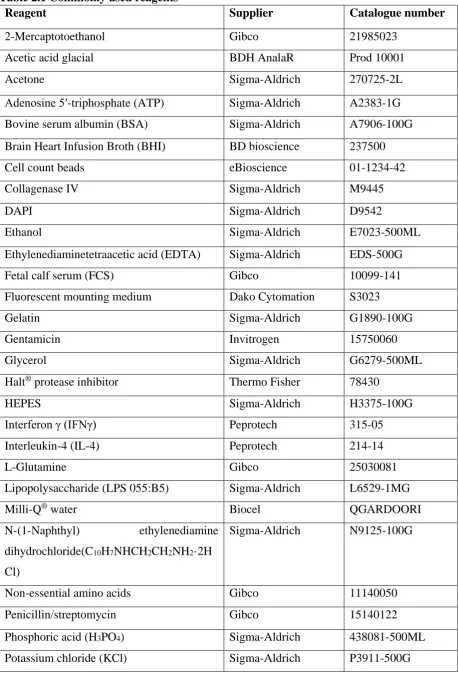
Table 2.1 Commonly used reagents
Reagent Supplier Catalogue number
2-Mercaptotoethanol Gibco 21985023
Acetic acid glacial BDH AnalaR Prod 10001
Acetone Sigma-Aldrich 270725-2L
Adenosine 5′-triphosphate (ATP) Sigma-Aldrich A2383-1G Bovine serum albumin (BSA) Sigma-Aldrich A7906-100G Brain Heart Infusion Broth (BHI) BD bioscience 237500
Cell count beads eBioscience 01-1234-42
Collagenase IV Sigma-Aldrich M9445
DAPI Sigma-Aldrich D9542
Ethanol Sigma-Aldrich E7023-500ML
Ethylenediaminetetraacetic acid (EDTA) Sigma-Aldrich EDS-500G
Fetal calf serum (FCS) Gibco 10099-141
Fluorescent mounting medium Dako Cytomation S3023
Gelatin Sigma-Aldrich G1890-100G
Gentamicin Invitrogen 15750060
Glycerol Sigma-Aldrich G6279-500ML
Halt® protease inhibitor Thermo Fisher 78430
HEPES Sigma-Aldrich H3375-100G
Interferon γ (IFNγ) Peprotech 315-05
Interleukin-4 (IL-4) Peprotech 214-14
L-Glutamine Gibco 25030081 Lipopolysaccharide (LPS 055:B5) Sigma-Aldrich L6529-1MG
Milli-Q® water Biocel QGARDOORI
N-(1-Naphthyl) ethylenediamine dihydrochloride(C10H7NHCH2CH2NH2·2H Cl)
Sigma-Aldrich N9125-100G
Non-essential amino acids Gibco 11140050
Penicillin/streptomycin Gibco 15140122
Reagent Supplier Catalogue number
Potassium dihydrogen phosphate (KH2PO4) Sigma-Aldrich P9791-500G
Proteinase k Sigma-Aldrich P6556-5MG
Rat serum Sigma-Aldrich R9759-10ml
RPMI 1640 medium Invitrogen 11875093
Sodium azide (NaN3) Sigma-Aldrich S2002-100G
Sodium chloride (NaCl) Sigma-Aldrich S5886-500G Sodium nitrite (NaNO2) Sigma-Aldrich 237213-500G Sodium phosphate dibasic (Na2HPO4) Sigma-Aldrich 30435-500G
Sodium pyruvate Gibco 11360070
Sulfanilamide (H2NC6H4SO2NH2) Sigma-Aldrich S9251-100G
Thioglycollate BD bioscience 211716
TMB ThermoFisher N301
Tris base Sigma-Aldrich T1378-1KG
[image:43.595.75.532.497.767.2]Triton X-100 Sigma-Aldrich X100-100ML
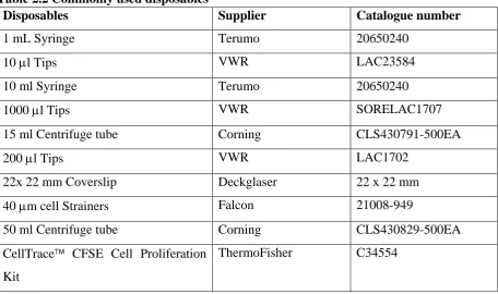
Table 2.2 Commonly used disposables
Disposables Supplier Catalogue number
1 mL Syringe Terumo 20650240
10 l Tips VWR LAC23584
10 ml Syringe Terumo 20650240
1000 l Tips VWR SORELAC1707
15 ml Centrifuge tube Corning CLS430791-500EA
200 l Tips VWR LAC1702
22x 22 mm Coverslip Deckglaser 22 x 22 mm
40 m cell Strainers Falcon 21008-949
50 ml Centrifuge tube Corning CLS430829-500EA
CellTrace CFSE Cell Proliferation Kit
Disposables Supplier Catalogue number
CellTrace Violet Cell Proliferation Kit
ThermoFisher C34557
Elisa 96-well plate Sarstedt 82.1583.200
Eppendorf tube Eppendorf 80-1500
Flat-bottomed 96-well microplate Corning NUN167008 Fluorescent mounting medium DakoCytomation S3023
FOXP3 fix/perm kit eBioscience 00-5523-00
iScript gDNA Clear cDNA Synthesis Kit
Bio-Rad 1725034
iScript gDNA Synthesis Kit Bio-Rad 172-5035
Light microscope Leica DM2500
Live/dead fixable Aqua dead cell stain kit
ThermoFisher L34957
Microscope slide Livingstone 7105-U
Needle 21G Terumo 20650030
Needle 26G Terumo 20650050
NucleoSpin® RNA XS kit Macherey-Nagel 740902.50 NueleoSpin RNA XS kit Macherey Nagel 740902.50
NuPAGETM gel Life technologies NP0322BOX
Protein broad range standard Bio-Rad 161-0317 ReliaPrep RNA Cell Miniprep
System
Promega Z6011
ReliaPrep RNA Cell Miniprep System
Promega Z6011
Round-bottomed 96-well microplate Corning CLS3799-50EA SsoAdvanced Universal SYBR
Green Mix
Bio-Rad 172-5271
Table 2.3 Commonly used equipment
Equipment Supplier
– 80 °C Freezer Sanyo
Autoclave Atherton
Bench top centrifuge Beckman Coulter Bench top microcentrifuge Thermo scientific BX50 Fluorescent microscope Olympus
Canto II flow cytometer BD biosciences Class II biological safety cabinet Gelman Sciences
Confocal microscope Nikon
Cyan ADP flow cytometry Beckman Coulter iBlot™ 2 dry blotting system Invitrogen
Incubator Sanyo
Light microscope Olympus
Lightcycler 480 qPCR instrument Roche
Microplate reader Bio-Rad
MoFlo Astrios cell sorter Beckman Coulter
pH meter inoLab
Platform mixer Ratek Instruments
Water bath Thermoline
2.4 Solutions
2.4.1 0.5 M EDTA stock
EDTA 46.53 g
2.4.2 0.5% Gelatine
Gelatine 0.5 g
Gelatine was dissolved in 100 ml PBS and stored at 4 C after autoclave.
2.4.3. 50% Glycerol stock
Glycerol 20 ml
PBS 20 ml
Under sterile condition, glycerol was mixed with PBS. The solution was stored at 4 ºC.
2.4.4. 10x Phosphate buffered saline (PBS)
NaCl 80.0 g
KCl 2.0 g
Na2HPO4 14.4 g
KH2PO4 2.4 g
Milli-Q® water 1000 ml
Using a magnetic stirrer, the reagents were dissolved in Milli-Q® water. The volume was adjusted to 1000 ml. The solution was diluted in 10 times before use.
2.4.5. 3x Western blot sample buffer (For 500 ml)
187.5 mM Tris–HCl (pH 6.8) 178 ml 0.5-M Tris–HCl pH 6.8 stock
6% SDS 150 ml 20% SDS stock solution
0.03% Phenol Red 150 mg
30% Glycerol 172 ml 87% Glycerol stock solution The reagents were dissolved in Milli-Q® water and stored at 4 ºC. The solution was diluted in 3 times with Milli-Q® water before use. 500 M DTT was added before freezing samples.
2.4.6. BHI broth media
BHI 3.7 g
BHI powder was dissolved in 100 ml dH2O. The medium was kept at 4 ºC after autoclave.
2.4.7. DFTD complete culture medium
Heat inactivated FCS 50 ml
L-glutamine 5 ml
Penicillin/streptomycin 5 ml
Under sterile condition, the RPMI-1640 medium was supplemented with the above solution and mixed by inversion. The solution was stored at 4 ºC.
2.4.8. ELISA coating buffer
NaHCO3 4.2 g
Na2CO3 1.8 g
The reagents were dissolved in 500 ml dH2O and pH was adjusted to 9.5. The solution was stored at room temperature.
2.4.9. FACS buffer
PBS 1.5 l
BSA 3 g
10% AZ buffer 3 ml
The reagents were mixed by inversion, and stored at 4 C.
2.4.10. Freeze L. monocytogenes
50% Glycerol 100 l
PBS 400 l
L. monocytogenes to be frozen was centrifuged at 5,000 rpm for 5 min and the supernatant was discarded. The tube containing L. monocytogenes pellet was added in 100 l 50% Glycerol. The solution was mixed and stocked at -80 ºC.
2.4.11. Freezing medium
Complete DFTD culture medium 20 ml
DMSO 20 ml
FCS 60 ml
2.4.12. Griess assay reagent I
1% Sulphanilamide 0.5 g
2.5% H3PO4 1.25 ml
The reagents were dissolved in 50 ml Milli-Q® water. The solution was stored at room temperature and protected from light.
2.4.13. Griess assay reagent II
NH2CH2CH2NH2. 2HCl 0.05 g
The reagent was dissolved in 50 ml Milli-Q® water, stored at room temperature and protected from light.
2.4.14. Heat inactivated fetal calf serum (FCS)
FCS was thawed at room temperature and heated for 1 h at 57 C. Aliquots were stored into sterile 10 ml tubes at -20 ºC.
2.4.15. Immunofluorescence blocking buffer
BSA 1% (10% BSA stock) 10 ml
Sodium Azide 100 mg
Glycine 2.25 g
The reagents were dissolved in 100 ml PBS and the solution was kept at 4 ºC.
2.4.16. Macrophage complete medium
RPMI 1640 500 ml
Heat inactivated fatal bovine serum 50 ml L929 hybridoma supernatant 50 ml Heat inactivated horse serum 25 ml
L-Glutamine (100x) 5 ml
Non-essential amino acids (100x) 5 ml Penicillin/streptomycin (100x) 5 ml
Sodium pyruvate (100x) 5 ml
Under sterile condition, the RPMI 1640 medium was supplemented with the above solution and mixed by inversion. The medium was stored at 4 ºC.
2.4.17. Mice tail lysis buffer
1 M Tris pH 8 10 ml
2 M NaCl 10 ml
0.5 M EDTA pH 8 1 ml
10% SDS 2 ml
The reagents were dissolved in 100 ml dH2O, stored at room temperature. The solution was diluted with Proteinase k (10 mg/ml) at 50:1 before use.
2.4.18. Macrophage harvesting media
PBS 500 ml
BSA 5 ml 10% stock
2 mM EDTA 2 ml 500 mM stock
Under sterile condition, the solution was mixed and kept at 4 ºC.
2.4.19. Ponceau staining solution (For 500 ml)
0.05% Ponceau S 250 mg
3% Trichloroacetic acid 15 g
The reagents were dissolved in 500 ml dH2O, and kept at room temperature.
2.4.20. Red blood cells lysis buffer
0.17 M Ammonium chloride 9.0933 g/l
0.02 M HEPES 4.766 g/l
The reagents were dissolved in 1000 ml dH2O and stored at room temperature.
2.4.21. Spleen lysis buffer
PBS 2.445 ml
1mM EDTA 5 l 500 mM stock
0.05% TritonX 100 25 l 5% TritonX 100
Set up 2.5ml spleen lysis buffer and freeze at -20 ºC.
2.5 Methods 2.5.1 Animals
The gene-targeted C57BL/6 (B6.TNF-/-) mouse strains deficient for TNF were generated on a genetically pure C57BL/6 (B6.WT) background as described [220]. The screening procedure followed the protocols described previously [220]. All animals were housed in pathogen-free conditions. Mice aged 8-16 weeks were used in all experiments following approval of the Animal Ethics Committee of University of Tasmania (UTAS) under the ethics number A13933, A13934 and A13936.
2.5.2 Cell culture
2.5.2.1 Bone marrow derived macrophages (BMDMs) culture
BMDMs from B6.WT and B6.TNF-/- mice were generated from pelvic and femoral bones. BMDMs were cultured in macrophage complete medium for 7-9 days.
For the experiment of inflammasome, BMDMs were harvested and were activated with LPS at 100 ng/ml in 96 well plates for 3 hours. After washing with PBS twice, BMDMs were co-cultured with L. monocytogenes at Multiplicity of infection (MOI) 5, 10, 20, 40 for 40 min. ATP was added for positive control and KCl was for negative control as described previously [221]. BMDMs were incubated with 10 g/ml gentamicin to kill extracellular bacteria. Cell culture supernatants were collected after 6 hours’ incubation and analysed with ELISA and western blot. Cells were lysed using western blot buffer and kept at -80 C.
For the experiment of phagocytosis assay, BMDMs were treated with the following substances: 20 ng/ml IFNγ, 100 ng/ml LPS or a combination of IFNγ and LPS for 24 h or 5 ng/ml IL-4 for 48 h. Control macrophages were cultured in medium alone. The macrophages were identified as CD11b+F4/80+ and the purity of macrophages obtained was always at least 90%.
2.5.2.2 Thioglycollate-elicited peritoneal macrophages collection
0.2% gelatine coated 12 mm coverslips at 5x10⁴ macrophages/well. The macrophages were subsequently incubated in fresh medium supplemented with 10% FCS or stimulated with 20 ng/ml IFNγ and 100 ng/ml LPS for 24 h or 5 ng/ml IL-4 for 48 h, and infected with L. monocytogenes as indicated.
2.5.2.3 Devil Facial Tumour Disease cell culture
The Devil Facial Tumour Disease cell line, C5065, was established from primary tumour samples [222]. Cells were grown in DFTD complete culture medium and maintained in a humidified 5% CO2 incubator at 35 C. Phagocytosis assays required co-culturing of mouse macrophages with DFTD cells were maintained at 37 C.
2.5.3 L. monocytogenes culture
L. monocytogenes strain EGD was grown overnight in brain heart infusion (BHI) broth at 37 °C with shaking. Bacterial suspension was diluted at 1:50 in fresh BHI broth and shaken at 37 °C for 2 h to obtain an OD600 of 0.10. 1 ml bacterial culture has 2x10⁸ L. monocytogenes. Bacteria were washed by pelleting and washed with PBS for two times. L. monocytogenes were suspended in PBS for infection in vitro or in vivo.
2.5.4 Cell collection from L. monocytogenes infected mice 2.5.4.1 Splenocytes and bone marrow cells dissociation
Spleens were dissociated with collagenase V in PBS for 10 min at room temperature. Red blood cells were lysed using ACK lysis buffer. Splenocytes were filtered through 40 m cell strainers to remove cell debris. Cell pellets were suspended in 10 ml FACS buffer for cell counting. Bone marrow cells were collected by flushing from femurs with ice-cold PBS. Red blood cells were lysed using red blood cells lysis buffer and centrifuged at 500 g for 5 min. Cell pellets were suspended in 10 ml FACS buffer for cell counting.
2.5.4.2 Isolation of peritoneal cavity cells
volume of fluid was recorded. Peritoneal cells were centrifuged at 500 g for 5 min and suspended in FACS buffer for cell counting.
2.5.5 CFUs assay
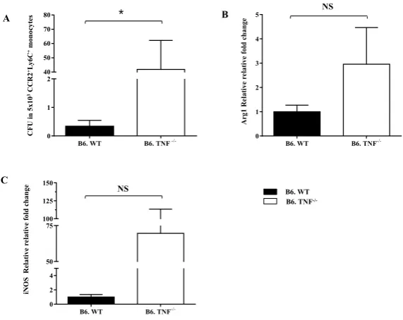
Spleen and liver were removed from animals and homogenized in 5ml lysis buffer (PBS with 0.05% Triton X-100). Serial 10-fold dilutions of homogenate were made in 96 well plates. Twenty µl undiluted homogenate was added into 180 µl lysis buffer and mixed thoroughly before starting any dilution. Fifty µl homogenate was spread in BHI agar plate and cultured at 37°C overnight. Colony forming units (CFUs) between 20 and 200 were counted and recorded as CFU/organ. Alternatively, to determine the bacterial load in cells, cells were isolated by flow cytometry and 5000 cells were lysed in 50µl buffer. The homogenate was plated in BHI agar plates and CFU were recorded as CFU/5000 cells.
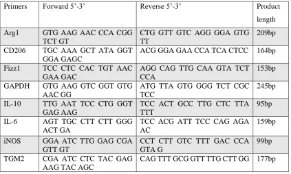
2.5.6 Quantitative real-time PCR
The qPCR primers were designed using Primer-BLAST in PubMed. DNA template was entered, to avoid amplification of contaminating genomic DNA, the option ‘Primer must span an exon-exon junction’ was selected. Primers with GC content of around 50-60% and primer-dimer were needed to be avoided. A standard curve was used to determine qPCR efficiency. Fivefold dilution series of cDNA were run in duplicate. The slope of a graph where log (dilution factor) is on the x-axis and Ct on the y-axis. Efficiency was calculated using the formula: efficiency = 10^ (-1/slope). The efficiency from 90-110% was preferred.
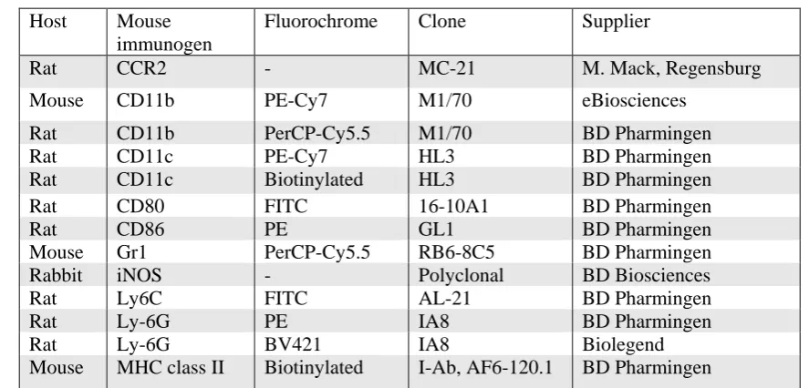
2.5.7 Flow cytometric analysis of splenic monocytes and cell sorting
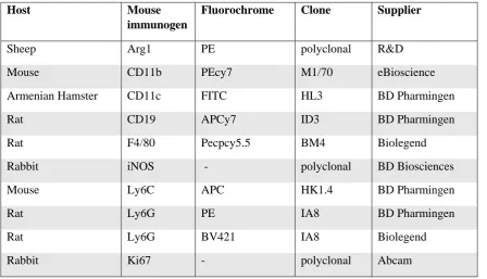
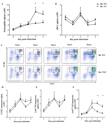
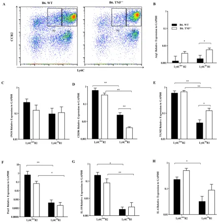
[image:53.595.85.509.97.350.2]1 million splenocytes were stained with anti-CCR2 antibody for 30 min on ice. After the application of secondary antibody, the cells were washed and blocked with 10% rat serum for 10 min. 1 million cells were stained for other surface markers on ice for 30 min. 20 µl DAPI (100 ng/ml) was added just prior to flow cytometry analysis for excluding dead cells. For intracellular staining, cells were fixed according to the manufacturer’s instruction with FOXP3 fix/perm kit. Live/dead fixable Aqua dead cell stain kit was used for dead cell exclusion. Data were acquired using CyAn ADP. Fluorescence minus one control was used for gate strategy to identify interest cells. Neutrophils were identified as Ly6G+, DCs were identified as CD11c+, and monocytes were gated with the expression of Ly6C and CCR2. Splenic monocytes were sorted by Beckman Coulter MoFlo Astrios cell sorter. Ly6Chi and Ly6Clow monocytes or Ly6C+ monocytes were sorted by the same gate strategy. The purity of sorted cells were detected and it was > 95%.
Table 2.4: Primers used for quantitative PCR characterizing monocytes
Primers Forward 5’-3’ Reverse 5’-3’ Product
length
Arg1 GTG AAG AAC CCA CGG TCT GT
CTG GTT GTC AGG GGA GTG TT
209bp
CD206 TGC AAA GCT ATA GGT GGA GAGC
ACG GGA GAA CCA TCA CTCC 164bp
Fizz1 TCC CTC CAC TGT AAC GAA GAC
AGG CAG TTG CAA GTA TCT CCA
153bp
GAPDH GTG AAG GTC GGT GTG AAC GG
ATG TTA GTG GGG TCT CGC TCC
245bp
IL-10 TTG AAT TCC CTG GGT GAG AAG
TCC ACT GCC TTG CTC TTA TTT
95bp
IL-6 AGT TGC CTT CTT GGG ACT GA
TCC ACG ATT TCC CAG AGA AC
159bp
iNOS GGA ATC TTG GAG CGA GTT GT
CCT CTT GTC TTT GAC CCA GTA G
99bp
TGM2 CGA ATC CTC TAC GAG AAG TAC AGC
