BACTERIAL AND FUNGAL PROFILE OF ACUTE
EXACERBATION OF CHRONIC OBSTRUCTIVE
PULMONARY DISEASE
Dissertation submitted to
THE TAMILNADU DR.M.G.R.MEDICAL UNIVERSITY
In partial fulfillment of the regulations for the award of the degree of
M.D.(MICROBIOLOGY)
BRANCH – IV
MADRAS MEDICAL COLLEGE
THE TAMILNADU DR. M.G.R. MEDICAL UNIVERSITY
CHENNAI – TAMILNADU
CERTIFICATE
This is to certify that this dissertation titled “BACTERIAL AND FUNGAL PROFILE OF ACUTE EXACERBATION OF CHRONIC
OBSTRUCTIVE PULMONARY DISEASE ” is a bonafide record of work done by DR.V.R.YAMUNADEVI, during the period of her Post Graduate study from MAY 2012 to APRIL 2015 under guidance and supervision in the Institute of Microbiology, Madras Medical College and Rajiv Gandhi Government General Hospital, Chennai- 600003, in partial fulfillment of the requirement of M.D MICROBIOLOGY degree Examination of The Tamilnadu Dr. M.G.R Medical University to be held in April 2015.
Dr.R. VIMALA M.D Dean
Madras Medical College & Government General Hospital, Chennai – 600 003.
Dr.G. JAYALAKSHMI,M.D.,DTCD, Director,
DECLARATION
I declare that the dissertation entitled “BACTERIAL AND FUNGAL PROFILE OF ACUTE EXACERBATION OF CHRONIC OBSTRUCTIVE
PULMONARY DISEASE ”submitted by me for the degree of M.D. is the record work carried out by me during the period of October 2013 to September 2014 under the guidance of Dr. Sheila Doris, M.D., Professor, Institute of Microbiology, Madras Medical College, Chennai. This dissertation is submitted to the Tamilnadu Dr.M.G.R. Medical Unversity, Chennai, in partial fulfillment of the University regulations for the award of degree of M.D., Branch IV (Microbiology) examination to be held in April 2015.
Place : Chennai Signature of the candidate Date: (Dr. V.R.YAMUNADEVI)
Signature of Guide,
Prof.Dr.T.SHEILA DORIS.,MD,
Professor,
Institute of Microbiology,
ACKNOWLEDGEMENT
I humbly submit this work to the Almighty who has given me the health and ability to pass through all the difficulties in the compilation and proclamation of this blue print.
I wish to express my sincere thanks to our Dean, Dr.R.Vimala M.D., for permitting me to use the resources of this institution for my study.
I owe special thanks to Prof. Dr. G. Jayalakshmi, M.D.,DTCD, Director and Professor, Institute of Microbiology for her support, invaluable suggestions, erudite guidance in my study and for being a source of inspiration in my endeavour
My sincere thanks to former Director Prof.Dr.N.MEENAKSHI,MD.,DTCD., Department of Thoracic Medicine, Former Director ,Prof.Dr.SIVA SUBRAMANIAM, MD., Institute of Internal Medicine for permitting me to carry the study.
My special thanks to Dr.GUNASEKARAN M.D., Director, King institute of preventive medicine, Guindy. Prof. Dr.VASANTHI M.D.,and Prof.Dr.RAJESHWARI HOD, Diagnostic Department (Bacteriology) for providing Horse blood for my dissertation.
I express my gratitude to our former Director, Prof.Dr. M. Mohammed Meeran, MD.,DVL.,for their guidance and support.
I whole heartedly thank my Guide Prof. Dr.T. Sheila Doris M.D.,to permit me to take this topic for my dissertation . I feel fortunate to work under her guidance with her valuable suggestions and great support throughout my study.
I would like to thank my Professors Dr.S.Vasanthi M.D., Dr. K. Muthulakshmi M.D., Dr. S. Thasneem Banu M.D., Dr. U. Uma Devi M.D., for their valuable assistance in my study.
I extend my whole hearted gratitude to our Assistant Professor Dr.R.Deepa M.D., Coguide in spending her valuable time to encourage me and also help me to complete my dissertation in time .She also gave her valuable suggestions and guidance in my study..
I also express my thanks to our Assistant professors Dr.Lata Sriram, M.sc., Ph.D., Dr.N.RathnaPriya M.D., Dr.R.Ushakrishnan M.D., Dr.K.G.Venkatesh
M.D., Dr.N. LakshmiPriya M.D., Dr.C.Sri Priya M.D., Dr.David Agatha M.D.,
and Dr.B.Natesan M.D.,DLO., for their immense support in my study.
I hereby express my gratitude to all the technical staff for their help throughout my study.
I would like to thank my department colleagues and friends for their constant support and co-operation.
Finally I am indebted to my family members especially my husband Dr.A.D.Edukondalu who took all family burden and relieved me to concentrate on my study. I also thank my son, Hemanth and daughter Renu who cooperated me for this work. Iam sincerely thankful to my parents, Ramanathan and Usharani who took responsibility of day to day work and also encourage me to focus on my study. All my family members have been solid pillars of everlasting support and encouragement.
TABLE OF CONTENTS
S.NO TITLE PAGE.NO
1 INTRODUCTION 1
2 REVIEW OF LITERATURE 5
3 AIMS AND OBJECTIVES 32
4 MATERIALS AND METHODS 33
5 RESULTS 56
6 DISCUSSION 86
7 SUMMARY 100
8 CONCLUSION 104
9 COLOUR PLATES
APPENDIX –I ABBREVIATIONS
APPENDIX-II STAINS AND REAGENT
ANNEXURE-I ETHICAL COMMITEE
ANNEXURE-II PROFORMA
ANNEXURE –III MASTERCHART-1
MASTERCHART-2
BACTERIAL AND FUNGAL PROFILE OF ACUTE
EXACERBATION OF CHRONIC OBSTRUCTIVE
PULMONARY DISEASE
ABSTRACT
BACKGROUND AND OBJECTIVE: Acute exacerbation of Chronic obstructive Pulmonary disease (AECOPD) is defined as a sustained worsening of the patient’s condition ,from the stable state in the patient’s baseline dyspnoea and cough or sputum ,or both and beyond normal day to day variation ,that is acute in onset and necessitates a change in regular medication in a patient with underlying COPD as per Gold guidelines ..It leads to significant increase in morbidity and mortality in COPD patients. Bacteria are responsible for 60% of exacerbation. The aim of our study was to determine the bacterial and fungal isolates in AECOPD and Stable COPD patients with special reference to antibiotic susceptibility and their resistance pattern from hospital data.
SETTINGS AND DESIGN : It was a Cross sectional study carried out at the Institute of Microbiology, Madras Medical College in association with Departments of Internal Medicine, Thoracic Medicine, Intensive Medicare Care Unit at Rajiv Gandhi Government General Hospital, Chennai from October 2013 to September 2014
MATERIALS AND METHODS : The Study population consisted of 150 in patients presenting with signs and symptom of AECOPD and 50 stable COPD out patients .All the respiratory samples were subjected to direct gram staining , culture ,biochemical reactions and the isolates were identified ac-cording to standard techniques. Antibiotic sensitivity was done by Kirby-Bauer method according to CLSI standards.
bacteria 21.6.Among Gram negative organisms Klebsiella pneumoniae 33.3% was the most commonly and significantly isolated organism followed by Pseudomonas aeruginosa .In stable COPD patients only 19% Klebsiella pneumoniae was isolated. Non fermenters were significantly isolated in Severe type and Staphylococcus aureus from Moderate type. Klebsiella pneumoniae showed Multi drug resistance(MDR) of 29.7% The prevalence of Carbapenemase production in Klebsiella pneumoniae was 28.5%This implies that Klebsiella pneumoniae was one of the important drug resistant pathogen isolated among AECOPD patients. Presence of MRSA and ESBL isolates were higher in Moderate AECOPD patients (66.6%, 66.6% , respectively) than in severe group(33.3%,33.3%). Penicillin resistant Streptococcus pneumoniae , Imipenem resistance, Carbapenamase producing isolates,Amp C producing strain were significantly higher in severe AECOPD patients MDR pathogens were present both in moderate and severe type of COPD.
Haemophilus influenzae and Fungus were not isolated.
1
BACTERIAL AND FUNGAL PROFILE OF ACUTE
EXACERBATION OF CHRONIC OBSTRUCTIVE
PULMONARY DISEASE
INTRODUCTION
Chronic Obstructive Pulmonary Disease (COPD ) is defined as a preventable
and treatable disease with pulmonary component characterised by airflow limitation
that is of not fully reversible which is usually progressive and associated with an
abnormal inflammatory response of the lungs to noxious particles or gases and some
significant extrapulmonary effects that may contribute to the severity in individual
patients (1-3). It includes: Emphysema, Chronic Bronchitis, Small airway disease
AECOPD(acute exacerbation of COPD): This condition is defined as a sustained worsening of the patient’s condition ,from the stable state(in the patient’s
baseline dyspnoea and cough or sputum ,or both and beyond normal day to day
variation ,that is acute in onset and necessitates a change in regular medication in a
patient with underlying COPD as per Gold guidelines (4). It is characterized by
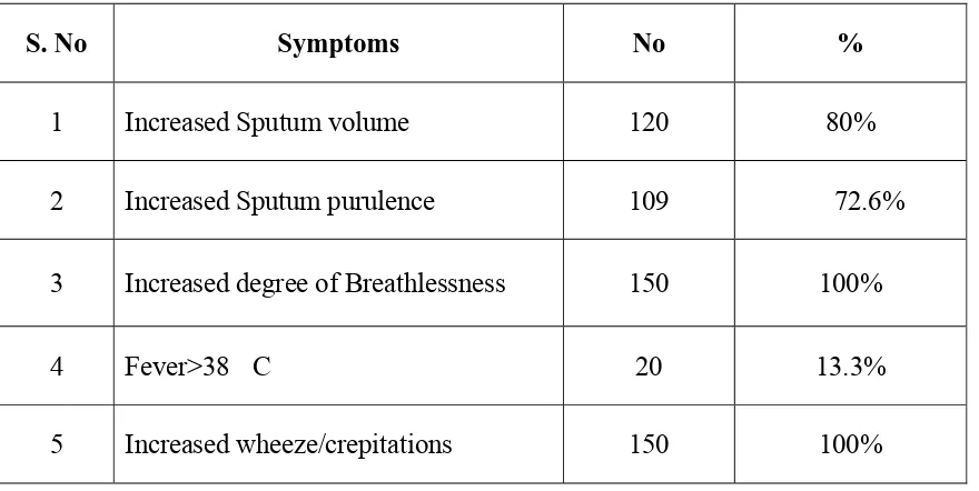
presence of increased sputum volume, sputum purulence and dyspnoea.
Burden of the disease : Chronic obstructive pulmonary disease (COPD) is associated with significant morbidity and mortality, with the World Health
Organization estimating its rise from being the fourth to the third leading cause of
2
Almost 95% of mortality due to chronic respiratory disease in India can be assigned
to COPD.
Exacerbations of COPD have considerable impact on health care system at
both primary and tertiary care levels as they are the major reason for antibiotic use and
admissions. WHO has estimated that 600 million people worldwide have COPD.
Additionally, exacerbations lead to indirect costs because of days lost from work.
COPD affects 30% of patients seen in chest clinics and constitutes 1-25% of hospital
admissions all over India(6).
AE-COPD is a common cause of emergency room (ER) visits and is a major
cause of morbidity and mortality (7.8).
A gross underestimate of COPD Prevalence had been estimated as 17 million
and it is likely to increase by over 30% in next decade. Highest prevalence (9.4%) was
reported from North Indian rural population from a study conducted by Jindal et.el
from 1964-1995.
Causes of AE-COPD:
Exacerbations are caused or triggered by a variety of factors including viruses, bacteria ,and air pollutants, and are associated with acutely increased worsening of existing (acute-on-chronic) airway inflammation and also due to defects in host defence mechanisms. Alterations produced in the bronchial epithelium by the damaging action of smoking favour bacterial adhesion and colonization. In turn,
3
damage via the action of proinflammatory substances in what is known as the “vicious
circle theory(9).
Infections are the important cause of acute exacerbation. Bacteria are
responsible for causing 60% of exacerbations. Viral infections are the likely cause
of approximately 30% of exacerbations, while PCR studies have suggested that up to
40% of acute respiratory infections in COPD are associated with viruses. Fungal
isolates have not been reported(3,10).
Exacerbations, mostly of an infectious etiology, are a frequent cause of
morbidity in COPD patients. Furthermore, infection was the most common observable
cause of death in prospectively followed-up COPD patients(11).
This condition is highly serious in our country as the prevalence of smoking
and air pollution is very high which are the main cause for COPD and increase the
frequency of exacerbation. Little has been documented about this problem from India.
Exacerbations punctuate the clinical course of COPD in many patients. Since it
is a vicious cycle recurrent exacerbation will lead to rapid deterioration of lung
parameters and early death due to respiratory failure and increased economic burden.
These episodes of acute exacerbation can vary considerably in severity as part of the
exacerbations will remain unreported while some episodes require admission. A
European survey found that sputum analysis of exacerbated patients is requested only
4
Antimicrobial therapy: Over 90% of patients with AECOPD are treated with antibiotics, on empirical basis without proper sputum analysis so the effectiveness of
treatment is uncertain due to emerging new strains and their resistant pattern thereby
leading to recurrent exacerbation(113). It would be useful to find the proper etiology of
COPD exacerbations, thereby facilitating the orientation of antibiotic treatment and
reducing the high number of failures recorded with empiric treatment, which in some
cases, is as high as 26%(114).
This study is taken up to find out the Bacteriological & Fungal profile and their
sensitivity pattern in AECOPD and Stable COPD patients as the knowledge of
possible bacterial & fungal etiology and sensitivity patterns of COPD exacerbations,
facilitates the orientation of antibacterial and antifungal treatment so that timely
institution of correct management is important for better prognosis of disease and to
5
REVIEW OF LITERATURE
HISTORICAL REVIEW:
The term exacerbation has its origin in the Latin descriptive acerbus, meaning harsh, bitter, sharp and more at outer edge.
Hippocrates (460-377BC): The Father of Medicine described an old men
suffering from breathlessness associated with cough and catarrhal
Earliest references to COPD
In 1679 Bonet described Chronic Obstructive Pulmonary Disease (COPD) as
“voluminous lungs”. It was corroborated around a century later in 1769 by Morgagni
who described cases in which the lungs were “turgid”, particularly from air.
Baillie in 1789 published a series of illustrations of the emphysematous lung
putting forth the pathology of the disease. Thus emphysema was known to be a part
of COPD.
It was much later that chronic bronchitis got included in COPD.
Badham in 1814 used the word catarrh to refer to the chronic cough and
increased mucus secretion as symptoms of bronchiolitis and chronic bronchitis that
could be part of COPD.
Laënnec described emphysema of the lungs in 1821 in his Treatise of diseases
6
were excessively inflated that did not empty well. Laënnec went on to describe a
combination of emphysema and chronic bronchitis. Our present knowledge of the
disease is founded on the clinical work of Laennec.
In 1855 Bierner was given the credit for studying the sputum in
Bronchopulmonary disease.
In 1846 John Hutchinson invented the spirometer. This was the key to
diagnosing COPD. The spirometer is still used today for diagnosis and regular
assessment regarding response to therapy in COPD. Hutchinson’s instrument only
measured vital capacity.
In 1915 Dass and Luetscher studied and described the application of
bacteriological sputum examination and recognised Haemophilus influenza as a
common cause of acute and chronic Bronchitis
In 1947 Tiffeneau and Pinelli added the concept of timed vital capacity as a
measure of airflow.
In 1964 Eriksson showed that people with a severe congenital deficiency of
serum α1 antitrypsin developed Emphysema.
William Briscoe is believed to be first person to use the term COPD in
discussion at the 9th Aspen Emphysema conference. This term became established
7
DEFINITION :
Chronic Obstructive Pulmonary Disease (COPD ) is defined as a preventable
and treatable disease with pulmonary component characterised by airflow limitation
that is of not fully reversible which is usually progressive and associated with an
abnormal inflammatory response of the lungs to noxious particles or gases and some
significant extrapulmonary effects that may contribute to the severity in individual
patients. (1,2)
It was also defined in a joint statement of American Thoracic Society and the
European Respiratory Society as a disease characterised by and diagnosed with
spirometric measurement of airflow limitation that is not fully reversible which is also
supported by GOLD.(13)
COPD includes Emphysema, Chronic Bronchitis, Small airway disease.
Emphysema : It is defined as an abnormal ,permanent enlargement of the distal
airspaces ,distal to the terminal bronchioles ,accompanied by destruction of their walls
and without obvious fibrosis.
Chronic Bronchitis: A clinically defined condition with presence of chronic
productive cough on most days for 3 months in each of 2 consecutive years in a
patient in whom other causes of chronic cough have been excluded.(3)
Small airway disease : It is a condition in which small bronchioles are
8
DISEASE CLASSIFICATION:
COPD is a heterogenous disease which has many hypotheses like British,
American, Dutch, Swedish and all the hypotheses probably have elements of truth as
COPD is a classic gene –by-environment disease.
In newer literature COPD severity is classified as per 2006 revision of GOLD
criteria that is based on post bronchodilator lung function
GOLD 1(mild) FEV1/FVC <0.70 and FEV1 >= 80% predicted
GOLD 2 (moderate) FEV1/FVC <0.70 and 80% > FEV1 >= 50% predicted
GOLD3(severe) FEV1/FVC <0.70 and50% > FEV1 >= 30% predicted
GOLD 4(very severe)
FEV1/FVC <0.70 and FEV1 <30% predicted or FEV1 <
50% predicted plus chronic respiratory failure or signs of heart failure
People with FEV1/FVC >=0.70 and respiratory symptoms of chronic cough and
sputum production are no longer included as COPD stage (formely GOLD stage
0).Patients with FEV1/FVC >=0.70 but an FVC <80% predicted meet spirometric
criteria for a restrictive process. Although this is not regarded as COPD ,patients
might present with several symptoms similar to those seen in COPD ,and these
patients have an increased risk of death.(14)
9
baseline dyspnoea and cough or sputum ,or both and beyond normal day to day
variation ,that is acute in onset and necessitates a change in regular medication in a
patient with underlying COPD as per Gold guidelines (4)
Anthosien criteria :(15)
Increased breathlessness
Increased volume of sputum
Increased purulence of sputum
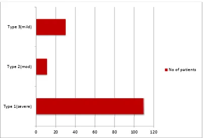
There are 3 types of acute exacerbations of chronic bronchitis which are based
upon 3 cardinal symptoms which include worsening of dyspnea, increase in sputum
purulence, and increase in sputum volume. The 3 types are defined as follows:
1. Type 3: Mild exacerbation with 1 of 3 cardinal symptoms PLUS 1 of the following:
a. Upper respiratory tract infection in the past 5 days
b. Fever without other apparent cause
c. Increased wheezing
d. Increased cough
e. Increased respiratory rate or heart rate by 20% above baseline
2. Type 2: Moderate exacerbation with 2 of 3 cardinal symptoms
3. Type 1: Severe exacerbation with all 3 cardinal symptoms
Cardinal symptoms include worsening of dyspnea, increase in sputum
10
BURDEN OF DISEASE :
COPD is a growing global epidemic and it is estimated to kill around 3 million
people every year .It is currently the 4th largest killer disease in the world and
expected to climb to 3rd position by the year 2030 .
WHO has estimated that 600 million people worldwide have COPD. It affects
around 5-10% of population over the age of 40 years but still there wide variations in
the prevalence between countries.(17)
ECONOMIC BURDEN:
The National Heart , lung and Blood Institute provides a estimate of 38.8
billion US $ for US and 38.6 billion US $ for Europe .COPD accounts for 56% of
total health care budget.
According to National Commission on macroeconomics and health published
in India that per capital expenditure on COPD is Rs.42664 in 2006 and expected to
increase to Rs. 62630 by 2016. Upto 84% of the costs spent on COPD is due to
inpatient hospitalization due to which the loss in productivity due to COPD account
for between 40% and 67% of the overall costs across the world .Hence it is a severe
economic burden for countries throughout the world.(10)
Morbidity: In Canada 1 in 4 people older than 35 years was likely to be
diagnosed with COPD. The burden was more in rural men ,with lower socioeconomic
11
In Canada by about 80 years about 25% of women and about 30% of men will
be diagnosed with COPD.(18)
8-22% of adults aged more than 40 years and older is the leading cause of
hospitalization and health care cost incurrence (19.)It is a common and leading cause of
morbidity which is the major cause of worsening of Quality of life.50% of patients
who survive first hospitalization get readmitted within 6 months . Estimated
prevalence rates for people more than 30 years vary between 0.6% and 4% in men and
0.2 – 32% in women(yin an et al.Passive smoking exposure and risk of COPD among
adults in China .(20)
INDIA:
A gross underestimate of COPD Prevalence had been estimated as 17 million
and it is likely to increase by over 30% in next decade. Highest prevalence(9.4%) was
reported from North Indian rural population from a study conducted by Jindal et.el
from 1964-1995(10) .
MORTALITY:
Global mortality: WHO states that more than 3 million people die of COPD per
year and 5% of all deaths globally and 160% in South east asian region over the next
two decades. India itself contributes to over half of a million deaths second only to
12
SOUTH EAST ASIAN REGION :(5)
Almost 5,56,000 deaths are attributable to COPD as estimated by WHO in
South east asian region which majorily comprises INDIA .So almost 95% of
mortality due to chronic respiratory disease in India can be assigned to COPD. State
wise data is at present available for Maharashtra .As the data for other states are not
available , studies on COPD is a must to assess the burden.
DALYS (Disability adjusted life year):
In 1990 COPD accounted for 2.1% of DALY’S which ranked 12th most
common cause .This is expected to increase upto 4.1% and it is assumed to move to
5th rank by 2020.(20,21).Overall COPD was estimated to have resulted in more than 26
million DALYS in 2000.
In the year 2002 6740 thousand DALYs were lost due to COPD in South East
Asian region.
In India COPD account for 3% of DALYS but this is likely to be
underestimated .(17)
DEFENCE MECHANISM OF NORMAL LUNG RESIDENT DEFENSES
1. Airway architecture
2. Epithelial barrier
3. Mucociliary clearance
4. Soluble factors in airway secretion –complement,immunoglobulins
13
Recruited Defenses
If resident defence system is not able to control the Polymorphonuclear
neutrophils and lymphocytes are recruited to augment host response by the production
of Leukotriene β 4,TNF- α.(22)
RISK FACTORS FOR COPD(106,107):
1. Smoking-cigarrete or Bidi-50% of smokers develop COPD.73% of mortality in
COPD is due to smoking among which 40% is from low and middle
socioeconomic status. Smoking leads to ciliary destruction and hypermucus
secretion and decreased mucociliary clearance.
2. Aging-As the lung function starts to decline by third and fourth decade of life
3. Tuberculosis (this is very common in India)
4. Respiratory infection in early life
5. Passive or second hand smoking
6. Ambient air pollution-WHO estimates 1% of COPD cases in high income
countries is due to urban air pollution where as it is 2% in nations of low and
middle income.
7. Occupational exposure-coal mining,cotton textile dust, mining
8. House hold exposure-biomass fuels -Ninety percent of rural households and
32% of urban households cook their meals on a biomass stove.3 billion people
14
risk of developing COPD as tobacco smoke. WHO states that 35% of
population in countries of low and middle income develop COPD due to its
inhalation and 36% of mortality from lower respiratory disease is due
biomass inhalation.
9. Low Socioeconomic status
10.Genetic factors-α1 antitrypsin deficiency leads to emphysema in 1-3% of patients
11.Gender-In high income countries COPD prevalence is similar in both sexes due
to smoking where as condition is different in low and middle income countries
as smoking in female is low .(23)
PATHOLOGICAL CHANGE IN COPD:
Triggers – Cigarrette smoke, Biomass fuel
Subepithelial infiltration Inflammatory
mediators Macrophages Growth factors
Neutrophils Proteinases , Oxidative stress
CD 8
CD 47
Emphysema Chronic Bronchitis Air trapping dyspnoea Cough and hypermucus
15
PATHOPHYSIOLOGY OF AECOPD:(3.10)
1. In COPD patients due to defective phagocytosis of macrophages it leads to
constant bacterial colonization of respiratory tract and frequent ,recurrent
exacerbation of COPD. This can amplify and lead to increase airway and
systemic inflammation in Stable COPD patients and this vicious cycle
increases progressively with disease severity. As airflow obstruction progresses
the frequency of exacerbation also increases.(24,25)As an increase in
concentration of bacteria that colonize the lower respiratory tract constitutes to
the pathogenesis of exacerbation and the acquisition of new bacterial strain
are also crucial in the pathogenesis of exacerbation.(26)
2. Alveolar macrophages from COPD patients phagocytose lesser number of
apoptotic epithelial cells .Therefore there is chronic bacterial colonization
with Streptococcus pneumonia ,Haemophilus influenzae which attributes to
acute infectious exacerbation .(27)
EFFECT OF COPD EXACERBATION WITH FREQUENT
EXACERBATION:(Vicious cycle)
HIGHER MORTALITY POOR QUALITY OF LIFE
PATIENTS WITH FREQUENT EXACERBATION
FASTER DECLINE IN LUNG FUNCTION
16
CAUSES OF ACUTE EXACERBATION OF COPD:(3,28)
INFECTIOUS AGENTS :
Virus , Gram positive and gram negative aerobic bacterial, atypical bacteria
ENVIRONMENTAL CONDITIONS:
Sudden change in temperature ,humidity,air pollution exposure,tobacco smoke
exposure,noxious gases or irritating chemicals
HOST FACTORS:
Patients with poor general health, poor nutrition, immunocompromised state
,lack of compliance to prescribed medicines,adoption of unhealthy life styles modes,
poor level of personal hygiene,lack of compliance with long term oxygen therapy,
failure to participate in pulmonary rehabilitation.
FACTORS THAT POTENTIALLY MODIFY RISK FACTORS OF AECOPD\
INTRINSIC FACTORS
Impairement of lung functions
Active smoking
Bronchial hyperresponsiveness
Chronic Mucus secretion
Impairement of defence mechanism
17
EXTRINSIC FACTORS:
Type of bacterial infection and changing strains of bacteria
Decreased environmental temperature
Air pollution
Type of treatment for Stable and exacerbation of COPD
TRIGGERS OF COPD EXACERBATIONS AND ASSOCIATED PATHOPHYSIOLOGICAL CHANGES LEADING TO INCREASED
EXACERBATION SYMPTOMS
Bacteria viruses Pollutants
Effects Inflammed COPD airway
Greater airway inflammation
Systemic inflammation Bronchoconstriction , edema
Expiratory flow limitation
Cardiovascular comorbidity Exacerbation Dynamic hyperinflation
Symptoms
Among all causes infection is the most common precipitating factor .Molecular
diagnostics have given strong evidence that Microorganisms are involved in 80% of
18
Viral infections:
It accounts for 30% of infection but it could be an underestimate as it is
difficult to isolate. The common viruse are Rhinovirus, Coronavirus, Influenza virus,
Parainfluenza virus, Adenovirus, Respiratory syncytial virus
Bacterial infections:
It accounts for 60% of infection. Most common are Haemophilus influenzae
nontypable, Moraxella Catarrhalis, Streptococcus pneumoniae.A number of studies have shown that virulent organisms are isolated in severe AECOPD patients like
Staphylococcus aureus, Pseudomonas aeruginosa and members of Enterobactericeae family.(10)
Atypical bacteria:
The role of Chlamydia , Legionella and Mycoplasma is conflicting in causing AECOPD as these microorganisms might also interact with airway bacteria and
viruses. A study done by using real time PCR by Diederen .et.al. found no role for
these atypical bacteria in AECOPD.(29)
Potter et.al. proved that although a variety of commensal bacteria inhabits
nasopharynx of all healthy individuals but still lower airways are usually found to be
sterile by standard culture techniques(30).
In Stable COPD:
The precise role of bacteria in causing exacerbation has been difficult to assess
19
isolated at exacerbations like Haemophilus influenzae nontypable, Moraxella
Catarrhalis, Streptococcus pneumoniae , Pseudomonas aeruginosa and also consists of other oropharyngeal commensal bacteria like-Streptococcus viridans,
Streptococci,Neisseria sp,Corynebacterium sp,and Candida sp. A study by Wilkinson TM Donald son et.al.found that bacteria was isolated in 48.2% of stable COPD
patients which rose to 70% in AECOPD patients.(31)A study by Berenson .et. al.
isolated bacteria in 25% of stable COPD and 50% in AECOPD(32).
Purulent sputum is the surrogate marker of bacterial infection since isolation
rate of bacteria is more in purulent sputum as compared to mucoid sputum.
Since in this study we have correlated every sample with proper history,
predominant growth , sputum quality ,gram staining so this can solve the dispute.
Clinical features:(10)
Clinical features include increase in cough, chest pain, Increase in
breathlessness, Increase in sputum volume and change in it’s colour(white, green,
yellow or blood streaked), Fever ,Increased fatigue ,Increase in oxygen requirement.
Physical findings-tachycardia, tachypnoea, expirtory wheeze, medium to coarse
crackles, pursed lip breathing, central and peripheral cyanosis, neck vein distention,
hepatomegaly, peripheral edema, hyperinflation of lungs with increased AP diameter
20
COMPLICATION:
Hypoxemic Type 1 respiratory failure
Hypercapnic Type 3 respiratory failure
Compensated Metabolic alkalosis
Cor pulmonale
Left ventricular failure
Multi organ failure
Arterial oxygen desaturation
Altered sensorium
As per the National clinical guideline for the treatment of COPD (33).
LABORATORY DIAGNOSIS:
Proper diagnosis of AECOPD depends on clinical history, Physical
examination and associated with Microbiological investigations. Microbiological
investigations are important to isolate the organisms and to determine it’s resistance
pattern in order to prescribe the appropriate drug and to reduce the development new
resistant strains and to prevent spread of existing resistant strains.
OTHER INVESTIGATIONS:
Pulmonary Function test, Complete blood count , Chest radiography, Complete
metabolic profile, Arterial Blood gas analysis, Oxygen saturation, ECG,
21
Microbiological investigations :(34,35,36,106,109)
The major benefit of microbiological investigations lies in the proper
etiological diagnosis of AECOPD and to identify the resistance and susceptibility
pattern of microorganisms. Different samples are collected from lower respiratory
tract to perform different microbiological investigations. The most samples are:
A. Non invasive:
1. Sputum (Expectorated): Collected under direct supervision, to minimise
contamination with oropharyngeal secretions.It is the easy and basic sample to
assess the lower respiratory tract.
2. Induced sputum: Collected in patients who are unable to produce sputum.
Those are the material directly obtained from alveolar spaces and should be
accepted in laboratory without prescreening.
B. Invasive (Bronchoscopic techniques):
1. Bronchial washings
2. Bronchoalveolar lavage(BAL)
3. Protected catheter bronchial brush(rare sample)
Other Samples:
Endotracheal aspirate:
D. Blood :
22
A. Microscopy:
Direct microscopy examination aids to determine the quality of sample ,and to
determine the severity and type of inflammatory response. To screen the likely
pathogens directly in the clinical specimen and to correlate with the organisms grown
in culture .With this initial screening ,the laboratory can help the clinicians to make
early decisions to initiate antibiotic therapy early.
1. Staining methods :(34,35,36) Direct staining provides a differential staining and
they enhance the chance of identifying the microbes and other details in the sample.
i) Gram’s stain: it is meant for evaluating the quality of the specimen ,
presence of bacteria and it’s arrangement, morphology, gram reactions ,
presence of neutrophils and also help to identify fungal elements .
ii) Acid Fast stain: Helps to identify acid fast bacilli directly in clinical
samples
2. 10% Potassium hydroxide mount: It is used to identify fungal elements in the
23
SCREENING OF SPECIMENS REQUESTED FOR ROUTINE BACTERIAL
CULTURE TO ENSURE QUALITY (37,106,109)
SPECIMEN SCREENING
METHOD ACCEPTABLE FOR CULTURE NO FURTHER TESTING; request another sample Sputum Microscopic
examination of gram stained smear <10 squamous epithelial cells/average 10xfield >10 squamous epithelial cells/average 10xfield Endotracheal Aspirate Microscopic
examination of gram stained smear
<10 squamous epithelial cells/average 10xfield and bacteria seen in atleast 1 of 20 oil immersion fields
>10 squamous epithelial cells/average 10xfield and no bacteria seen in atleast 1 of 20 oil immersion fields
Bronchoalveolar lavage or
Bronchial wash
Microscopic
examination of gram stained smear
<1% of cells present are
squamous epithelial cells
>1% of cells present are squamous epithelial cells
24
REQUIREMENTS FOR CULTURE AND CONDITIONS:(34,35,38)
Media Incubation Culture reading Pathogens that grow Time Temperature Atmosphere
5%sheep Blood agar
Upto 48
hrs
37 degree C 5-10% Co2 Daily
Streptococcus species, Staph.aureus, Moraxella Catarrhalis Chocolate agar Upto
48 hrs 37 degree C 5-10% Co2
Daily Streptococcus spp. 5% Horse chocolate agar Upto 48-72 hrs
37 degree C 5-10%CO2 with humidity Daily Haemophilus spp
MacConkey agar
Upto
48 hrs 37 degree C Ambient air
Daily Enterobacteriac eae,
Non fermentors
Tryptic Soy broth
Upto
72 hrs 37 degree C Ambient air Daily
Blood culture broth Sabouraud Dextrose Agar with antibiotics Upto 4 weeks 25°C and
37°C Ambient air
25
C) Antigen detection :
Counter current immunoelectrophoresis can be used to detect Streptococcus
pneumoniae antigen in sputum. (56)
D) Serology: (40,41,42)
Indirect immunofluorescent assay for the simultaneous diagnosis in human
serum of IgM antibodies of the main infectious agents of the respiratory tract is used
for detection of atypical pathogens.Serologic assays for the detection of Specific Ig M
antibody ,has been found to be very useful in detecting AECOPD caused by atypical
pathogens and viruses ,which are difficult to grow by culture methods. It can be
determined by ELISA. Biological markers like determination of Interleukines
tumour necrosis factor α (TNFα), interleukin‐1β (IL‐1β), and the chemoattractants
leukotriene B4 (LTB4), interleukin 8 (CXCL8), and growth‐related oncogene α
(GROα), Procalcitonin,C-reactive protein by ELISA .
E) Polymerase Chain Reaction :
PCR is an important technique which gives confirmatory result and higher
diagnostic yield along with conventional diagnostic methods.PCR is useful for
available for Streptococcus pneumoniae, Haemophilus influenzae, Moraxella
catarrhalis, and for many respiratory viruses
ANTIMICROBIAL THERAPY:(3,10)
Antibiotic prevention of exacerbations is a highly researched topic in COPD.
26
Clinical Microbiology and Infectious disease(ESCMID) guidelines for lower
respiratory tract infections management suggest that antibiotics should be given to
AECOPD patients according to Anthosien criteria with1)all 3 cardinal
symptoms.2)with either of two symptoms in which increased sputum purulence is one
among. 3) mechanically ventilated patients .4)patients with severe COPD. The
recommended duration for antibiotics is 3-7 days. Patients with type 1 and type 2
exacerbations are most likely to benefit from antibiotic therapy. Giving long term
antibiotic treatment to a patient may have consequences; the development of
antimicrobial resistance is by far the most important one. As the new emerging strains
and the older strains develop emerging resistance pattern to old classes of antibiotics
it has become still more important to determine antimicrobial sensitivity pattern.
Treatment should be worked towards three important goals particularly against
bacterial infection:
1. Prevention of transient loss of pulmonary function
2. Relief of symptoms
3. To reassess the cause of disease to reduce the risk of further exacerbation
Drug treatment should always aim on decreasing the bacterial load, to prevent
respiratory infection to decrease airway inflammation, to decrease work of breathing
and to prevent further exacerbation ,to select proper and appropriate antibiotic therapy
27
The Antibiotics should be chosen as per patients affordability, severity of
exacerbation, bacterial spectrum, the most important is to have the knowledge of local
bacteriological profile and it’s sensitivity pattern especially prevalence of MRSA,
ESBL, AmpC, MBL producers .
MDR are organisms which are resistant to 3 or more group of antibiotics with
different mechanisms of action. Organisms isolated commonly with the following
resistance pattern-(43)
1. Methicillin resistant staphylococcus aureus (MRSA) 2. Beta lactamases producing GNB-ESBLs, AmpC, MBL
3. Drug Resistance Streptococcus pneumoniae (DRSP).
1. BETA LACTAMASES IN GRAM NEGATIVE BACILLI:(44,45)
a) EXTENDED SPECTRUM BETA LACTAMASES (ESBL)
ESBL’s are Bush class A plasmid mediated βlactamases capable of hydrolysing Penicillins and Monobactams and inhibited by βlactamase inhibitors(Clavulanic acid, Sulbactam, Tazobactam) but have no detectable activity
against Cephamycins or Carbapenems(Imipenem, Meropenem) produced mainly by
members of family enterobacteriaceae(Klebsiella pneumonia,Klebsiella
oxytoca,E.coli,Proteus mirabilis) and some non fermentors.They also exhibit carry resistance for other group of antibiotics(like aminoglycosides, fluroquinolones,
cotrimoxazole etc) which actually narrowes down the choices of antibiotics available
28
Recent surveys have identified ESBLs in 70—90% of Enterobacteriaceae in
India (105).Study done by SMART 2007 stated ESBL rates in India for
Klebsiella
pneumoniae, and Klebsiella oxytoca were , 69.4%, and 100%, respectively which is
higher as compared to our study.(46)
DETERMINATION METHODS FOR EXTENDED SPECTRUM
BETALACTAMASES :(63)
1. Screening methods: using cefotxime/Ceftriaxone cefpodoxime /ceftazidime/
aztreonam discs by disc diffusion method.
2. CLSI phenotypic confirmatory methods: by broth microdilution method/disc
diffusion method.
3. Other methods: Inhibitor potentiated disc diffusion test,double disc diffusion
synergy test, ESBL E test, automated methods.
4. Molecular methods: PCR,DNA probes, PCR-RFLP,PCR-SSCP, Oligotyping,
nucleotide sequencing.
b) AmpC PRODUCTION IN GRAM NEGATIVE BACILLI:(48)
Amp C βlactamases are Bush class C β lactamases (plasmid or chromosomal
mediated),that are resistant to all beta lactamases (including Cephamycins) and are
poorly inhibited by beta lactamase inhibitor (Clavulanic acid)combinations except
Carbapenems.
The main Amp C producing microbes are Acinetobacter species and
29
Multicentric study conducted in India by Laghawe et al showed that the prevalenc of Amp C producing GNB in india is 15.97% .(47)
DETECTION METHODS FOR AmpC BETA LACTAMASES :(48)
1. Screening methods: using cefoxitin disc by disc diffusion method, Cefoxitin
agar method, Inhibitor based methods, AmpC disc test, Modified three
dimensional test, Amp C β lactamase E test. 2. Molecular methods: PCR based methods.
c) METALLO BETA LACTAMASES IN GRAM NEGATIVE BACILLI :(49) MBL are Bush class B β lactamases capable of hydrolysing carbapenems, other β lactams and βlactamase inhibitors with the exception of aztreonam. They are predominantly found in Acinetobacter baumanii and Pseudomonas aeruginosa. Resistance to Carbapenems may be due to impermeability through cell wall due to
loss of omrD porin, up regulation of an active efflux system ,Carbapenemase
production ,or production of MBL’s. In Acinetobacter species and Pseudomonas
aeruginosa the prevalence of carbapenam resistance in various parts of our country was found to be 48-80% and 31 to 64% respectively.(50)
DETECTION METHODS FOR MBL :(64)
1. Screening methods:using carbapenem disc(imipenam, meropenam, ertapenam
etc)
2. Confirmatory methods: Imipenam –EDTA combined disc method, Imipenam
EDTA double disc synergy test (DDST),EDTA disc potentiation test, HODGE
test, MBL E test
30
2. METHICILLIN RESISTANT STAPHYLOCOCCUS AUREUS:
These are β lactamases which rendeer the organism resistance to all available
βlactamases, βlactam/βlactamase inhibitor combinations, monobactams abd carbapenems. A study conducted by INSAR group ,showed that the prevalence of
MRSA in our country is about 40 %.(51)
Detection methods for MRSA (52):
1. Screening methods: using cefoxitin/oxacillin disc by disc diffusion method
2. Confirmatory methods:Oxacillin MIC detection (by broth dilution,agar
dilution,E test method),Oxacillin screen agar
3. Molecular methods:detection of mecA gene or PBP2protein(its protein
product)
3. DRUG RESISTANT STREPTOCOCCUS PNEUMONIAE (53)
In the past, S. pneumoniae was almost uniformly susceptible to penicillin,
allowing most physicians to treat persons who had severe infections with penicillin
alone without testing for resistance. Resistance to penicillin and other antimicrobial
agents has spread rapidly and was first reported in Australia in 1967, in New Guinea
in 1969, in South Africa in 1977, and in many other countries throughout Africa, Asia,
and Europe.MDR strains resistant to penicillin, tetracycline, erythromycin,
cotrimoxazole and chloramphenicol were identified.
Investigations of outbreaks by CDC have revealed that Pneumococcal isolates
31
In a study conducted by Kurien et al. Pneumococcal infection is found in the
prevalence of about 53.90% in India, with resisitance strains ranging from
4%-15.12%,in different parts of our country .(55)
DETECTION METHODS FOR DRUG RESISTANT TREPTOCOCCUS
PNEUMONIAE :(56)
1. Screening method : by disc diffusion method using MHA supplemented by 5%
sheep blood.Penicillin sensitivity is detected by using oxacillin (1μg) disc. 2. Confirmatory methods: MIC detection methods – Isolates found to be
nonsusceptible by oxacillin disk should then be subjected to quantitative MIC
testing against penicillin. MIC detection methods –broth/agar dilution method,
or antimicrobial gradient E strips using Mueller Hinton broth supplemented
AIMS AND OBJECTIVES
32
AIMS AND OBJECTIVE
To identify the bacterial and fungal agents causing acute exacerbation in
COPD and in Stable COPD .
To compare the isolates between AECOPD and Stable COPD patients
To determine the antibacterial and antifungal susceptibility pattern of the
MATERIALS AND
33
MATERIALS & METHODS
This study was conducted at the Institute of Microbiology, Madras Medical
College in association with Departments of InternalMedicine, Thoracic Medicine,
Intensive Medicare Care Unit at Rajiv Gandhi Government General Hospital,
Chennai. This study samples were selected as per GOLD criteria(14) .
Study design & period:
Cross sectional study. One year (from October 2013 to September 2014)
Study population:
The Study population consisted of 150 in patients presenting with signs and
symptom of AECOPD and 50 stable COPD out patients.
Ethical clearence:
Before the commencement of the study, approval was obtained from
Institutional Ethical Committee. Informed consent was obtained from the study group.
The Patients were interviewed with structured questionnaire.
Inclusion criteria:
Patients older than 18 years were selected as per FEV1/FVC and FEV1 with
34
For AECOPD as per Anthosien criteria (15)
Patients with history of chronic bronchitis , emphysema, and small airway
disease presenting with two of the following symptoms of acute exacerbation
such as:
Increased cough
Increased purulence and/or volume of expectorations
Increased severity of dyspnoea.
4.Fever and Leucocytosis
For Stable COPD patients
Patients with history of COPD visiting OPD without the above symptoms of
AECOPD were included in the Stable group.
Exclusion criteria:
Patients with asthma, interstitial lung disease, present or past history of
tuberculosis and all cases who had evidence of pneumonia or bronchiectasis
clinically or on chest radiography (PA view) which developed as a sequelae to
other disease.
Exacerbation due to noninfectious causes (pulmonary thromboembolism, air
pollutants).
Patients already taking antibiotics for past 24 hrs .
Collection of data:
Data were collected from patients who satisfied the inclusion criteria, using
35
address,date of admission, clinical data like presenting complaints, personal history,
past medical history, history suggestive ofimmunization , chest radiographic findings,
physical examination findings and details of clinical diagnosis were collected.
Basic blood investigation such as Haemoglobin estimation, Total leukocyte
count, Differential count,ESR,Blood glucose level,Blood urea and Serum creatinine
levels were documented.
Sample collection and Transport:
All the samples were collected under strict aseptic precautions in sterile
containers, properly labelled and were transported to the laboratory in appropriate
conditions and processed within one hour of collection.
Samples collected:
1) Sputum (expectorated /induced)
2) Bronchial wash
3) Endotracheal aspirate
4) Blood
Procedure of collection &transportation of samples(21,106,109)
1a) Expectorated Sputum:
The patient was instructed regarding the method of using the sterile sputum
cups and the importance of collecting deeply coughed specimen. Patients were
instructed to brush their teeth , rinse their mouth with saline or water, just before
36
supervision even before the patient had any food intake. Samples were taken to the
laboratory for processing, within 1 hour of collection.
1b) Induced sputum:
These samples (aerosol induced specimen) were collected after allowing the
patients to inhale aerosolized droplets of solution containing 15% sodium chloride for
10 minutes or until a strong cough reflex is induced. The samples were collected in
sterile sputum cups and were taken to the laboratory for processing, as soon as
possible.
2) Bronchial wash:
These samples were collected when the pulmonologist, via a fiber optic
bronchoscope, was not able to visualize purulent secretions or diseased part of the
lung segment. Small amounts of physiological saline were infused and the reaspirated
sample is collected into the sterile container for further processing.
3) Endotracheal aspirate:
The fraction of inspired oxygen was set at 90% or more. None of the patients
received local anesthetics. A blind endotracheal aspiration sample was obtained first
by sterile means using a 22-inch suction catheter and collected in a mucus collector
37
4) Blood:
With strict aseptic precautions 10ml of blood sample was collected into a
sterile screw capped blood culture bottle containing 50 ml of sterile Trypticase Soy
Broth.
Direct Microscopy:
All the respiratory samples like sputum, bronchial wash,Endotracheal aspirate
were subjected to the following microscopic examination as per Standard Operating
Procedures.
10% potassium hydroxide mount-to detect the presence of fungal elements
Gram’s stain-to detect the presence of bacterial cells, their gram reaction,
morphology, arrangement and also to detect fungal elements
Acid Fast stain-to detect the presence of Mycobacterium tuberculosis bacilliin
the Sample
Processing of sample and Culture : (35-37)
1) Sputum: (55,39,56 )
A few glass beads (2.5-3.5 mm) and an equal volume of 2% (w/v)
N-acetyl-l-cysteine (NAC) were added to each specimen. The NAC solution was freshly
prepared each day by dissolving 2 g NAC in 13 ml 1N NaOH and diluting to a final
volume of 100 ml with PBS. The pH of the solution was adjusted to 7.3 . The caps on
the universal containers were securely tightened, the NAC-sputum mixtures were
38
minutes, and finally vortex mixed for a further 15 seconds. Homogenised sputa were
processed within 30 minutes.
All the sputum samples were prescreened with Gram’s stain , using Bartlett
scoring system. Only those samples which met the acceptance criteria (a final score 0f
>0) were further processed for culture. Rest of the samples was discarded and repeat
sample was obtained in all possible cases.
Sputum samples were mechanically homogenized with sterile glass beads
using vortex machine. Tenfold serial dilutions of the homogenised sample were made
in brain heart infusion broth and with 0.01 ml loop were plated out onto the surface of
a range of different media including blood agar, chocolate agar, MacConkey agar.
MacConkey agar plate, at 37 oC in ambient air for 24 hrs
5% sheep Blood agar plate, with 5-10% Co2, 37 o C for 24 hrs
Chocholate agar plate ,with 5-10% Co2, 37 o C for 24 hrs
5% Horse chocolate agar plate, with 5-10% CO2, at 370 C for 24 hrs
Horse Blood Collection:
Horse was brought to plasmapheresis block and name,group,bleeding date were
verified before bleeding. Jugular vein was raised by means of a neck rope .The area at
the site of jugular furrow was shaved and sterilised .A cannula of 10G autoclaved
needle is inserted into the vein .The blood was collected by using sterilised 5L/10L
39
(As per CPCSEA norm – Blood collected as per body weight of animal :0-5%
of body wt every 2 wks with plasmapheresis or 1.5% of body wt for every 4 wks with
plasmapheresis ie 300ml)
After i ncubation, bacterial colonies were counted .The number of colony
forming units/ml sputum was calculated from the number of colonies obtained and the
dilution of the sputum.
Evaluation of bacterial culture plates:
Colonies grown on cultured plates were evaluated at 24 and 48 hrs
a) Detection of Colony Forming Units in the sample:(38)
After the specified period and condition of incubation, the number of colonies
grown in the culture plate is counted and the CFU/ml in the original sample was
calculated using the following formula,
CFU/ml=number of colonies X dilution factor
CFU >106/ml was accepted as potential pathogen except for Streptococcus
pneumoniae CFU >105/ml was considered as significant.
Induced sputum sample was accepted for culture without prescreening , as they
usually contain material directly from alveolar spaces with very little contamination
40
2) Bronchial wash:
These samples were concentrated by centrifugation and the sediment were used
for Gram staining and culture. Prescreening is not done for these samples,as they
contain lower respiratory tract material.
3) Endotracheal Aspirate(EA)(57):
Endotracheal aspirate samples were mechanically liquefied and homogenized
by vortexing for 1 min with glass beads, followed by centrifuging at 3,000 rpm for 10
min .EA cultures were quantified using calibrated loops. 100-fold diluted EA were
evenly streaked with 1 µl loop on entire surface of a chocolate agar plate, a sheep
blood agar plate, and a MacConkey agar plate. Plates are incubated overnight in a 5%
CO2 atmosphere at 35°C. Colonies were then counted and bacterial concentrations
(cfu/mL) were calculated. Microorganisms with counts > 104 cfu/mL were submitted
for identification and antimicrobial susceptibility testing. If no growth was detected on
any plate, the incubation was extended for 24 hr.
4) Blood:
Blood sample along with Trypticase Soy Broth in blood culture bottle is
incubated for 24 hrs at 37oC and then further subcultured onto specific media plates.
For fungal culture the samples were inoculated into two sets of culture
media-Sabouraud’s Dextrose agar (SDA) with cycloheximide and antibiotics,
41
b) Interpretation of bacterial cultures:
The isolated colonies were identified by means of Gram’s stain, motility,
catalase test, oxidase test, coagulase test and by of various other biochemical
reactions like Indole test, Methyl red test, Voges proskauer test, Citrate utilisation
test, Urease test, Triple sugar iron agar, Nitrate reduction test, Hugh-Leifsons
oxidation fermentation test, coagulase production (for Staphylococcus), Optochin Sensitivity (for Streptococcus pneumoniae)were performed. Sugar fermentation tests with sugars viz: Glucose, Lactose, Sucrose, Maltose, Mannitol, Xylose, Arabinose and
Dulcitol, inositols etc were done to identify the isolate according to standard
laboratory procedures.
c) Interpretation of fungal cultures:
Inoculated SDA slants were inspected daily for first one week and then twice
weekly for the next 3 weeks. Filamentous fungal isolates were identified by LPCB
mount preparation, based on the hyphal and conidial arrangement and morphology.
Antimicrobial susceptibility testing :(58)
Done to identify the sensitivity and resistant patterns of all isolates according to
CLSI guidelines.
Non fastidious organisms(59,60):
Antimicrobial susceptibility testing was done by disc diffusion method using
Kirby bauer technique on Mueller Hinton agar (HiMedia,Mumbai) ,using appropriate
42
Inoculum: Growth method suspension, equivalent to a 0.5 McFarland standard
Incubation: 35 ± 2 °C; ambient air ,for 16 to 18 hours
Quality control tests were done every week for testing the performance of
media & drugs using the following standard ATCC control strains.
ATCC control strains:
Staphylococcus aureus–ATCC 25923
Escherichia coli-ATCC 25922
Pseudomonas aeruginosa-ATCC 27853
Klebsiella pneumoniae (ESBL)-ATCC 700603
Interpretation of Zone of inhibition diameters were done according to CLSI
guidelines.
Fastidious organism(61,62):
Antimicrobial susceptibility testing was done by disc diffusion method using
Kirby bauer technique on Mueller Hinton agar supplemented with 5% sheep blood ,
using antimicrobial drugs ,as directed by CLSI guidelines .
Inoculum: Direct colony suspension, equivalent to a 0.5 McFarland standard
Incubation: 35 ± 2 °C; in 5% CO2 for 20 to 24 hours
Quality control tests were done every week for testing the performance of
43
Interpretation of Zone of inhibition diameters were done according to CLSI
guidelines for all isolates .
Panel of antibiotics included for testing antimicrobial sensitivity of Gram
negative bacilli.
Antibiotic Disc content in µg Gram Negative Bacilli
Diameter of Zone of Inhibition in mm Break points
Sensitive Intermediate Resistant
Amikacin 30 ≥17 15-16 ≤14
Cefotaxime 30 Enterobacteriaceae ≥≥26 23 23-25 15-22 ≤≤22 14 Acinetobacter
Ceftazidime 30
Enterobacteriaceae ≥21
≥18 18-20 15-17
≤17
≤14 Acinetobacter
P.aeruginosa
Cotrimoxazole 1.25/23.75 ≥16 11-15 ≤10
Ciprofloxacin 5 ≥31 21-30 ≤20
Gentamicin 10 ≥15 13-14 ≤12
Imipenem 10 Enterobacteriaceae ≥23 ≥19 ≥22 20-22 16-18 19-21 ≤19 ≤15 ≤18 P.aeruginosa Acinetobacter Piperacillin- Tazobactam
44
Panel of antibiotics included for testing antimicrobial sensitivity of Gram positive
and Gram Negative Cocci.
Antibiotic
Disc content
µg
Organisms
Diameter of Zone of Inhibition in mm Break points
Sensitive Intermediate Resistant
Amikacin 30 ≥17 15-16 ≤14
Penicillin 10units Staphylococcus
aureus
≥29 - ≤28
Amoxycillinclavulanic
acid 20/10
Moraxella
catarrhalis ≥20 - ≤19
Ciprofloxacin 5 ≥21 16-20 ≤15
Cotrimoxazole 1.25 / 23.75 Staphylococcus aureus ≥16 ≥19 11-15 16-18 ≤10 ≤15 S.pneumoniae
Cefotaxime 30 Streptococcus sp. ≥24 - -
Chloramphenicol 30 S.pneumoniae
≥21 ≥21 - 18-20 ≤20 ≤17 Streptococcus sp
Cefoxitin 30 Staphylococcus
aureus ≥22 - ≤21
Erythromycin 15
Staphylococcus
aureus ≥23 14-22 ≤13
S.pneumoniae ≥21 16-20 ≤15
Ofloxacin 5 S.pneumoniae ≥16 13-15 ≤12
Oxacillin 1 S.pneumoniae ≥20 - -
Optochin 5 S.pneumoniae ≥14 - <14
Tetracycline 30
Staphylococcus
aureus ≥19 15-18 ≤14
S.pneumoniae ≥28 25-27 ≤24
45
Methods of detection of βlactamase production among gram negative bacilli:
A)Extended Spectrum βLactamase - detection methods :(44,45)
1)Screening test:
Gram negative bacilli isolates showing the following zone of inhibition
diameters to the respective drugs were considered to be possible ESBL producers.
Antibiotic Break point zone diameter for possible ESBL strains
Cefotaxime(30μg) ≤27 mm Ceftazidime(30μg) ≤22 mm
2) Phenotypic confirmatory method:
To 5ml of nutrient broth , 3-5 colonies of isolates grown on a non selective
culture medium was added and incubated for 2-4 hrs at 35o C and the resulting
turbidity is matched with 0.5 Mcfarlands standard. The test was lawn cultured onto
Cation adjusted MHA Plate(HiMedia,Mumbai). Ceftazidime (30μg) disc and Ceftazidime/Clavulanic acid disc (30μg/10μg) (Himedia, Mumbai) were placed on the surface of the plate and incubated overnight at 350 C. A ≥ 5mm increase in zone diameter for Ceftazidime tested in combination with Clavulanic acid versus its zone
when tested alone confirmed an ESBL producing organism.
3) Double disk diffusion synergy test:
On a lawn culture of 0.5 Mcfarlands test lsolate on Cation adjusted MHA plate,
Augmentin disc(Amoxycillin Clavulanic Acid) was placed in the centre of the plate
46
centre .After incubation , a clear increase in the zone of inhibition towards Augmentin
disc was interpreted as positive for ESBL production.
B)AmpC βlactamases detection methods : (48) 1) Screening method:
A lawn culture of 0.5 Mcfarland suspension of test isolate was made on Cation
adjusted MHA plate. Ceftazidime (30μg) disc were placed adjacent to Cefoxitin (30μg) disc at a distance of 20 mm from each other. After overnight incubation at 350
C, isolates showing blunting of Ceftazidime zone of inhibition adjacent to Cefoxitin
disc or showing reduced susceptibility to Ceftazidime and Cefoxitin were considered
as screen positive.
2) AmpC disc test :
On a Cation adjusted MHA plate ,lawn culture of ATCC E.coli 25922 was
prepared.On a 6mm sterile disc ,which are moistened with sterile saline, several
colonies of test organism were inoculated. The inoculated disc was then placed beside
a Cefoxitin disc (30μg) (almost touching) on the inoculated plate. After incubation, flattening or indentation of the Cefoxitin inhibition zone in the vicinity of the test disc
were considered as AmpC positive isolate.
C) Metallo βlactamase (MBL) detection methods :(49) 1) Imipenem- EDTA double disc synergy test(DDST):
The organism was inoculated onto Cation adjusted MHA plates as
recommended by the CLSI .A 10 μg Imipenem disc was placed 20mm centre to
47
enhancement of zone of inhibition in the area between Imipenem and EDTA as
compared to the zone of inhibition on the far side of drug was interpreted as positive.
2) Imipenem –EDTA combined disc test:
Imipenem 10μg and 10μg Imipenem disc containing 750μg of EDTA solution, were placed on a lawn culture of test organism on Cation adjusted Muller Hinton Agar
plate and incubated overnight. If the increase in inhibition zone with Imipenem-
EDTA disc was ≥ 7mm than the Imipenem disc alone, it was considered MBL
positive.
1) Screening and confirmatory test for suspected carbapenase production: (64)
A) Initial screen test :
0.5 Mcfarland’s suspension of test isolate was lawn cultured on cation adjusted
MHA plates. 10 μg Meropenem disc was placed on the surface of lawn culture,
incubated at 33–35 °C; in ambient air for 16–18 hours.
Interpretation : Meropenem 16-21mm
The zone diameter of inhibition indicates Carbapenamase production and was
confirmed by Modified Hodge test.
B) Phenotypic confirmatory test: Modified Hodge test(MHT)
It is a phenotypic confirmatory test for Carbapenemase production in
Enterobacteriaceae.
The Confirmatory test recommendations are largely derived from US isolates
48
(>90%) in detecting KPC-type carbapenemase in these isolates .No data exist on the
usefulness of these tests for the detection of Carbapenemase production in
non-fermenting gram –negative bacilli.
Method:
0.5 Mcfarland’s suspension of ATCC E.coli 25922 isolate in broth or saline
,and dilute 1:10 in saline or broth was lawn cultured on cation adjusted MHA plates.
10 μg Meropenem disc was placed on the surface of lawn culture .With 10 µl loop
3-5 colonies of positive control Klebsiella pneumoniae ATCC –BAA-1705 ,of negative control Klebsiella pneumoniae ATCC –BAA-1706 and of test isolates grown overnight on Blood agar plate were inoculated in a straight line out from the edge of
the disc .The streak length of 20-25mm were made. Incubated at 33–35 °C; in ambient
air for 16–18 hours.
Interpretation : The presence of distorted zone of inhibition was interpretated
as positive for Carbapenemase production.
Note :Not all Carbapenemase producing isolates of Enterobacteriaceae are
MHT positive and MHT –positive results may be encountered in isolates with