A STUDY OF THE CLINICAL PICTURE,
VISUAL PROGNOSIS AND TREATMENT
OUTCOME OF UVEITIS IN BEHÇET’S
DISEASE
Dissertation submitted for
M.S Degree (Branch III) Ophthalmology
April 2015
THE TAMIL NADU DR. M.G.R MEDICAL
UNIVERSITY
CERTIFICATE
This is to certify that this dissertation entitled “A Study of the Clinical Picture, Visual Prognosis And Treatment Outcome of Uveitis in Behçet’s Disease” is a bonafide work done by Dr.Tushar Grover under our guidance and supervision in the UVEA Department of Aravind Eye Hospital and Post Graduate Institute of Ophthalmology, Madurai during
the period of her post graduate training in Ophthalmology for May 2012 - April 2015.
DR.S.R. RATHINAM DR.S.ARAVIND
Guide Head of the Department,
Chief Aravind Eye Hospital,
Uvea Clinic Madurai. Aravind Eye Hospital,
Madurai.
Dr. M.SRINIVASAN
Director,
DECLARATION
I, Dr.Tushar Grover, hereby declare that this dissertation entitled, “A Study of the Clinical Picture, Visual Prognosis And Treatment Outcome of Uveitis in Behçet’s Disease””.
Is being submitted in partial fulfilment for the award of M.S.in Ophthalmology Degree by the Tamil Nadu DR.MGR Medical University
in the examination to be held in April 2015.
I declare that this dissertation is my original work and has not formed the basis for the award of any other degree or diploma awarded to
me previously.
DR.TUSHAR GROVER
ACKNOWLEDGEMENT
I would like to take this opportunity to acknowledge all the people who have made this course of M.S Ophthalmology a joyous adventure and a pleasurable experience. All their support rendered has aided in the compilation of this study and culmination of this book.
I thank God Almighty for bringing me into this world and showering all his graces upon me till date. It’s with his grace that I have been able to choose this path and achieved what little I have today.
First and foremost I take this opportunity to pay my respect and homage to Dr. G.Venkatasamy our founder and visionary, who dedicated his life to eliminating needless blindness through patient
outreach and affordable care.
I am grateful towards my guide and mentor Dr. Rathinam who has been a pillar of support without whom this thesis would be impossible. Her constant guidance from the break to dawn of this journey has made
this a reality.
My heartful gratitude goes to Dr. N.V.Prajna, Director of Academics, Aravind Eye Care System for being a wonderful teacher, an
A word of gratitude to Dr. R.D.Ravindran , Chairman of Aravind Eye Care System; Dr. P. Namperumalsamy, Chairman Emeritus and Director of Research; Dr.G. Natchiar, Director Emeritus (Human Resource Department); Dr. M.Srinivasan Director Emeritus and other scholars of Ophthalmology at Aravind Eye Care system.
I am thankful to the statistician Mr. VijayaKumar who has helped with the task of compiling the statistics and enabled in the preparation of results. My sincere thanks to Mrs. Kumaragurubari, librarian for her prompt and innumerable request for articles and information.
I thank the patients who have patiently listened to and compiled
with this study, for without them, this study would not have been possible.
I am indebted to my family without whom I wouldn’t be the person I am today and none of this would be a reality. I am ever so
CONTENTS
PART-I
1. Introduction 1
2. Review of Literature
a. Epidemiology 3
b. Classification 6
c. Immunology 8
d. Pathology and Pathogenesis 11
e. Clinical Features 14
Ø Oral Aphthae 14
Ø Genital Ulcers 16
Ø Skin Lesions 18
Ø Ocular Manifestations 20
Ø Ocular Complications 26
Ø Other Systemic Manifestations 29
f. Management 33
Ø Testing for Behçet’s Disease 33
PART –II
1. Aims and Objectives 47
2. Materials and Methods 48
3. Results 51
4. Discussion 89
5. Conclusion
102 6. Bibliography 7. Annexure
Clinical Photos Proforma Master Chart
INTRODUCTION
Behçet’s disease, first introduced by Hulusi is a chronic
multisystem idiopathic inflammatory disorder. The most common clinical presentation of Behçet’s disease is in the form of recurrent Oral or genital ulceration, uveitis and skin lesions. Ocular involvement can be seen in 70 % of individuals as a remitting – relapsing uveitis.
Ocular Behçet’s causes significant morbidity throughout the world. The highest prevalence however has been reported in the Mediterranean
along with the countries of Far and Middle East. Males are typically more commonly affected, though geographic location plays a role in the variation between sexes.
The pathology that underlies Behçet’s disease is in the form of a necrotizing and obliterative vasculitis affecting veins as well as arteries of all the organ systems of the body.
Apart from the typical picture, Behçet’s disease may also present rarely as extraocular muscle paralysis, conjunctival ulcers, episcleritis,
Though certain components of phagocytic system of polymorphonuclear leukocytes such as levels of acute phase proteins, erythrocyte
sedimentation rate, rheumatoid factor, C-reactive protein, neopterin, ASO, α 1-antitrypsin, and α 2-macroglobulin tests will help to determine the level of disease activity, the diagnosis of Behçet’s disease remains
clinical as there is no sensitive or specific pathologic finding or laboratory diagnosis for definitive diagnosis1.
There are a substantial number of studies that have been done on the clinical picture, visual prognosis and treatment outcome of Behçet’s disease. However very few have been done on the Indian population, and
none have sufficiently covered the South Indian population.
This study was performed at Aravind Eye Hospital and Postgraduate institute of Ophthalmology, a tertiary eye care centre in the
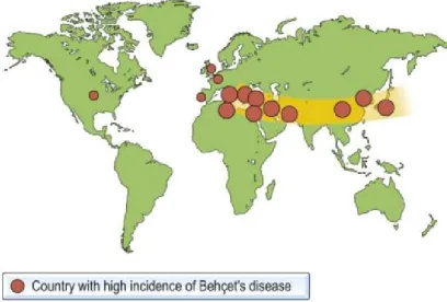
EPIDEMIOLOGY
Behçet’s Disease has been traditionally known to be associated
with the ‘Silk route’, which includes the Mediterranean and the Middle and Far East, but is a cause of ocular morbidity throughout the world.
In the Mediterranean countries, the Middle and Far East the
prevalence varies between 1/10,000 and 1/1,000. Japan has a prevalence Rate of about 1/10,000 with the occurrence more common in the temperate northern parts as compared to the subtropical southern parts of
Fig.1 : Showing countries with the highest incidence of Behçet’s disease
In Asia, the prevalence varies from 13.5 to 30 per 100,000 which is
Young adults are typically affected, with prevalence believed to be more in males. However the sex ratio varies with geographic location and
women have been found to be more commonly affected in USA and Western Europe9-13.Men usually tend to have a younger age of onset and this in turn is related to higher prevalence of ocular disease, and worse
potential visual acuity. Age and sex however have been found to be independent risk factors for the development of visual loss.14,15Although Behçet’s Disease affects primarily young adults, typically presenting in
the second to fourth decade of life, the onset however can occur at any age ranging from infants to the elderly, though it is rarely seen in children.16-21
Familial occurrence has been rarely reported with no conjugal transmission. There have been reports of lymphocytic antibodies in healthy children of parents with Behçet’s disease. Transient neonatal
CLASSIFICATION
INTERNATIONAL STUDY CRITERIA
Recurrent oral ulcerations Plus two of:
· Recurrent genital ulceration
· Eye lesions
· Skin lesions
· Positive Pathergy test
Diagnostic criteria proposed by Behçet Research Committee of Japan (1972).
MAJOR CRITERIA
1. Ocular lesions (iridocyclitis, chorioretinitis and there sequelae) 2. Recurrent aphthous ulcerations of the oral mucosa
3. Genital ulcers
MINOR CRITERIA
1. Arthritis
2. Gastroenteritis 3. Epididymitis
4. Vascular symptoms
5. Neuropsychiatric involvement
TYPES
1. Complete: all four major criteria simultaneously or at different times (ocular lesions are recurrent hypopyon iritis or typical retinitis) or three major symptoms simultaneously or at different
times
2. Incomplete: Ocular and one major or three minor criteria 3. Suspect: Two major criteria
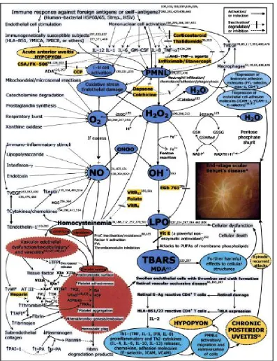
IMMUNOLOGY
Behçet disease is believed to have an autoimmune origin based on
its vasculitic nature as well as the cellular characteristics of the infiltrate in associated lesions. The main features include perivascular infiltrates consisting of lymphomononuclear cells,22,23 swelling/ proliferation of
endothelial cells which partially obliterate small vessels and lead to fibrinoid degeneration.24
Inflammation is believed to be mediated in part with cytokines which are associated with the Th1 subset of T lymphocytes. A significant number of inflammatory markers have been found to be increased in the
Fig.2 : Pathogenesis of Uveitis in Behçet’s disease
Other subsets of T and B cells have also been found to be elevated in affected patients. There is an increase in the circulating T cells of
Behçet patients.26,27 These cells have a role in recognizing autologous as well as bacterial antigens. γ – δ T cells are also elevated28 with an associated increase in the production of interferon-γ by these cells.
Finally, there is an increase in the level of activated and memory B cell subsets in affected patients29Although there has been found evidence of activation of the cellular (T cell) and humoral (B cell) limbs of immune
response in a number of these patients, the significance of this and the exact antigens triggering this response remains unknown. At the cellular level, Lehner30 has shown evidence of cell-mediated immunity to
antigens from oral mucosa. In addition, Rogers et al31 have shown increased cell mediated cytotoxicity to cultured oral epithelial cells in patients suffering from Behçet disease. Circulating immune complexes have been demonstrated by the Raji cell method.32
Other investigators have found circulating antibodies to oral mucous membrane antigens, particularly during relapse.33 Although it is
showed the presence of IgM in the walls of synovial veins and capillaries.34 It is however not clear whether the fluorescence seen is due
to the deposition of IgM or immune complex.
Other auto-antigens have been proposed, one of them being the retinal S antigen.35Weighing against this autoimmune hypothesis
however is the lack of association with other autoimmune diseases as well as the lack of production of more typical organ nonspecific auto-antibodies like antinuclear auto-antibodies.
PATHOLOGY AND PATHOGENESIS
The pathogenesis of Behçet disease still continues to be poorly
understood.
Both environmental and instrinsic factors are believed to play a role in the development of the disease. Although the disease is associated
with HLA-B51 locus, not all people who develop this disease have the genotype.
also be associated. The common pathergy skin reaction, though not present in all patients, gives an insight into the activation of this disease.
In this reaction, minor trauma to the skin of affected patients produces a rapid inflammatory response which indicates some of the same characteristic cellular infiltrates as found in systemic lesions of Behçet
disease.36 It is hypothesized that, in a genetically susceptible individual, all of these potential trigger factors for the disease can cause tissue stress or lead to the activation of heat shock proteins, thereby causing the
induction of Th1 T cell responses, with the characteristic Behçet inflammatory response as the end result.
The pathologic changes of Behçet’s disease are a lot better understood. The underlying histopathologic lesion common to all organ symptoms is a vasculitis primarily involving the venules.37,38 The chief Pathologic features include perivascular infiltrates of mononuclear cells.
Also, there is swelling/proliferation of endothelial cells leading to obliteration of the vessels, fibrinoid degeneration along with surrounding tissue necrosis. This cellular infiltrate is similar to that in the skin
The pathologic changes in Behçet vasculitis in the eye have been studied at early as well as late stages.37 In the early stage, the most
commonly seen feature is vasculitis in the retina and uveal tissues. This leads to the occurrence of retinal hemorrhage and neuroretinal degeneration. Granulation tissue and fibrous scar formation in the
vitreous and retina lead to neovascularization at the ora serrata and pars plana where cyclitic membranes develop. In the late stages, there is an occlusive vasculitic in the vessels of the cyclitic membrane. The
CLINICAL FEATURES
BD can produce distressing symptoms with systemic involvement
in varying combinations. It is charecterised by exacerbations and remissions and the healing time varies from patient to patient. The disease severity tends to diminish as time progresses and may stabilize at any moment. It however may become chronic in one of the multiple
systems involved in its course.41,42,43,44
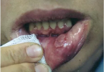
1. Oral Aphthae
These are the most universal sign of Behçet’s disease and usually the first to appear. A majority of the mild cases present in the form of aphthous ulcers involving the oral mucosa and it may not be possible to differentiate them from common aphthae – canker sores, similar in
Fig.3 : Oral Ulceration seen in patient with Behçet’s Disease
These are often painful and cause a significant amount of discomfort. These may progress fast from a tiny flat looking ulcer to a
large sore. These may occur well before other systemic manifestations and the time period between recurrences may be highly variable, between weeks to months. These lesions may be found not only on the gums but
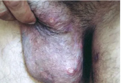
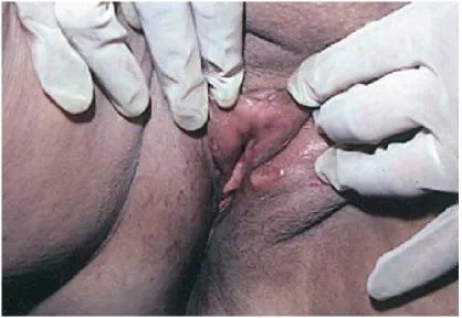
2. Genital Ulcers
These are also extremely commonly seen, reported to occur in
[image:23.595.94.510.216.500.2]nearly 80–90% cases, and are similar in appearance to oral aphthae.45,47
Fig.4 : Genital ulcers in a male patient
In males the lesions may appear on the scrotum and penis and are
therefore quite evident, sending the patient for professional help early in the disease. Females usually have their lesions on the vulva or on the vaginal mucosae. Other lesions may be perianal. Kobayashi and colleagues48 reported that lesions on the vulva occur in the premenstrual
scars. Thus an examination of the genital region in a patient with suspected Behçet's disease can be useful as signs of old disease may be
[image:24.595.92.510.183.471.2]present.
Fig.5 : Genital ulcers in a female patient
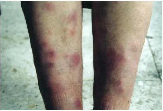
3. Skin Lesions
Skin involvement can be seen in nearly 80% of patients and is seen
in different combinations through the disease course.45,49 Papulo-vesiculo-pustular eruptions along with lesions similar to erythema nodosum are seen. These most commonly involve the lower limbs, and
those involving the anterior surface are the most commonly seen lesions and are particularly common in females .50,51
These lesions can however also be seen on the face, neck, buttocks and elsewhere. They involute after several weeks without ulceration. Acne-like lesions or folliculitis are also commonly seen. These usually
Fig. 6 : Erythema – Nodosum like lesions on the lower extremeties
Cutaneous hypersensitivity is characteristically seen in Behçet's
disease. The scratching of skin with a needle or even taking a blood sample frequently results in a pustule at that site. This phenomenon known as Pathergy – has become the basis for the ‘prick’ test, with some
OCULAR LESIONS
The eye is the most frequently affected internal organ in Behçet
Disease. In some patients ocular disease may be present at the outset.53,54 The vast majority of patients however have the onset of uveitis at 2 to 4 years of disease course
In about 1/5th of cases patients present with ocular disease as the initial manifestation. It runs a chronic, relapsing course and may show bilateral (80 %) or unilateral (20 %) hypopyon, iridocyclitis or
panuveitis.
Initial attacks have the tendency of being unilateral and more anterior. Subsequent attacks tend to involve the vitreous and posterior
segment, and are more commonly bilateral
Ocular Behçet Disease can be classified into anterior and posterior
uveitis. This is needed for therapeutic purposes as well prognostication, as the involvement of posterior segment indicates a persistent and chronic nature, and also a more severe and progressive visual loss. Most
The symptoms the patient presents with, therefore depend on the site involved.
a. Anterior Involvement
The typical presentation in the anterior segment is in the form of
Recurring and frequently Bilateral Anterior Uveitis which may be mild to severe, and can present with blurred vision associated with periorbital/global pain, lacrimation and photophobia. Circumcorneal
[image:28.595.127.474.439.715.2]ciliary injection and vasodilation are seen associated with a violoaceous hue (ciliary flush). The onset may vary from sudden, developing in a period of hours or days.
It classically produces a recurrent acute relapsing iridocyclitis
which may persist for up to 2 to 3 months.
On slit lamp examination, cells (leukocytes) can be seen in the anterior chamber as a result of inflammation and damage to the vascular
endothelium, which in turn causes increase in permeablity.
The presence of cells is indicative of active anterior segment
inflammation. Similarly, flare or Tundall effect caused due protein leakage as a result of breakdown of blood – ocular barrier can be observed. This may be present even when active inflammation has
subsided and indicates persistent vascular damage
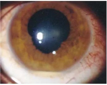
Keratic precipitates, typically seen on the lower cornea are also a frequent manifestation. They are a collection of inflammatory cells,
typically polymorphonuclear cells and lymphocytes on the endothelium of the cornea
Patients with chronic uveitis may present with floaters and mild redness associated with mild pain and photophobia. Following recurrent
attacks of acute iridocyclitis, adhesions may develop between the pupil or posterior surface of the iris with the anterior surface of the lens, forming
[image:30.595.100.500.279.554.2]posterior synechiae and leading to an irregular and distorted pupil
Fig. 8 : Posterior synechiae in a case of Behçet’s Disease
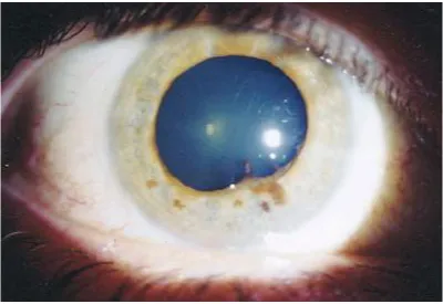
Recurrent inflammation may lead to the formation of a cyclitic membrane formation which in turn may lead to the development of 360 degrees ring synechiae or secclusio pupillae. As a result pupillary block
(peripheral anterior synechiae) may also be seen along with atrophic patches on the iris
In some instances, a macrohypopyon may be seen with the naked eye, and is caused due to the massive inflammatory and leukocytic
response. In the others, microhypopyon observed on slit - lamp biomicroscopy and angle hypopyon (demonstrated on gonioscopy) may be seen .The hypopyon is mobile in Behçet's disease which may move
with gravity, posture and head movements as the concentration of fibrinoid exudates in BD is not very high. Hypopyon may be the presenting feature in 6 % of the patients55,56 and may be missed if the
patient does not present immediately after the attack of acute inflammation.
b. Posterior Involvement
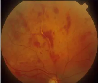
Attacks of retinal vasculitis are dreaded complications of Ocular BD. Vitritis and retinal vasculitis are the most common posterior segment manifestations of Behçet's disease and may involve the veins
Fig. 9 : Retinal Vasculitis seen in Behçet’s disease
Vitritis, on the other hand typically presents with cellular reaction in the vitreous and fewer cells in the anterior chamber, in the absence of focal lesions in the retina
Retinitis is another common manifestation of ocular Behçet's disease and presents in the form of scattered yellowish - white infiltrates
with indistinct margins causing clouding of the retina which may be superficial or deep. Superficial exudates obscure the view of blood vessels but are usually transient and self-resolving. Deep exudates on the
The severity and frequency of exacerbations of posterior segment inflammation determine the extent of permanent ocular structural damage
and irreversible visual loss.
Recurrent severe exacerbations of posterior segment inflammation
determine the extent of permanent ocular structural damage and may lead to irreversible visual loss
However, the most common form of presentation of Behçet’s disease is Panuveitis, which may present with any or all the manifestations listed above
c. Ocular Complications
Behçet’s disease can give rise to a wide range of ocular complications, which contribute to the grave morbidity which is typical
of uveitis in Behçet’s disease
Cystoid Macular Edema is reported as the most common complication seen in uveitis associated with Behçet’s Disease.57 Reported
Elevation of intraocular pressure may be seen. This may occur due to multiple mechanisms. These include plugging of the trabecular
meshwork by inflammatory cells, trabeculitis, peripheral anterior synechiae, neovascular glaucoma, seclusio pupillae, and also due to the prolonged use of corticosteroids, topical or systemic.58 Intraocular
Pressure may also be reduced in some cases due to reduced aqueous secretion
Secondary glaucoma was reported in 11% of patients by Elgin et al.59 Nearly half these cases were either steroid – induced or due to ocular inflammation, one – fourth had angle closure glaucoma associated with
peripheral anterior synechiae while one-fifth of the patients had pupillary- block angle-closure glaucoma. 10% had Neovascularization as the cause of glaucoma
Cataract formation can also be induced by inflammation or induced by the prolonged usage of corticosteroids. Posterior subcapsular cataracts are the most commonly seen form of cataract. Anterior
In advanced cases, iris atrophy may ensue, Vascular occlusion, both central as well as Branch Retinal Vein occlusion may be seen. These
are usually caused by the vasculitic changes, i.e periphlebitis and gliotic sheathing of the vessels, macular degeneration may be seen. There may be scarring in the retina, which when present in the macular region can significantly impair vision. epiretinal membrane is commonly seen.
Optic disc changes include papillitis, disc edema and optic atrophy in advanced cases. Neovascularization of the iris (rubeosis) as well as in the disc and elsewhere in the retina may be seen. These in turn can cause a vitreous haemorrhage and a secondary tractional retinal detachment60,61
After recurrent inflammatory episodes, end-stage disease with eventual complete blindness may ensue. This is charecterised by optic atrophy and sclerosed vessels that look like white cords. Retinal pigmentation accompanied by atrophy and scarring in the retina is seen.
OTHER SYSTEMIC MANIFESTATIONS
Behçet disease is a multisysytemic disorder and the other systems that may be involved include the following
1). Articular
This may be present in nearly half the affected patients, and typically presents in the form of non – migratory arthritis affecting one or two joints. There is arthralgia, occasionally with swelling and redness.
The inflammation and ulceration can subsequently lead to radiologically visible erosion of cartilage of the joint
2). Vascular Involvement
Vascular involvement is usually seen in the form of superior and inferior vena cava thrombosis. Thrombophlebitis may also be seen,
3). Gastrointestinal
Liver may be involved in the form of hepatic outflow impairment
and hepatic vein obstruction. This is secondary to hepatic venous thrombosis. Budd – Chiari syndrome or hepatic hemorrhagic infarction may also be seen in nearly 2 % individuals
Spleen may be enlarged due to splenic vein thrombosis. Ileum and Colon may be involved in the form of an inflammatory vasculitis that can
be similar in presentation to Crohn’s disease
4). Cardiovascular
These include disturbances in the conduction system, pericarditis, myocarditis, acute myocardial infarction and vascular occlusions and aneurysms. Valvular disorders like Mitral and Aortic Regurgitation have also been reported rarely
5). Renal
Microscopic haematuria as well as proteinuria may be seen, and
6). Genitourinary
In about 6 % of patients, epididymitis may be seen. Urinary
Tuberculosis and non – infective Urethritis may be seen rarely
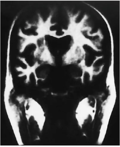
8). Central Nervous System
Central Nervous System Behçet disease presents in the from of Cerebral Venous Thrombosis involving either the cerebral veins or the dural sinuses. Pappiloedema may be seen and this may mimic Idiopathic
Intracranial Hypertension. The treatment involves use of steroids, heparin, cytotoxic drugs and in severe cases CSF shunting may have to be attempted
Fig.10 :Grey – matter lesions showing involvement of the Central Nervous
MANAGEMENT
Testing for Behçet’s Disease
At present there is no specific laboratory investigation that can accurately diagnose Behçet Disease. HLA B-51 genotype has been used for screening in some patients. The sensitivity of association however is lacking in the test which renders it to be not useful diagnostically.
Additionally, the skin pathergy reaction may occasionally be used for confirmation in Behçet patients. This procedure involves the intradermal injection with a 20-gauge needle, which leads to the development of a
[image:40.595.165.475.454.662.2]papular erythematous skin reaction within 48 hours of infection, at the site of injection
In the 1980s, a test was developed which helped detect antibodies against mucosal antigens by indirect immunofluorescene.64
Serum of Behçet patients was incubated along with other retinopathies with tissue sections. These were washed and then overlaid
with goat antibody to human immunoglobulin and conjugated with fluorescein. The serum in patients suffering from Behçet syndrome was seen to produce a typical reaction with the epidermal cytoplasm. External
surface of guinea pig lip showed the strongest reaction; Importantly however, the human oral mucosa was also positive, though to a lesser degree. On comparison with subjects with Behçet’s disease, it was found
that 80 % of patients with probable Behçet disease in an American population, and 60 % of the patients with definite Behçet disease in a Turkish population showed positive staining with this assay. The false positivity in normal subjects seen with this test was 6 %.The high cost
TREATMENT
Various treatment regimens have been attempted in Behçet disease
and many have shown promising results. None of the agents however has emerged as the single most effective treatment modality. The treatment to be adapted varies based on individual patient circumstances. Also, the
disease has a natural unpredictable, intermittent course which makes it difficult to evaluate with certainty, of various treatment regiments.
There are some generalities however about various classes of
Fig. : General outline of treatment of uveitis in Behçet’s disease Corticosteroids
by their effect on macrophage and neutrophil migration along with the activation of lymphocytes. These may be given topically, as injections in
the periocular region or given systemically. Subtenon’s injections have a significant role in the treatment of posterior segment inflammation.66
Intravenous pulse corticosteroids have been found to be effective in treating retinitis causing severe loss of vision.67
Intravitreal triamcinolone has found application in the treatment of CME(Cystoid Macular Edema) .68 Corticosteroids may also be applied in the form of mouthwash or cream for the treatment of ulcers and relieve
pain. Intra articular injection of corticosteroids is also occasionally done
Systemic administration finds a significant role in patients presenting with severe thrombophlebitis and vasculitis, CNS
involvement, or retinal neovascularization.66,69
Acute manifestations can be treated by daily administration of
Though corticosteroids form a mainstay of treatment in the acute stage, they are not as successful in preventing recurrences. They are
therefore commonly used in adjunct with other immunosuppressives There has been found to be a synergistic role between cyclosporine and corticosteroids in the treatment of acute inflammation.70
In the presence of CNS involvement persistent uveitis not responding to administered treatment, a longer duration of corticosteroid
administration is indicated. Corticosteroids are used in tapering doses as an abrupt discontinuation can in turn result in rebound increase in intraocular inflammation. However, due to adverse effects, long time use
of corticosteroids should be avoided. Adverse effects of ocular administration include intraocular pressure rise, infection, formation of cataract. Side effects associated with systemic administration include hypertension, diabetes mellitus, increased chances of infection,
gastrointestinal ulcers and osteoporosis.
Cyclosporine and FK506
extent explains their additive effect with steroids as both have separate mechanisms of action. Studies have shown that cyclosporine decreases
the severity as well as the frequency of uveitis.71,72
When combined with prednisone, improvement in visual acuity
was seen in 75% of studied eyes at a follow up period of 2 years.73 Tacrolimus has also been found to be useful in the treatment of uveitis74.
The systemic side effect profile however limits the use of both medications to severe uveitis.
Cyclosporine is also reported to play a role in the treatment of thrombophlebitis and hearing loss, which may be associated with Behçet’s disease.72. Concomitant use of cytochrome inhibitors has been found to increase the efficacy of treatment with cyclosporine76
Both cyclosporine as well as Tacrolimus should however not be used in patients having Central Nervous System manifestations. They can
Cyclosporine has been reported to result in a higher incidence of central nervous system involvement and the adverse effects caused by the
drug can be hard to distinguish with manifestations of neuro-Behçet. Nephrotoxicity and hypertension are other side effects associated with Behçet’s disease
Cyclophosphamide, Methotrexate, Azathioprine, and Chlorambucil Cyclophosphamide and Chlorambucil which are alkylating agents, along
with the antimetabolites Methotrexate and Azathioprine are used primarily in the treatment of severe manifestations, which though initially treated with steroids and cyclosporine have not been successfully
treated. These indications include refractory eye disease and Central Nervous System Vasculitis. In a randomized clinical trial which compared cyclophosphamide with cyclosporine at a dose of 5 mg/kg/day, it was noted that cyclosporine was more effective in terms of visual
acuity, though this effect could not be maintained at a review period of 2 years.77
not only in the incidence but also in the recurrence rate and severity of ocular disease.79 Also, there was found to be a long – term improvement
in the prognosis of patients with Behçet disease80
It is however, Chlorambucil that is being believed to be the agent
most effective in the treatment of retinal vasculitis81 and many neurologists favor its use for CNS vasculitis as well.
However, as mentioned before, the side effect profile of these agents limits their widespread use. These side effects include malignancies and hepatotoxicity. Bone marrow suppression and
infertility are also possible complications. The possiblity of iatrogenic teratogenicity must not be neglected as many of these patients are women of child – bearing age.
Interferon-α
Interferon-α has been reported to play a role in the treatment of mild to moderate forms of uveitis in Behçet’s disease. It however acts as
One of these studies showed response to treatment in 46 out of 50 patients having non resolving ocular disease82.Also, after an average of
16 months in 20 patients treatment was stopped, and over an observation period of 30 month no relapse was seen.82 Interferon-α can also be used in the treatment of joint and skin manifestations of Behçet’s disease. A
clinical trial showed that it was useful in treating oral and genital ulcerations and supported its empirical use.83
However, a recurrence of mucocutaneous disease is frequently seen once treatment has been discontinued.
The side effect profile of interferon – α has, in most studies not
been found to be as severe as many other agents. Flu like symptoms are commonly seen and may be troublesome in many patients. Severe side effects like psychosis and seizures have however been reported. In the
clinical trial conducted, 30 % patients had to discontinue interferon – α
due to toxicity 84
Infliximab
The mechanism of action of Infliximab is by inhibition of TNF. Though initially it was used only to treat very severe forms of the disease
and occasionally for gastrointestinal disease, it has now been documented to play a significant role in its ability to sustain the patient in a state of remission
In a study, the largest conducted so far, of 25 patients, 24 showed immediate remission on only a single treatment with this agent.90
Also, of a total of 15 patients who were given another round of treatment over a period of 32 weeks, there were no further recurrences in
60 %. However, despite the encouraging results so far, data regarding it s efficacy and safety over a long-term period is still not adequate. Infliximab has also been found to be having an effective role in treating non – ocular manifestations of Behçet’s disease, particularly
mucocutanous ulcers and Central Nervous System disease.91,92
The adverse effect profile of this agent is however incompletely
Surgical Options
In Behçet disease, phthis bulbi is an unfortunate but commonly
seen complication. This may be caused either as result of chronic inflammation causing degeneration of tissue, or by the detachment of ciliary body which in turn may be caused by inflammation or surgical
intervention. Vitreoretinal surgery has been reported to improve not only the visual prognosis but also prevent recurrence in uveitis associated with Behçet’s disease96
One such study showed improvement in visual acuity of 5 out of 10 eyes, from hand movement to finger counting or 3/60 on a Snellen
scale .97
Also, amongst these 10 eyes, none showed severe postoperative inflammation or developed phthisis bulbi. There was also a decrease of
recurrent inflammatory episodes in these patients.
Behçet’s patients were also found to be well tolerant to
Complications were seen in in 12% to 17% of eyes and included severe postoperative inflammation, posterior synechiae formation, and
cystoid macular edema. Another study showed that the visual outcome was to a great extent dependent on the extent of inflammation seen post operatively in patients undergoing extracapsular cataract surgery.99
In patients who underwent surgery without preoperative ocular inflammation, there was no significant rise in inflammation
Prognosis
Behçet’s disease as already mentioned typically shows remissions
followed by relapses. There is no well defined uniform disease period and there is no external factor known yet that can control the period of remission in these patients. Mucocutaneous manifestations are the commonest to occur and rarely cause significant permanent impairment.
Morbidity is usually as result of the ocular inflammation, thrombophlebitis, arthritis and Central Nervous system and other neurologic complications, all of which may be seen in patients with
segment involvement usually show a good prognosis on treatment with immunosupressives and as time progress, the frequency of relapses
lessens and the disease course stabilizes at 15 – 20 years with no further attacks. The major causes of disability in patients are blindness which is seen in 25 %, and neurological defects along with paralysis which is seen
in nearly 10 % patients. Death from complications of Behçet’s disease is rare, with the commonest causes being severe Central Nervous system involvement, ruptured aneurysm and intestinal perforation.
Although the treatment of Behçet disease continues to be a struggle, there is significant improvement being made. As we begin to
understand the disease better and our treatment options continue to expand there has been a significant improvement in the treatment outcome of patients suffering from Behçet’s disease, particularly in terms of preservation of vision.100
AIMS & OBJECTIVES
AIM
To study the clinical picture, visual prognosis and treatment outcome of uveitis in Behçet’s disease
OBJECTIVES
· To study the demographic patterns
· To the study the pattern of uveitis in Behçet’s disease
· To study the complications of Behçet’s disease
MATERIALS AND METHODS
TYPE OF STUDY
Hospital Based, Observational Prospective Study
INCLUSION CRITERIA
Patients who met the 1990 classification criteria of the International Study Group for Behçet’s disease were included in the study.
EXCLUSION CRITERIA
· Ocular involvement other than uveitis
· Any history of trauma
· Patients with uveitis other than Behçet’s disease
METHODOLOGY
· A detailed history was obtained from each patient which included
¨ Ocular complaints
· A complete ocular examination was performed at each visit which
included
¨ Best corrected visual acuity on a snellen scale
¨ Slit lamp biomicroscopy
¨ Tonometry
¨ Indirect ophthalmoscopy
· Reports from the patient’s primary ophthalmologists,
dermatologists, and rheumatologists were evaluated and patients were referred to the related specialist when required during the follow – up visits
· A data form was prepared which collected information on
¨ Demographics including age at presentation
¨ Male or female sex
¨ Extraocular clinical manifestations of Behçet’s disease
· Ophthalmic data recorded which included
¨ Age at onset of uveitis
¨ Type of uveitis (namely anterior, posterior or panuveitis)
¨ Ocular findings
¨ Ocular complications
¨ Visual acuity
· The treatment advised for each patient was documented
· The patient was examined on each follow up visit
RESULTS
This descriptive study included a total of 55 eyes of 30 patients
and was conducted at the Uvea clinic of Aravind Eye Hospital and Postgraduate Institute of Ophthalmology, Madurai in the period of May 2013 to May 2014
1. Demographic Pattern a. Age at presentation
The mean age of patients in the study was 28.6 with a standard
deviation of ± 8.8 and the range was 13-49.
b. Age at onset
The mean age of studied patients at onset was 26.27 ± 9.0 and the
range was 13-49.
c. Sex
Sex
Female
Male
Total
93.3%
1. Sex
No. of patients %
2 6.7
28 93.3
30 100
6.7%
93.3%
Sex
2. Complaints Complaints
Complaints No. of eyes %
Defective vision Present Absent 44 11 80.0 20.0 Pain Present Absent 13 42 23.6 76.4 Watering Present Absent 3 52 5.5 94.5 Photophobia Present Absent 8 47 14.6 85.4 Redness Present Absent 20 35 36.4 63.6 Floaters Present Absent 13 42 23.6 76.4
The most common complaint patients presented with was defective vision (44 eyes or 80 %), followed by redness (20 eyes or 35 %), pain (13 eyes or 23.6 %), floaters (13 eyes or 23.6 %), photophobia (8 eyes or
3. Laterality
Laterality No of patients %
Unilateral 5 9.1
Bilateral 25 90.9
90.9%
9.1%
90.9%
Laterality
4. Location
Location No. of eyes %
Anterior 1 1.8
Intermediate 1 1.8
Posterior 1 1.8
Panuveitis 52 94.6
Total 55 100
Panuveitis was the presentation in a large majority of patients. Of a total of 55 involved eyes, 52 or 94.6 % showed Panuveitis. Anterior uveitis was seen in 1 patient (1.8%), Intermediate in 1 patient (1.8%), and
1.8% 0%
20% 40% 60% 80% 100%
Anterior Intermediate
1.8% 1.8%
94.6%
Intermediate Posterior Panuveitis
5. Duration of first presentation
The duration of first presentation in our study ranged from 7 to
3650 days and Median duration of symptoms was 365 days.
6. Severity Severity
Severity No. of eyes %
Mild 7 12.7
Moderate 17 30.9
Severe 31 56.4
Total 55 100
12.7%
0% 20% 40% 60% 80% 100%
Mild
30.9%
56.4%
Moderate Severe
7. Chronicity
Chronicity No. of eyes %
Acute 3 5.4
Chronic Recurrent 52 94.6
Total 55 100
The most common presentation was in the form of chronic recurrent uveitis, seen in 52 (94.6 %) of eyes. Only 3 eyes (5.4 %)
8. Visual acuity
Baseline vision category
Vision category No. of eyes %
6/6-6/18 29 52.7
6/24-6/60 7 12.7
5/60-3/60 6 10.9
<3/60 13 23.6
Total 55 100
In our study, at presentation a majority of the patients had a visual acuity in the range of 6/6 – 6/18(29 eyes or 52.7 %). 7 patients or 12.7 % fell in the range of 6/24 – 6/60, 6 or 10.9 % in 5/60 – 3/60, and 13 eyes or
52.7%
Baseline Vision Category
6/6-12.7%
10.9%
23.6%
Baseline Vision Category
-6/18 6/24-6/60 5/60-3/60 <3/60
10.9%
Final vision category
Vision category No of eyes %
6/6-6/18 31 57.4
6/24-6/60 8 14.8
5/60-3/60 2 3.7
<3/60 13 24.1
Total 54 100
57.4%
Final Vision Category
6/6
14.8%
3.7%
24.1%
Final Vision Category
6/6-6/18 6/24-6/60 5/60-3/60 <3/60
52.7% 57.4%
0% 20% 40% 60% 80% 100%
6/6-6/18
Baseline Vision Category Vs Final Vision
Baseline Vision Category 12.7%
10.9%
23.6%
14.8%
3.7%
24.1%
6/24-6/60 5/60-3/60 <3/60
Baseline Vision Category Vs Final Vision
Category
Best Corrected Visual Acuity Log mar
vision
N Median (Snellen’s equivalent)
Mean (SD)
Min-Max P-value*
Baseline 55 0.48(6/18) 0.81(0.9) 0-2.9
0.481 Final 54 0.48(6/18) 0.78(0.9) 0-2.9
*Wilcoxon signed rank test
Between the baseline and final visual acuity, the P- value was
found to be 0.481 which was not found to be statistically significant
There were a total of 7 eyes which showed a visual recovery of 2
9. Ocular findings – Anterior Segment Variable No. of eyes %
Scleritis Yes No 1 54 1.8 98.1 Conjunctival ulcer Yes No - 55 - 100 Keratitis Yes No - 55 - 100 Keratic Precipitates Yes No 36 19 65.4 34.6 Hypopyon Yes No 9 46 16.4 83.6 Cells Yes No 44 11 80.0 20.0 Flare Yes No 45 10 81.8 18.2 Iris nodules Yes No 1 54 1.8 98.2 Lens status Clear lens Cataract Pseudophakia Aphakia 35 12 6 1 63.6 21.8 10.9 1.8 Extra ocular
Iridocyclitis presenting as cells and flare was the most commonly seen manifestation in our study, seen in 44 (80.0 %) and 45 Eyes (81.8 %
patients) respectively.
Keratic precipitates were seen in 36 eyes, which constituted 65.4
% of the eyes
Hypopyon was seen in 9 eyes (16.4 %). 1 eye (1.8 %) showed
1.8% 0.0% 0.0% 98.1% 100%
0% 20% 40% 60% 80% 100%
0.0%
65.4%
16.4%
80.0% 81.8%
1.8% 100%
34.6%
83.6%
20.0% 18.2%
98.2%
Anterior Segment
Yes No
Lens status
Clear lens
Cataract Pseudophakia
Aphakia
35
12 6 1
63.6
21.8 10.9 1.8
63.6%
0% 20% 40% 60% 80% 100%
Clear Lens
21.8%
10.9%
1.8%
Cataract Pseudophakia Aphakia
10.Posterior segment Posterior segment
Variable No.of eyes %
Media Clear Hazy 39 16 70.9 29.1 Vitreous cells Present Absent 49 6 89.1 10.9 Snow bank Present Absent 5 50 9.1 90.9 Snow balls Present Absent 8 47 14.5 85.5 Retinitis Present Absent 13 42 23.6 76.4 Vasculitis Present Absent 35 20 63.6 36.4 Vitreous opacities Present Absent 24 31 43.6 56.4 Neovascularization Present Absent - 55 - 100 Optic nerve edema
Present Absent 7 48 12.7 87.3 Macular edema Present Absent 18 37 32.7 67.3
The most common posterior segment manifestation was in the form of vitreous cells seen in 89.1
%) eyes.
70.9%
0% 20% 40% 60% 80% 100%
Clear
Posterior Segment
common posterior segment manifestation was in the form of vitreous cells seen in 89.1(49 %) eyes, and vasculitis in 35 (63.6
29.1%
Hazy
Posterior Segment - Media
Snow balls and snow banking were seen in 5 (9.1 %) and 8 (14.5 %) patients respectively.
Retinitis was seen in 13 (23.6 %). Vitreous opacities were seen in 24 (43.6 %) eyes, retinitis in 13 (23.6 %) eyes. Optic nerve edema and
macular edema were seen in 7(12.7 %) eyes and 18(32.7 %) respectively.
89.1% 9.1% 14.5% 10.9% 90.9% 0% 20% 40% 60% 80% 100%
Posterior Segment Manifestations
14.5% 23.6% 63.6% 43.6% 0.0% 12.7% 85.5% 76.4% 36.4% 56.4% 100% 87.3%
Posterior Segment Manifestations
Present Absent
32.7% 87.3%
11.Extraocular manifestations
Extraocular manifestation Present Absent
Recurrent oral ulcer 28(93.3) 2(6.7)
Genital ulcer 17(56.7) 13(43.3)
Arthritis 11(36.7) 19(66.7)
Skin lesions 4(13.3) 26(86.7)
Thrombophlebitis - 30(100.0)
Recurrent oral ulceration was present in 28 of the 30 patients studied (93.3%). Genital ulceration was the next most common
manifestation seen in 17 (56.7 %) patients. Arthritis was %) patients, Skin lesions in 4 (13.3 %) patients,
only in 1 (3.3 %) patients.
93.3% 56.7% 6.7% 43.3% 0% 20% 40% 60% 80% 100%
Extraocular Manifestation
Recurrent oral ulceration was present in 28 of the 30 patients studied (93.3%). Genital ulceration was the next most common
manifestation seen in 17 (56.7 %) patients. Arthritis was seen in 11 (36.7 kin lesions in 4 (13.3 %) patients, and CNS involvement
36.7% 13.3% 0.0% 3.3% 43.3% 66.7% 86.7% 100%
Extraocular Manifestation
Present AbsentRecurrent oral ulceration was present in 28 of the 30 patients studied (93.3%). Genital ulceration was the next most common
seen in 11 (36.7 and CNS involvement
12.Past treatment
The patient’s previous treatment history was documented and it
was observed that a large number of patients were undertreated (24 or 43.6 %). 18 (32.7 %) were adequately treated whereas 13 (23.6 %) had received no previous treatment at all.
Past treatment No. of eyes %
Not treated 13 23.6
Under treated 24 43.6
Adequately treated 18 32.7
43.6%
Past Treatment of Patients
Not treated
23.6% 43.6%
32.7%
Past Treatment of Patients
Immunosuppressives Administered to Patients outside Prior to First Visit with us
Immunosuppressive No. of eyes %
None 32 58.2
Methotrexate 8 14.5
Cyclophosphamide 2 3.63
Azathioprine 5 9.09
Cyclosporine a 7 12.7
Colchicine 6 10.9
In the treatment given outside to the patients, most commonly used immunosuppressive (8
cyclosporine A (7 or 12.7 %),
or 9.09 %), Cyclophosphamide (2 or 3.63 %) and Mycophenolate (1 or 1.82 %)
14.5% 3.63%
9.09%
ImmunosuppressivesAdministeredtoPatients
outsidePriortoFirstVisitwithus
None
Azathioprine
Mycophenolate Mofetil
In the treatment given outside to the patients, Methotrexate wa immunosuppressive (8 or 14.5 %), followed by
7 or 12.7 %), Colchicine (6 or 10.9 %), Azathioprine (5 or 9.09 %), Cyclophosphamide (2 or 3.63 %) and Mycophenolate Mofetil
58.2% 12.7% 10.9% 1.82%
ImmunosuppressivesAdministeredtoPatients
outsidePriortoFirstVisitwithus
Methotrexate Cyclophosphamide
5Cylosporin A Colchicine
Mycophenolate Mofetil
Methotrexate was the or 14.5 %), followed by
6 or 10.9 %), Azathioprine (5 Mofetil
58.2%
Treatment Topical steroid Oral steroid PSI steroid Immunosuppressive 69.1% 30.9% 0% 20% 40% 60% 80% 100% 13.Treatment
USED NOT USED
38(69.1) 17(30.9)
51(92.7) 4(7.3)
18(32.7) 37(67.3)
Immunosuppressive 39(70.9) 16(29.1)
At our centre, Oral steroids were the most commonly used treatment regimen (51 or 92.7 %). Immunosupressives were used in 39 or
70.9%. Posterior subtenon injection of Triamcinolone acetonide were administered in 18 eyes (32.7 %) and topical steroids were used in 38 (69.1 %) of eyes
Immunosuppressive No.of eyes %
None 14 25.45
Methotrexate 29 52.72
Cyclophosphamide 3 5.45
Azathioprine 6 10.91
Cyclosporine A 3 5.45
Colchicine 8 14.54
In those administered immunosupressives, Methotrexate was the most common (29 or 52.72 %) followed by
Azathioprine (6 or 10.91 %), Mycophenolate cyclosporine A (3 or 5.45 %) and
5.5% 10.9% 5.5%
Immunosuppressive
None Azathioprine Mycophenolate MofetilIn those administered immunosupressives, Methotrexate was the most common (29 or 52.72 %) followed by Colchicine (8 or 14.54 %),
r 10.91 %), Mycophenolate Mofetil (4 or 7.27 %), cyclosporine A (3 or 5.45 %) and cyclophosphamide (3 or 5.45 %)
25.5%
52.7% 5.5% 14.5% 7.3%
Immunosuppressive
Methotrexate Cyclophosphamide Cylosporin A Colchicine
Mycophenolate Mofetil
In those administered immunosupressives, Methotrexate was the 8 or 14.54 %),
25.5%
52.7%
0% 20% 40% 60% 80% 100%
5.5% 10.9% 5.5% 14.5% 7.3%
Immunosuppressive
14.Complications
Complication Present Absent
Rubeosis 2(3.6) 53(96.3)
Anterior synechiae 2(3.6) 53(96.3)
Posterior synechiae 14(25.5) 41(74.55)
Cataract 17(30.9) 38(69.1)
Vitreous haemorrhage 3(5.4) 52(94.6)
Optic atrophy 5(9.1) 50(90.9)
Macular edema 16(29.1) 39(70.9)
Macular degeneration 9(16.4) 46(83.6)
Macular hole 1(1.8) 54(98.2)
Epiretinal membrane 17(30.9) 38(69.1)
Neovascularisation - 55(100)
Retinal tear - 55(100)
Retinal detachment 3(5.4) 52(94.6)
Phthisis bulbi 2(3.6) 53(96.4)
The most common complications observed in the study were Cataract and epiretinal membrane which were each seen in 17(30.9 %) of
the eyes.
Synechiae were seen in 16 eyes (29.1 %) where 2 (3.64 %) showed
anterior synechiae and 14 (25.5 %) showed posterior synechiae.
Macular edema was present in 17(30.9 %) eyes, macular
degeneration in 9 (16.4 %) eyes, macular hole in 1(1.8 %).
2 patients (3.6 %) showed rubeosis iridis. Optic atrophy was seen
in 5(9.1 %) eyes Vitreous haemorrhage and retinal detachment were each seen in 3(5.4 %) eyes.
4 patients (3.6 %) were seen to have an end stage disease and 2
3.6%3.60% 25.50%
30.9%
0% 20% 40% 60% 80% 100%
5.4%9.1% 29.1%
16.4%
1.8% 30.9%
0.0% 0.0% 5.4%
Complications
DISCUSSION
Age Distribution
In our study the distribution of age of onset in patients varied from a range of 13-49 and had Mean of 26.27 with a standard deviation of ± 9.0. Tutkun et al10 reported a mean of 30 years ranging from 9 years to 72
years. Barra et al101 reported the mean age of 29.6 years which ranged from 9 to 61 years. Uragancioglu et al104reported the mean age of 15.4 ± 3.3 years from 9 to 27 years. Rohtagi et al reported the mean age of 25.7
years ranging from 15 to 46 years. Colvard et al103 reported the mean age of 33.3 years with a range of 10 to 66 years.
Sex Distribution
Male to female ratio in our study was 14 : 1. Tutkun et al10 reported a male : female ratio of 2.1 : 1, males (68%) more frequently involved than females (29%) with a male to female ration of 2.5 : 1.
Laterality
Comparative Analysis of Laterality Laterality Tutkun et
al10
Barra et al101
Uragancioglu et al104
Present study
Unilateral 193(21.9) 2(4.0) 6(16.6) 5(9.1)
Bilateral 687(78.10) 47(96.0) 30(83.3) 25(90.9)
A large majority of the patients in our study showed bilateral presentation (25 patients or 90.9 %). This was comparable to other
Extraocular Manifestations
Comparative analysis of extraocular clinical manifestations
Manifestations Tutkun et al10
Barra et al101
Rohatgi et al102
Colvard et al103
Present study
Recurrent oral ulcers
880(100) 48(98) 19(100) 32(100) 28(93.3)
Genital ulcers 526(59.8) 27(55.1) 18(94.7) 27(84.0) 17(56.7)
Arthritis 299(34.0) 22(44.9) 17(89.5) 18(56.0) 11(36.7)
Erythema 387(55.4) 25(51.0) 11(57.9) 22(69.0) 4(13.3)
Thrombophlebitis 41(4.7) 2(4.1) 0(0)
Central Nervous system
37(4.2) 3(6.1) 7(22.0) 1(3.3)
The commonest extraocular manifestation seen in our study was
Genital ulceration was seen in 17(56.7 %) of our patients, which was comparable to that reported by Tutkun et al10 and Barra et al.
Rohatgi et al and Colvard et al103 reported a higher incidence of 94.7 % and 84 % respectively.
Arthritis was seen in 11 of our patients. Tutkun et al10 showed an incidence of 34 %, Barra et al101 44.9%, Rohatgi et al102 showed 89.5 % and Colvard et al103 showed an incidence of 56 % .
Erythema nodosum was seen in only 4(13.3 %) of our patients. Other studies however reported a higher incidence of the same. Tutkun et
al10 showed 55.4 %, Barra et al101 51.0 %, Rohatgi et al102 57.9 %, and Colvard et al103 69 %.
None of our patients showed Thrombophlebitis but the same was
reported in 4.7 % by Tutkun et al10 and 4.1 % by Barra et al101.
Only 1 of our patients (3.3%) showed Central Nervous system
Pattern of Uveitis
Comparative analysis of type of Uveitis
Type of Uveitis
Tutkun et al10
Uragancioglu et al104
Barra et al101
Present study
Anterior 172(11.0) 5(13.8) 7(14.8) 1(1.8)
Posterior 452(28.8) 1(1.8)
Panuveitis 943(60.2) 31(86.2) 42(85.7) 52(94.6)
The most common presentation was of Panuveitis (94.6 %) in our
Comparative analysis of Ocular Findings – Anterior Segment
Anterior segment findings
Tutkun et al10
Barra et al 101
Rohatgi et al102
Colvard et al103
Present study
Iritis 42(85.7) 2(10.5) 21(65.0) 44(80.0)
Hypopyon 188(12.0) 17(34.7) 3(15.2) 3(9.3) 9(16.4)
Iritis presenting in the form of cells in the anterior chamber was
Comparative Analysis of Ocular Findings – Posterior segment
Posterior segment findings
Tutkun et al10
Barra et al101
Uragancioglu et al104
Colvard et al103
Present study
Vitritis 1395(89.0) 42(86.0) 32(88.8) 21(65.1) 49(89.1)
Vasculitis 1395(89.0) 49(100) 59(89.3) 13(40.6) 35(63.6)
Retinitis 808(51.6) 45(68.2) 17(89.4) 13(23.6)
Papillitis 86(5.5) 8(16.3) 7(12.7)
Vitritis was the most common posterior segment finding in our study seen in 49(89.1 %) eyes. The same was reported in 89.0 % by Tutkun et al10, 86 % by Barra et al101, 88.8 % by Uragancioglu et al104,
Vasculitis was the second most common finding, seen in 35(63.6 %) eyes in our study. Tutkun et al10 reported the same in 89.0 %, Barra et
al101 in 100 %, Uragancioglu et al104 in 89.3 %, Colvard et al103 in 40.6 %.
Retinitis was seen in 23.6 % eyes in our study, whereas Tutkun et al10 reported the same in 51.6 %, Uragancioglu et al104 68.2 %, Colvard et al 103 89.4 %
COMPLICATIONS Complications Tutkun
et al10
Barra et al101
Uragancioglu et al104
Present study
Cataract 604(38.5) 40(41.7) 31(46.9) 17(30.9) Synechiae
Posterior
409(26.1) 14(21.2) 14(25.5)
Epiretinal membrane
266(17.0) 17(30.9)
Macular edema 697(44.5) 6(12.2) 16(29.1)
Intraocular pressure rise
216(13.8) 0(0) 7(10.6) 13(23.64)
Optic Atrophy 370(23.6) 18(18.8) 26(39.4) 5(9.1) Intravitreal
hemorrhage
36(2.3) 3(5.4)
Retinal Detachment
23(1.4) 0(0) 4(6.1) 3(5.4)
Macular Degeneration
304(19.4) 15(5.5) 30(45.4) 9(16.4)
Rubeosis 19(1.2) 2(3.6)
Phthisis bulbi 28(1.8) 2(2.1) 1(1.5) 2(3.6)
Corneal opacity 1(1.5) 0(0)
Neovascularisation 68(4.3) 1(1.5) 0(0)
Macular hole 41(2.6) 5(5.2) 1(1.8)
Cataract was the most commonly complication seen in our study seen in 30.9 % eyes in our study. The same was reported in 38.5 % by
Tutkun et al10, 41.7 % by Barra et al101, and 46.9 % by Uragancioglu et al104.
Posterior synechiae were seen in 25.5 % patients in our study, which was comparable to 26.1 % by Tutkun et al10 and 21.2 % by Uragancioglu et al104.
Epiretinal membrane was seen in 30.9 % of our patents while Tutkun et al10 reported the same in 17.0 %.
Macular edema was seen in 16(29.1 %) eyes in our study, while the incidence was reported as 44.5 % by Tutkun et al10 and 6(12.2 %) by Barra et al101.
Optic atrophy was seen in 9.1 % of our patients, while it was higher i