A Dissertationon
A STUDY ON THE PRESENCE OF ACCESSORY
MAXILLARY OSTIUM
Submitted to the
THE TAMILNADU DR. M.G.R. MEDICAL UNIVERSITY
In partial fulfilment of the requirements For the award of the degree of
M.S.BRANCH IV
(OTORHINOLARYNGOLOGY)
GOVERNMENT STANLEY MEDICAL COLLEGE & HOSPITAL
THE TAMILNADU DR. M.G.R. MEDICAL UNIVERSITY, CHENNAI, TAMILNADU
DECLARATION
I, Dr. VRINDA .B. NAIR, solemnly declare that the dissertation, titled “A Study On The Presence Of Accessory Maxillary Ostium” is a bonafide work done by me during the period of January 2013 to July 2014 at Government Stanley Medical College and Hospital, Chennai under the expert supervision of PROF. DR. T. BALASUBRAMANIAN, M.S., D.L.O., Professor and Head, Department Of Otorhinolaryngology, Government Stanley Medical College and hospitals, Chennai.
This dissertation is submitted to The Tamil Nadu Dr. M.G.R. Medical University in partial fulfilment of the rules and regulations for the M.S. degree examinations in Otorhinolaryngology to be held in April 2015.
Place: Chennai-1 DR.VRINDA .B. NAIR
CERTIFICATE
This is to certify that the dissertation -“A Study On The Presence Of Accessory Maxillary Ostium” presented BY DR.VRINDA .B. NAIR , is an original work done in the Department of Otorhinolaryngology, Government Stanley Medical College and Hospital, Chennai in partial fulfillment of regulations of the Tamil Nadu Dr. M.G.R. Medical University for the award of degree of M.S. (Otorhinolaryngology) Branch IV, under my supervision during the academic period 2012-2015.
THE DEAN,
Govt. Stanley medical college,
Chennai-1.
PROF.DR.T.BALASUBRAMANIAN
PROFESSOR AND HEAD OF THE DEPARTMENT
Govt. Stanley Medical College and Hospital Chennai-1
ACKNOWLEDGEMENTS
I wish to express my sincere thanks to Prof. Dr.AL. MEENAKSHI SUNDARAM, MD, DEAN, Government Stanley Medical College and Hospital for having permitted me to utilize the facilities of the hospital for conducting this study.
My heartfelt gratitude to Prof.Dr.T. BALASUBRAMANIAN, M.S., D.L.O., Professor and Head of the Department, Department of Otorhinolaryngology, Government Stanley Medical College and Hospital for his constant motivation, valuable suggestions, and expert supervision during the course of this study.
I express my whole-hearted gratitude to Prof.Dr.N. SEETHALAKSHMI M.S.,D.L.O.,D.N.B, Professor and Chief of ENT UNIT II of Otorhinolaryngology, PROF. DR. F.ANTHONY IRUDHAYA RAJAN M.S.,D.L.O, Professor, for supporting, guiding and encouraging me in this study.
I wish to thank my Assistant Professors DR.K.ATHIYAMAN M.S, DR.C.KARUPPASAMY M.S.,D.L.O., DR.M.P.CHANDRAMOULI M.S., DR.SARAVANA SELVAN M.S., DR.C.BHARANIDHARAN D.L.O. for their valuable tips and guidance.
I also thank the staff nurses, theatre personnel, OPD staff, Department of Otorhinolaryngology, Government Stanley Hospital for their co-operation and assistance in the conduct of this study.
I wish to extend my gratitude to my statistician for his expert assistance.
Last but not the least, I am indebted and grateful to all the
TABLE OF CONTENTS
1. ABSTRACT 1
2. OBJECTIVE 3
3. INTRODUCTION 5
4. REVIEW OF LITERATURE 7 5. MATERIALS AND METHODS 38
6. RESULTS 51
7. DISCUSSION 64
8. CONCLUSION 109
9. ANNEXURES 111
A.BIBLIOGRAPHY B. PROFORMA
C. ETHICAL COMMITTEE APPROVAL LETTER
D. PATIENT INFORMATION SHEET
E. INFORMED CONSENT FORM F. PLAGIARISM
1.
ABSTRACT
The association between accessory ostium and chronic sinusitis is still a subject of controversy as various studies have given a wide range of variable results. The prevalence of accessory ostium varies between 2 – 44 %.
Objective
To study the prevalence of Accessory ostium in those with Chronic Sinusitis and those without the disease.
Methods
83 patients with chronic sinusitis and 83 subjects without the disease were studied by nasal endoscopy for the presence of accessory ostium.
Results
Page | 2 the diseased individuals had double ostia. Only 12 % showed
circular phenomenon. However symptoms like headache , facial pain , post nasal drip and halitosis , and purulence on examination (Rhinosinusitis Task Force Criteria) were not found to have any specific association with the presence of accessory ostia.
Conclusion
To conclude we would say that the presence of an accessory ostium can be considered as an indicator of maxillary sinus disease, along with the other criteria for chronic sinusitis and that it can help surgeons in their decision making as to whether a surgery is required or not.
Page | 3
2.
OBJECTIVE
To study,
Page | 4 INCLUSION CRITERIA:
1. Age:15-45 years.
2. Patients with clinical symptoms of chronic maxillary sinusitis. 3. Radiological diagnosis of chronic maxillary sinusitis.
4. Persistence of symptoms for more than 12 weeks even after medical line of treatment.
EXCLUSION CRITERIA:
1. <15 or>45 years of age.
2. History of previous maxillary sinus surgeries. 3. Patients with sinonasal neoplasms.
4. Patients with sinonasal encephaloceles.
5. Patients with known ciliary dysfunction such as primary ciliary dyskinesia and Kartagener Syndrome .
6. Patients with sinonasal polyposis. 7. Patients with fungal sinusitis.
8. Patients with Cystic Fibrosis .
Page | 5
3. INTRODUCTION
Chronic sinusitis is a group of disorders characterized by the inflammation of nasal and paranasal sinus mucosa which is now extremely prevalent, affecting 32 million adults , or 16.3 % of the adult population[9]. Its diagnosis is mainly based on clinical symptoms and signs, supported by diagnostic nasal endoscopy and CT scan.
Maxillary sinusitis is the side effect of evolution as a result of which man acquired an erect posture. The secretions of the maxillary sinuses have to be transported against gravity by the mucociliary action. Hence the normal functioning of a sinus depends on – patency of the ostium , normal mucociliary transport and the normal quality and quantity of secretions.
Page | 6 Majority of the accessory ostia occur singly. A few multiple ostia have also been observed. They are mostly acquired and rarely congenital. However there is still a controversy over its etiology.
Page | 7
4. REVIEW OF LITERATURE
Hippocrates in the 5th century B.C itself has described sinusitis when he quoted “in a person having a painful spot in head, with intense headaches, pus or fluid running from the nose removes the disease.”
Leonardo da Vinci in his period had accurately given the description of the maxillary antrum and the frontal sinus. [1]
In 1869, Wertheim made a Conchoscope to examine the anterior and middle thirds of the nasal cavity. [1]
In 1879, Nitze-Leiter developed the Cystoscope, and this marked the second stage in the developement of endoscopy. [1]
Hirschmann is considered the Father of Endoscopy. In 1902, Hirschmann and Valentin introduced a modified cystoscope into a maxillary sinus through an enlarged dental alveolus.[1]
Page | 8 In the 1950s Messerklinger developed a systematic diagnostic approach to the lateral wall of nose. [1]
HISTORY OF STUDIES ON MAXILLARY ACCESSORY OSTIUM
Page | 9 result from rupture of fonatanelles due to the blockage in the primary maxillary ostium. Scheaffer (1920) reported the AMO to number between 1 -3. But this is uncommon and hence there is not much in the literature regarding multiple openings. Stammberger & Kennedy (1995) came up with an incidence of 4-5 % in the general population as opposed to 25 % in those with chronic sinusitis.
Incidence of AMO ranges between 2 - 44 % , lower values being obtained from in vivo studies whereas higher ones from cadaver studies. The reason for this wide variation may be that the AMO that may be covered with a mucous film and in inaccessible locations are often missed out in in vivo studies. Whereas in cadaver studies, due to drying and fixing, the moist nasal mucosa can undergo shrinkage, also the fontanelle mucosa may be damaged during drying and investigations, giving an impression of higher incidence.
Page | 10 Myerson (1932), van Alyea (1936), Lang & Wurzburg (1991), Kumar. H et al (2001, Lady Hardinge medical college, New Delhi ) in their studies on cadavers came up with an incidence of 31 %, 23 %, 28%, 30 %[5] respectively.
Page | 11
INCIDENCE AND LOCATION OF
ACCESSORY MAXILLARY OSTIA
S. No. REFERENCE LOCATION INCIDENCE
%
STUDY MATERIAL
1 Schaeffer (1920) ANF or PNF 43 Cadavers
2 Myerson (1932) Not Specified 31 Cadavers
3 Van Alyea
(1936)
Not Specified 23 Cadavers
4 May et al (1990) PNF 0 Cadavers 10 Endoscopic 5 Kennedy
&Zinreich (1991)
Not Specified 15 Endoscopic
6 Lang &
Wurzburg (1991)
Not Specified 28 Cadavers
7 Stammberger & Kennedy (1995)
ANF or PNF 5 General population 25 Diseased 8 Kumar et al
(2001)
Page | 12 An experimental animal study was conducted by Genc et al in New Zealand type rabbits where they induced rhinogenic sinusitis using Streptococcus pneumonia in the right nasal cavities. After sacrificing the rabbits on the 21st day , their lateral nasal walls were examined. An accessory ostium was observed in 40% in the fontanelle like regions. The opposite nasal cavity showed no pus or accessory ostium. Histopathological examination of the mucosa showed - eosinophils, lymphocytes, plasma cells, ciliary loss, epithelial degeneration, areas of ulceration and lymphoid follicle hyperplasia.
RELEVANT ANATOMY
EMBRYOLOGY OF THE LATERAL NASAL WALL[2]
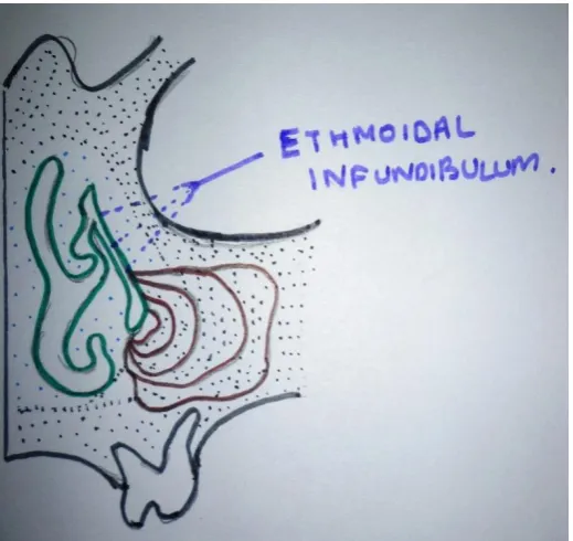
Page | 13 The Maxilloturbinal ridge arises inferior to these structures and gives rise to the Inferior turbinate. Primary furrows between the ethmoturbinals form nasal meati and recesses. The first furrow lies between the first and second ethmoturbinals, the descending part of which forms the Ethmoidal infundibulum, Middle meatus and Hiatus semilunaris. Its ascending part forms the Frontal recess. The second and third primary furrows form the superior and supreme meati respectively. Secondary lateral nasal wall Evagination gives rise to the Bulla ethmoidalis, and the Secondary lateral wall Invagination gives rise to the Supra and Retrobullar recesses.
Page | 14 Figure 1:Development of maxillary sinus from ethmoidal infundibulum.
Page | 15
ANATOMY OF LATERAL WALL[2]
The complex ethmoidal labyrinth can be reduced into a series of lamellae which corresponds to the following - 1st- uncinate, 2nd – ethmoidal bulla, 3rd – basal or ground lamella and 4th – lamella of superior turbinate.
Agger Nasi
It is seen as a prominence anterior to the attachment of the middle turbinate to the lateral nasal wall. This region may be pneumatised by an anterior ethmoidal cell giving rise to the Aggernasi cell. It takes origin from the superior aspect of the infundibulum or the frontal recess region.
Ethmoidal Bulla
Page | 16
Hiatus Semilunaris
Hiatus Semilunaris Inferior of Grunwald is a 2 dimensional sagittaly oriented crescent shaped gap between the posterior free margin of the uncinate process and the anterior free margin of the bulla ethmoidalis. It communicates with the ethmoidal infundibulum.
Hiatus Semilunaris Superior
It is cleft between the posterior wall of bulla and the basal lamella where the middle meatus communicates with the Lateral Sinus ( Retrobullar and Suprabullar recess)
Ethmoidal Infundibulum
It is a 3 dimensional funnel shaped passage through which secretions from Anterior ethmoid, Maxillary and frontal sinus are transported and channeled into the middle meatus.
Frontal Recess
Page | 17 anteriorly by the posterosuperior wall of Agger nasi and posteriorly by the anterior wall of the ethmoidal bulla.
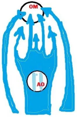
Osteomeatal Unit
It is a functional designation (Naumann) referring to all the middle meatal structures ,viz. – the uncinate , ethmoid infundibulum , anterior ethmoidal cells , ostia of anterior ethmoidal , maxillary and frontal sinuses.
Uncinate process
Page | 18
Fontanelles[1,2,3,8,35]
Page | 19 have been found to be 4-5 % in the general adult population and 25 % in those with Chronic rhinisinusitis[3].
MUCOCILIARY CLEARANCE[2]
Paranasal sinuses are lined by pseudostratified ciliated columnar epithelium with goblet cells. The maxillary sinus has the highest density of goblet cells compared to other paranasal sinuses ( 9700/mm2 ).[3] Seromucinous glands are relatively infrequent , concentrated around the ostium. The thickness of the sinus mucosa is 0.2 – 0.8 mm.
Page | 20 mucosal samples showing absent ciliary activity. Isotope methods show that there is a lesser drainage of tracer substances in sinuses with
a. Retention of fluid
b. Thick mucosa
Quantitative and qualitative changes in the secretion, including the delicate periciliary fluid layer is of greater importance for the impairment of mucociliary transport during sinus inflammation than the structural abnormality of the cilia or their retarded beat rate. Ciliary impairment in the presence of purulent secretions is due to-
a. High proteolytic enzyme activity
b. Low pH
c. Anaerobic mucosal metabolism
Page | 21
SECRETION AND TRANSPORTATION[1]
Principles
Drainage and ventilation are two important factors for the normal physiology of the paranasal sinuses and their mucus membranes. Drainage depends on secretion and transport mechanisms which in turn depend on
-· Amount of mucus
· Composition of mucus
· Effectiveness of ciliary beat
· Mucosal resorption
· Condition of ostia and ethmoidal clefts
· Free flow of inspired air
· Mucosal pulsations and movements of fontanelles (in case of inflammation)
Page | 22 quantity are important and requires an intact blood supply and nervous system. It is composed of -
1. Water & ions
2. Glycoproteins (sialomucins , fucomucins , sulphomucins)
3. Enzymes (lysozymes, lactoferrin)
4. Circulatory proteins (Complement , α 2 macroglobulin , CRP)
5. Ig- IgA , IgE , IgG , IgM , IgD
6. Cells – surface epithelium , basophils , eosinophils , leucocytes.
The mucus film has two layers – the sol phase , which is the
inner serous layer (water and ions) and the gel phase , which is the
Page | 23
· Humidity
· Pollution
· Airborne irritants
The mucosal glands are controlled mainly by the parasympathetic fibers. The nerve fibers from the Superior Salivatory Nucleus via the Greater Petrosal Nerve reach the Pterygopalatine ganglion, from where the postganglionic fibers supply the mucosal glands.
The sympathetic fibers arise from the Lateral Horn of the spinal cord. The postsynaptic fibres via the Carotid plexus form the DeepPetrosal Nerve which joins the Greater Petrosal Nerve to form the Vidian Nerve which ultimately supplies the nasal and sinus mucosa.
Substance P secreted from type C fibers via local reflexes also have great effect on mucosal glands. They are found to produce Hypersecretion , Vasodilation and extravasation of plasma.
Page | 24 the viscous layer is found to be thicker as secretions from the whole sinus converge there.
When mucosal surfaces come close to each other leaving a recess in between, the gap is filled by the mucosal blanket by a the
Bridging phenomenon, whereby the cohesive forces in the gel phase bridge the gap, while the sol phase fills the recess in between. Similarly the flow over a small mucosal defect also goes unhindered due to cohesive nature of the mucus carpet.
But when the mucus is too viscous, this defect can prove to be an obstacle with the secretion being retained at the site of the defect. Similarly at the region of crests a thick secretion will be retained for a while and finally drain away under the influence of gravity. A highly viscous mucus can block the primary maxillary ostium ,later fall down into the sinus only to be transported again towards the primary ostium. If the sinus ostium is oval or oblong , theciliary beat works on the mucus from two or three sides and the mucus passes through the corners of the ostium.
Page | 26
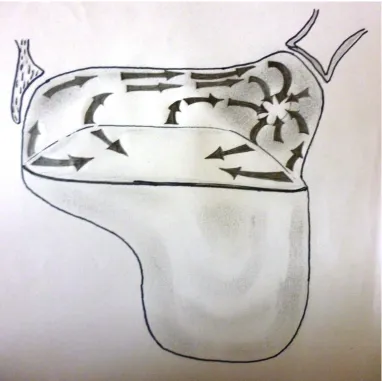
Figure 2Mucous Blanket moving over and across
Page | 27
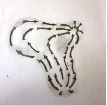
Figure 3Blue arrow shows circular transport of mucous.
SECRETION TRANSORT PATHOLOGY
Page | 28 direction of transport in the corresponding areas are not opposing each other.
Figure 4 A half filled maxillary sinus. The cohesional forces of the
gel phase are holding back the transportation of the mucus. The
still intact cilia are not powerful enough to ‘tear off ’ the gel layer
Page | 29 In case of decreased secretions or reduced humidity at the surface , sol phase becomes rather thin. The mucus becomes very viscous and the gel phase comes in direct contact with the cilia , thereby impeding their action resulting in a worm like movement of the mucus layer.
During inflammation of the sinuses , the mucosa gets inflamed rapidly and even pulsate. Also increased movements in the region of the membranous portions of the fontanelles may occur. Such mucosal movements may also assist the transport of secretions out of the maxillary sinus.
There is another phenomenon known as “secretion expressways” which is found in both cases of abnormal secretions as well as apparently normal sinuses. This means that the transport of secretion is not uniform throughout a sinus. From time to time the mucosa of one region transports secretion faster than the neighboring areas. In some time the slower areas catch up speed and the faster region slows down.
Page | 30
· Normal ventilation
· Humidification
· Normal Metabolism
· Osmotic pressure
· Optimal pH – 7-8
· Optimal temperature – around 330 C
· protection from noxious stimuli
TRANSPORT OF SECRETION IN THE MAXILLARY SINUS
Page | 31 ethmoidal sinuses usually join. The ethmoidal infundibulum via the Hiatus
Figure 5Mucous Transportation Pathways inside Maxillary Sinus
Page | 32 ciliated and squamous epithelium active transport continues after which secretions move downwards by gravity aided by the swallowing mechanism.
Secretions from the maxillary sinus always tend to pass
through the natural ostium even in the presence of a single or
multiple accessory ostia or a surgically created inferior meatal
antrostomy.
MECHANISM OF MAXILLARY SINUSITIS
Page | 33 Also poor ventilation decreases the pH of the sinus , further slowing down its ciliary movement with the resultant formation of viscous mucous. Such viscous mucous may remain in the sinus for a considerable length of time due to the blockade in the prechamber. This again is a favorable condition for microbial growth, toxins of which may cause further impairment of the mucosal function. This is an instance where an otherwise normal sinus ends up diseased due to a pathology that lies in the outflow tract.
Inhaled pathogens deposited at the entrance of the middle meatus adhere to the mucus. Due to the confluence of the mucus pathways of the ethmoidal infundibulum and the frontal recess , the microbes get transported into either of the sinuses. In the presence of a suboptimal self healing capacity of the sinus mucosa or inadequate antibiotic therapy an Acute or a Chronic recurring sinusitis results.
Page | 34 mucus may again re enter the sinus via the accessory ostium and this may continue endlessly. As long as the natural ostium is patent this is not of much significance. But if it is blocked or in the presence of a nasal infection this plays an important role in the transport of pathogens into the maxillary sinus from the nose. Due to ostial blockade infected secretions are unable to leave the sinus resulting in maxillary sinusitis.
Thus the cause of infections of the large sinuses like maxillary sinus is mostly rhinogenic. It usually spreads from the nose through the ethmoid clefts and prechambers into the maxillary and frontal sinuses. An exception to this is dentogenic sinusitis (<2%). Other such exceptions are foreign bodies in the maxillary sinus (aberrant root filling material) , blood in the sinus following trauma, cholesterol or mucus retention cysts (depending on their location).
OSTIAL PATENCY AND GAS STUDIES[2]
Page | 35 aberration of pO2 and pCO2 which results in mucosal exudation
and development of local pathology. Mucosal exudation is preceded by extravasation and interstitial spreading of plasma derived liquids and proteins leaving the lamina propria with a pool of inflammatory mediators.
Clinical initiation of sinusitis is a 3 step process -
· Ostial obstruction
· Pathogenic bacteria
· Impaired local defense.
Page | 36 and sinus, diffusion is the dominant transport mechanism. Whereas for larger ostia and smaller concentration differences it is convection. Contrary to Proetz’s findings , they have proved that the presence of one or more accessory ostia increases the sinus ventilation rates by several folds. In their study model, a standard
double ostium geometry (ostium 1 &ostium 2 with 3mm diameter & 6 mm length each ) has a pressure difference of ~0.1 Pa between the ostia.[4] This pressure difference produces an inflow to the sinus through the upstream ostium and an outflow from the sinus through the downstream ostium of 7.2 x 10⁻⁷m3/s.[4] The time to replace 90 % of sinus air (T90) ~31.9 seconds. In the various other double
ostium models , which varied in the ostial diameter and length , this T90 varied from 8.9 seconds to 31.9 seconds.[4]
LIMITATIONS OF SINUS VENTILLATION[4]
The natural ventilation rate in a single ostium sinus is extremely slow. This slow ventilation is protective as it
-- helps prevent drying of mucosal secretions
Page | 37 Whereas the transport rates are higher in a double ostium sinus with ostia exposed to even very slightly different pressure changes compared to a single ostium sinus. When a sinus with a single ostium can be considered as a reservoir of fluid attached to the nose, only a sinus with multiple ostia can have a net flow through it as it offers an alternative flow path in parallel with the nose. Thus their study contradicts Proetz’s conclusion that an accessory ostium cannot increase the ventilation of a sinus.
ADVERSE EFFECTS OF INCREASED VENTILATION
1. Decreased NO concentration - (when convective transport exceeds production) leading to impaired mucociliary function resulting in increases pathogen entry into the sinus
2. Mucosal drying - ( as density of goblet cells in sinus is less than nasal cavity). Risk of mucosal drying is more when the upstream ostium is closer to the nostril.
Page | 38
5. MATERIALS AND METHODS
83 Patients (Male and female in the age group of 15-45 years) with Chronic Maxillary Sinusitis who attended the ENT Out Patient Department and 83 subjects , who attended the ENT Out Patient Department for reasons other than Chronic Sinusitis and normal volunteers, during the period January 2013 to July 2014 , who satisfied the inclusion criteria were enrolled for the study after getting an informed written consent.
Study design : Cross Sectional study
Study place : Department of ENT, Government Stanley Medical College and Hospital.
Study and Follow-up period : January 2013 to July 2014.
Page | 39
METHODS OFSTUDY
HISTORY
CLINICAL EXAMINATION
DIAGNOSTIC NASAL ENDOSCOPY
DIAGNOSTIC CRITERIA FOR CHRONIC
RHINOSINUSITIS(1997 RHINOSINUSITIS TASK FORCE)[9]
(>12 weeks)
Major Factors
1.Facial pain/pressure
2.Nasalobstruction
3.Nasaldischarge/discolored postnasal drip
4.Hyposmia/anosmia
Page | 40
Minor Factors
1.Headache
2.Fever (all nonacute)
3.Halitosis
4. Dental pain
5. Fatigue
6. Cough
7. Ear pain/pressure/fullness
DIAGNOSIS[9]
Presence of either;
Ø 2 major factors, or,
Page | 41
Facial pain/pressure alone does not constitute a suggestive
history for diagnosis in the absence of another major symptom or
sign.
Since in our present study we had normal subjects as well , we did not use the CT findings as a criteria in order to avoid unnecessary radiation exposure. Hence in our study we only adhered to the Task Force Criteria and Nasal Endoscopic findings for diagnosing Chronic Sinusitis.
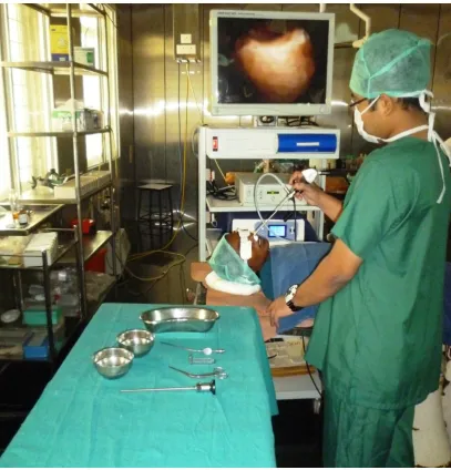
EQUIPMENT USED:
1. 4mm – wide angled zero degree, 30o Karl Storz endoscopes.
2. Stryker HD camera ( 24 mm )
3. High definition LED monitor
Page | 42
Figure 61.HD camera , 2. 300 Karl Storz Endoscope , 3. 00 Karl Storz endoscope , 4. Tilley’s nasal dressing forceps , 5. Thudicum speculum , 6. Metal suction tip ,
7. Kidney tray , 8. Nasal packs dipped in decongestant solution , 9. Defog solution – Savlon
Page | 43
Page | 44 PROCEDURE[10,11]
Topical anesthetic, about 7 ml of 4% xylocaine was mixed with 10 drops of xylometazoline. Cotton pledgets dipped in the solution, squeezed dry and used to pack the nasal cavity. Pledgets were packed in the inferior, middle and superior meati and left in place for full 5 minutes. Diagnostic endoscopy is performed using a 4mm30 degree Hopkin nasal endoscope.
First pass :
The endoscope was introduced along the floor of the nasal cavity. First the inferior meatus came into view. In cases where the inferior turbinate was lateralized , the same was medialised by applying more topical anaesthetic. Endoscope was advanced posteriorly to identify adenoid tissue. Eustachean tube identified. The entire nasopharynx was visualized by rotating the 300 endoscope.
Second pass:
Page | 45 The scope is gently slipped medial to the middle turbinate to view the sphenoethmoidal recess.
Third pass:
The shape and size of the middle turbinate as well as its relationship to the lateral nasal wall and septum was evaluated. The middle turbinate is gently medialised and the attachment of the uncinate process is carefully noted. Any discharge in this area also recorded. If accessory ostium is present it comes into view now. Accessory ostium is present more posteriorly.
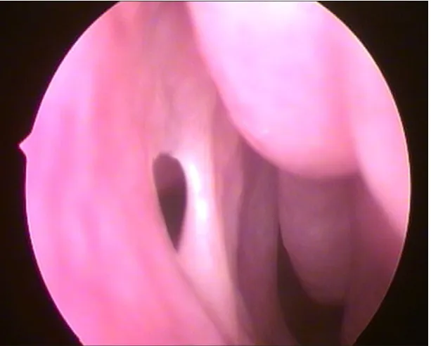
Normal ostium is actually not visible during diagnostic nasal
endoscopy. Accessory ostium is spherical in shape and
oriented anteroposteriorly, while the natural ostium of
maxillary sinus is oval in shape and oriented transversely.
PMO AMO
Ovoid in shape Circular
Page | 46
CROSS SECTIONAL STUDY
A cross-sectional study examines the relationship between disease (and other variables of interest as they exist in a defined population at a single point in time or over a short period of time. Cross-sectional studies can be thought of as providing a snapshot of the frequency of a disease or other health related characteristics (e.g. exposure variables) in a population at a given point in time.
Cross-sectional studies are used to assess the burden of disease or health needs of a population and are particularly useful in informing the planning and allocation of health resources.
SAMPLE SIZE CALCULATION
Page | 47 Sample size was calculated using OpenEpi Version 3 , open source calculator.
Sample Size:X-Sectional, Cohort, & Randomized Clinical Trials
Two-sided significance level(1-alpha): 95 Power(1-beta, % chance of detecting): 90 Ratio of sample size, Unexposed/Exposed: 1 Percent of Unexposed with Outcome: 5 Percent of Exposed with Outcome: 22
Odds Ratio: 5.4
Risk/Prevalence Ratio: 4.4
Risk/Prevalence difference: 17
Kelsey Fleiss Fleiss with CC
Sample Size – Exposed 85 83 95 Sample Size-Nonexposed 85 83 95
Total sample size: 170 166 190
Prevalence = number of cases in a defined population at a given period of time number of persons in the given period of time
Using the formula the prevalence of accessory ostium in chronic sinusitis (Pc)and the prevalence of accessory ostium in
Page | 48
Pc = 30 %
Pn= 10 %
In order to find the significance of the values , a Chi Square test was done on the sample.
[image:54.595.97.520.326.437.2]The data captured in the sample size was arranged in the following table to arrive the X2
Data type 1 Data type 2 Totals Category 1 A b a + b Category 2 C d c + d Total a + c b + d a + b + c + d = N
The formula for Chi Square Distribution is
X2= (ad-bc)2 (a+b+c+d) (a+b) (c+d) (b+d) (a+d)
AO + AO - Totals
Exposed (CRS +) 25 58 83
Not Exposed(CRS
-) 8 75 83
Total 33 133 166
Page | 49 probability level (alpha)
Df 0.5 0.10 0.05 0.02 0.01 0.001
1 0.455 2.706 3.841 5.412 6.635 10.827
2 1.386 4.605 5.991 7.824 9.210 13.815
3 2.366 6.251 7.815 9.837 11.345 16.268
4 3.357 7.779 9.488 11.668 13.277 18.465
5 4.351 9.236 11.070 13.388 15.086 20.517
Applying the formula above we get:
Chi square = 166[(25)(75) - (8)(58)]2 / (33)(133)(83)(83) = 10.93058
When a comparison is made between one sample and another, a simple rule is that the degrees of freedom equal (number of columns minus one) x (number of rows minus one) not counting the totals for rows or columns.
For our data this gives (2-1) x (2-1) = 1.
Page | 50 10.93058) lies near 10.827. The corresponding probability is less than 0.001. Since a p-value of 0.001 is lesser than the conventionally accepted significance level of 0.05 (i.e. p < 0.05) we reject the null hypothesis.
In other words, there is a statistically significant difference in the proportion of AO in patients with Chronic Sinusitis and in those without.
Page | 51
6. RESULTS
Prevalence Of AO (Accessory ostium) In The Exposed Population
AO No. in exposed population (CRS)
YES 25
NO 58
TOTAL 83
Yes, 30%
No, 70%
Page | 52
Prevalence Of AO In The Unexposed Population
AO No. in unexposed population
YES 8
NO 75
TOTAL 83
Yes 10%
No 90%
Page | 53
Bar Chart Showing In Difference In Prevalence In Chronic Sinusitis
&Normal Population
Among the 83 CRS patients enrolled for study , 25 ( 30 % ) had an Accessory ostium and in the 83 unexposed persons it was 8 ( 10 % )
AGE DISTRIBUTION OF AO IN CHRONIC SINUSITIS (CRS)
Subjects in the age group 15 – 45 years were chosen for the study. The distribution of CRS in the various age groups , as obtained from our recent study has been shown in the table and chart are given below.
AO [VALUE]
AO [VALUE]
0% 5% 10% 15% 20% 25% 30% 35%
Page | 54
0 2 4 6 8 10 12 14 16 18 20
15 - 20 21 - 25 26 - 30 31 - 35 36 - 40 > 40
No. of CRS patients in each age group
Age distribution of CRS
Age group Frequency of CRS
15 - 20 19
21 - 25 14
26 - 30 18
31 - 35 9
36 - 40 12
Page | 55 Age Distribution of AO in CRS
CRS AO % of AO
Age < 30 51 14 27.45
Age > 30 32 11 34.38
SIDE DISTRIBUTION OF AO IN CHRONIC SINUSITIS
The following are the tables and charts showing the side on which the AO was present - right / left / bilateral in the group with CRS.
27.45%
34.38%
0.00% 5.00% 10.00% 15.00% 20.00% 25.00% 30.00% 35.00% 40.00%
< 30 > 30
Page | 56
SIDE FREQUENCY
Right 9
Left 10
B/L 6
Right 36%
Left 40%
B/L 24%
Page | 57
SIDE DISTRIBUTION OF AO IN THE UNEXPOSED
The following are the table and chart showing the side on which the AO was present - right / left / bilateral in the unexposed group.
SIDE FREQUENCY
Right 3
Left 4
B/L 1
Right 37%
Left 50%
B/L 13%
Page | 58
DISTRIBUTION OF AO ACCORDING TO ITS SITE ON THE
LATERAL WALL – ANF / PNF
AO is more commonly found on the posterior nasal fontanelle (PNF) than the anterior nasal fontanelle (ANF). Following is the frequency of the sites of occurrence in the two groups.
IN CHRONIC SINUSITIS
SITE FREQUENCY
ANF 6
PNF 19
AF 24%
pF 76%
SITE OF AO IN CRS
Page | 59
IN THE UNEXPOSED
SITE FREQUENCY
ANF 0
PNF 8
AF 0%
pF 100%
SITE OF AO IN UNEXPOSED
Page | 60
DISTRIBUTION OF DOUBLE OSTIA IN
CHRONIC SINUSITIS
The occurrence of AO is usually single and occasionally multiple. In our study we found only 2 such cases and that too only in those with CRS. Double ostia were not present in the unexposed group in the present study.
NO. OF OSTIA FREQUENCY
DOUBLE 2
SINGLE 23
Double, 8%
One, 92%
Page | 61
Distribution of signs & symptoms among CRS patients
with and without AO
The following tables show the variation in percentage of symptoms and signs like discolored postnasal drip , halitosis , purulence on examination.
Also Recirculation phenomenon in the accessory ostium was found only in 2 patients of CRS.
Frequency CRS with AO Percentage
Discolored
postnasal drip 24 25 96.00%
Halitosis 16 25 64.00%
Purulence on
examination 19 25 76.00%
Page | 62 96.00%
76.00%
64.00%
12.00%
0.00% 20.00% 40.00% 60.00% 80.00% 100.00% 120.00%
Discolored postnasal
drip purulence on examination Halitosis recirculation
symptoms & signs - CRS with AO
Frequency CRS without
AO Percentage
Discolored
postnasal drip 56 58 96.55%
Halitosis 24 58 41.38%
Purulence on
examination 42 58 72.41%
Page | 63 96.55%
72.41%
41.38%
0.00%
0.00% 20.00% 40.00% 60.00% 80.00% 100.00%
Discolored postnasal
drip purulence on examination Halitosis recirculation
Page | 64
7. DISCUSSION
INTERPRETATION AND ANALYSIS OF DATA
PREVALENCE OF ACCESSORY OSTIUM
[image:70.595.155.462.467.713.2]Analysis of our present study shows that 25 out of 83 ie. 30 % CRS patients had an AO and that 8 out of 83 ie. 10 % , subjects without CRS had an AO. All of them were round in shape. As mentioned earlier prevalence of AO shows a wide range between 2 – 44 % , with cadaveric studies showing a higher incidence than that on live subjects. The various studies and results have been tabulated in the section ‘Review of Literature’.
Page | 65
Figure 9 A closer look at the same AO showing the interior of maxillary sinus
[image:71.595.166.463.417.706.2]Page | 66 Here we are quoting a few more studies that were not discussed in the previous section.
In a prospective cohort study by Jog and Mc Garry, on Rhinology clinic patients and General ENT clinical controls, they reported that 7 % of Rhinology patients and 2% of the controls had AO. Of the rhinology patients with rhinitis and sinusitis , 8 % showed AO.[20]
AlperSindel et al [21] Manju et al [19] Kolvekar et al[22] Manjula Patil et al[23]in their cadaver studies have reported the prevalence as 13.8% , 18.5 % , 22.5% and 26% respectively.
AGE
Subjects chosen for the study belonged to the age group between 15 and 45 years. In the present study the frequency of CRS in each group was found to be the following –
Page | 67 accessory ostium, ie. 27.45 %. Of the 32 patients above 30 years, 11 had accessory ostia, ie. 34.38 %.
In the unexposed group, of the 35 individuals below 30 years 4 (11.42 %) had AO, and out of 48 above 30 years, 4 (8.33 %) had AO.
Though literature says that AO can develop with advancing age, such an association was not obtained in our study[24].
GENDER
According to the review of literature gender had no effect on the development of AO. Hence no conscious effort was taken to equalize the number of males and females while enrollment.
SIDE
In our study, of the CRS patients with AO, 10 (40 %) on the
Left, 9 (36 %) were on the Right and 6 (24 %) were present
Page | 68
Figure 11 Bilateral AO (PNF in the a 16 year old male with CRS
In a study by Mladina et al , 68.3 % AO were bilateral in patients with CRS as opposed to none the normal subjects.[17]
Page | 69
bilaterally in those with post nasal discharge as against none in the group of healthy subjects. There
is no mention on the right / left distribution in either of these studies.[18]Kolvekar et al[22] have reported on a 2.66% laterality.
In a cadaver study by Kumar et al the findings were as follows – right 66.7 % , left 33.3 %. There were no bilateral cases.[5]
In a cadaver study Manju et al they found that AO occurred on the Right in 60% and on the Left in 40%. [19]
[image:75.595.104.513.167.344.2]A radiological study by Sheetal et al showed 13 % Right and 11 % left.
Page | 70
LOCATION OF ACCESSORY OSTIUM – ANF / PNF
In the group with CRS , 19 (76 %) AO were found on the posterior nasal fontanelle and 6 ( 24 % ) in the anterior nasal fontanelle. In the unexposed individuals , all the 8 AO were found on the posterior nasal fontanelle.
Page | 71
Figure 13AO in PNF
NUMBER OF OSTIA
Page | 72
Figure 14 Double Accessory Ostia
Kumar et al have reported a 44.4 % incidence of double AO in their cadaver study. Manju Singhal et al have reported a 35 % incidence of double AO , and all of them were in the anterior nasal fontanelle. [19]
Page | 73 Again all these were cadaver studies. Of all the studies , cadaver studies have given a larger incidence of accessory ostia and here in particular the incidence of multiple / double ostia are also larger in these studies. This could be due to the fact that moist nasal mucosa undergo shrinkage after death , and following drying and fixing the fontanelles undergo damage resulting in the formation of accessory ostia.[5]
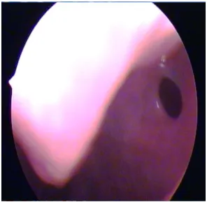
Figure 15 Lower ostia was not visualized initially as
Page | 74
RECIRCULATION
[image:80.595.139.466.231.507.2]Recirculation was found only in 3 ( 12 % ) cases of AO in this study. All the three cases had post nasal drip and also purulence on examination.
Figure 16 mucous moving out of AO
Page | 75 towards it, only to repeat this vicious cycle. This results in persistence of sinus infection.
[image:81.595.138.480.336.649.2]In the figure above, secretion appears to be moving out of the accessory ostium into the middle meatus. In the normal course, though an accessory ostium is more advantageously placed than the natural one, it does not take part in the physiological drainage of the maxillary sinus secretions.
Figure 17 Circular phenomenon – the mucous secretion re entering
Page | 76 Only in the presence of any blockade or obstruction of the natural ostium does secretions get transported out through the accessory ostium, under the effect of gravity.
ANALYSIS OF THE SYMPTOMS AND SIGNS
95 % of CRS patients complained of headache and 42 % had facial pain. 80 out of 83 (96.4 % ) patients complained of post nasal discharge. Hence there was no significant difference in its presence between those with and without AO. Halitosis was a complaint only for 49.3 %. However it was higher in those with AO ( 64 % ) than those without AO (41.38 % ). Purulence on examination was slightly higher in the presence of an AO ( 76% ) in contrast to those without ( 72.41 % ).But both these associations could not be proved statistically as the p value was > 0.05 for both.
CHRONIC RHINOSINUSITIS[9],[12]
Chronic rhinosinusitis is one of the most common otorhinolaryngologic problems affecting 32 million adults, or 16.3 % of the adult population.[9]
Page | 77 osteitic changes. Since the mucosal lining is contiguous between the nose and sinuses, one can’t spared if the other is involved. Hence the term Sinusitis has now been expanded into Rhinosinusitis.
There are two categories of changes in Chronic Rhinosinusitis
1. Polypoid mucosal changes with eosinophilia ( which causes more damage to the nasal mucosa )
2. Submucosal serous gland hyperplasia.
The Rhinosinusitis Task Force of the American Academy of Otolaryngology Head and Neck Surgery have developed a classification.[12]
CLASSIFICATION DURATION
ACUTE RHINOSINUSITIS (ARS) 7 days to ≤ 4 weeks SUB ACUTE RHINOSINUSITIS 4 weeks to 12 weeks
RECURRENT ACUTE
RHINOSINUSITIS
≥ 4 episodes of ARS / year
CHORONIC RHINOSINUSITIS (CRS)
≥ 12 weeks
Acute exacerbation of Chronic Rhinosinusitis
Page | 79 The 1997 Task Force of Rhinosinusitis report also described the following physical findings which they divided into 2 groups. These are important but not required as a part of TFR criteria. They are -
1. those findings that are accessible to all clinicians ( examination of face and anterior rhinoscopic findings )
2. those accessible only to specialists ( nasal endoscopy)
The specificity of endoscopy is 85 %.
External Findings Anterior Rhinoscopy Nasal Endoscopy
Swelling and erythema of maxillary, frontal, ocular, orbital areas
Hyperemia Blue discoloration of turbinates
Edema OMC / ostia purulence
Crusts Polyp
Purulence Septal deviation Polyp Concha Bullosa Changes in symptoms
after topical decongestion
Paradoxical Middle Turbinate
Page | 80 The Chronic Rhinosinusitis Task Force has published another set of guidelines for diagnosing adult Chronic Rhinosinusitis where it recommends to continue the use of 1997 TFR CRS symptoms and to add the existence of physical findings – Polyps , Purulence , Polypoid changes [9]
[13]There is another staging system which can be considered
the most accepted CT staging system called the Lund-Mackay CT staging system. It is a very simple system and has a high degree of interobserver and intraobserver agreement. It is the only system recommended by the Task Force on Rhinosinusitis for outcomes research.
Scoring is based entirely on CT findings. Each sinus is given a of 0/1/ 2:
0 = no opacification,
1 = partial opacification,
Page | 81 There is a separate grading for frontal, maxillary, anterior ethmoid, posterior ethmoid, and sphenoid sinuses. The ostiomeatal complex is also included in the score. The total possible score is 24.
LUND-MACKAY COMPUTED TOMOGRAPHY STAGING SYSTEM
No abnormality Partial opacification Total opacification Anterior ethmoid L R 0 0 1 1 2 2 Posterior ethmoid L R 0 0 1 1 2 2
Maxillary L
R 0 0 1 1 2 2
Frontal L
R 0 0 1 1 2 2
Sphenoid L
R 0 0 1 1 2 2
Non obstructed Obstructed
Page | 82
MANAGEMENT ALGORITHM FOR CHRONIC SINUSITIS[9]
DIAGNOSIS OF CRS SUGGESTED BY HISTORY
SINONASAL ENDOSCOPY
POSITIVE ENDOSCOPY NORMAL
PURULENCE CT PNS
OBTAIN CULTURE POSITIVE NORMAL
- Consider other
diagnosis
POLYP -Allergy evaluation
MANAGEMENT OF POLYP
INITIATE TREATMENT
Page | 83
TREATMENT OF CHRONIC RHINOSINUSITIS[14]
Our upper respiratory tract which includes the nasopharynx is a storehouse for pathogenic bacteria which cause respiratory tract infections including rhinosinusitis.
An Upper Respiratory Tract Infection has several phases - An initial viral infection which lasts about 10 days undergoes complete recovery in the majority. A minority develop an acute secondary bacterial infection by facultative aerobic bacteria. This if not resolved pave way for anaerobic bacteria of the oral flora.
EARLY VIRAL INFECTION (~ 10 DAYS)
Complete recovery(majority) 0.5 %
Secondary acute bacterial infection [Streptococcus pneumoniae, H.influenzae, Moraxella catarrhalis]
Not resolved
Page | 84
MICROBIOLOGY OF ACUTE SINUSITIS[14]
The community acquired acute purulent maxillary, frontal and ethmoid sinusitis are caused by Streptococcus pneumoniae, Hemophilus influenzae, Moraxella catarrhalis, beta hemolytic streptococci.
Staphylococcus aureus and Hemophilus influenza are the causative agents in acute sphenoid sinusitis.
In nosocomial infections , pseudomonas and gram negative rods are the culprits.
Whereas fungal sinusitis is seen in the immunocompromised and diabetics.
MICROBIOLOGY OF CHRONIC SINUSITIS[14]
Anaerobes like Prevotella , Fusobacterium , Peptostreptococcus and aerobes like Staphylococcus aureus , Moraxella catarrhalis and Hemophilus sp. are the pathogens in chronic sinusitis.
Page | 85 Pseudomonas aeroginosa and gram negative aerobic bacilli are seen in sinus infections following sinus surgeries.
In chronic sinusitis, Polymicrobial infection is more common. Hence it is more difficult to eradicate with narrow spectrum antibiotics.
In chronicity, aerobes and facultative species are replaced by anaerobes.
This is thought to be due to –
1. Selective pressure of antibiotics that enable resistant organisms to survive.
Page | 86
TREATMENT OF CHRONIC SINUSITIS[14]
MEDICAL MANAGEMENT
Antibiotics should be effective against both aerobes and anaerobic Beta Lactamase Producing Bacteria. The following antibiotics can be used for treatment.-
Oral & Parenteral Forms Only Parenteral
Amoxicillin Clavunate Cefoxitin
Clindamycin Cefotetan
Chloramphenicol Cefmetazole Macrolide + Metronidazole Imipenem Newer generation - Trovafloxacin
Along with antibiotics, topical steroid sprays, nasal saline irrigation and mucolytics is also useful.[15]
In infections by aerobic gram negative bacteria like pseudomonas aeroginosa
1. parenteral aminoglycosides
Page | 87 3. Fluoroquinolones ( oral / parenteral ) in post pubertal age groups.
These antibiotics should be given for 21 days. The treatment may be extended up to 10 weeks.
When a patient does not respond to medical management , surgical drainage should be done. Antibiotic treatment alone without surgical drainage of pus may not result in eradication of disease.
The reason for failure of medical treatment in chronic sinusitis may be due to the fact that chronically inflamed mucosa with a lesser blood supply is a poor medium of transport of antimicrobial agents to the affected tissue even in the presence of a therapeutic blood level. Also the reduced oxygen tension and acidic pH of the inflamed sinuses interfere with antimicrobial activity of the drugs.
[16]Another reason for drug resistance in chronic sinusitis are
Page | 88 cooperative manner. The matrix is slime like made of polysaccharide, nucleic acids and proteins.
In a study on New Zealand white rabbits, sinusitis was induced by pseudomonas aeroginosa. On days 1, 5, 10, 20 – pus was cultured to get pseudomonas aeroginosa. The mucosa on scanning under a microscope revealed growth and biofilm.
Another study was conducted on the sinonasal specimens from patients undergoing revision sinus surgery or office based debridement. All were antibiotic non responders. Their specimens also showed findings consistent with presence of biofilms.
SURGICAL MANAGEMENT
INDICATIONS[42]
Absolute Indications
1. complications of sinusitis
2. expansile mucoceles
3. allergic / invasive fungal sinusitis
Page | 89 Relative Indications
1. Symptomatic nasal polyps unresponsive to medical therapy
2. Chronic / Recurrent acute sinusitis unresponsive to medical therapy
Functional Endoscopic Sinus Surgery has shown to be the approach of choice in chronic sinusitis. Though the association between anatomical variations and recurrence of disease is still under dispute , surgery is acceptable when the site of obstruction complements the area of recurrent symptomatology.
Page | 90 PREOPERATIVE EVALUATION
Prior to surgery, a patient requires culture directed antibiotics which according to the severity of the disease may be given for up to 2 or more weeks. Steroids may put into use in case of polyposis or hyperactive mucosa. A preoperative course of 20 – 30 mg prednisone for 3 – 10 days will suffice.
Before surgery, the patient should undergo diagnostic nasal endoscopy to review the anatomy and pathology, to rule out any acute infection and to take cultures for intraoperative and postoperative antibiotic selection. [42]
Preoperative CT evaluation of each anatomic site for any variation should be done closely. The areas to stress are - the skull base , the medial orbital wall, ethmoid vessels, posterior ethmoid, maxillary sinus medial wall, sphenoid sinus, frontal recess and frontal sinus [42].
Extent of Surgery [42]
Page | 91 denuded bone leads to delayed healing. Ciliary action may not come back to normal in those sites where the bone remains bare for more than 6 months.
The surgery should extend one stage beyond the disease established by the Computed Tomogram , or that identified during surgery. It has been shown that the underlying bone is also involved in Chronic sinusitis , and that removal of the mucosa alone does not solve the problem unless the osteitic bone is removed. This is particularly important in areas like the uncinate which is more severely involved.
ANTROSTOMY [42]
Theoretically speaking the size of the antrostomy is to be kept small. This is to protect it from over ventilation and its consequences, viz., nitrous oxide washout , slowing of ciliary activity and decreased bacteriostatic activities.
Page | 92 disease , a small opening of the ostium is all that is necessary. Whereas in case of a long standing and diffuse chronic sinusitis , with evidence of osteitis on CT scan or during surgery, a complete removal of the uncinate and a wide middle meatal antrostomy is advisable. If the medial wall of the maxillary sinus behind the antrostomy is displaced into the nasal airflow because of a medially extending maxillary sinus , air will directed into the maxillary sinus during inspiration. To avoid this , the medially plasced wall is to be removed up to the pterygoid plate.
What is to be done in the presence of an accessory ostium will be discussed in the coming section.
Page | 93 Rest of the steps of FESS are not being discussed here as the discussion is being limited to Chronic Maxillary sinusitis.
COMPLICATIONS OF ENDOSCOPIC ANTROSTOMY[42]
Complications are fortunately rare in middle meatal antrostomies. If present they are -
1. Bleeding
2. Facial pain
3. Numbness ( injury to alveolar nerves supplying the meatal wall of maxillary sinus)
4. Nasolacrimal duct injury / epiphora
5. Synechiae
6. Blindness( possible, but usually associated with ethmoidectomy)
MAXILLARY SINUSITIS& ACCESSORY OSTIUM
Page | 94 in the maxillary sinus draining against gravity. Maxillary sinusitis is the result of non dependent drainage and impedance of mucociliary action. The natural ostium also opens in an angle to the coronal plane.
Sinus disease and Accessory Ostium – How are they related ?
If in the presence of an intact uncinate process , you are able to see an opening which takes you into the maxillary antrum, it is rather an Accessory Ostium than a natural ostium.[32,37]Accessory ostium is also known as Giralde’s orifice[43]. They are found over the weak areas on the lateral nasal wall called fontanelles which are devoid of bone and covered only by mucosal membrane of the nasal cavity on one side and that of the maxillary sinus on the other side with intervening connective tissue.
It may be assumed that
Page | 95 commonly through the larger posterior fontanelle, which results in the formation of AO. This is the Acquired Development Hypothesis of accessory ostium.[6]
2. AO may be a cause of sinus disease by causing disturbance in the mucociliary transport and also by allowing pathogens to easily enter the maxillary sinus.
3. If looked at this way , it can also be said that an acute nasal or sinus infection damages the fontanelles which do not have a bony component , thus creating an AO , which in turn leads to recurrent sinusitis.
Mucous clearance in maxillary sinus is exclusively by Mucociliary action. It happens against gravity. The cilia in the sinus beat only towards the Primary maxillary ostium. Though an Accessory maxillary ostium is located in a more advantageous position with respect to gravity , the secretions are not transported out through it as one might assume.
Page | 96 through the center of the accessory ostium alone gets transported out into the middle meatus. The portion of the mucous in the periphery passes along its margins are taken into the natural ostium. On the other hand the secretion that has already been transported out through the natural ostium can reenter through the accessory ostium , and this time when it enters the mucous carries with it all the pathogens that have got adhered to its outer viscous layer. Thus the pathogens in the nasal cavity gain free access into the antrum via this ‘extra hole’. This recirculation can go on and on , thus perpetuating the infection and working as a vicious cycle.
[6,26]A similar scenario can happen following endoscopic sinus
Page | 97 Shaffer, Ramadan et al conducted a study on chronic sinusitis patients with and without accessory ostium. They studied the ciliary area on maxillary sinus biopsy specimens taken during endoscopic sinus surgery. Electron microscopic examination of the specimens showed a significant difference in the ciliary area in the two groups. Those with accessory ostium showed a reduced ciliary area than those without it.
APPLICATIONS OF THE AWARENESS OF ACCESSORY OSTIUM
Why is the awareness about accessory ostium important ?
In today’s scenario where CT scan nasal endoscopy play a major role in the diagnosis and treatment of sinusitis, it is advisable to be aware of all the anatomical and pathological variants of the nasal cavity and paranasal sinuses, one such being, our Accessory ostium.
Page | 98 it can aid in your decision making and also in planning the surgery.
The radiologist should be aware of this condition as in a CT of the paranasal sinuses, it will as an additional communication between the maxillary sinus and the nasal cavity. sometimes when stacked one above the other, it may appear as two openings in a coronal film.
Van Alyea ( 1936 ) stated that in 20 % cases , the primary maxillary ostium is unapproachable due to the altered configuration of the uncinate or bulla , or due to the size of the ostium. In such cases when you fail to canulate the primary maxillary ostium , the accessory ostium can be used to irrigate the maxillary sinus ( Levine et al , 1993 ). Or one can even use the fontanelles to create an alternate pathway to reestablish the ventilation.