A COMPARATIVE STUDY OF INTERNATIONAL CLASSIFICATION FOR LUNG CANCER AND WHO CLASSIFICATION IN HISTOLOGICAL
DIAGNOSIS OF LUNG CANCER IN SMALL BIOPSIES Dissertation submitted in
partial fulfilment of the requirements for the degree of M.D. (PATHOLOGY)
BRANCH - III
GOSCHEN INSTITUTE OF PATHOLOGY AND ELECTRON
MICROSCOPY
MADRAS MEDICAL COLLEGE
CHENNAI – 600 003
THE TAMIL NADU
DR. M.G.R. MEDICAL UNIVERSITY CHENNAI
CERTIFICATE
This is to certify that this Dissertation entitled “A COMPARATIVE STUDY OF INTERNATIONAL CLASSIFICATION FOR LUNG CANCER AND WHO CLASSIFICATION IN HISTOLOGICAL DIAGNOSIS OF LUNG CANCER IN SMALL BIOPSIES” is the bonafide original work of Dr. NITHYA. I, in partial fulfillment of the requirement for M.D., (Branch III) in Pathology
examination of the Tamilnadu Dr.M.G.R Medical University to be held in April
2015.
Prof. Dr RAMAMOORTHY, M.D., Prof. Dr. M.SARASWATHY, M.D., PROFESSOR OF PATHOLOGY, DIRECTOR & PROFESSOR,
Institute of pathology, Institute of Pathology ,
Madras Medical College, Madras Medical College
Chennai – 600003. Chennai – 600003.
Prof. Dr. R.VIMALA, M.D.,
DEAN,
Madras Medical College and
Government General Hospital,
DECLARATION
I, Dr. Nithya .I, solemnly declare that the dissertation titled “A COMPARATIVE STUDY OF INTERNATIONAL CLASSIFICATION FOR LUNG CANCER AND WHO CLASSIFICATION IN HISTOLOGICAL DIAGNOSIS OF LUNG CANCER IN SMALL BIOPSIES” is the bonafide work done by me at Institute of Pathology, Madras Medical College under the expert
guidance and supervision of Prof. Dr. RAMAMOORTHY, M.D., Professor of Pathology,Institute of pathology, Madras Medical College. The dissertation is
submitted to the Tamilnadu Dr. M.G.R Medical University towards partial
fulfillment of requirement for the award of M.D., Degree (Branch III) in
Pathology.
Place: Chennai
ACKNOWLEDGEMENT
I express my sincere thanks to Prof. Dr.R.VIMALA , M.D., Dean, Madras Medical College and Government General Hospital, for permitting me
to utilize the facilities of the Institution.
I take the opportunity to express my thanks to Prof. Dr.M.SARASWATHY, M.D., Director and Professor, Institute of Pathology, Madras Medical College, Chennai for her keen interest, constant
encouragement and valuable suggestions throughout the study.
I am extremely thankful to Dr. RAMAMOORTHY, M.D., Professor of Pathology, Institute of pathology, Madras Medical College, for her valuable
suggestions, constant support, advice and encouragements throughout the
study.
I am thankful to Prof. Dr. P.KARKUZHALI, M.D., Professor and former Director of Institute of Pathology, Madras Medical College for her
initial guidance and valuable suggestions during the study.
Rama M.D., Prof.Dr.Rajavelu Indira Prof. Dr. S. Pappathi M.D., D.C.H., for their valuable suggestions and encouragement throughout the study.
I express my heartfelt sincere thanks to all my Assistant Professors for
their help and suggestions during the study.
I would like to thank the Institutional Ethics Committee for
approving my study.
On a personal level, I extent my gratitude to my parents, my daughter
B.SAHANA and all the members of the family, with a special mention to
Er.I.Mayilan, M.Tech and Er. N.Arunkumar, M.B.A., for their
love,friendship,advice and support in my personal and professional endeavors.
I sincerely acknowledge the cooperation of my husband Dr.P.Baskaran
,M.S.,Mch ,who ,as ,always ,has patiently guided me at every stage , offering
me helpful criticisms and suggestions.
I thank my Friends, Colleagues, Senior Postgratuate, Junior Postgraduate,
Technicians and the Staffs for their continuing support and helpful advice.
I dedicated the entire thesis work to my late son, B.Siddharth, our
bundle of joy, alive in our heart. He continues to live in this world, till our
ABBREVIATIONS
SCC : Squamous cell carcinoma
ADC : Adenocarcinoma
EGFR : Epidermal growth factor receptor
WHO : World Health Organisation
IASLC/ATS/ERS : International Association for the Study of Lung
Cancer/American Thoracic Society/European
Respiratory Society
NSCLC : Non Small Cell Lung Carcinoma
NSCLC-NOS : Non Small Cell Lung Carcinoma-not otherwise
specified
TTF-1 : Thyroid Transcription Factor-1
AB/PAS : Alcian Blue-Periodic Acid Schiff Reagent
IHC : Immunohistochemistry
H & E : Hematoxylin & Eosin
CIS : Carcinoma in situ
BAC : Bronchoalveolar Carcinoma
AIS : Adenocarcinoma Insitu
CONTENTS
S. NO. TITLE PAGE NUMBER
1 INTRODUCTION 1
2 AIMS AND OBJECTIVES 3
3 REVIEW OF LITERATURE 4
4 MATERIALS AND METHODS 47
5 OBSERVATION AND RESULTS 52
6 DISCUSSION 94
7 SUMMARY 109
8 CONCLUSION 112
ANNEXURES
BIBLIOGRAPHY
A COMPARATIVE STUDY OF INTERNATIONAL CLASSIFICATION FOR
LUNG CANCER AND WHO CLASSIFICATION IN HISTOLOGICAL
DIAGNOSIS OF LUNG CANCER IN SMALL BIOPSIES
ABSTRACT
BACKGROUND :
Lung cancer is a highly aggressive malignancy causing
high morbidity and mortality. An increasing incidence of lung cancer has been
observed in India. Currently, the classification of lung carcinoma has gone
beyond small cell lung carcinoma and non
‑
small cell lung carcinoma (NSCLC).
Precise subtyping of poorly differentiated NSCLC into adenocarcinoma and
squamous cell carcinoma has a direct impact on patient management and
prognosis.70% of lung cancers are unresectable, as patients present in advanced
stages. Hence small biopsy and cytology specimens are the primary method of
diagnosis for the majority of lung cancers.Also, prior 2004WHO classifications
primarily addressed resection specimens, they did not propose standardized
terminology and criteria for small biopsies and cytology. Hence this new 2011
IASLC classification provides for the first time a proposed set of terms and
criteria for all major histologic types of lung cancer in small biopsies and
cytology.
AIMS AND OBJECTIVES:
To classify lung cancer according to International
classification based on morphology, special stains and IHC in small biopsies
and compare the same with previous WHO classification. And finally to
determine the diagnostic supremacy of one classification over the other and its
MATERIALS AND METHODS:
151 cases Paraffin sections of small biopsy
samples diagnosed as Non small cell lung carcioma will be subjected to routine
H&E staining and supplemented to special stain for mucin (alcian blue/PAS)
and IHC markers p40(marker of SCC) and TTF1(marker of adenocarcinoma)
RESULTS :
Of the total 151 cases studied on morphological basis, 121 Cases
were diagnosed as adenocarcinoma and squamous cell carcinoma. The
remaining 30 cases diagnosed as NSCLC-NOS. In this study, According to
IASLC/ATS/ERS, the percentage of NSCLC – NOS was minimised with the
use of alcian blue/PAS and the IHC markers p40 and TTF 1, from 19.86% to
1.86%
CONCLUSION :
According to this study we conclude that multidisciplinary
International Classification For Lung Cancer is superior to 2004 WHO
1
INTRODUCTION
Worldwide Lung carcinoma is the leading cause of cancer related
mortality. It occurs most often in the age group of 40 to 70 yrs. It constitutes
12.5% of all newly detected cancers and 17.8% of cancer related deaths(1).
It has been classified mainly as two clinical subgroups as
1. Non- small cell carcinoma of lung and
2. Small cell carcinoma of lung, with the incidence of 80- 85% and 15-
20% respectively(2).
Non- small cell carcinomas of lung are further sub typed as
1. Adenocarcinoma (40% of lung cancers),
2. Squamous cell or epidermoid carcinoma (25-30%),
3. Large cell or undifferentiated carcinoma (10-15%),
4. Adenosquamous and
5. Sarcomatoid type ( less common types ) (2)
Around 68-72% of lung malignancies detected are not resectable at
diagnosis, as most of these patients seek treatment with advanced disease.
Now the primary method of diagnosing lung cancer are small biopsies and
cytology. Until recently further classification of NSCLCs were not done, as it
2
options available today for patients with squamous, adenocarcinoma or
NSCLC-NOS, further sub-classification of NSCLC based on small tissue
biopsy and cytology using tumour markers and special stains have become
necessary. (3,4)
WHO classification of lung cancer(2004) is based on resection
specimens and primarily addressed only them. This classification did not
propose any standardised criterias for small biopsies and cytology in lung
cancer(1). But the new multidisciplinary classification proposed by
INTERNATIONAL ASSOCIATION FOR THE STUDY OF LUNG
CANCER / AMERICAN THORACIC SOCIETY / EUROPEAN
RESPIRATORY SOCIETY ( IASLC/ATS/ERS 2011) provides, for the first
time, certain terms and criteria for all major types of lung cancer based on
cytology and small biopsy.
This study is aimed at classifying the lung cancer with small biopsies
using special stains and immuno-histochemistry (IHC) markers according to
IASLC/ATS/ERS proposed new international multidisciplinary classification,
and to compare with previous standardised 2004 WHO classification and its
3
AIMS AND OBJECTIVES
To classify lung cancer in accordance with the new classification
proposed by International association for the study of lung
cancer/American thoracic society/European respiratory society
(IASLC/ATS/ERS 2011) .
To compare with the previous 2004 WHO classification of lung
tumours.
To determine the diagnostic supremacy of one classification over the
other.
4
REVIEW OF LITERATURE
Epidemiology:
Worldwide carcinoma of lung is the most common cancer for several
decades. In 2012 ,it is estimated to be 1.8 million new cases of lung
carcinoma detected.this constitutes about 12.9% of all cancer cases. among
these 58% of cases occured in less developed countries. Lung cancer is the
most common malignancy among men worldwide (1.2million cases) with
highest age standardised rates in east and central Europe ( 53.5 per 1lakh
population) and eastern Asia (50.4 per 1lakh population). Low incidence rates
are observed in western and middle Africa ( 1.7 & 2 respectively per 1lakh
population).
In women, the incidence of lung cancer are generally low. the
geographical pattern of lung cancer is little different and it mainly reflects the
different historical exposure to tobacco smoke. Regarding incidence of this
disease, the highest recorded are in north America ( 33.8% ) and northern
Europe ( 23.7%) with relatively high rate in Asia ( 19.2% ) and the lowest
incidence in middle and western Africa. Lung cancer remains the commonest
cause of cancer related mortality worldwide and it is estimated nearly 1 in
every 5cases of deaths are due to lung cancer (about 1.59 million deaths
5
risk of survival variability in different world regions, the geographical pattern
in mortality from lung cancer closely follows those with incidence (5).
In India lung cancer remains the most common and severe form of
cancer among males. It accounts for 10.9% of cancer cases and 13% of
cancer related deaths ..Its incidence is low among Indian women.(5)
Etiology and Pathogenesis:
Tobocco smoke:
Among the risk factors, Smoking is the leading cause for lung cancer.
At least 80% of lung malignancy related deaths are due to tobacco smoke. lung
cancer occur mostly (86% ) in active smokers and those quit smoking
recently. There has been proven statistical association between the incidence
of lung malignancy and smoking and it depends on
1. The duration
2. The quantity of cigarettes and
3. Depth of inhalation of smoke
Smokers are at increased risk(10 fold) and heavy smokers ( > 40
cigarettes per day) have sixty times more risk for lung cancer ,when
compared to non smokers,. susceptibility to tobacco carcinogens is more in
women when compared to men . Despite all this only 11% of heavy smokers
6
genetic factors also plays a role. Studies shows that industrial exposure to
tobacco and second hand smoke also contains many human carcinogens.(6,7,8)
Clinical evidence is obtained from habitual smokers through
observation of all histological changes in their respiratory tract epithelium.
Linear correlation is observed between the intensity of tobacco smoking and
the appearance of changes in epithelial lining of lung. This change begins
with squamous metaplasia, CIS and then invasive carcinoma, in that
sequence(9).
Radon:
It is a gas which occurs naturally and is obtained from breakdown of
substances like uranium present in earth . Radon is identified as second
leading risk factor for lung cancer in united states.(8,10,11)
Industrial hazards:
Exposure to occupational hazards accounts for about 5-10% of lung
carcinoma cases, in industrialised countries. industrial exposure such as
ionising radiation, , asbestos, arsenic, cadmium, beryllium, vinyl chloride ,
silica ,nickel compounds, mustard gas ,chromium, coal products, diesel
exhaust etc increases the risk of lung cancer . Exposure to ionising radiation is
a moderate risk factor for this type of malignancy as in patients treated with
7 Asbestos:
Asbestos is an important risk factor for Lung cancer and the most
common cancer in peoples exposed to asbestos is lung cancer. Smoking in
Asbestos workers have 50 to 90 times increased risk than do
non-smokers.(12,13)
Air pollution:
Air pollution increases the risk for lung cancer. It may be due to both
Outdoor or Indoor cause. Worldwide it constitutes about 5% of all lung cancer
cases. Indoor air pollution may be responsible for increased risk among non
smoking women in Asia including some parts of china. This risk is highest
among women living in poorly ventilated homes where wood, coal and other
solid fuels are burnt regularly. Fumes from unrefined vegetable oils also
increases the risk.(11,12,14)
Radiation therapy:
Radiation therapy to the chest as treatment for other malignancies ( eg:
as for Hodgkins disease or Carcinoma breast etc ) are at higher risk for lung
cancer.(14)
Geographical location:
People living in certain areas of south-America and south-Asia with
8 Family history:
Inheritance of certain DNA changes on a specific chromosomes are at
increased risk for carcinoma of lung.
Dietary supplements:
Studies showed that smokers taking beta-carotene supplements develops
lung cancer with increasing frequency. The possible role of other vitamins in
decreasing the risk of lung malignancy, is not promising so for.(14)
Molecular genetics:
Exposure to risk factors will cause genetic alterations in lining epithelial
cells of lung, which accumulate and lead to malignancy. Some molecular
lesions are common for both clinical types ( small cell and non small cell lung
cancer ), but some are very specific.
Genes frequently associated with lung cancer are ,
Dominant oncogenes such as MYC, EGFR, MET, KRAS and
c-KIT(15) are inactivated and / or deleted.
Tumour suppressor genes such as p53, p16( INK4a) , RB1 and many
9
So far the identified genetic factors involved in lung cancer are ,
1. In small cell carcinoma, C-KIT ( ~40-70%), 3p (100%) , p53(~90%),
BCL2(70-90%), RB(~90%) MYCN and MYCL (20-30%) are
commonly involved.
2. In non-small cell lung carcinoma KRAS(10-15%), p53(50%),
p16INK4a(70%), EGFR(25%) , ALK(5%) are involved .(15-18)
ANATOMY AND HISTOLOGY OF LUNGS
GENERAL CONSIDERATIONS:
The lungs are paired intra thoracic organs that in turn are divided into
lobes. On right side it is divided into three as upper, middle and lower lobes.
On left side into two as upper and lower lobes. There is a rudimentary
appendage arising from upper lobe of left lung called lingula which is the
analog of the middle lobe on the right side. The lobes are divided by fissures
and each have their own pleura investments. The lobes were further subdivided
into broncho pulmonary segments. In an normal individual there were
approximately 20 generations extending from trachea upto respiratory
10 Trachea : Major Cartilagenous airway,
Bronchi : Cartilagenous airway and are usually greater than 1mm .
Bronchioles : These airways lack cartilage and are usually less than
1mm.
Non-respiratory bronchioles : Represent all bronchioles proximal to
respiratory bronchiole.
Terminal bronchiole: The last non respiratory bronchiole is termed as
terminal bronchiole.
Respiratory bronchioles: Airways where gaseous exchange takes place
and are lined with alveoli in their walls.
HISTOLOGY:
The respiratory tract is lined by pseudo stratified columnar epithelium
composed primarily of ciliated columnar cells interspread with mucous cells,
and less number of brush cells, neuro-endrocrine cells, and migrated
inflammatory cells. The height of the pseudo stratified epithelium decreases
progressively towards the periphery of lung. The importance of this
segmental anatomy for pathologist, radiologist, bronchoscopy specialist is in
11 ORIGIN:
The site of origin for lung cancer refers to the type of tissue from which
the cancer cells develops (18,19.) Usually lung cancer is categorized by its site of
origin into hilar and peripheral types, as these structures from where the
disease originates are different . The majority of the early lung cancers arising
in hilar regions are squamous cell carcinoma, whereas those early stage
cancers arising in the peripheral areas of lung are adenocarcinomas.(19).
Adenocarcinomas usually originates in glandular tissue whereas squamous cell
carcinoma originates in the tissue that lines the organs and tubes of the lungs
called epithelial tissues (20). NSCLCs such as adenocarcinomas and large cell
lung carcinoma are located typically in the peripheral areas of lungs and can
present as either solitary nodule or masses (19). Squamous cell carcinoma and
small cell carcinoma are normally found to arise in the central portions of the
lung and may be misdiagnosed as collapsed lung (Atelectasis) or
pneumonia(19). Small cell carcinoma are usually located in the main bronchi.
this type of malignancy appears to originates from the Kulchitsky cells, which
in turn is a component of the bronchial epithelium.(19).
Precancerous lesions of lung:
According to the recently publicised tumour classification system of
12
Squamous dysplasia / carcinoma in-situ:
This represents the precursors of SCC of lung.
Atypical Adenomatous hyperplasia (AAH) :
This adenomatous hyperplasia represents the precursor lesion for
adenocarcinoma . it usually seen in peripheral lesions and
Diffuse idiopathic pulmonary neuroendocrine cell hyperplasia:
This may progresses to carcinoids. Other possible pre-neoplastic lesions
are Squamous metaplasia (which progresses to squamous dysplasia,carcinoma
in situ ), Adenomatous hyperplasia (precursor to AAH), Basal cell
hyperplasia, pulmonary fibrosis, angiogenic squamous dysplasia , etc.(21)
No precancerous lesion is identified for small cell carcinoma so far. But
sometimes precursors of NSCLSs such as squamous dysplasia or carcinoma
in-situ be seen in the nearby airway mucosa.(21-27)
Carcinoma of lung:
Like any other malignancy, Lung cancers arise by accumulation of
genetic changes which alter and modify the normal bronchial epithelium to
malignancy. Usually these tumours found to arise in and around the hilum of
lung. About 75% of these tumours arises from first to third order bronchi.
13
LUNG CANCER TYPES
Small cell carcinoma:
Of all lung cancers, Small cell carcinoma constitutes about 10-20% of
them. Mostly found to arise in male patients and in that more than 85% are in
smokers. Small cell carcinoma of lung is an highly aggressive malignant
tumour. Most commonly it appears as lesion in the central portion of lung , but
it may also seen in peripheral regions. Macroscopilly it appears as greyish
white to tan, soft, friable and extensively necrotic. Microscopically it appears
as solid pattern, but there are other growth patterns like ribbon and streams, or
ductules and tubules, rosettes and pseudo rosettes may also be seen .(28,29)
according to betticher et al(30)
The neoplastic cells are small round, oval or spindled with scant
eosinophilic cytoplasm, finely granular chromatin ( salt and pepper
chromatin), almost absent or inconspicuous nucleoli and nuclear moulding. (30)
Combined small cell carcinoma :
A tumour with characteristics of small cell variety with additional small
components of either squamous cell carcinoma or adenocarcinoma. This type
usually appears as hilar or periphral mass lesions or often presents with
14
Among all lung cancers, Small cell variety is the most aggressive
malignant tumour. It would have metastasized widely at diagnosis and it is
almost incurable surgically.
Non small cell lung carcinomas
Squamous cell carcinoma:
Most cases of squamous cell carcinoma occurs in men. Among smokers
it is the most common malignant lung tumour. This tumours are usually large
at presentation arising in the central portions of lung, either from segmental or
sub-segmental bronchi.(31,32) But now the incidence of this tumours in
peripheral of lung is also increasing. Usually it may occurs as hilar or perihilar
mass lesion. In central type lesions lobar or entire lung collapse may occur. It
has a special tendency to undergo cavitation, central necrosis,etc .
According to suprun et al(33), histologically, these tumours shows
keratinization (individual cells or pearl formation) with or without intercellular
bridges. The degree of differentiation of tumours influence the presence of
these features, being more prominent in well differentiated ones and seen
15
Variants of scc:
Papillary variant:
This variant of squamous cell carcinoma shows exophytic and
endo-bronchial growth invasion in most of the cases. But sometimes limited
intra-epitheial spread without invasion is seen.(34)
Clear cell variant:
This variant of scc contains most malignant cells featuring classical
clear cytoplasm.(35)
Small cell variant:
These are poorly differentiated SCC with small tumour cells which
retains the morphological characteristics of NSCLCs but with focal squamous
differentiation.(36,37)
Basaloid variant:
This squamous cell carcinoma variant shows peripheral palisading of
nuclei which is the prominent finding and it usually presents with very
aggressive clinical course. Squamous cell carcinoma have better survival rate
than adenocarcinoma.(29)
Adeno carcinoma of lung:
Adenocarcinoma is the most common type of lung tumour seen among
16
peripherally located at presentation. But rarely it may also presents in central
location as hilar or peri hilar mass lesions. Cavitation is seen rarely.
Adjacent structures like pleura and chest wall are involved in 15% of
cases. Presentation with hilar lymphadenopathy is less common with
adenocarcinoma than with other types . Grossly the size varies widely and
may appear as solitary or multiple mass lesions.
Based on location ,Six macroscopic patterns are recognised.
1. Peripheral tumour ( most commom type),
2. Central or endobronchial tumour.
3. Diffuse or lobar pneumonia like tumour. In this variety the underlying
architecture is preserved which is a typical feature of mucinous BAC.
4. Bilateral diffuse lung disease.
5. Diffuse interstitial fibrosis or localised scar.(38)
6. Tumour invades widely along the visceral pleura.(39).
Histologically it may appears as well differentiated tumours with well
developed glandular pattern to poorly differentiated solid mass.
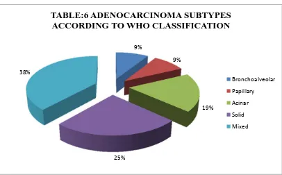
Histological subtypes of adenocarcinoma:
1. Mixed Type :
Most common type of adenocarcinoma representing 80% of resected
17 2. Acinar Pattern:
It contains tubules and acini composed of columnar or cuboidal cells
which may secrete mucin.(29)
3. Papillary Pattern :
In this type secondary and tertiary papillary structures are seen which
replaces the underlying lung architecture. Tissue invasion and necrosis may be
present. The lining cells may be mucinous and non-mucinous secreting
cuboidal to columnar cells. Micropapillary pattern of adenocarcinoma, are
usually prognostically unfavourable varieties.(40)
4. Bronchoalveolar pattern :
In this pattern malignant cells will grow along the alveolar structures
(this is known as lepidic growth). but without vascular, stromal, or pleural
invasion.(29)
5. Solid Pattern:
This variety composed usually of polygonal cell sheets which lacks
18 Variants of Adenocarcinoma of lung:
Mucinous (or colloid) adenocarcinoma.
Fetal adenocarcinoma.
Clear cell adeno carcinoma.
Signet ring adeno carcinoma.
Large cell carcinoma:
These are undifferentiated carcinoma that lacks the architectural and
cytologic features of squamous cell or small cell or glandular pattern. 9% of
lung cancer patients are due to large cell carcinoma.(41-43) It usually presents as
large peripheral mass. Histologically it consists of sheets of large polygonal
cells with characteristic vesicular nuclei, nucleoli and moderate cytoplasm.
Variants :
1. Large cell neuroendocrine carcinoma :
This type constitutes 3% of lung cancers.(42). The malignant cells are
arranged in various patterns such as organoid, nesting, trabecular rossetes or
peritubular palisading paterns. (29,44). The cells are usually large with
19
2. Combined large cell neuro endocrine carcinoma :
This tumor shows combination of features of squamous cell carcinoma,
adenocarcinoma, giant cell carcinoma and may be spindle cell carcinoma too.
3. Basaloid carcinoma:
Here, the tumor cells are arranged in many patterns as nodular, solid,
trabecular, and invasive growth pattern . May be Peripheral palisading of
cells are noted. The cells are monomorphic, small cuboidal to fusiform with
nuclei showing moderate hyperchromatism. (45)
4. Lymphoepithelioma like carcinoma:
They show growth pattern, with tumor cells having large vesicular
nuclei, and prominent nucleoli. This type of carcinoma show heavy lymphatic
infiltration .(42-44,46)
5. Clear cell carcinoma:
Large polygonal tumour cells with clear, foamy cytoplasm.(47,48)
6. Large cell with rhabdoid phenotype:
Rhabdoid cells containing tumour in which this rhobdoid cells should
20 Adeno squamous carcinoma:
It occupies about 0.4 to 4 % of all lung malignancies. Common at
periphery of the lung. Microscopy shows features of both squamous cell
carcinoma and adeno carcinoma, in which, each type should contribute atleast
10% of the tumour.(49-51)
Sarcomatoid carcinoma:
It occupies about 0.3 to 1.3% of all lung malignancies. Microscopically
this shows components of sarcoma or sarcoma like differentiation. Five
subtypes in this are pleomophic type, spindle cell type, giant cell type,
carcinosarcoma and pulmonary blastoma type.(52-56)
Carcinoid tumours:
Typical and atypical carcinoids are the major types of carcinoid tumor.
Other types of lung malignant neoplasms:
Other rare types include adenoid cystic carcinoma, mucoepidermoid
carcinoma, lymphomas, sarcomas, and epithelial- myoepithethelial
carcinoma.
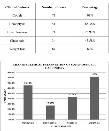
CLINICAL FEATURES:
Symptoms include cough , chest pain, hoarseness , haemoptysis,
,shortness of breath , fatigue, weight loss, new onset of wheeze (common in
21
Adenocarcinomas are usually asymptomatic, and more often a
incidental radiologic finding.
Small cell carcinoma is present with symptoms pertaining to distant
metastasis.
The local effects of lung tumor can be obstructive pneumonia, pleural
effusion, chest pain/back pain which is due to mediastinal invasion by the
tumor itself.
Nerve entrapment by the tumor, to say, recurrent laryngeal nerve
causing hoarseness, sympathetic nervous system leads to horner syndrome,
phrenic nerve leads to diaphragmatic paralysis.
Superior vena cava syndrome due to SVC obstruction by tumor
Pericardial involvement produce pericarditis and cardiac tamponade
Dysphagia due to invasion into oesophagus.
Metastasis leads to:
The symptoms related to the organs involved. Involvement of liver,
pancreas and adrenals produce symptoms like loss of weight, abdominal pain.
22
CNS involvement causes neurological symptoms such as dizziness, head
ache, and vomiting.
Paraneoplastic syndromes common with lung carcinoma are
hypocalcemia, hypercalcemia, carcinoid syndrome , cushing syndrome,
gynacomastia etc.
Other systemic manifestations :
Lambert-Eaton myasthenic syndrome due to autoantibodies directed
against neuronal calcium channels(57). It is charecterised by peripheral
neuropathy, acanthosis nigricans, hypertrophic pulmonary osteoarthropathy,
hematologic abnormalities like anemia, thrombocytopenia, eosinophilia,
leukemoid reaction, leukoerythroblastosis, etc.,
Course of the Disease:
Lung carcinomas are usually preceded by dysplasia lasting for years
followed by carcinoma in situ for several years and presents as a mass lesion
which becomes symptomatic. The lesion appears as a gray white ,firm to hard,
which may show haemorrhage and necrosis at some foci.(20,21)
The neoplasm can grow intraluminally and present as a mass within the
lumen. The tumour can also penetrate the wall of the bronchus and involve
23
Local extension to pleura, pericardium, and the adjacent lymph nodes
such as tracheal, bronchial, mediastinal lymph nodes may occur.
Distant metastasis occurs via both lympatic and haematogenous routes.
All tumours except squamous cell carcinoma will metastasize early . Distant
metastasis to adrenals is more common (>50%), followed by liver (30-50%),
bone(20%), and brain(20%).(46)
Imaging in Lung Cancers:
Pre and post diagnostic imaging done for various reasons
1. For finding a suspicious area that might be malignant.
2. For staging of the disease.
3. Inorder to assess the effectiveness of treatment
4. For the surveillance for recurrence of tumor
T.V Colby et al(59) found that the various imaging modalities that have
their role in diagnosing lung cancer are
X-ray:
The primary investigation to detect and characterise the lung mass. It
helps in assessing the involvement main bronchi / trachea. Also , helps in
24 Computed Tomogram:
This helps to locate the tumor. helps in assesing the size and shape of
tumours. Also the adjacent areas of involvement and any metastastic lesions
in adrenals, liver , brain and other internal organs can be found.
Magnetic Resonance Imaging:
Metastatic lesions of brain and spinal cord etc., can be detected
Ultrasonogram:
It detects pleural effusion. also guides for thoracocentesis, and
diagnostic biopsy of peripheral lung and lesions of mediastinum.
Positron Emission Tomography( PET scan):
A form of nuclear imaging which detects biochemical changes in body
tissues. Often, it is used as a whole body scan to detect early metastatic lesions
and tumour recurrence.
Literature stated that staging of cancer patients has been improved with
the use of PET (60-62)
Bone Scan:
25
INVESTIGATIONS TO DIAGNOSE LUNG CANCER:
The gold standard investigation for the diagnose of lung cancer is tissue
diagnosis under microscopy. Many diagnostic techniques are available to
obtain tissues are
1. Sputum cytology
2. Thoracentesis
3. Excisional biopsy of accessible nodes
4. Flexible bronchoscopy (FOB) with or without transbronchial needle
aspiration
5. Transthoracic needle aspiration
6. Video-assisted thoracoscopy, and
7. Thoracotomy (63).
In view of selecting the appropriate test procedure, the diagnosing
physician, should determine which type of lung cancer is suspected.
Thoracotomy can be done in patients suspected to have early stage
disease, which appears amenable to surgery. (63-67). Staging and tissue
diagnosis can be done with this (68).
To diagnose with sputum cytology, at least 3 samples of sputum must be
collected; since this is a non-invasive test, even if it is negative, further testing
26
centrally located tumors needs sputum cytology. Specificity of sputum
cytology in diagnosing lung cancer is 99% and its sensitivity for centrally
located lung tumors is 71%, and for peripheral tumors is <50%. (60,66,69) It also
aids in diagnosing squamous cell carcinoma.
Thoracocentesis can be performed in case of pleural effusion.
Malignacies of lung can be diagnosed with pleural fluid sampling. The
sensitivity of thoracocentesis in diagnosing lung cancer is 80% and its
specificity is < 90%.
In order to get a tissue sample, biopsy of an accessible lymph node may
also be taken.
If the stage of the cancer types are not clear, sputum cytology, flexible
bronchoscopy (FOB), and transthoracic needle aspiration are the
recommended test procedures.
Flexible bronchoscopy is done by passing scope along the bronchial
lumen and taking tissue samples by bronchial washings and/or by biopsies.
The sensitivity of flexible bronchoscopy in detecting the lung cancer is 88%.
According to De Wever W et al(60) Placing catheters into patients lung
should never be attempted without the guidance of Computerized tomography
27
of tumor and site of tissue sampling. The sensitivity in diagnosing the centrally
located tumors with the help of flexible bronchoscopy is 88%. The specificity
for same is 90%. The sensitivity for peripherally located tumors falls to 60 to
70% with this technique.
According to Rivera MP et al, With the guidance of CT or fluoroscopy,
transthoracic aspiration with the use of appropriate needle is the procedure of
choice recommended for peripherally located tumours with sensitivity of
90% and specificity of 97%. If the transbronchial needle aspiration done in a
patient with peripheral tumour is not conclusive, and if the patient is not
suitable for surgery, this technique is recommended. A major complication of
this procedure is pneumothorax, which is seen in 25 to 30% of the patients
undergoing this procedure .
Small peripherally located tumours with size < 2 cms in diameter,
pleural effusion, pleural tumours can be proceeded with Video assisted
thoracoscopy. Endoscopes are helpful in visualising the space between lungs
and parietal pleura. It also helps in detecting the small lesions in inter pleural
space, to take tissue biopsy and to resect some lung cancers in early stage. It
can prevent the attempt of thoracotomy, which is the major advantage of this
28
Lastly in all cases where the tumour is resectable, thoracotomy is
routinely recommended for diagnosis and as treatment for early stage
disease(60,70).
SCREENING:
Since we know many types of lung cancer histologically, finding a
single biomarker is in fact a great challenge. Several such biomarkers are
being evaluated. The effective screening programs could help for early
detection of lung cancers which may increase the survival. One of the research
projects at the Moffitt Cancer Research Center, has such an objective (71,72).
Present research emphasises microscopic examination of sputum sample
staining patterns. One of the possible screening tool is monoclonal antibodies
(Mabs). The pattern and the intensity of stain of the Mabs and varying cell
characteristics are now being analysed. The genetic and protein markers helps
in more understanding of tumor biology(72). The process of epithelial
carcinogenesis can be due to mutation of some particular genes, which can
alter its control over abnormal cell growth. Heterogeneous nuclear
ribonucleoprotein (hnRNP) is found to be useful marker for early detection of
the disease in sputum cytology. Datas suggest that hnRNP is being expressed
in most of the lung cancers before any morphologic abnormalities detected.
Other important biological markers found in lung cancers include:
29
as c-myc, c-erB-1, K-ras , HER-2, HGF and growth factors such as TGF-b,
GRP/BN, PTHrP, IGF-I & II , FDGF, apoptotic factors and factors favouring
angiogenesis such as Bcl-2, VEGF, respectively and gene amplification factor,
HER-2 (71) These molecular markers are important in diagnosing pulmonary
malignancies.It also helps to determine the prognosis as well as treatment
regimen. According to the study presented by Duarte,et. al, 2005, many
biological markers are found to be associated with greater frequency with
various tumors(73) . Rb is found to be associated with 30% of NSCLC, whereas
Rb gene is found to be associated with positive in 100% of SCLC.
The present clinical trial conducted by National Cancer Institute on a
large scale called as Prostate Lung Colorectal and Ovarian Screening Trial
(PLCO)(74,75). Its main objective is to ascertain the efficacy of screening tools
used in trial and to evaluate the mortality rate associated with the specific type
of malignancy under study (142). The main disadvantage of this trial is that the
conventional chest x-ray fails to detect the lung malignancies in early
stages(74). Another such study is the the National Lung Screening Trial
(NLST). This trial compares spiral CT scans with the conventional chest
x-rays and determines which screening tool is more effective in reducing the
mortality due to lung cancers. Spiral CT detects the lung nodules which are be
30
since spiral CT has been proved to detect the lung cancers at early stages as
compared to chest x-rays (76).
CLASSIFICATION OF LUNG CANCER
WHO Classification (ANNEXURE II)
IASLC/ATS/ERS Classification in small biopsy and cytology
(ANNEXURE III)
Prognostic factors:
Early detection of cancer favours increased survival; but unfortunately,
none of the screening programmes are proved to be successful(79). Due to lack
of early detection of this malignancy, it has become one of the most lethal
among all cancers; Mortality rate in lung cancer have superceded the same
due to colorectal, prostate and breast cancer combined. Patients are
asymptomatic till the advanced stage, which is the major drawback. American
Cancer Society observed that only 15% of these cancers are diagnosed in
early stages , i.e. Stage I. In patients with lung cancers, the five year survival
rate is 15%, which is due to the lack of programmes for early diagnosis .
National cancer institute showed that, in lung cancer, the 5 yr survival
31 Age :
Lung cancers in patients < 40years will have poor prognosis. The
aggressiveness of the cancer and presentation at advanced stages are the
probable cause for this..(80)
Sex :
Women have worse prognosis than men. This is partially due to the fact
that, women have high incidence of lesions in advanced stage and
adenocarcinoma is the commenest type in women.(81,82)
Location :
Superior sulcus tumors have better prognosis rather than tumors at other
site. Peripherally located squamous cell carcinomas have better prognosis than
those located centrally.(83-85)
Stage of the disease :
Tumour size :
Large tumours have worser prognosis than small tumours of same
histological type. In adenocarcinoms showing both in-situ and invasive
components, the site of the invasive component is an independent predictor of
32 Cell type and degree of differentiation:
Squamous cell carcinoma is found to be the most type of lung cancer
among other lung cancer types.(89-92) For well differentiated tumours, 5
year survival rate is 40% for those undergoing resection, for
moderately differentiated tumours, it is 20% and for poorly
differentiated tumours 5 year survival rate is 7% .
Among adenocarcinomas, papillary carcinoma showing micropapillary
pattern have the worst prognosis than other types. Prognosis of bronchio
alveolar carcinoma seems better than ordinary adenocarcinoma.(93-94)
The presence of tumour giant cells in large cell carcinoma have worse
prognosis.(95)
Small cell carcinomas have worse prognosis. The 5 year survival rate
for small cell carcinoma patients is only < 2%(95)
If the lymphoplasmacytic component is found prominently in tumor
under microscopy, then it favours good prognosis.(94)
Stronger expression of TTF -1 in patients with NSCLC, indicates better
survival.(96,97)
Other poor prognostic factors are :
Local and regional extension of the tumor, to say, chest wall invasion,
vascular invasion, regional lymph node involvement .(98)
33
Peripherally located adenocarcinomas and also undifferentiated large
cell carcinomas which are found to be in association with fibrotic
scar.(100,101)
The presence of rhabdoid cells.(78)
RAS and P21 expression in NSCLC, and NMYC gene expression in
small cell carcinomas(102-105)
p53 and HER 2/neu over expression (102-105)
TREATMENT FOR LUNG CANCER
Four basic modes of treatment available for lung cancer
1. Sugery
2. Radiation therapy
3. Chemotherapy
4. Targeted therapy
Treatment plan varies with many factors such as the type of cancer,
stage at presentation, side effects of particular treatment, patients preference,
health of the patient.
If NSCLC is confined to the lung, it is considered as early stage of the
disease and surgical resection can be done.(106) Post operative radiotherapy with
or without chemotherapy can be considered.(107). If patient cannot withstand
34
Various types of surgeries performed are lobectomy, wedge resection,
segmentectomy and pneumonectomy.
Early stage samll cell lung carcinoma should be treated with
chemotherapy and concurrent radiotherapy.
Once the tumor started spreading beyond hemithorax with metastasis to
mediastinal lymph nodes, surgery is no longer recommended and the treatment
of choice includes radiotherapy and chemotherapy.
External beam radiotherapy and radioisotope therapy are the two types
of radiotherapies given in lung cancers.
Chemotherapy includes platinum based Cisplatin and Carboplatin(107)
and non platinum based drugs such as Docetaxel, Paclitaxel, Gemcitabine,
Irinote can.
Distant metastasis is treated with chemotherapy alone.(107)
MOLECULAR TESTING AND TARGETED THERAPY
Molecular testing reveals various specific charecters of the tumor which
influences the diagnosis, prognosis and treatment with targeted therapy.
Targeted therapy have promising benefits if given to appropriate
35
Anti angiogenesis therapy blocks the formation of new blood vessles.
Bevacizumab, a monoclonal antibody targeted against VEGF is a potential
inhibitor of angiogenesis.
EGFR mutations are seen in 10% - 15% of NSCLC, and common in
adenocarcinomas. Geftinib and Erlotinib are the tyrosine kinase inhibitors
targeted against intracellular tyrosine kinase domain of EGFR. Hence,
tyrosine kinase inhibitors are the first line treatment for NSCLC.(108)
Rearrangement of ALK gene is seen in 5% of NSCLC and is most
common in nonsmokers with adenocarcinoma. Drugs that are targeted against
ALK mutation includes, Crizotinib and Certinib.(109-111)
Several new markers are evolved which may be associated with the
outcome on targeted therapy. They are ROS1, BRAF , FGFR, HER2,
36
Treatment for NSCLCs in advanced stage:
RECOMMENDATIONS BY IASLC/ATS/ERS NEW
MULTIDISCIPLINARY INTERNATIONAL CLASSIFICATION, FOR
SMALL BIOPSY AND CYTOLOGY SPECIMENS WERE:
For cytology as well as small biopsy specimens, if a clear
differentiation can be done, which satisfies the standard morphologic
criteria, further specific typing of NSCLC into squamous cell
37
The term NSCLC - NOS must be used as infrequently as possible and
it should only be used if the diagnosis cannot be made out by
morphology and /or by special staining / IHC.
When small biopsy / cytology specimen is used in addition with
special stains for diagnosis, it should be clearly noted whether the
diagnosis is achieved with only light microscopy or in combination
with special stains.
The term non-squamous cell carcinoma which is used by clinicians,
should not be used by pathologists while reporting. Pathologists should
report NSCLC only as ADC , SQCC and NSCLC - NOS.
The tissue specimens received by pathologists just be used judiciously
and preserved to the maximum, as more tissues will be needed for
further molecular studies.(113-115)
In small biopsies / cytology specimens, if any invasive pattern is found
in adenocarcinoma ,it is to be reported as a lepidic growth pattern . The
term minimally invasive ADC and ADC- in situ should not be used.
The term large cell carcinoma, should be used only in resected
specimens as thorough sampling of tumour is not possible in small
38
If the tumor shows sarcomatoid features characterised by malignant
giant cells or spindle cells with nucleus showing pleomorphism should
be classified according to guidelines above as NSCLC favouring ADC
or NSCLC favouring SCC based on features of glandular pattern or
squamous features respectively. when these features are absent it is to
be reported as NSCLC - NOS with a word about sarcomatoid features..
Only if the tumor shows neuro endocrine morphology, neuro endocrine
IHC markers are performed then.
Further classification of NSCLC- NOS is possible with the use of IHC,
into NSCLC favouring ADC and NSCLC favouring SCC.
It is advised to use minimal stains for further subclassification of
NSCLC-NOS.
It is recommended to use only one marker for adenocarcinoma or one
marker for squamous cell carcinoma.
Currently, the single best marker for diagnosing adenocarcinoma is
TTF-1. Staining with diastase - periodic acid schiff, alcian blue/ PAS
39
The specific marker for diagnosing SCC is Polyclonal p40 rather than
the monoclonal p63 . p40 is likely to surpasses p63 as a best IHC
marker in diagnosing squamous cell carcinoma.
In NSCLC -NOS , the cases which shows TTF-1 positive and /or
mucin positive, but p40 and p63 negative are termed as NSCLC
favouring adenocarcinoma. similarly those cases with p40 and/or p63
positive but TTF-1 and mucin stain negative are termed as NSCLC
favouring SCC with comment on whether special stains are used to
arrive at diagnosis.
In case, one population of tumour cells show TTF-1 reactivity and
another population of tumor cells show postive for squamous cell
markers, possibility of adenosquamous carcinoma should be
considered.
But if TTF-1 as well as p40 are negative and fails to show any
squamous or glandular morphology, the diagnosis still remains as
40
ALTERATIONS SUGGESTED BY IASLC/ERS/ATS INTERNATIONAL
CLASSIFICATION OF LUNG MALIGNANCY IN RESECTED SPECIMENS:
1. The term bronchoalveolar carcinoma is discarded.
In the new multidisciplinary classification, BAC is discarded. Originally,
broncho alveolar carcinoma is defined as a non-invasive lesion, but since
then, it is used to denote broad group of tumours which includes
Nonmucinous BAC. This is defined as solitary non invasive small
peripheral adenocarcinoma. This type will have 100% 5 year survival
rate.(116)
Minimally invasive small peripheral adenocarcinoma with 5 year
survival upto 100%.(117,118)
Invasive adenocarcinoma with mixed subtype.(119,120)
Nonmucinous and mucinous adenocarcinoma, which is known as BAC
earlier.(120)
Advanced mucinous adenocarcinoma( stage 4) with low survival rate.(4,6)
In the new multidisciplinary classification, ‘BAC’ is referred to as "former
BAC"
41
Small solitary peripheral adenocarcinoma with size less than or equal to
3cm,with pure lepidic growth without invasion with 100% disease specific
survival as adenocarcinoma in situ(AIS).(116)
Small, solitary peripheral adenocarcinoma with size less than or equal to
3cm, with predominantly lepidic growth with invasion, with 100% disease
specific interval as minimally invasive adenocarcinoma( MIA).(117,118)
3. Former invasive adenocarcinoma with mixed subtype is replaced by
predominant pattern..
According to 2004 WHO classification more than 90% of lung
adenocarcinoma are of mixed subtypes. In this new international
classification this mixed subtype is replaced with predominant pattern. It is
recommended to choose one predominant pattern based on recording of
patterns in 5% increments.(121,122)
4. In multiple lung adenocarcinoma patients it is recommended comprehensive
histological subtyping of heterogenous, complex lung adenocarcinoma to
determine if the tumours are synchronous, metachronous or metastasis.(123)
5. It recommends the term lepidic predominant adenocarcinoma in place of
previously classified as mixed subtype ,predominantly non mucinous
42
6. New histological type of Micropapillary predominant adenocarcinoma is
introduced . This is associated with poor prognosis.(122)
7. In new international classification, invasive mucinous adenocarcinoma,
fetal, enteric and colloid adenocarcinomas are introduced as new
variants.(124)
Acccording to Edwards et al (127), only 10-15% of lung cancer
patients undergo resection and the preoperative diagnosis confirmed. So
treament for most of the patients is based on diagnosis with small
biopsy/cytology specimens alone.
According to Suprun et al, (33 )
The criteria for diagnosis of SCC are
The presence of Keratin formation and/or Intercellular bridges. In cases
if the tumor lacks such features, the intraepithelial in- situ like
extensions along the bronchus are present in SCC. Both adeno and
small cell carcinoma do not replace the bronchial epithelium to a
considerable extent. And most of these cases are either well
differentiated or moderately differentiated. This feature aids in
histological typing of lung cancers in small biopsy specimens.(125)
The grading of squamous cell carcinoma cannot be done in small biopsy
43
The diagnosis of adenocarcinoma seems to be more challengeable as the
presence of mucin and gland formation are frequently not present in
small biopsies, which calls for mucin stains to demonstrate the presence
of glandular elements.
According to the study of Edwards et al,(127) the diagnosis of large cell
carcinoma is possible only with resected specimens and not on small
biopsies. This is because, they are the poorly differentiated forms of
adenocarcinoma, squamous cell carcinoma, or neuroendocrine
carcinoma and also most major types of lung carncers contain foci of
features of large cell carcinoma .
Recent data showed that the high percentage (30-50%) of NSCLC-NOS
has been diagnosed in small biopsies. (128-130) and data from the registry
of epidemiology surveillence shows the increasing frequency of this
diagnosis.(131)
ALCIAN BLUE/PAS ( Mucin stain) :
Mucins are a glycoprotein with high molecular weight, that was
synthesized and secreted by epithelial mucosal cells, mainly the goblet
cells (132). Mucins are classified into acidic mucins and neutral mucins
44
Many adenocarcinomas like cancers of colon, breast, ovary,
lung, etc will increase mucin production(133).
Acid mucins and neutral mucins will be differentiated using
Alcian blue- PAS staining.(134). This property will be of value in
demonstrating even minimal acid mucin. Mucin stains is a valuable
markers but its use in lung carcinoma in terms of sensitivity and
specificity may be variable. This is a defining characteristics of lung
adenocarcinoma, although not positive in all cases.(134)
THYROID TRANSCRIPTION FACTOR-1:
TTF-1 is a DNA binding protein expressed normally in thyroid,
lungs, and also in specific locations of diencephalon(135). Its role in lung tissues
is, it regulates the gene expression of surfactant and clara cell secretory
protein. the study of such transcription factors in human lung tumours are
helpful in understanding the molecular events of the noeplastic transformation
of such lung tumors (136).Several reports says that in lung cancers the
expression of TTF-1 varies with different histological subtypes(137-140).
According to J.Korean et al(143), TTF-1 was expressed in 62.5 - 90% of
ADC and in 89-100% of small cell carcinomas, but it was not found or very
less frequently found in SCC ( 0-25%) and large cell carcinomas ( 0-24%).
45
from non pulmonary tumours.TTF-1 appears to be more sensitive and specific
marker for diagnosing adenocarcinomas and small cell carcinoma of lung. It is
a useful prognostic marker and used in conjucation with proliferative marker
ki 67.(143)
p40(p63 delta):
p40 (p63 delta), a new marker, is the shortest variant of human p53. It
is a valuable marker in cases where p63 has been traditionally used. p40 has
equal sensitivity as p63 in detecting squamous cell carcinoma.
But p40 exhibits superior specificity when compared with p63.
Moreover, p40 helps in the early detection of breast cancers , prostate cancers
and lung cancers.
In lung cancer, the p40 is a very specific marker for squamous basal
cells. It helps to differentiate squamous cell carcinoma from ADC. In breast
cancers, the p40 stains the myoepithelial cells along the ducts of mammary
glands, and helps in detecting ADH and DCIS.
In prostate cancer, p40 staining of basal cells inside the ducts of the
prostatic glands will indicates PIN.
p40 and p63 have equal sensitivity in pulmonary squamous cell
carcinomas, but former is more specific. In rare instants, p40 is positive for
46
cells, easily distinguisable from squamous cell carcinoma which is diffusely
reactive.
p40, a nuclear marker has excellent sensitivity and specificity, makes
itself a more specific marker for SCC. Diffuse labelling of p40 indicates SCC,
47
MATERIALS AND METHODS
This study is a retrospective and prospective study of lung cancer in
small biopsies conducted in the Institute of pathology,Madras Medical College
and Rajiv Gandhi Government General Hospital, Chennai during the period
between July 2013 to June 2014.
A total of 13042 cases were submitted to our department the Institute of
pathology, Madras medical college during the period of July 2013 to June
2014 for histopathological examination. Out of them, 392 were lung cases.
Among them 19 cases were pneumonectomy specimens, 50 were lobectomy
specimens and 323 were small biopsies (Transbronchial, Endobronchial, Open,
Ultrasound guided biopsies and Computed tomography guided core biopsies).
Inclusion criteria:
FOB biopsies and CT Guided biopsies from clinically and radiologically
suspected cases of lung cancer.
Exclusion criteria:
1) Cases treated with prior Chemotherapy and radiotherapy.
2) Small cell carcinoma.
48 METHOD OF DATA COLLECTION:
Detailed history of the cases regarding age, sex, site, tumor location,
radiological findings, FOB findings, cytological findings were obtained for all
the 323 cases. Out of these
Inadequate for opinion - 23
Non neoplastic - 95
Suspicious of malignancy - 12
Malignancy - 189
Among the malignant cases,
NSCLCs - 153 cases
Small cell lung carcinoma - 13
Others - 23
These 153 Non small cell lung carcinoma cases were reviewed and sub
classified based on H&E morphology according to WHO classification
criteria. Tumours were sub typed as adenocarcinoma if it showed features of
gland formation and/ or mucin production, squamous cell carcinoma if it
showed features of keratinazation or intercellular bridges , large cell
carcinoma ( undifferentiated non small cell carcinoma) if it lacked both
49
because they were resected specimens. Out of these 151 cases, based on
morphology seen in biopsy further segregation done as follows:
Group 1: Cases that could be subtyped by morphology alone as
adenocarcinoma or squamous cell carcinoma (121 cases),
Group 2: Cases that are poorly differentiated non small cell lung
carcinoma (NSCLC-NOS) (30 cases).
All 30 cases of poorly differentiated non small cell lung carcinomas
(NSCLC-NOS) from group 2 and 10 cases each of poorly differentiated
adenocarcinoma and squamous cell carcinoma selected randomly from group 1
were included for special staining and IHC study for further classify the
NSCLC-NOS group into favouring adeno or squamous subtypes and to study
the efficency of special stains( ALCIAN BLUE/PAS) and markers ( TTF-1,
p40).
Five –micron thick paraffin sections were cut and stained with
combined mucin stain, TTF-1 and p40 and the tumours were sub-typed based
50
Alcian Blue / PAS staining was done as per protocol given in
ANNEXURE-V.
IMMUNOHISTOCHEMICAL EVALUATION:
Antigen Vendor Species Dilution Positive control
TTF- 1 BIOGENIX Mouse Ready to use lung
Adenocarcinoma
P40 BIOGENIX Mouse Ready to use Lung squamous
cell carcinoma
Immunohistochemistry was done as per protocol given in
ANNEXURE-VI.
INTERPRETATION AND SCORING SYSTEM:
The immunohistochemically stained slides were analyzed for the
presence of reaction, cellular localization(nuclear), percentage of cells stained
and intensity of reaction.
In this study, for evaluation of TTF1 and p40 proteins, greater than
10% expression of the tumour marker within tumour cells is considered as
positive. Cases with no focal areas of positive staining and with less than 10%
51 STATISTICAL ANALYSIS:
Immunohistochemical analysis was done in paraffin embedded tisse
samples using the statistical package for social science software version
15.5 which consisted computing the frequency counts and percentages
for qualitative variables and mean for quantitative variables.
P value of 0.005 was taken as cut-off point to determine statistically
52
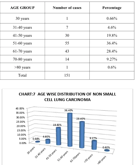
OBSERVATION AND RESULTS
In the study period of 12 months from July 2013 to June 2014,a total of
13042 specimens were received in the Institute of Pathology, Madras Medical
College for histopathological examination.
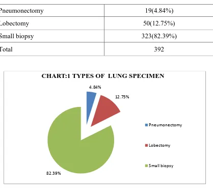
Total numbers of lung specimens received were 392 cases, of these 19
cases were pneumonectomy specimens,50 cases were lobectomy
[image:68.612.117.535.354.723.2]specimens,323 were small biopsy specimens. (TABLE:1 CHART:1)
TABLE 1 : TYPES OF LUNG SPECIMEN:
Pneumonectomy 19(4.84%)
Lobectomy 50(12.75%)
Small biopsy 323(82.39%)
Total 392
4.84%
12.75%
82.39%
CHART:1 TYPES OF LUNG SPECIMEN
Pneumonectomy
Lobectomy
53
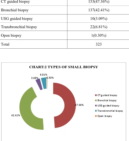
Among the 323 small biopsy specimens ,number of bronchial biopsy
were 137,CT guided biopsy were 153,USG guided biopsy were10,open biopsy
[image:69.612.111.533.213.674.2]was 1 and transbronchial biopsy were 22. (TABLE:2,CHART:2)
TABLE :2 TYPES OF SMALL BIOPSY
CT guided biopsy 153(47.36%)
Bronchial biopsy 137(42.41%)
USG guided biopsy 10(3.09%)
Transbronchial biopsy 22(6.81%)
Open biopsy 1(0.30%)
Total 323
47.36%
42.41%
3.09% 6.81%
0.30%
CHART:2 TYPES OF SMALL BIOPSY
54
Totally 189 malignant cases have been reported with the percentage of
56.6%, among which 183 malignancies were found to be in small biopsy
cases. Among the 183 small biopsy cases 152 were non small cell carcinoma
of lung, 13 were small cell lung carcinoma, 2 were malignant mesothelioma ,
11 were metastatic deposits in lung, 1 was solitary fibrous tumour, 1 was
carcinoid tumor. Out of 6 resected specimen, 1 was giant cell tumor, 1 was
large cell lung carcinoma, 1 was spindle cell carcinoma, 1 was SCC, 2 were
typical carcinoid tumors.
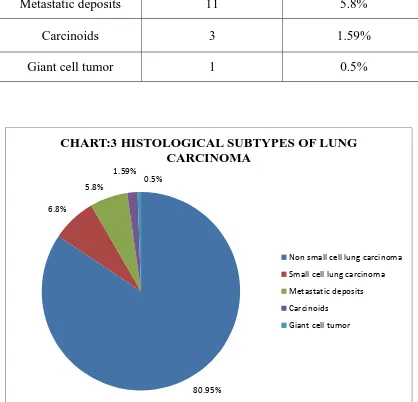
The most common lung malignancy were NSCLCs (Adenocarcinoma,
SCC, adenosquamous cell carcinoma, spindle cell carcinoma, large cell
carcinoma) which constitutes 153 cases (80.95%),small cell lung carcinoma
constituted 13 cases which accounts for 6.8%.
Metastatic carcinoma constitutes 11cases which accounting 5.8%,the
other histological subtypes accounts for 2% .(carcinoids-1%,giant cell
55
TABLE 3:HISTOLOGICAL SUBTYPES OF LUNG CARCINOMA
HISTOLOGICAL
SUBTYPES NUMBER OF CASES PERCENTAGE
NSCLC 153 80.95%
Smallcell lung
carcinoma 13 6.8%
Metastatic deposits 11 5.8%
Carcinoids 3 1.59%
Giant cell tumor 1 0.5%
80.95% 6.8%
5.8% 1.59%
0.5%
CHART:3 HISTOLOGICAL SUBTYPES OF LUNG CARCINOMA
Non small cell lung carcinoma Small cell lung carcinoma Metastatic deposits Carcinoids
56
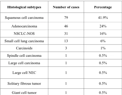
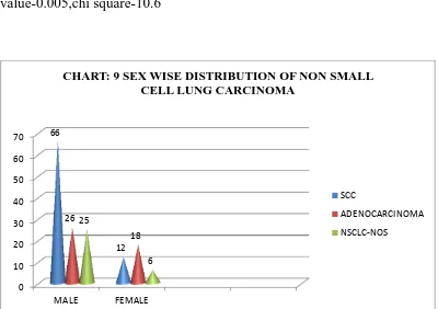
Of the total 189 lung tumours, Squamous cell carcinomas were most
common accounting for 79 cases which accounts for
41.9%,adenocarcarcinoma constituted 46 cases accounting for 24%,30 cases
were poorly differentiated non small cell lung carcinoma(16%) ,small cell lung
carcinoma were 13 cases constituted 6%,11 were metastatic
deposits(4%),carcinoids were 3 cases(1%), giant cell carcinoma, spindle cell
carcinoma, solitary fibrous tumor and large cell carcinoma each constitutes 1
[image:72.612.108.535.352.685.2]case and accounting for o.5%. (TABLE: 4& CHART:4 )
TABLE:4 HISTOLOGICAL SUBTYPE OF LUNG TUMOURS:
Histological subtypes Number of cases Percentage
Squamous cell carcinoma 79 41.9%
Adenocarcinoma 46 24%
NSCLC-NOS 31 16%
Small cell lung carcinoma 13 6%
Carcinoids 3 1%
Spindle cell carcinoma 1 0.5%
Large cell carcinoma 1 0.5%
Large cell NEC 1 0.5%
Solitary fibrous tumor 1 0.5%
57
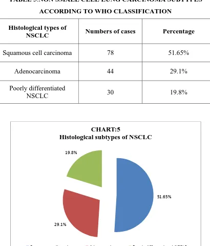
In small biopsy specimen the non small cell carcinoma of lung
(NSCLCs) was classified according to WHO classification into squamous
cell carcinoma, adenocarcinoma, poorly differentiated non small cell lung
carcinoma (which lack squamous or adeno differentiation). Among this SCC
of lung were the most common type which accounts for 78 cases (51.65%),
adenocarcinoma constituted 44 cases (29.1%), poorly differentiated NSCLC
were 30 cases(19.8%), large cell carcinoma was 1 case (0.65%) and large cell
neuroendocrine carcinoma was 1 case (0.65%) .large cell neuroendocrine
carcinoma was already confirmed by neuro endocrine marker such as NSE and
41.9% 24% 16% 6% 1% 0.5% 0.5%0.5% 0.5% 0.5%