A Dissertation on
“A STUDY TO DETERMINE THE PREVALENCE OF PRIMARY OPEN
ANGLE GLAUCOMA IN PATIENTS WITH SYSTEMIC HYPERTENSION"
Submitted to the
THE TAMILNADU DR. M.G.R. MEDICAL UNIVERSITY
In partial fulfilment of the requirements For the award of degree of
M.S. (Branch – III)
OPHTHALMOLOGY
GOVERNMENT STANLEY MEDICAL COLLEGE & HOSPITAL
THE TAMILNADU DR. M. G. R. MEDICAL UNIVERSITY,
CHENNAI, TAMILNADU
CERTIFICATE
This is to certify that the study entitled “A STUDY TO DETERMINE THE PREVALENCE OF PRIMARY OPEN ANGLE GLAUCOMA IN PATIENTS
WITH SYSTEMIC HYPERTENSION"
is the result of original work carried out byDr. INDHU. C, under my supervision and guidance at STANLEY MEDICAL COLLEGE, CHENNAI. The thesis is submitted by the candidate in partial fulfilment of the requirements for the award of M.S Degree in Ophthalmology, course from May 2011 to April 2014 at Stanley Medical College, Chennai.
Prof.Dr. K.BASKER, M.S., D.O., Prof.Dr.K.KANMANI,M.S.,D.O., Head of the Department, Professor,
Department of Ophthalmology, Department Of Ophthalmology, Stanley Medical College and Hospital, Stanley Medical College and Hospital,
Chennai- 600001. Chennai – 600001.
Prof.Dr.S.GEETHALAKSHMI, M.D, Ph.D
Dean
DECLARATION
I hereby declare that this dissertation entitled “A STUDY TO DETERMINE THE PREVALENCE OF PRIMARY OPEN ANGLE GLAUCOMA IN
PATIENTS WITH SYSTEMIC HYPERTENSION"
is a bonafide and genuine research work carried out by me under the guidance ofProf.
Dr. K.KANMANI M.S.,D.O., Professor, Department of Ophthlamology,
Government Stanley Medical College and Hospital, Chennai- 600001.
Date : Signature
ACKNOWLEDGEMENT
I express my deep gratitude to PROF. Dr.S. GEETHALAKSHMI, M.D., Ph.D., Dean, Stanley Medical College for permitting me to do this study.
With overwhelming respect and gratitude, I thank PROF & HOD Dr.K.BASKER,M.S,D.O., for his valuable advice and guidance in conducting this study.
I am very grateful to PROF.Dr.K.KANMANI,M.S,D.O., for his constant encouragement, valuable suggestions and support throughout the study. I would also like to thank ASSOCIATE PROF.Dr.THANGERANI.M.S., for her continuous support and guidance.
My sincere gratitude to my assistant professors Dr.S.Venkatesh, M.S., Dr.A.Nandhini,M.S., Dr.P.Geetha,M.S,D.O., Dr.B.Meenakshi, M.S., and
Dr.T.R.Anuradha,M.S., for their kind assistance and help throughout the study.
I am thankful to all my colleagues for their support.
I would sincerely like to thank all the patients for their kind cooperation for completion of this study.
ABSTRACT
“A STUDY TO DETERMINE THE PREVALENCE OF PRIMARY OPEN
ANGLE GLAUCOMA IN PATIENTS WITH SYSTEMIC HYPERTENSION"
Key Words Primary Open Angle Glaucoma, Intraocular Pressure, Systemic Hypertension
OBJECTIVES OF THE STUDY:
The aim of the study was to find the prevalence of primary open angle glaucoma in patients with systemic hypertension presenting to Stanley OPD to find the association of systemic hypertension with the occurrence of POAG.
METHODS:
In this cross-sectional case control study, 100 patients with systemic hypertension and 100 non hypertensive subjects were evaluated at department of ophthalmology at Stanley medical college for the presence of primary open angle glaucoma based on predetermined criteria and the
prevalence of primary open angle glaucoma in both the groups was found and analysed to find the asscociation of primary open angle glaucoma and systemic hypertension.
RESULTS:
Systemic hypertension was not found to have a statistically significant
association with the occurrence of primary open angle glaucoma{ odds ratio 1.936 with 95% confidence interval being from 0.687 to 5.457. The mean intraocular pressure of hypertensive group was mildly higher than the mean intraocular pressure non hypertensive group and the difference in the mean IOP of both the groups was statistically significant { P value – 0.006}. The
occurrence of POAG was more in patients with systemic hypertension for a longer duration i.e 55.5% of persons with POAG in this group was diagnosed with hypertension for 10 years. The prevalence of POAG also increased with increasing age in both the groups with 66.6% of subjects being above 60 years of age.
In our study systemic hypertension was not found to have a
statistically significant association with POAG,but persons with long duration of hypertension and advancing age, need to be monitored for high IOP and optic nerve head changes to aid in early diagnosis and also to minimize visual
CONTENTS
PART – I PAGE NO
INTRODUCTION 1
HISTORICAL REVIEW 3
ANATOMY 5
GRADING OF ANGLE & GONIOSCOPY 10
AQUEOUS HUMOR 16
INTRAOCULAR PRESSURE & TONOMETRY 18
OPTIC NERVE HEAD 25
VISUAL FIELDS & PERIMETRY 35
PRIMARY OPEN ANGLE GLAUCOMA 41
PATHOPHYSIOLOGY OF POAG 44
RISK FACTORS 48
HYPERTENSION AND POAG 52
PART II
OBJECTIVES OF THE STUDY 57
MATERIALS AND METHODS 58
OBSERVATIONS AND RESULTS 62
DISCUSSION 77
CONCLUSION 82
ANNEXURES BIBLIOGRAPHY PROFORMA PHOTOGRAPHS
INTRODUCTION
Glaucoma is a potentially blinding disease affecting a large number of
people worldwide. It is a frequent cause of permanent blindness in the world.
Glaucoma is a progressive optic neuropathy characterised by
recognizable patterns of alterations in the structure of the optic nerve head and
nerve fiber layer of the retina and consequent visual field defects6. Glaucoma
can be subdivided into primary glaucoma in which the mechanism for the
disease is unknown and secondary if the glaucoma is secondary to another
ocular or systemic disease.
Among all types of glaucoma, primary open angle glaucoma is said to
the most frequent one. Asians tend to have higher rates of angle closure
glaucoma. This may be true, or, in some instances, might be an artefact because
angle closure glaucoma may cause acute symptoms, therefore, it is more readily
diagnosed but POAG is relatively asymptomatic till late stages and they readily
go undiagnosed.
POAG is often asymptomatic & patients may suffer substantial vision loss
prior to diagnosis and treatment7. Therefore it is essential to identify at-risk
populations & develop accurate screening tests to make the diagnosis so that
sight-preserving treatment can begin early in the disease process. Several
POAG and to identify risk factors for the disease. Age8,9,10, race12, myopia17,
systemic HT11,15, diabetes11,13,14, family history18 etc have long been considered
as risks for occurrence of primary open angle glaucoma but recent studies have
questioned the strength of these associations.
Blood pressure increases with age in most populations but the relationship
between systemic hypertension and POAG is yet to be successfully elucidated.
Several large-scale studies have yielded dissimilar & even contradictory results.
Systemic HT causes direct microvascular damage and can impair blood flow to
the optic nerve, precipitating POAG. Significant ethnic & racial variation exists
in the prevalence and complications of HT. Evaluation of hypertension as a risk
factor for POAG in our population is essential so that the disease can be
REVIEW OF LITERATURE
HISTORIC REVIEW:
Glaucoma has been known since Roman era. The word "glaucoma" was
first described in Hippocrates' Aphorisms about 400 BC as an ailment of old
men.!
In Hippocratic writings,’ glaucosis’ derived from the Greek word
glaucos, meaning glaze , later more specified as a greenish glaze of the lens.
In 1626 Dr.Banister first established the concept of elevated intra-ocular
pressure in glaucoma.
Impairment of the aqueous drainage of the eye as the cause of glaucoma
was described by Hermann Boerhave in 1708.Inflammation as a cause of
glaucoma was put forth in 1722 by Charles Saint.
Emphasis was put on the intraocular pressure in the diagnosis of
glaucoma till the introduction of ophthalmoscope in 1853 following which optic
In 1862, Donder published his classical paper in which he described
‘Glaucoma simplex’ as a disorder with increasing tension of the eye, excavation
of the disc, shift of the vessels in the disc, loss of visual field and loss of vision.
Today glaucoma is defined as a disease causing progressive atrophy of
the optic nerve with loss of ganglion cells and their axons along with specific
ANATOMY
Aqueous humor is produced in ciliary body and drained via anterior
chamber angle. These two structures play an important role in maintaining
aqueous humour dynamics and also intraocular pressure.
CILIARY BODY
In the eye uveal tract is composed of iris,ciliary body and choroid. Ciliary
body is seen between iris and choroid and is attached with sclera spur anteriorly.
Histologically it is composed of epithelium, vascular structures and ciliary body
muscle.
PARTS OF THE CILIARY BODY Pars plicata:
Pars plicata contains the ciliary processes which are the site of aqueous
production, they are nothing but ridges which are radially oriented and
projecting into the posterior chamber. Pars plicata forms anterior one third of
ciliary body.
Pars plana:
Pars plana is smooth, devoid of ridges and forms the posterior two third
HISTOLOGY OF CILIARY PROCESS
Ciliary process is made up of a core of connective tissue substance with
numerous capillaries and a overlying epithelium. Capillary network in ciliary
processes are fenestrated.
Epithelium of ciliary process is double layered with an inner layer of
non pigmented epithelium and outer layer of pigmented epithelium lying
adjacent to the stroma. Blood aqueous barrier is formed by the tight junctions
ANGLE OF ANTERIOR CHAMBER
Schwalbe’s line, trabecular meshwork, canal of Schlemn and the scleral
spur are the main structures forming the anterior chamber angle.
SCHWALBE’S LINE
The anteriormost structure of the anterior chamber angle is the
Schwalbe’s line and it is the point where descemet’s membrane ends , it also
marks the point where corneal endothelium continues as trabecular
endothelium.
TRABECULAR MESHWORK
The trabecular meshwork is an important structure of the anterior
chamber angle playing a vital role in the maintainence of IOP, it is triangular in
outline with its apex directed towards Schwalbe’s line and base towards sclera
spur.
PARTS OF TRABECULAR MESHWORK
Uveal meshwork
Juxta-canalicular meshwork
The inner layers of trabecular meshwork is formed by uveal meshwork,
the next layer is formed by corneoscleral meshwork and it extends from anterior
part of sclera sulcus to sclera spur.
Juxtacanalicular meshwork is considered as the main site of resistance for
aqueous outflow and it is made up of proteoglycan matrix with embedded
SCHLEMN’S CANAL
Schlemn’s canal is a vascular channel lying within the sclera sulcus. It
consists of two layer of endothelial cells. The outer layer is attached to sclera.
The inner wall plays an important role in IOP maintenance by its invaginations
called ‘ giant vacuoles’6,7. These giant vacuoles increase in size and number
when IOP increases facilitating aqueous outflow thus reducing IOP.
Schlemn’s canal also contains collector channels which drain aqueous
into the aqueous veins which in turn drain into episceral and conjunctival veins.
These collector channels are of three types namely direct (4-6) which connects
directly with aqueous veins, indirect type (15-20) which forms an intrascleral
plexus and then drain into aqueous veins and an intermediate type (4-6)
SCLERAL SPUR
The scleral spur is nothing but a wedge shaped projection from the
anterior sclera and attaching itself anteriorly onto the trabecular meshwork.
Posteriorly is attached to sclera and longitudinal ciliary muscle fibres.
As sclera spur is attached posteriorly to ciliary muscle, when ciliary
muscle contracts it pulls the sclera spur posteriorly which in turn pulls the
spaces and also prevents the schlemn’s canal to get narrowed or collapsed, in
this way they help in aqueous humor outflow.
OUTFLOW OF AQUEOUS CONVENTIONAL OUTFLOW
Outflow of aqueous from the anterior chamber angle through the
trabecular meshwork into the schlemn’s canal which in turn drains into the
episcleral veins via aqueous veins is considered as the conventional outflow
pathway and as it drains through the schlemn’s canal it is also termed the
canalicular pathway.
Contraction of the ciliary muscle through its insertion into the trabecular
meshwork increases pore size in the meshwork and hence the rate of aqueous
drainage. Passage of aqueous into Schlemn's canal depends on cyclic formation
of transcellular channels in the endothelial lining. The collector channels from
Schlemm's canal drain into the episcleral veins via aqueous veins which are
UVEOSCLERAL OUTFLOW
It is estimated that up to 15% (much higher values suggested by recent
studies) of aqueous outflow occurs via an alternative route known as
UVEOSCLERAL OUTFLOW described in 1960s by Bill.
In this pathway aqueous enters the ciliary body course through the loose
connective tissue and then pass between ciliary muscle fibres to enter
supraciliary space, subsequently into suprachoroidal space. From here aqueous
is drained by the veins draining the uvea. But this pathway is not affected by
changes in IOP and remains the same both in high and low intraocular pressures
but outflow can be modified pharmacologically. Uveoscleral outflow is
decreased by pilocarpine and increased by drugs like adrenaline and
prostaglandin analogs.
GRADING OF ANGLE
In an open angle structures seen from posterior to anterior are the root
of the iris,ciliary body, scleral spur, trabecular meshwork and the Schwalbe’s
Anterior chamber angle is graded by various systems namely,
SHAFFER GRADING SYSTEM
SCHEIE GRADING SYSTEM
SPAETH GRADING SYSTEM
SHAFFER’S GRADING SYSTEM
In Shaffer’s grading system anterior chamber angle is graded by taking
into account the angle formed between the anterior iris surface and posterior
corneal wall.25
Grade Angle Width Description
4 45-35 Wide open
3 35-20 Wide open
2 20 Narrow
1 10 Extremely narrow
S <10 Slit
0 O Closed
SPAETH GRADING SYSTEM
Spaeth grading of anterior chamber angle is based on three variables
a) Insertion of iris root
b) Angular width
c) Configuration of iris
A - Anterior to Schwalbe’s line.
B - Behind Schwalbe’s line.
C - Centered at scleral spur
D - Deep to scleral spur
E - Extremely deep in ciliary body.
Configuration of iris
s - ‘Steep’ or convexly configured
r - ‘Regular’ or flat
q - ‘Queer’ for deeply concave
SCHEIE GRADING SYSTEM
This system is based on the extent of angle structures visualized
All structures seen -Wide open
Iris root not seen - Grade I narrow
Ciliary body band not seen - Grade II narrow
Posterior trabeculum obscured - Grade III narrow
SCHEIE GRADING SYSTEM
GONIOSCOPY
Gonioscopy refers to the technique used to view and define the
structures and abnormalities of anterior chamber angle or iridocorneal angle.
PRINCIPLE
As the light emitted from angle structures undergo total internal
reflection, it is not possible to visualise the angle through intact cornea. Contact
lenses have an index of refraction similar to that of the cornea, allowing light to
enter the lens and then be refracted (goniolens) or reflected (gonioprism)
beyond the contact lens-air interface. Gonioscopy can be done by two methods
Direct gonioscopy:
In this method,anterior curve of the contact lens i.e configuration of the
contact lens is such that exiting light rays strike the contact lens/air interface at a
steeper angle than critical angle so that they will pass directly to the observer
without reflection inside the lens e.g is koeppe lens.
Indirect gonioscopy :
In indirect gonioscopy light rays from the angle are reflected by a mirror
such that they exit the lens at an angle much less than the critical angle. So the
angle viewed is the angle exactly opposite to the mirror. Indirect gonioscopy is
done with the help of a slit lamp. Examples are Goldmann and Zeiss types of
lenses.
AQUEOUS HUMOR:
Aqueous humor flows from its production site( nonpigmented epithelium
of ciliary body) into the posterior chamber, then into the anterior chamber
passing between the posterior iris surface and lens .It exits from the anterior
chamber via trabecular and nontrabecular routes. The trabecular route is at the
angle of the anterior chamber flowing through the trabecular meshwork (TM) of
the sclera, into Schlemm's canal and then via its efferent channels,the aqueous is
carried to the episcleral vessels, where aqueous mixes with blood. A balance
between aqueous humor production and the resistance to its outflow is essential
AQUEOUS HUMOUR DYNAMICS
It is important to understand the aqueous humour dynamics for the
evaluation and management of glaucoma. Aqueous humour is formed in the
ciliary process by three important mechanisms namely simple diffusion,
ultrafiltration and active secretion. Active secretion of ions across the
epithelium of the ciliary body is considered as the primary mechanism for the
formation of aqueous these days. Active secretion creates an osmotic gradient
which in turn leads to passive flow of water into the posterior chamber. This
process is decreased by hypoxia, hypothermia and any inhibitor of active
metabolism.
Aqueous humor exits the eye via two pathways. The trabecular
meshwork is considered the conventional, pressure-dependent pathway, while
the uveoscleral pathway is considered the unconventional, pressure-independent
pathway.
Mean outflow facility is 0.22 to 0.33 microlitre/min/mmHg. Outflow
facility decreases with age, surgery, trauma, medications, endocrine factors etc.7
Trabecular outflow accounts for 5 to 95% of aqueous drainage. This flow
is pressure dependent, meaning that the flow is proportional to the difference
In the uveoscleral outflow pathway, aqueous humor exits the eye through
the interstitial spaces between the ciliary muscle fibres into the supraciliary and
suprachoroidal space where it is absorbed into the venous system. There are
age-related changes to both the TM and the uveoscleral outflow pathways,
including decreased TM cellularity with age and increased extracellular
depositions in both the TM and uveoscleral pathways that are associated with
age-dependent decreased aqueous outflow.
INTRAOCULAR PRESSURE
Although Intraocular Pressure is not part of the definition of glaucoma,
reduction of IOP remains the only proven and approved means of glaucoma
management and is the single most important modifiable risk factor. To cause
glaucomatous optic neuropathy, there is a complex interaction between IOP and
other risk factors. Therefore the study of those elements that contribute to the
production and drainage of aqueous humor, maintenance of intraocular pressure
and variation of intraocular pressure is material to understand the
DETERMINANTS OF INTRAOCULAR PRESSURE
Intraocular pressure (IOP) is determined by three factors namely aqueous
formation (F), facility of outflow (C), and episcleral venous pressure (Pv) .
Goldmann equation relates these factors by the formula
Po= F/C + Pv ,
or if solving for F then
F = (Po - Pv) C
in which Po represents the IOP in the undisturbed eye in mmHg, F represents
aqueous formation in ul/min, C is aqueous outflow facility in ul/min/mmHg
and Pv stands for episcleral venous pressure in mmHg.
From the equation, it is clear increase in IOP occurs when the aqueous
formation rate increases the episcleral venous pressure increases or the outflow
facility decreases.
NORMAL INTRAOCULAR PRESSURE:
The range of intraocular pressures in the normal population is fairly wide;
deviation of 2.5. The ‘statistical’ normal range, defined as the mean two
standard deviations, would therefore be approximately 11–21 mmHg.
IOP is influenced by number of factors like age, sex, race, heredity,
obesity, posture, exercise etc. IOP is also altered by cholinergic and adrenergic
inputs Corticosteroids raise IOP; diabetes associated with increased IOP;
myopic individuals have higher IOP.
Diurnal variation -Most people have a diurnal pattern, IOP varies with
an average of 3–6 mmHg in normal individuals.
TONOMETRY
Intraocular pressure is measured clinically using tonometers. Tonometers
work on the principle that the pressure within the globe is directly related to the
force required to deform the same.
Tonometers developed for the purpose of measuring intraocular pressure
fall into two categories:
Indentation Tonometers – Here the amount of corneal or globe
deformation in response to an externally applied weight is determined.
Applanation tonometers – Here the force required to flatten a surface area
In both applanation and indendation tonometers, the factors that decide
the measured IOP includes the actual intraocular pressure, central corneal
thickness and corneal deformability.Applanation tonometer is considered as the
gold standard tool for IOP measurement.
INDENTATION TONOMETERS
Shiotz in 1905 described the indendation tonometer. The Schiotz
tonometer is an indentation instrument which measures IOP by registering the
depth of indentation of the cornea produced when the instrument with a known
weight is applied to the eye. The weight is carried on a plunger and when the
weight is applied to the eye, the intraocular pressure provides a
counterbalancing force which pushes back up on the plunger. This causes a
deflection of the pointer along the inclined scale. Each unit on the scale, which
ranges from 1 to 20, corresponds to an indentation of 1/20 of a mm in the
cornea. High intraocular pressure resists indentation, resulting in low scale
readings, while low intraocular pressure allows for easy indentation, manifested
by high scale readings. The intraocular pressure is determined by referring to
the calibration chart and reading the pressure that corresponds to the scale
Advantages:
Low cost
Portability
Disadvantages:
Ocular rigidity tends to influence the tonometry readings, example in
patients with low ocular rigidity like myopia it tends to give a falsely low IOP.
APPLANATION TONOMETERS
Applanation tonometers are of two types :
1. Variable area – Here force is kept constant by using a fixed weight and
the diameter of the area flattened is measured and this value gives an
estimate of IOP. The prototype of this principle is Maklakov tonometer.
2. Variable force - Here area applanated is kept constant and the force
required to applanate this fixed area determines the IOP. The prototype is
the Goldmann applanation tonometer, which was introduced in 1954.
Others include Perkins applanation tonometer, Mackay Marg tonometer,
GOLDMANN APPLANATION TONOMETER
Goldmann applanation tonometry is still considered the gold standard
technique for measurement of IOP.
In using the Goldmann applanation tonometer, the flattening force to the
cornea is supplied by a coiled spring contained in the instrument, controlled by
a rotating knob at the base, and is applied to the anesthetized eye by the tip of a
split prism device through which the cornea may be viewed with the slit lamp.
The area of cornea flattened is 3.06 mm in diameter. Topical anesthetic and
fluorescein are applied to the eye, and the tear film illuminated using the cobalt
blue filter of the slit lamp. As the instrument is applied to the eye, the
applanating head creates a circular tear film meniscus which may be viewed
through the slit lamp. The prism in the applanating head splits the circular
image into two semicircles, and the end point of the measurement is determined
by adjusting the knob until the inside edges of the semicircles are just touching.
The intraocular pressure is then determined by multiplying the reading on the
scale on the knob by ten.
Source of errors of applanation tonometry:
Astigmatism >3 diopters can give false values. Too much of flourescein
staining as well as inadequate staining can cause thick or thin mires and show
inaccurate intraocular pressure.
Advantages:
Accurate method of intraocular pressure measurement
Accurate IOP measurement in patients with scleral rigidity.
Disadvantages:
Cannot be used in infants and children
Cannot be used in operative room
Goldmann tonometry will not be accurate if the cornea is very much
thinner or thicker than average, highly astigmatic, irregular, edematous or
NONCONTACT TONOMETERS
In non contact tonometers, cornea is not touched and flattened with a puff
of air and the time required to flatten it is measured and correlated with IOP.
Ocular response analyser a type of non contact tonometer is gaining
popularisation in recent years.
Advantages:
No corneal trauma
OPTIC NERVE HEAD ANATOMY AND BLOOD SUPPLY
Glaucoma is essentially an optic neuropathy and damage to the optic
nerve is occurs within the neural, cellular and connective tissues of the optic
nerve head. Knowledge about the anatomy of the normal optic nerve and the
pathological changes that occur in glaucomatous optic neuropathy (GON) is
essential for the early detection and monitoring of these diseases.
The optic nerve (cranial nerve II) is formed by the axons of the ganglion
cells of the retina; it then traverses the scleral canal to exit the eye. Optic nerve
ends at the level of optic chiasma where the axons of one side merge with axons
of the contralateral optic nerve.
From its origin in the eye until it reaches the optic chiasma in the anterior
cerebral fossa the ON can be divided into four segments
Intraocular part: 1 mm
Intraorbital part: 30 mm
Intracanalicular part: 6-9 mm
Intracranial part: 10 mm
The intraocular portion of the ON is also referred to as the optic nerve
head (ONH). The visible most anterior part of the ONH is known as the optic
Optic nerve head consists of the following zones:7
Surface nerve fibre layer
Prelaminar region
Lamina cribrosa region
Retrolaminar region
SURFACE NERVE FIBRE LAYER
Anteriormost part of the optic nerve head is called as the surface nerve
fiber layer and it consists of approximately one million ganglion cell axons (also
known as nerve fibers) from all over the retina that converge onto this part and
exit through the scleral canal. Small branches from retinal arterioles and
cilioretinal arteries supply the superficial nerve fibre layer.
PRELAMINAR REGION
Arterial branches from the peripapillary choroid and short posterior ciliary
arteries supply the pre-laminar region.
LAMINAR REGION
The lamina cribrosa consists of a meshwork formed by interlinked
connective tissue plates (cribriform plates).Central retinal vessels and bundles
The 300–400 pores that transmit axon bundles show a considerable variation in
size, with the largest pores typically found in the superior and inferior
quadrants. Banches of short posterior ciliary arteries either directly or its
branches from circle of Zinn –Haller supplies lamina cribrosa.
RETROLAMINAR REGION
Axons within the post laminar optic nerve are myelinated, which
principally accounts for the doubling of the optic nerve diameter from 1.5mm at
the pre-laminar and laminar levels to 3.0 mm in the post-laminar region.
Retrolaminar part supplied by centrifugal branches from central retinal artery
and centripetal branches from pial vascular plexus.30
Venous drainage occurs via the central retinal vein and there may also be
OPTIC NERVE HEAD FEATURES
Upon clinical examination the optic disc is generally described as
consisting of Neuraretinal rim (the nerve axons)
Optic cup(the central area of the disc surrounded by the
neural rim).
CUP DISC RATIO
The optic cup is relatively devoid of nerve fascicles and normally appears
as a round to oval depression of variable size, usually of a lighter color.
The diameter of the optic cup divided by the diameter of the optic disc is
known as the cup/disc ratio (C/D ratio) and is expressed in decimal notation
(0.1, 0.2, etc.). Ninety per cent of normal individuals have an average C/D ratio
of less than 0.5 measured by direct ophthalmoscopy.
The size of the optic cup changes proportionally with the size of the optic
disc; a larger cup occurs in larger disc and vice versa. Assuming discs of equal
size, both optic cups usually appear fairly symmetric in normal individuals.
NEURORETINAL RIM
The neuroretinal rim is normally pink in colour.The rim is broadest in the
inferior disc, then the superior disc, then the nasal disc, and thinnest in the
temporal disc and it correlates with the morphology of the lamina cribrosa – the
largest pores and the least amount of interpore connective tissue are in the
inferior and superior poles, compared with the nasal and temporal sectors.
OPTIC NERVE HEAD IN GLAUCOMA
1. Progressive enlargement and/or deepening of the cup (‘cupping’ is
the direct result of axonal loss and the resulting structural alterations in the
lamina cribrosa ie backward bowing lamina cribrosa)31
2. Neuroretinal rim is formed by the extension of the superficial
nerve fiber layer into the optic disc and hence it is important to evaluate
neuroretinal rim. NRR changes in glaucoma are notching of the rim, NRR loss
and thinning in one or more quadrants, asymmetry of NRR between the two
eyes and haemorrhages crossing the rim.
3. VASCULAR SIGNS
“ Bayonneting,” occurs when a vessel take a sharp 90° bend as it passes through an acquired pit formed as a consequence of neuroretinal rim loss and then emerges out from the edge of optic disc.
“Nasalization” of blood vessels – Normally blood vessels at the optic nerve head emerge centrally, in glaucoma owing to diffuse neuroretinal rim loss ,major vessels appear to emerge more nasally and this appearance is named as nasalisation of major blood vessels.
Splinter haemorrhages in the margin of ONH. These hemorrhages are more common in eyes with norm al-tension glaucoma, usually indicating progression of disease.
4. NERVE FIBRE LAYER DEFECTS
Nerve fiber layer defects appear as a series of striations radiating in an
arcuate fashion from optic nerve head above and below macula and not crossing
the horizontal raphe.
Localized defects – These are wedge shaped areas occurring wider
peripherally radiating from the optic nerve head.
Diffuse loss – Diffuse loss of nerve fiber layer is seen in advanced
glaucoma.
5. PERIPAPILLARY ATROPHY
Atrophy around the disc consists of two zones –
Beta zone - Zone of chorioretinal atrophy immediately surrounding the
choriocapillaris, with more clear visibility of the choroidal vessels and
sclera.
Alpha zone - alpha zone surrounds the beta zone and represents areas of
chorioretinal thinning occurring as irregular areas of hypo and
hyperpigmentation.
OPTIC NERVE HEAD ASSESSMENT DIRECT OPHTHALMOSCOPY
It is the most commonly used method for assessing the optic nerve head
by general ophthalmologists and it provides a non stereo view of the optic
nerve head.
SLIT LAMP BIOMICROSCOPY
Optic nerve head can be assessed with a slit lamp biomicroscopy using
hand held high power convex lenses ( 78 D & 90 D) and fundus contact lenses
like Goldmann three mirror lens to provide a stereoscopic view of the disc
FUNDUS PHOTOGRAPHY
Fundus photography is the most widely used technology nowadays to
document the optic nerve head appearance objectively and also helps in
CONFOCAL SCANNING LASER OPHTHALMOSCOPY
Confocal scanning laser ophthalmoscopy takes transaxial laser scans (
around 64) through the optic nerve head and peripapillary retina to reconstruct a
high resolution three dimensional imaging of optic nerve head and nerve fiber
layer.
Heidelberg Retinal Tomograph (HRT) is a prototype of Confocal
scanning laser ophthalmoscopy.
Advantage – Rapid image acquisition time, reduced need for pupillary
dilation or clear media.
OPTICAL COHERENCE TOMOGRAPHY
OCT uses low-coherence laser light (850 nm) and based on its property
of optical backscattering,it takes axial cross-section of tissues as it passes
through layers of different optical density. OCT assess optic nerve head using
this laser interferometry principle and quantifies the amount of nerve tissue in
optic nerve.
NERVE FIBER LAYER ANALYSIS
In Scanning laser polarimetry ,a laser beam is passed to the posterior
retina and changes in the polarization of the reflected beam is measured and is
used to quantify peripapillary RNFL .The software used in nerve fiber analyzer
is named as GDx. VCC stands for variable corneal compensator which
accounts for the variable corneal birefringence.
OPTICAL COHERENCE TOMOGRAPHY
Optical coherence tomography uses the reflected and backscattered light
to create reflected images of various retinal layers and is displayed as quadrants,
clockhours and overall mean as measured by computer analysis.
Fourier-domain OCT yields up to five times higher resolution with faster
imaging speed (60 times faster) than conventional time-domain OCT.
VISUAL FIELDS
Visual field testing gives a non-invasive direct measure of visual
function. The distribution and arrangement of the retinal nerve fibres govern the
pattern of visual field loss in glaucoma and scotomas follow the shape of these
nerve fibre bundles, giving rise to characteristic glaucomatous field defects
which may be absolute, or relative, or a combination of both.
Early visual loss within the arcuate area ( starts from blind spot and arches above or below fixation), they most often appear as localised defect or PARACENTRAL SCOTOMAS
Early arcuate defect may connect with the blind spot giving rise to SIEDEL SCOTOMA.
The localised defect then coalesce filling the entire arcuate area from blind spot forming ARCUATE OR BJERRUM SCOTOMA
Further disease progression leads on to DOUBLE ARCUATE OR RING SCOTOMA.
Loss of nerve fibers rarely proceeds at the same rate in the upper and lower portions of the horizontal meridian creating a step like defect along the horizontal raphe referred to as the NASAL STEP.
Continued damage results in a temporal island and central island in advanced glaucoma.33
PERIMETRY
There are two main types of perimetry namely
1. Kinetic perimetry
2. Static perimetry
KINETIC PERIMETRY
A stimulus of given size and brightness is selected and moved from
where it is not seen until its threshold is crossed. Island of vision is kinetically
STATIC PERIMETRY
Automated static perimetry is the most reliable and sensitive means of
testing a visual field. Here relative differential light sensitivity throughout the
field of vision is determined based on either age corrected normal data or
patients response to preliminary spot tests.
Testing Strategies:
1.Suprathreshold:
In this strategy a stimulus that is slightly brighter than the anticipated
normal for the corresponding retinal location is presented. It is mainly used in
screening of glaucoma.
2.Threshold:
A staircase procedure is applied in which a retinal threshold is crossed by
increasing or decreasing stimulus and is then crossed second time with smaller
increments of change in luminosity.
Types of threshold testing :
1. Standard Full Threshold Testing
The differential light sensitivity is determined at 76 locations
phenomenon.Here the threshold is crossed twice , initially in 4dB
increments followed by 2 dB increments.
2. Fastpac
Threshold is estimated with single crossing in 3 dB in contrast to
standard double threshold crossing with 4dB and 2 dB, so as to reduce
time
3. Swedish Interactive Threshold Algorithm (SITA)
Swedish Interactive Threshold Algorithm (SITA) uses standard
24-2 or 30-24-2 patterns to assess visual field based on the probability analysis of
patterns of glaucomatous damage .It reduces testing time to half and is more
time efficient than standard full threshold strategy.
There are two versions off SITA currently available now namely
SITA Standard
SITA Fast
SITA standard and SITA Fast are so fast that they take approximately half
the time to complete as compared to standard full threshold and FASTPAC
algorithm respectively.
Also known as white on white perimetry , here white stimulus is
projected on white background. The target is presented randomly at
54 locations within the central 24 degree or 76 locations in central
30 degree.
SHORT WAVE AUTOMATED PERIMETRY(SWAP)
Short wave automated perimetry uses blue wave length as stimulus and a
specific colour and brightness of yellow is used as back ground illumination.
The red and green cones are desensitised by yellow back ground. The 440nm
blue stimulus falls on blue cones.
Blue yellow ganglion cells are selectively damaged in early glaucoma
hence early diagnosis is possible by SWAP by 3-5 years earlier than standard
achromatic perimetry.
FREQUENCY DOUBLING PERIMETRY
Frequency doubling perimetry is designed based upon a frequency
doubling illusion that occurs when viewing a grating with a low spatial
frequency and a high temporal rate. The target in frequency doubling perimetry
is a sinusoidal grating (0.25 cycles per degree )that subtends an angle of 10
degree. Early glaucomatous defect are detected early than standard perimetry
PRIMARY OPEN ANGLE GLAUCOMA
The mechanistic classification is probably the most common scheme for
sorting out the various glaucomatous diseases. The main division of this
classification is open-angle and angle closure glaucoma based on gonioscopic
findings. The other important differentiation in this classification scheme is
between primary (primary disease of the eye with no associated conditions or
diseases) and secondary glaucomas (where the glaucoma is attributed to some
underlying condition or disorder).
Primary open-angle glaucoma is an optic neuropathy, which is chronic
and progressive in nature. It is usually a bilateral and asymmetrical disease of
unknown cause where optic nerve is damaged by IOP and other unknown
factors leading on to loss of ganglion cells of the retina.
Idiopathic open-angle glaucoma,chronic open-angle glaucoma (COAG)
and chronic simple glaucoma are the other synonymous terms used in context
with primary open angle glaucoma.
Three elements are needed to make the diagnosis POAG
(1) Anterior chamber angles must be ‘open’ by gonioscopy
(3) There must be optic nerve damage, manifested by optic disc changes
or nerve fiber layer changes consistent with glaucoma or characteristic visual
field abnormalities.
It is most notable that intraocular pressure is not part of the definition but
increased IOP is a risk factor associated with the development of disease and is
TERMINOLOGY
Normal-Tension Glaucoma
Normal tension glaucoma is one where patients have optic disc and visual
field changes suggestive of glaucoma but their IOP is always below 21mmHg.
Such patients with the above said findings with a open anterior chamber angle
are said to have normal tension glaucoma.
Ocular Hypertension
Patients who have an IOP consistently above 21 mm Hg for which there
is no apparent cause with normal optic nerve head and visual fields are said to
have ocular hypertension.
Glaucoma suspect
A person with at least one of the following findings in either eye is named
as Glaucoma Suspect.
Elevated IOP over 22 mmHg.
Glaucomatous Optic nerve head changes or nerve fiber layer defect.
Visual field abnormality consistent with glaucoma.
• In 2010, it is estimated that glaucoma affects approximately 60.5 million
people worldwide.
• There were 44.7 million people with primary open-angle glaucoma of the
total predicted 60.5 million people with glaucoma.
• Glaucoma in India:
In India 11.2 million people were affected with glaucoma, among them
POAG is estimated to affect 6.48 million persons in 2010.
PATHOPHYSIOLOGY
Two pathophysiological issues to be addressed in primary open angle
glaucoma includes:
(1) Pathophysiology of elevation of intraocular pressure.
(2) Pathophysiology of cupping and atrophy of the optic nerve.
PATHOPHYSIOLOGY OF INCREASED INTRAOCULAR PRESSURE:
Elevated IOP in open angle glaucoma occurs secondary to structural
greatest phagocytic activity and high mucopolysaccharide content is considered
as the main site of aqueous outflow resistance.
Mechanics of outflow obstruction have been explained by various theories .
Some of them are as follows:
1. Materials like glycosaminoglycans, amorphous material, red blood cells,
extracellular lysosomes ,pigment, protein etc can get deposited and cause
obstruction of the trabecular meshwork.
2. A normal constituent which is synthesized excessively or not catabolized
properly can also obstruct the meshwork. Added on to it normal phagocytic
activity is lost in this type of glaucoma trabecular meshwork to self clean
itself leading on to increased resistance.
3. Giant vacuoles found in the the inner wall of Schlemm’s canal has an
important role in maintenance of IOP and their reduction in size and number
seen in open angle glaucoma is thought to be responsible for elevated IOP.
4. Trabecular endothelial cells are involved in functions like synthesis and
degradation of macromolecules and phagocytosis. These functions are lost
HISTOPATHOLOGY
Histopathologic studies of the aqueous outflow pathway in patients with
primary type of open angle glaucoma has revealed abnormalities like fused
trabecular beams,fragmentation and long-spacing of collagen in the trabecular
beams, decreased number of trabecular endothelial cells, decreased number of
giant vacuoles in the inner wall of schlemn’s canal, narrowing or collapse of
schlemm’s canal, thickened scleral spur etc.
Despite extensive research the precise mechanism of outflow obstruction
in this condition cause of the still remains unclear.
PATHOPHYSIOLOGY OF GLAUCOMATOUS OPTIC NEUROPATHY
Features characteristic of glaucomatous optic neuropathy includes:
Characteristic cupping of the optic disc
Apoptosis of retinal ganglion cells and their axons.
The mechanisms of glaucomatous damage and the initial site of
involvement is not well understood till date. It is thought that multiple factors
individually .Final end result will be permanent visual loss as a result of retinal
ganglion cell death.
Various theories have been put forth to explain the pathophysiology of ganglion cell death namely
Backward bowing of the lamina cribrosa cause mechanical kinking of the axons as they exit through the lamina pores.This in turn lead to focal ischemia, interfere with axoplasmic flow ,deprive the axons of neurotrophins and trigger death of axons.
Vascular theories propose that cell death is triggered by ischemia.
Genetic theories propose that cell death is triggered by genetic
predisposition.
Following the death of individual axons, substances may be released into
the environment that causes a secondary triggering of apoptosis in neighboring
cells, including glutamate (a neurotransmitter that may cause excitotoxicity),
calcium, nitric oxide, and free radicals.
RISK FACTORS
Risk factors involved in the occurrence of primary open angle glaucoma
Demographic factors
Ocular factors
Systemic factors
DEMOGRAPHIC RISK FACTORS AGE
POAG is becomes more prevalent as age advances and is seen most often
in patients around 60 years of age (and rarely before 40 years old). Prevalence
tends to roughly double for each decade over 40 years and is about 10 fold
higher above 80 years of age.8,9,10
GENDER
There is discordance among various studies about gender association in
glaucoma. Frammingham study62 , studies by Ramakrishnan et al 9, Leske et al
etc have reported increased prevalence of POAG in males whereas Blue
mountain eye study , St lucia ,Andhra Pradesh eye study etc have reported
RACE
POAG is more common in people of African ancestry and the disease
tends to more severe in them.
FAMILY HISTORY
Family history of glaucoma is associated with an increased risk of POAG.
Genetic influence occurs through polygenic or multifactorial transmission.
Recent studies have demonstrated the association of juvenile-onset open-angle
glaucoma with GLC1A gene located on chromosome 1 in the q23–25 region
and adult-onset open-angle glaucoma with GLC1B gene located on
chromosome 2.
OCULAR RISK FACTORS INTRAOCULAR PRESSURE
Intraocular pressure is considered to be the major and treatable risk factor
involved in the development and worsening of glaucoma. Prevalence of nerve
CENTRAL CORNEAL THICKNESS
Central corneal thickness has been shown in many recent studies, not
only to affect the accuracy of intraocular pressure measurements, but also to
perhaps act as its own intrinsic risk factor for glaucoma damage.There is
increased risk of conversion from ocular hypertension to open-angle glaucoma
in patients with thin corneas. People of African ancestry had thinner corneas
and this is explained as the factor responsible for increased risk for conversion
from ocular hypertension to open-angle glaucoma among black population.
MYOPIA
Many studies have associated myopia with open angle glaucoma but the
mechanisms are not fully explained, increased IOP and large cup disc ratioseen
in myopic patients is thought of as a risk factor.46
SYSTEMIC RISK FACTORS DIABETES MELLITUS
The prevalence of POAG have been reported to be higher in diabetic
HYPERTENSION
Hypertension may contribute to the risk of development of glaucoma
through vascular mechanisms and increased IOP occurring with increased
systemic blood pressure may also play a role.
VASOSPASM
Associations between vasospastic phenomenon (cold extremities,
migraine, Raynaud’s syndrome) and glaucoma have led to an ischaemic
hypothesis for glaucoma.
SMOKING
Blue mountain eye study reported a small increase in IOP in smokers but
other studies have reported no difference in the prevalence of POAG among
smokers and non smokers.
HYPERTENSION AND PRIMARY OPEN ANGLE GLAUCOMA
There are a number of risk factors involved in the development and
progression of primary open angle glaucoma but treatment for glaucoma till
date is targeted towards IOP reduction. But despite sufficient IOP reduction,
explains the fact that there are risk factors other than IOP which can also cause
disease development and progression. Risk factors like advancing age, positive
family history and ethnicity (especially African ancestry) have long been
associated with glaucoma. Recently risk factors which alter the blood supply of
the optic nerve head like systemic HT, atherosclerosis and vasospasm have been
evaluated. Evaluation of the role of systemic hypertension in the development
and progression of primary open angle glaucoma has attracted attention in
recent years as it represents a potentially modifiable risk factor and thus
provides option for new treatment strategies beyond IOP reduction.
OPTIC NERVE HEAD PERFUSION
Blood flow to the optic nerve head is given by the formula
Blood flow = Ocular perfusion pressure / vascular resistance
OCULAR PERFUSION PRESSURE
Blood flow to the capillary network of the lamina cribrosa region of the
optic nerve can be described by a parameter known as ocular perfusion pressure
(OPP). Ocular perfusion pressure is given by the formula i.e
OPP = BP – IOP.
This relationship between IOP and blood pressure is important clinically
Abnormalities in blood flow and the ability of the eye to maintain its blood
flow against changes in perfusion pressure are central to the pathogenesis of
glaucoma. When the capacity of the eye to maintain blood flow in the face of
variations of OPP by a process called autoregulation decreases, the risk of
impaired oxygen and nutrients supply to the eye increases and this in turn can
result in neuronal dysfunction.
BLOOD PRESSURE AND INTRAOCULAR PRESSURE
Many population based studies have proved an association between high
blood pressure and IOP . Each 10 mmHg rise in systolic blood pressure is
associated with but only a small increase in IOP (approximately 0.27 mmHg).
The physiological basis of this relationship between blood pressure and IOP
remains unclear.
Various hypotheses has been proposed for the same
Both elevated IOP and blood pressure might be driven by a common
extrinsic factor such as an age-related increase in the sympathetic tone.
elevated BP rises ciliary artery pressure which in turn results in an
increase of ultrafiltration component of aqueous production, resulting in
Small increase in venous pressure occurs as arterial pressure increases,
this increase in venous pressure reduces aqueous clearance resulting in
higher IOP.
BLOOD PRESSURE AND VASCULAR RESISTANCE
Vascular resistance in a circulation is directly related to the state &
calibre of the vessels in the circulation which in turn depends on many factors
like autoregulation, vessel wall changes like arteriosclerosis etc.
Blood pressure can influence vascular resistance by various mechanisms.
There is increased vascular resistance in terminal arterioles all over the
body, the basic pathology in hypertension.
Autoregulation is the inherent ability of an organ to maintain constant
blood flow despite changes in perfusion pressure. Systemic hypertension
causes endothelial cell damage/ dysfunction and abnormal release of
vasoactive substances resulting in an alteration of autoregulatory
The vascular endothelial cells release various known endothelial
vasoactive agents of which nitric oxide (NO; vasodilation) and
endothelin-1 (ET-1; vasoconstriction) are perhaps the most important
factors and have opposing actions in ocular blood flow.
Abnormalities in endothelial derived vasoactive agents particularly
reduction in production of NO can occur in hypertension
Higher systemic concentrations of catecholamines and circulating
vasoconstrictor agents like angiotensin in arterial hypertension can have a
direct effect on ONH blood flow.
So changes in blood flow and blood pressure play a vital role in the
development and progression of glaucoma but the mechanisms involved are not
OBJECTIVES OF THE STUDY
To determine the prevalence of primary open angle glaucoma in
patients with systemic hypertension.
To find the association of primary open angle glaucoma, intraocular
MATERIALS AND METHODS
This cross sectional case control study was conducted at Stanley medical
college from November 2012 to October 2013.Patients with documented history
of hypertension and on antihypertensive medication attending hypertensive
clinic at Stanley medical college were included in the study. Informed consent
was obtained from all patients.
Sample size – 100 hypertensive patients and 100 age-sex matched non
hypertensive controls.
INCLUSION CRITERIA
1. Patients with documented systemic hypertension on antihypertensive
medications.
2. Patients above 40 yrs of age
EXCLUSION CRITERIA
1. Patients less than 40 yrs of age
2. Patients with secondary glaucoma
3. Patients with diabetes and patients with family history of glaucoma.
4. Myopia
5. Corneal scarring/ opacity on which gonioscopy is not helpful.
6. Previous ocular trauma or diseases other than primary open angle
glaucoma.
METHODS
Patients with systemic hypertension and age and sex matched controls
meeting the criteria mentioned above were included in the study after taking
informed consent. A detailed history regarding past medical illness including
hypertension, duration of hypertension, diabetes mellitus, family history of
glaucoma, myopia was taken. Details regarding antihypertensive medications
and any other medications that the patient was on were also obtained.
Patients with family history of glaucoma, diabetic patients with a history
glucose level of >140 mgs % 61and myopic patients with a spherical equivalent
refractive error > 1.00 diopter were excluded from the study.
Systemic Hypertension was defined by documented diagnosed case of
hypertension with a blood pressure of 140/90 and on treatment with anti
hypertensive medications.
Primary open angle glaucoma was defined by the presence of open angles
in gonioscopy with any two of the following features;
IOP >21 mm Hg
Glaucomatous optic disc changes
Visual field changes suggestive of glaucoma.
All participants underwent detailed examination. Assessment included:
Systemic Blood Pressure recording using mercury sphygmomanometer
with average of 3 readings at an interval of 10 min in right upper limb in
supine position
Visual Acuity was assessed using Snellens chart.
Slit lamp examination to evaluate anterior segment.
Gonioscopy was done with Goldmann’s single mirror goniolens and
angle was graded according to Modified Shaffer’s grading.
IOP measured using Goldmann’s applanation tonometer.
Visual fields assessed using octopus perimetry.
Fundus examination by direct, indirect ophthalmoscopy followed by
+90D assessment of optic nerve head with slit lamp.
OBSERVATION AND RESULTS
A total of 100 patients with systemic hypertension and 100 age & sex
matched controls were studied. Patients were divided accordingly into two
groups,
Group 1 – with systemic hypertension, Group 2 – without systemic
hypertension, for the purpose of determining the prevalence of primary open
angle glaucoma in the two groups and analysing the statistical significance.
The statistical analysis was performed with SPSS (statistical analysis of
social science) Version 16. Tests for statistical significance were done using
Annova test. Pvalue of less than 0.05 was considered significant. The numbers
of glaucoma patients with hypertension were compared to the number of control
subjects with the same conditions using the Fisher’s exact test and odds ratio
AGE DISTRIBUTION
The study population (200) consists of patients in the age group of 40 to
80 years. In group 1, there were 22 patients in the age group of 40-49 years, 31
patients in the age group of 50-59 years, 35 patients in the age group of 60-69
years and 12 patients were above 70 years of age. In group 2, there were 23
patients in the age group of 40-49 years, 33 patients in the age group of 50-59
years, 34 patients in the age group of 60-69 years and 10 patients were above 70
years of age.
Age distribution within groups is given in table 1. The mean age of
[image:71.612.89.524.454.662.2]GROUP 1 is 57.94 years and mean age of GROUP 2 is 57.55 years.
TABLE 1: AGE DISTRIBUTION OF STUDY SUBJECTS
AGE GROUPS ( in years )
GROUP 1 (with systemic
hypertension)
GROUP 2 ( without systemic
hypertension)
TOTAL
40 -49 22 23 45
50 – 59 31 33 64
60 -69 35 34 69
70 12 10 22
FIGURE 1: AGE DISTRIBUTION OF STUDY SUBJECTS
AGE DISTRIBUTION OF PATIENTS WITH GLAUCOMA
There were 9 patients (9/100) with glaucoma in group 1 and 6 patients
(6/100) with glaucoma in group 2.Among these 15 patients with glaucoma, 5
were in the age group of 50-59 years, 7 were in the age group of 60-69 years
and 3 were above 70 years of age.
Among 15 patients with POAG – 33.3 % (5/15) were below 60 years of
age and 66.6% (10/15) were above 60 years of age. The mean age of patients
with glaucoma was 62.6 years.
0 5 10 15 20 25 30 35 40
40-49 50-59 60-69 >=70
GROUP 1(With systemic hypertension)
GROUP 2(Without systemic
hypertension)
N
O
OF ST
U
DY S
UBJ
E
CT
S
TABLE 2: AGE DISTRIBUTION OF GLAUCOMA PATIENTS
AGE GROUPS ( in years )
GLAUCOMA PATIENTS TOTAL GROUP 1 (with systemic hypertension) GROUP 2 (without systemic hypertension)
40 -49 - -
-50 – 59 3 2 5
60 -69 4 3 7
70 2 1 3
TOTAL 9 6 15
FIGURE 2: AGE DISTRIBUTION OF GLAUCOMA PATIENTS
0 1 2 3 4 5
40-49 50-59 60-69 >=70 GROUP 2 (Without Systemic Hypertension) GROUP 1 (With Systemic Hypertension) A G E G ROUPS (in y ears)
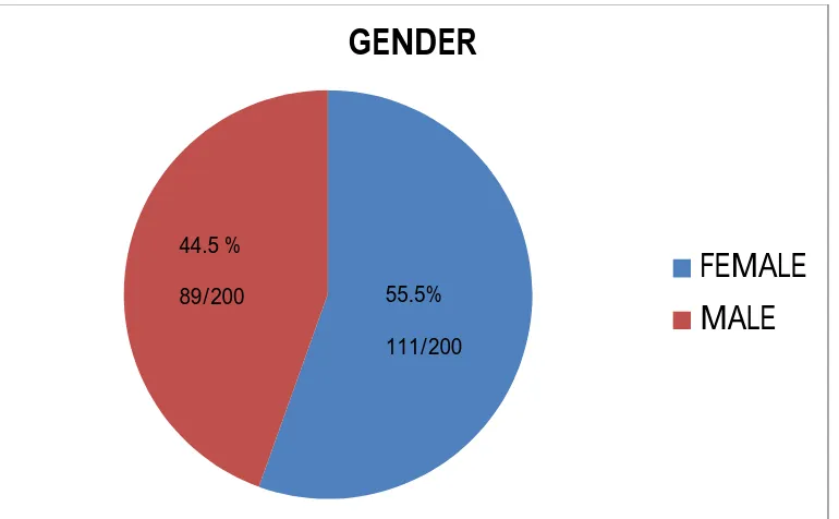
[image:73.612.102.465.423.679.2]GENDER DISTRIBUTION
There were 44 males and 56 females in group 1 and in group 2 there were
45 males and 55 females. The study subjects (200) in total consisted of 89 males
(89/200) and 111 females (111/200) i.e. 44.5 % were males and 55.5 % were
[image:74.612.103.488.287.525.2]females.
FIGURE 3: GENDER DISTRIBUTION OF STUDY SUBJECTS
GENDER
FEMALE
MALE
44.5 %
89/200 55.5%
PREVALENCE OF POAG IN DIFFERENT GENDERS
POAG was present in 5 males and 4 females in group 1 and in 4 males
and 2 females in group 2.Among 15 (15/200) patients with POAG, 9 were
males (9/15) and 6 were females (6/15). There was no statistically significant
difference in the gender distribution of POAG patients.
[image:75.612.91.525.332.505.2]P Value – 0.60.
TABLE 3: GENDER DISTRIBUTION OF POAG PATIENTS
GENDER GROUP 1 (with systemic
hypertension
GROUP 2 (without systemic
hypertension)
TOTAL
MALE 5 4 9
FEMALE 4 2 6
BLOOD PRESSURE
The mean systolic and diastolic blood pressure of the study subjects were
as follows:
POAG PRESENT:
HYPERTENSIVE GROUP:
SYSTOLIC = 143.11 mmHg
DIASTOLIC = 88.89 mmHg
CONTROL GROUP:
SYSTOLIC = 115 mmHg
DIASTOLIC = 75 mmHg
POAG ABSENT:
HYPERTENSIVE GROUP:
SYSTOLIC = 138.52 mmHg
DIASTOLIC = 88.13 mmHg
CONTROL GROUP:
SYSTOLIC = 116.59 mmHg
DIASTOLIC = 74.57 mmHg
0 20 40 60 80 100 120 140 160
SYSTOLIC BLOOD PRESSSURE
DIASTOLIC BLOOD PRESSURE
HYPERTENSIVE GROUP CONTROL GROUP
FIGURE 5: BLOOD PRESSURE IN PATIENTS WITHOUT POAG
TABLE 4:BLOOD PRESSURE AND POAG
GROUP POAG PRESENT POAG ABSENT
SYSTOLIC BLOOD PRESSURE DIASTOLIC BLOOD PRESSURE SYSTOLIC BLOOD PRESSURE DIASTOLIC BLOOD PRESSURE HYPERTENSIVE GROUP
143.11 88.89 138.52 88.13
CONTROL GROUP
115 75 116.59 74.57
0 20 40 60 80 100 120 140 160 SYSTOLIC BLOOD PRESSSURE DIASTOLIC BLOOD PRESSURE
HYPERTENSIVE GROUP CONTROL GROUP
[image:78.612.91.524.450.652.2]DURATION OF HYPERTENSION
Duration of hypertension of the 100 hypertensive subjects was grouped
[image:79.612.93.501.200.660.2]as follows:
TABLE 5: DURATION OF HYPERTENSION DURATION OF HYPERTENSION NO OF SUBJECTS
< 1 YEAR 20
1 YEAR TO < 5 YEARS 34
5 YEARS TO < 10 YEARS 30
10 YEARS 16
[image:79.612.91.472.415.659.2]TOTAL 100
FIGURE 6: DURATION OF HYPERTENSION
NO OF SUBJECTS
< 1 YEAR
>= 1 YEAR TO < 5 YEARS >= 5 YEARS TO < 10 YEARS >= 10 YEARS
DURATION OF HYPERTENSION
30 16
DURATION OF HYPERTENSION AND POAG
[image:80.612.89.478.233.431.2]Among glaucoma subjects in group 1, 5 were hypertensive for 10 years and the remaining 4 were hypertensive for less than 10 years. Duration of hypertension of glaucoma subjects were as follows:
TABLE 6: DURATION OF HYPERTENSION AND POAG
DURATION OF
HYPERTENSION
NO OF SUBJECTS WITH
POAG
< 1 YEAR
-1 YEAR TO < 5 YEARS 1 5 YEARS TO < 10 YEARS 3
10 YEARS 5
TOTAL 9
Out of 9 patients with glaucoma in hypertensive group, 55.5% (5/9) was hypertensive for 10 years.
FIGURE 7: DURATION OF HYPERTENSION AND POAG
NO OF SUBJECTS WITH POAG
< 1 YEAR
>= 1 YEAR TO < 5 YEARS
>= 5 YEARS TO < 10 YEARS
>= 10 YEARS
DURATION OF HYPERTENSION 1
3
[image:80.612.135.481.465.694.2]INTRAOCULAR PRESSURE
Mean IOP in hypertensive group was 17.30 with a standard deviation of
3.268. Mean IOP in control group was 16.02 with a standard deviation of
[image:81.612.129.482.249.367.2]3.250.
TABLE 7: MEAN IOP OF STUDY SUBJECTS
GROUP MEAN IOP
P VALUE =0.006
HYPERTENSIVE GROUP
17.30 ± 3.268
CONTROL GROUP 16.02± 3.250
STATISTICAL ANALYSIS
By applying annova table, there is a statistical significant difference in
GLAUCOMA AND INTRAOCULAR PRESSURE
There were 15 definitive cases of glaucoma meeting the defined criteria
for diagnosis of POAG. Nine of them had an IOP above 21 mm Hg and
remaining six had optic disc and field changes suggestive of glaucoma but the
IOP was < 21 mm Hg.
Among 200 subjects studied, 18 people had IOP above 21 mmHg. 10 of
them had POAG & remaining 8 were ocular hypertensive without optic disc or
[image:82.612.89.526.378.570.2]field changes.
TABLE 8 – IOP AND GLAUCOMA
IOP POAG PRESENT POAG ABSENT TOTAL
(%) GROUP 1
(WITH HT)
GROUP 2 (WITHOUT
HT)
GROUP 1 (WITH
HT)
GROUP 2 (WITHOUT
HT)
> 21 5 5 5 3 18(9%)
< 21 4 1 86 91 182(91%)
TOTAL 9 6 91 94 200
No of glaucoma patients
With IOP > 21mmHg - 10 With IOP < 21 mmHg - 5
FIGURE 8: INTRAOCULAR PRESSURE AND POAG
SYSTEMIC HYPERTENSION AND POAG
Out of 200 subjects studied, 15 of them was found to have glaucoma
amounting to an OVERALL PREVALENCE rate of 7.5%.
% PREVALENCE IN
HYPERTENSIVE GROUP - 9%
NON HYPERTENSIVE GROUP – 6%
0 5 10 15 20 25 30 35 40 45 50 55 60 65 70 75 80 85 90 95
IOP < 21 IOP > 21
TABLE 9: PREVALENCE OF POAG RISK FACTOR POAG
Present
POAG Absent
TOTAL
HYPERTENSION + 9 91 100
HYPERTENSION - 6 94 100
TOTAL 15 185 200
STATISTICAL ANALYSIS
Odds ratio and risk estimate was found to be 1.936 with a 95%
confidence interval ranging from 0.687 to 5.457 .Odds of hypertensive patient
developing POAG was 1.936 which was not statistically significant.
FIGURE 9:HYPERTENSION AND POAG
0 20 40 60 80 100 120 140 160 180 200
POAG + POAG
[image:84.612.126.487.409.655.2]DISCUSSION
Glaucoma is the most common cause of irreversible blindness in the
world and according to WHO estimate, 12.3% of blindness in the world is due
to glaucoma. POAG is often asymptomatic characterised by progressive retinal
ganglion cell death and visual field loss. IOP still remains the focus of therapy
but many patients experience continued progression of disease despite adequate
control of IOP. Extensive research is underway to identify other risk factors like
vascular factors, geneti