THE STUDY OF VARIATIONS IN THE ORIGIN
AND THE COLIC BRANCHES OF THE
SUPERIOR MESENTERIC ARTERY
Submitted to
THE TAMILNADU DR. M.G.R. MEDICAL UNIVERSITY
In partial fulfillment for
M.D. DEGREE EXMINATION
BRANCH – XXIII (ANATOMY)
STANLEY MEDICAL COLLEGE, CHENNAI
THE TAMILNADU DR.M.G.R. MEDICAL UNIVERSITY
CHENNAI
CERTIFICATE
This is to certify that the dissertation on ‘THE STUDY OF
VARIATIONS IN THE ORIGIN AND THE COLIC BRANCHES OF
THE SUPERIOR MESENTERIC ARTERY’ is a bonafide work done by
Dr. M.R. Manimekalai in the Department of Anatomy, Stanley Medical College, Chennai – 600 001, during 2013-2016 under my supervision and guidance in partial fulfillment of the regulation laid down by The Tamil Nadu Dr.M.G.R. Medical University, for the M.D. Anatomy, (Branch XXIII) examination to be held in April – 2016.
Dr. Isaac Christian Moses, M.D., Dr. S.Chitra, M.S.,
The Dean Professor and Head of Department,
Stanley Medical College, Stanley Medical College, Chennai – 600001. Chennai – 600001.
Date : Date :
CERTIFICATE OF THE GUIDE
This is to certify that the dissertation on ‘THE STUDY OF
VARIATIONS IN THE ORIGIN AND THE COLIC BRANCHES OF
THE SUPERIOR MESENTERIC ARTERY’ is a bonafide work done by
Dr. M.R. Manimekalai in the Department of Anatomy, Stanley Medical College, Chennai – 600 001, during 2013-2016 under my supervision and guidance in partial fulfillment of the regulation laid down by The Tamil Nadu Dr.M.G.R. Medical University, for the M.D. Anatomy, (Branch XXIII) examination to be held in April – 2016.
Dr. S.Chitra, M.S.,
Professor and Head of Department,
Stanley Medical College, Chennai – 600 001.
Date :
DECLARATION
I solemnly declare that this dissertation, “THE STUDY OF VARIATIONS IN ORIGIN AND THE COLIC BRANCHES OF SUPERIOR MESENTERIC ARTERY” was written by me in the
Department of Anatomy,Government Stanley Medical College and Hospital,Chennai under the guidance and supervision of Prof.Dr.S.CHITRA MS.,Professor and Head of the Department of Anatomy, Government Stanley Medical College,and Chennai-600001.
This dissertation is submitted to The Tamilnadu Dr.M.G.R.Medical University,Chennai in partial fulfillment of the university regulations of the award of Degree of M.D. Anatomy(Branch XXIII)examination to be held in April 2016.
Place : Chennai-1
ACKNOWLEDGEMENT
I wish to express my sincere thanks and gratitude to Dr. ISAAC
CHRISTIAN MOSES, M.D, FICP, FACP, Dean, Stanley Medical College
and Hospital, Chennai – 1 for having permitted me to utilize the facilities in this college for the conduct of the study.
My heartfelt thankfulness, gratitude and gratefulness to Dr. S.Chitra, M.S. Professor and Head of the Department of Anatomy, Government Stanley Medical College, Chennai for her invaluable guidance, motivation and persistent support, encouragement and for providing all necessary arrangement to make the study a reality.
I am grateful to Dr. C. Amarnath, M.D. (RD), Professor and Head of the Department of Radiology, Government Stanley Medical College, Chennai -1 and his faculty for their help in radiological study.
I sincerely thank Dr. T.Vasantha Kumar, M.S., Associate Professor
Dr. J.Thilagavathy, M.S., Associate Professor, Dr. K. Sujatha, M.S.,
Dr. V.RajaPriya, M.S. and Dr. B. Anbumalar, M.D., Dr. K.Raja, M.D.,
Dr. C.Adline, M.D., Dr. S.Elizabeth, M.D, Dr. M. Anuradha, M.D.,
I thank Dr. Rajkumar, Tutor in Anatomy for helping me in the study
I am also thank earnestly my seniors Dr.G.Sasi Krishnan,
Dr. R. Saranya, Dr. R. Senthamizh Selvi and my juniors Dr. S. Manonmani,
Dr. C. Sasikala, Dr. V. Santhi, and Dr. G.Senthil Kumar, and my colleague
Dr. F. Stelina Sophie Dina for their immense help rendered to me during the
study.
I am also thankful to lab technicians Smt. K.Rajalakshmi, Smt.
E.Jayanthi, and departmental staffs Thiru. C.Birammaiah, Thiru.A. Kadher
Basha, Thiru. G. Anbalagan, Thiru M. Jagadeswaran for helping me in
carrying out the study.
Last but not least I am thankful to my parents, husband and children for their constant unending possessive love,support and patience for putting up with me until this day.
CONTENTS
S.NO TITLE PAGE NO.
1. INTRODUCTION 1
2. ANATOMICAL AND EMBRYOLOGICAL CONSIDERATIONS
3
3. AIM OF THE STUDY 15 4. REVIEW OF LITERATURE 17 5. MATERIALS AND METHODS 42
6. OBSERVATION 44
7. DISCUSSION 64
8. SUMMARY 75
9. CONCLUSION 76
THE STUDY OF VARIATIONS IN THE ORIGIN AND
THE COLIC BRANCHES OF THE SUPERIOR
MESENTERIC ARTERY
INTRODUCTION
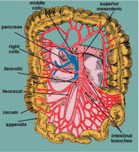
Superior mesenteric artery is one of the ventral branches of abdominal aorta. It arises from the front of the abdominal aorta behind the body of the pancreas, at the level of first lumbar vertebra, one centimetre below the coeliac trunk. It is the artery of mid gut.
It runs downwards to the right and it forms a curve with its convexity towards the left.
It lies first behind the body of pancreas and then in front of uncinate process of pancreas.
Then it crosses the third part of duodenum, enters the mesentery and runs between its two layers.
It supplies the duodenum below the opening of bile duct, jejunum, ileum, appendix, caecum ascending colon, right two thirds of transverse colon and lower half of head of pancreas.
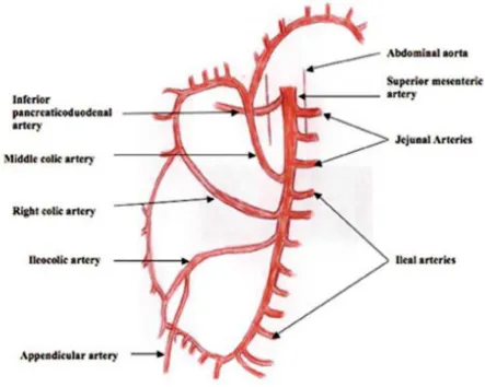
a) Branches from right side of superior mesenteric artery are :
i. Inferior pancreatico duodenal artery ii. Middle colic artery
iii. Right colic artery and iv. Ileo colic artery
b) Branches from left side of superior mesenteric artery are :
ANATOMICAL AND EMBRYOLOGICAL CONSIDERATIONS
Superior mesenteric artery arises from the front of the abdominal aorta about one cm below the coeliac trunk, opposite the lower border of first lumbar vertebra. It supplies the second part of the duodenum distal to the major duodenal papilla, third and fourth part of duodenum, lower half of the head of pancreas, jejunum, ileum, caecum, appendix, ascending colon and right two thirds of transverse colon. It is the artery of midgut.
It crosses the third part of duodenum, then enters the root of mesentery runs between its two layers and it terminates in the right iliac fossa. Above the root of mesentery, anteriorly, superior mesenteric artery is related to the body of the pancreas and to the splenic vein; posteriorly related to aorta, left renal vein, uncinate process of pancreas and third part of the duodenum. Within the root of the mesentery it crosses the inferior venacava and the right psoas major muscle. It is accompanied with the superior mesenteric vein on its right side. It is surrounded by the superior mesenteric plexus of nerves.
Branches of superior mesenteric artery
1.Inferior pancreatico duodenal artery
2.Middle colic artery
3.Right colic artery
4.Ileo colic artery
5.Jejunal and ileal branches
and dorsal arches after anaostomosing with the corresponding branches of the superior pancreatico duodenal artery which is a branch of gastro duodenal artery.
2. Middle colic artery: It comes from the superior mesenteric artery at the lower border of the pancreas and immediately enters the root of transverse mesocolon and divides into right and left branches. The right branch reaches the right colic flexure and anastomoses with the ascending branch of right colic artery; the left branch anastomoses close to left colic flexure, with the ascending left colic branch of the inferior mesenteric artery. Arches thus formed are 3 to 4 cm from the transverse colon and supply the right two thirds of the transverse colon.
4. Ileo colic artery: It is the terminal branch derived from the right side of the superior mesenteric artery. The artery passes retro peritoneally downwards and to the right; on reaching the right iliac fossa it divides into ascending and descending branches. Ascending branch anastomoses with right colic artery, descending branch continues as terminal part of superior mesenteric artery.
The descending branch of ileo-colic divides into four sets of branches at the superior border of ileo- colic junction.
1. Anterior caecal artery reaches the front of caecum along the superior ileo -caecal fold.
2. Posterior caecal artery supplies the back of the caecum and anastomoses with recurrent branch of the appendicular artery. 3. Appendicular artery – It gives a recurrent branch to the base of
appendix where it anastomoses with posterior caecal artery. 4. Ileal branch supplies the terminal portion of ileum .
Jejunal branches :
(occasionally double)tier of anastomotic arcs before giving rise to multiple vasa recta.These vessels run almost parallel in the mesentery and distributed alternatively to opposite aspects of its wall, where the two series form distinct leaves within the mesentery. Small twigs supply regional lymph nodes and other structures in the mesentery.
Ileal branches:
Ileal branches are more numerous than the jejunal branches but smaller in caliber.They arise from the left and anterior aspects of the superior mesenteric artery.The length of the mesentery is greater in ileum and the branches form 3,4 or 5 tiers of arcs within the mesentery before giving rise to multiple vasa recta that run directly towards the ileal wall. The ileal branches run parallel in the mesentery and are distributed to the alternate aspects of the ileum. They are longer and smaller than similar jejunal vessels particularly in the distal ileum, and do not form such definite parallel leaves of the vessels.
mesenteric artery they are often larger in caliber than the mid ileal vessels.
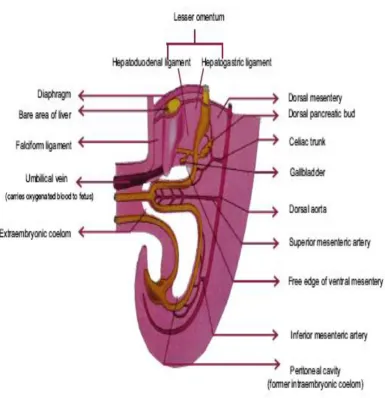
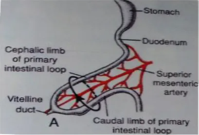
EMBRYOLOGICAL CONSIDERATIONS:
The superior mesenteric artery shows many embryological irregularities in its contour, origin and configuration.
The first part of the artery for an inch or more, may be, indented in a spiral form, a vestige of the primitive rotation of the midgut which takes place counter-clockwise around it, to 270 degrees, the artery itself making a 180 degree rotation.
Thereafter, from the concave side of the artery, the middle colic, right colic and ileocolic arteries arise.The terminal or ileal branch of ileocolic artery continues as superior mesenteric artery.
From the convex left side of the artery 5-10 jejunal and ileal branches arise, which in the unrotated gut, arise from the right side of the superior mesenteric artery and constitute its first branches.
The occurrence of a coeliaco mesenteric trunk has repeatedly been reported in the literature, its average incidence being about 1%. The mode of formation of the coeliaco mesenteric trunk can be accounted from both an ontogenetic and phylogenetic point of view.
Superior mesenteric artery
AIM OF THE STUDY
The aim of the study is to examine the pattern of variations in the origin of the superior mesenteric artery and its colic branches, mainly in South Indian Population and comparing the variations with the previous study.
Thorough knowledge of variations in origin and branching pattern of superior mesenteric artery is helpful for correct interpretation of any procedures such as Laparoscopic procedures and resection of colon for carcinoma, intestine transfers, resections of small and large intestines, appendicectomy and embolectomy. By ligating the arteries properly during surgeries and by knowing the anatomy completely , we can avoid injury to the blood vessels. Variations in branching pattern are common. A sound knowledge about the normal origin of superior mesenteric artery, its variation in the origin, branching pattern of the variations of superior mesenteric artery is important for surgeon, physician, Radiologist , Gastro enterologist and vascular surgeon.
Parameters:
Level of origin
Course of artery
In branching pattern
Relation with coeliac trunk
REVIEW OF LITERATURE
Galen (121-201 AD) was among the first to describe arteries carrying blood and their supply of colon (Buck, 1917, Garrison 1929, Singer 1925). This was followed by a period of dark ages when anatomy remained dormant for several hundred years .
It was Von Haler (1803) who between 1759 and 1966 described them main bloody supply to the colon by the superior mesenteric artery and its branches.
Quain in his study of 1040 bodies merely named the colic arteries without note of the differences in anatomical disposition.
Richead quain Published – The Anatomy of the Human body” in 1844. In the preface of this classis he wrote: The difficulties which have often occurred in the performance of those surgical operations in which the larger arteries are concerned have arisen in great point from want of sufficient acquaintance with the difference in anatomical disposition to which these vessels are subject.
The vascular supply of the small intestine was investigated by Cokkin’s (1930) in his study of mesenteric thrombosis. He stated that the collateral circulation stops with the terminal row of arcades in the mesentery. Beyond this there is absolutely no anastamoses either between the vasa recta in the mesentery or between the ramifying vessels of the gut wall.
Noer (1943) confirmed the findings of Eisberg, with the help of his liquid injected specimens.
In 1963 Veslinguise reported the superior mesenteric artery in his work, which was translated by Culpeper.
Richaed S. Snell Page 229 Basic Anatomy Clinical Anatomy by Regions- superior mesenteric artery, a branch of the abdominal aorta supplies all extensive territory of the gut from halfway down the 2nd part of the duodenum to the left colic flexure. Occlusion of the artery or one of its branches results in death of all or part of this segment of the gut. The occlusion may occur as a result of embolus, an aortic dissection or a thrombus or an abdominal aneurysm.
ORIGIN OF THE SUPERIOR MESENTERIC ARTERY :
Von Holer(1803) (Tripod of Haler) reported that the coeliac trunk may arise from superior mesenteric artery. The coeliac trunk give rise only to the splenic artery and left gastric artery. The hepatic artery arising from the superior mesenteric artery or the hepatic and left gastric arteries from coeliac, the splenic artery from the superior mesenteric artery.
Henle(1809-1885) and later Delannoy(1923) reported the occurrence of two superior mesenteric arteries.
Tandler(1904)gave the first comprehensive description of the embryogenesis of the coeliacomesenteric trunk in human beings.
Eaton (1917) studied 206 bodies and reported the origin of the hepatic artery from the aorta or superior mesenteric artery, left gastric and splenic arteries from a common trunk and classified this as type 1 coeliac trunk.
Coeliaco mesenteric trunk was reported by Lipstutz (1917).
1. The hepatic, the splenic and the superior mesenteric aretery arise as a common trunk from the abdominal aorta-1.2%.
2. The left gastric, the splenic, the hepatic and the superior mesenteric arteries arise as a common trunk from abdominal aorta.
Adachi(1928) published the book “Das arteriensystem Der Japanese”with
an extensive study of variational anatomy of the arteries.
Coeliaco mesenteric trunk was reported by Munger and Mangoushi(1941).
Michels(1955) stated that the pattern of the arteries are determined by internal and external factors. Development peculiarities formed in the arteries to the supramesocolic organs (coeliac and superior mesenteric artery) would be correlated with
a) Variations in the degree and the site of gut rotation
b)Persistence of differently interrupted sections of the primitive roots of the omphalomesenteric (vitelline) arteries (10,11,12,13 ventral segments and their longitudinal anastamosis.
Wright(1959)in a case of left sided vermiform appendix,found the superior mesenteric artery from the aorta, 3cm below the coeliac trunk.
Dr.Kalavathy, Director, Institute of Anatomy, Madras Medical College, carried out a detailed study in 75 cases (1980) and she observed that the superior mesenteric artery with coeliac artery arising as a common trunk in 3.3% cases.
Dr.Radhakrishnayya (1990) reported the distance of origin between the coeliac trunk and the superior mesenteric artery in 25 cases.
Yuksel et al6 reported the inferior phrenic artery arising from the coeliac trunk and an aberrant right hepatic artery arising from the superior mesenteric artery.Kaibojaku Zasshi;1998;73;497-503.
Remanuflacker reported the origin of the superior mesenteric artery is about 1.cm below the origin of the coeliac trunk, behind the pancreas and is crossed anteriorly by the splenic vein.
Kao et al studied 24 superior mesenteric artery angiograms. The locations of the origin of the coeliac trunk and the superior mesenteric artery were determianed in each case. The superior mesenteric artery arose at the level of 1st lumber vertebra in 24 angiograms (83%), below the pedicle of 1st lumber vertebra in 5 cases (21%), none arose the 1st lumber -2nd lumber inter space.
Covdar et al reported a case of coeliaco mesenteric trunk a variation found in only 1% to 2.7% population. Gastro hapato spleno mesenteric trunk 2 percent (coeliac and superior mesenteric combined)
Higashi N. and Hirai K observed that the hepatic artery arising from an unusual hepatomesenteric trunk of aorta immediately inferior to the gastro duodenal trunk was reported as type .2
LAST’S ANATOMY REGIONAL AND APPLIED : 10TH
EDITION 1999
“The superior mesenteric artery arises from the front of aorta,a
GRAY’S ANATOMY 40TH
EDITION 2011:
SUSAN STANDRING PAGE NO.1130, 1131
“The superior mesenteric artery originate from the aorta 1 cm below the level of the intervertebral disc between the first and second lumbar vertebra”.
CUNNINGHAM’S MANUAL OF PRACTICAL MEDICINE 15TH
EDITION: 2011 VOLUME 2:
“The superior mesenteric artery arises at the level of first lumbar
vertebra o.5cm inferior to the coeliac trunk,posterior to the body of pancreas and splenic vein”
COURSE OF ARTERY:
GRAY’S ANATOMY 40TH
EDITION: 2011
SUSAN STANDRING PAGE NO.1130, 1131
“The superior mesenteric artery originates from the aorta 1 cm
posterior to the splenic vein and the body of pancreas.The left renal vein lies behind it and separates it from aorta.The artery crosses anterior to the inferior venacava,right ureter and right psoas major as in the root of small bowel mesentery.Its caliber progressively decreases and successive branches are given off to the loop of jejunum and ileum,and its terminal branch anastomose with the ileocolic artery”.
CUNNINGHAM’S MANUAL OF PRACTICAL ANATOMY:
15th EDITION 2011 volume 2:
“The superior mesenteric artery arises at the level of first lumbar
T.S.RANGANATHAN:A TEXTBOOK OF HUMAN ANATOMY 2011TH EDITION:P-327,328:
“The superior mesenteric artery arising 1.25cm below the origin of
the coeliac axis.At its origin,it is behind the body of pancreas and crossed in front by splenic vein,posteriorly it is related to the left renal vein.Its emerges out between the body of pancreas and its uncinate process,crosses in front of the uncinate process and the 3rd part of duodenum to get into the root of mesentery.Then it is related to superior mesenteric vein on right side and it crosses the inferior venacava,right ureter,right psoas major and ends in the right iliac fossa by anastomosing with one of its branches.It presents a curved course with the convexity facing downwards and to the left side”
TERMINATION OF ARTERY:
A.K.DATTA HUMAN ANATOMY 9TH EDITION P-157,158: 2010
“The superior mesenteric artery arises from the front of aorta about
mesentery towards the right iliac fossa.It ends by anastomosing with its ileocolic branch”.
HENRY HOLLINSHEAD VOLUME2 ANATOMY OF SURGEON.2ND EDITION,1971.P-463,463,465:
“The superior mesenteric artery arises from the front of aorta just
JOHN E.SKANDALAKIS GEME L.COLBORN:
“The horizontal part of the duodenum may become compressed in
INFERIOR PANCREATICO DUODENAL ARTERY:
A.K.DATTA HUMAN ANATOMY 9TH EDITION, VOL-2: 2010
“Inferior pancreatico duodenal artery is usually the first branch of
the superior mesenteric artery.It often arises from the first jejunal branch.The artey passes to the right along the upper border of 3rd part of duodenum and immediately divides its anterior and posterior branches and form the ventral and dorsal arches after anastomosing with the corresponding branches of the superior pancreatico duodenal artery”.
T.S.RANGANATHAN : A TEXT BOOK OF HUMAN ANATOMY 2011TH EDITION:
“It is the branch of superior mesenteric artery.It runs upwards to
anastomose with superior pancreatico duodenal artery to form anterior and posterior pancreatico duodenal arcades”.
NEETA KULKARNI,2ND EDITION,CLINICAL ANATOMY, page no:705: 2012
“Inferior pancreatico duodenal artery may arise from the superior
branches made anastomoses with the corresponding branches of the superior pancreatico duodenal artery”.
“Michael observed inferior pancreatico duodenal artery had a
common origin along with superior mesenteric artery from the abdominal aorta in 2 cases(4%)”.
MIDDLE COLIC ARTERY:
Henle (1876) reported a case of presence of two middle colic arteries and several cases where branches of superior and inferior mesenteric arteries replace the middle colic arteries.
Moynhan (1913) reported an accessory middle colic artery running direcntly towards the middle of the transverse colon.
trifurcated or had 4 branches. In 2 cases it was absent (2%) being replaced by large branches from the left colic artery.
Steward and Rankin(1933) found, 27% an additional branch from middle colic artery running towards the left colic flexure, and it reinforces the marginal artery at that point 10% cases they found an accessory middle colic artery arising from superior mesenteric artery above the origin of major middle colic artery.
Michels (1955) and co workers observed an accessory middle colic artery in 8% and no middle colic artery in 3%.
Girffth’s found no middle colic artery in 22% of 100
cases.Ann.Roy.Coll.Surgeons,Eng.119:241,1956.
Vandamme and Schuren (1956) explored and reported single middle colic artery in 75%, two middle colic arteries with separate origins in 24% and three middle colic arteries in 1% of the cases. In one case, the middle colic artery was absent.
absence of middle colic artery in 3.6% (22 bodies in 600%). Single middle colic artery was present in 7% two middle colic arteries with two separate origin was found.
Nelson et al8 found in their study 2 rare variations in middle colic artery.In 4% cases, the middle colic artery was branching off from ilecolic artery. In 16% of cases, 2 middle colic arteries were present.Clinical anatomy 1988;1:75-91.
Waldeyer(1989/1900) describes a colica media (middle colic artery) and a colica media accessoria,but none of these branches were well defined.
Kerofi et al (1995) reported an anomalous middle colic artery from the proximal segment of the splenic artery.
Dr.Ashwini Hetal 6 found in their study that in 90% of cases the middle colic and in 66% the ileo colic artery arose directly from the superior mesenteric artery-a cadaveric study,Int J Biol Med Res.2013;4(1):3004-3006.
at:www.anatomyatlases.org.reported that the middle colic artery the first branch of superior mesenteric artery most commonly originates as an independent branch from superior mesenteric artery or arises along with the right colic artery as a common stem.
W. Henry Hollinshed, in his book of Anatomy for Surgeons, Vol.2 states that in 30 to 50% of cases the common stem which shares middle colic and right colic arteries.
Ridan found an accessory middle colic (Arc of Riolan) connecting superior mesenteric artery with superior left colic artery.
Benton and Coter observed that the superior mesenteric artery gave rise to one major trunk, which was divided into ileocolic and right colic branches only.
NEETA V.KULKARNI CLINICAL ANATOMY 2ND EDITION: 2012
“The middle colic artery arise from the right side of superior
HENRY HOLLINSHEAD VOLUME2 ANATOMY OF SURGEON.2ND EDITION:
“The superior mesenteric artery, enters the root of the mesentery before
so doing, it has typically given off inferior pancreatico duodenal artery unless these arise from the 1st jejunal branch and also before or as it emerges it gives rise to middle colic artery”.
T.S.RANGANATHAN:A TEXTBOOK OF HUMAN ANATOMY 2011TH EDITION:
“The middle colic artery passes between the layers of transverse
mesocolon,divides into left and right branches.The right branch anastomose with the ascending branch of the right colic artery.The left branch anastomose with the left colic branch of the inferior mesenteric artery.It supplies the right two third of the transverse colon”.
RIGHT COLIC ARTERY:
ileocolic is 12%.78% right colic artery arose as a single vessel.8.7% shows two right colic arteries. 0.7% had three right colic arteries.
Basmajian (1955) reported that the right colic artery arises more commonly with either the middle colic or the ileocolic artery.
Sonneland et al (1958) reported 12.6% of absence of right colic artery in a series of 600 bodies.
Michels and coworkers (1963) failed to identify the right colic artery in only 2%. They found in origin from the superior mesenteric artery in 38% an origin with middle colic in 52% and one with me liecolic in 8%.
Vadamme and Suhuren (1976) reported the presence of 32% right colic arteries in 156 cases and in 105 of the cases two right colic arteries, in one case it arises from the ileocolic.
Dr.Radhakrishnayya (1990) reported the normal origin of the right colic artery and its origin from the ileocolic artery.
Biswa Bhusan Mohantry et al10 reported that the right colic artery is the most variable branch. In his report there was no right colic artery.International journal of anatomical variations;(2013)6;26-27.
Dr.Ashwini H et al6 found in their study in 46% of cases. Right colic artery arose as a direct branch from superior mesenteric artery.In 10% of cases it originated as a common stem with middle colic artery and in 34%,it arose with ileo colic artery. Right colic artery was absent in 10% cases.Int J Biol Med Res.2013;4(1):3004-3006
T.S.RANGANATHAN:TEXTBOOK OF HUMAN ANATOMY 2011TH EDITION:
“The right colic artery divides into an ascending and a descending
branch.The latter anastomoses with the ascending branch of the ileocolic artery to form the marginal artery of the colon.It supplies the upper two third of the ascending colon and hepatic flexure of colon”.
“Bergman et al7
ILEOCOLIC ARTERY:
Michels (1955) reported the ileocolic artery which divides into three branches and the site of origin of the appendicular artery is extremely varied.
Michles(1955) and co workers found an origin of right colic artery with the ileocolic artery is 8%
Basmajian,J.V.The main arteries of the large intestine.Surg,Gynec.&Obst 101:585,1955. agreed that ileocolic artery arise more commonly with right colic artery.
Vandamme and schuren (1976) stated that the ileocolic artery is the most constant collateral of the superior mesenteric artery.
Garica et al (1996) reported the ileocolic artery to be the constant branch.
Dr.Ashwini H et al 6 found in their study the ileocolic artery 66% arose directly from superior mesenteric artery.
2. A.K.DATTA HUMAN ANATOMY 9TH EDITION: 2010
“It is the terminal branch from right side of the superior mesenteric
artery.The artery passes retroperitoneally to the right and on reaching the right iliac fossa,it divides into ascending and descending branches.The former anastomoses with the right colic artery and the latter with the termination of superior mesenteric aartery.The descending branch of ileocolic artery divides into 4 sets of branches.They are anterior and posterior coecal,appendicular and ileal branches”.
NEETA V.KULKARNI CLINICAL ANATOMY 2ND EDITION,2012:
“The ileocolic artery is actually the continuation of the superior
APPENDICULAR ARTERY:
SHAH AND SHAH STUDY:
It is a study of blood supply to the appendix,reported that among 60 bodies,70% had a single appendicular artery while 30% had more than 1. 11% from ileocolic trunk as from coeccal branches and least frequently from the ascending branch.
MICHEL’S AND CO WORKERS found a 2nd appendicular artery in only 8% of 132 specimens
HENRY HOLLINSHEAD VOLUME2 ANATOMY OF SURGEON.2ND EDITION:
“ The anterior and posterior coecal arteries may arise from a
common trunk or separately in 36% and 64% respectively”.
RELATION WITH COELIAC TRUNK:
T.S.RANGANATHAN:A TEXTBOOK OF HUMAN ANATOMY 2011TH EDITION:
“ Superior mesenteric artery is a ventral branch of the abdominal aorta
A.K.DATTA HUMAN ANATOMY 9TH EDITION: 2010
“The superior mesenteric artery arises from the front of aorta about 1cm below the celiac trunk,opposite the lower border of L1 vertebra”.
GRAY’S ANATOMY 40TH
EDITION: 2011
“The superior mesenteric artery originates from the aorta 1 cm
below the coeliac trunk at the level of the intervertebral disc between the first and second lumbar vertebra”.
NEETA V.KULKARNI,(CLINICAL ANATOMY) 2ND EDITION:
“The superior mesenteric artery arises from the front of the
abdominal aorta about 0.5cm below the coeliac trunk at the level of disc between L1 and L2 vertebra behind the body of pancreas”.
RELATION WITH PANCREAS:
A.K.DATTA HUMAN ANATOMY 9TH EDITION: 2010
“The superior mesenteric artery passes downwards,forwards and to
T.S.RANGANATHAN:A TEXTBOOK OF HUMAN ANATOMY 2011TH EDITION:
“The superior mesenteric artery lies behind the body of
pancreas,then its emerges out between the body of pancreas and its uncinate process,crosses in front of the uncinate process and the 3rd part of duodenum to get into the root of mesentery”.
HENRY HOLLINSHEAD VOLUME2 ANATOMY OF SURGEON.2ND EDITION:
“The superior mesenteric artery is the second ventral branch of
aorta,gives off slightly below the celiac trunk,opposite the lower border of L1 vertebra.The artery descends in the groove on the posterior surface of the neck of pancreas.Immediately below the origin,it crosses the left renal vein,which lies between it and the aorta.Below the inferior margin of neck of pancreas,it crosses anteriorly to the uncinate process and horizontal part of duodenum.These also separate it from the aorta”.
NEETA V.KULKARNI,(CLINICAL ANATOMY) 2ND EDITION: 2012
“The superior mesenteric artery arises from the front of the
MATERIALS AND METHODS
Materials:
A total number of 50 superior mesenteric arteries were studied. (30) superior mesenteric arteries were studied from the cadavers. Twenty (20) superior mesenteric arteries, pictures of CT angiogram.
• Venue of study:
• Department of Anatomy,Stanley Medical College.
• Department of Radiology,Stanley Medical College.
Methods:
1. Dissection method:
mesentery of small intestine in the infra colic compartment was exposed by turning the colon and its mesentery upwards.The attachment of the mesentery was traced. The small intestine was lifted to left. Superior mesenteric artery in the root of mesentery and its branches were exposed. Then the origin of superior mesenteric artery was identified, then the course, termination, branches, relation with coeliac trunk, relation with pancreas were traced and the photographs were taken.
2. Radiological study:
After getting consent from the patients, the (CT angiogram) pictures were taken, collected and studied for above mentioned parameters.
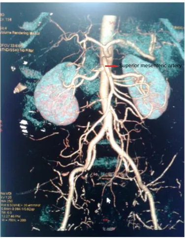
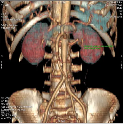
CT ANGIOGRAM (Computerized Tomography Angiogram)
OBSERVATION
LEVEL OF ORIGIN:
In the present study , the origin of superior mesenteric artery was normal in 47 specimens.
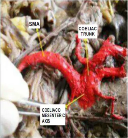
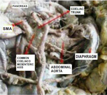
In two specimens, both the superior mesenteric artery and coeliac trunk arise from abdominal aorta by a common trunk at the level of first lumbar vertebra. The trunk was about 1.5 cm long and was divided into two as coeliac and superior mesenteric arteries.
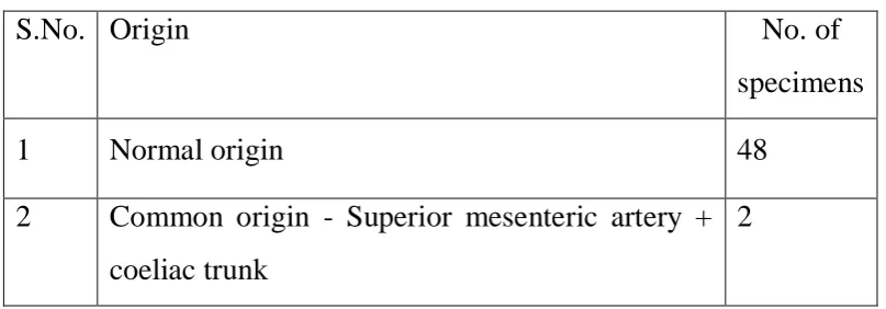
ORIGIN OF SUPERIOR MESENTERIC ARTERY:
S.No. Origin No. of
specimens 1 Normal origin 48
2 Common origin - Superior mesenteric artery + coeliac trunk
[image:53.595.97.501.356.497.2]2
Chart -1 Origin of Superior Mesnteric Artery 48 2 0 10 20 30 40 50 60
Normal origin Common origin - Superior mesenteric artery + coeliac trunk
N o . o f Sp e c im e n
COURSE OF ARTERY:
In the present study, in all cases superior mesenteric artery arises 1cm below the origin of the coeliac trunk at the level of first lumbar vertebra. It runs downwards and to the right forming a curve with its convexity towards the left. The artery runs anterior to uncinate process of pancreas and the third part of duodenum, posterior to the splenic vein and the body of the pancreas. The left renal vein lies behind it and separates it from the aorta. The artery crosses anterior to the inferior vena cava, right ureter, right psoas major it descends in mesentery and runs in between the two layers. It terminates in the right iliac fossa by anastomosing with a branch of ileo colic artery.
LEVEL OF TERMINATION:
It terminates in the right iliac fossa by anastomosing with a branch of ileocolic artery in all the 50 cases of present study.
BRANCHING PATTERN OF SUPERIOR MESENTERIC ARTERY INFERIOR PANCREATICO DUODENAL ARTERY:
S.No Origin Numbers 1. Normal origin 49
2. Superior mesenteric artery + inferior pancreatico duodenal artery
[image:58.595.105.482.123.235.2]1
Table 2 Inferior Pancreatico Duodenal Artery
Chart 2 Inferior Pancreatico Duodental Artery
98% 2%
INFERIOR PANCREATICO DUODENAL ARTERY
Normal origin
MIDDLE COLIC ARTERY:
In the present study ,in 49 specimens the middle colic artery originate from superior mesenteric artery and in one specimen it was absent.
Middle colic artery
S.No. Origin Specimens 1 Origin from superior mesenteric artery 49
[image:61.595.92.522.374.684.2]2 Absence of Middle colic artery 1
Table 3 Middle Colic Artery
Chart 3 Middle Colic Artery
49 1 0 10 20 30 40 50 60
Origin from superior mesenteric artery Absence of Middle colic artery
N o o f SPe c im e n
RIGHT COLIC ARTERY
In the present study, right colic artery was present in 47 specimens and absent in 3 specimens.
Out of 47 specimens, mentioned above, in 43 specimens, the right colic artery, had normal site of origin; in 4 specimens, abnormal origin were observed. Out of 4 specimens with abnormal origin, three specimens, the right colic artery and the ileocolic artery had common origin from the superior mesenteric artery. In one specimen, the right colic artery took origin from the ileocolic artery.
[image:63.595.85.499.486.598.2]In three specimens where the right colic artery was absent, it was replaced by branches from the middle colic artery and ascending branches of ileocolic artery
Table 4 Right Colic Artery
S.No. Origin No of specimens
1 Presence of right colic artery normal 47
Chart 4 Right Colic Artery 47 3 0 5 10 15 20 25 30 35 40 45 50
Presence of right colic artery normal Absence of right colic artery
N o . o f SPe c im e n
ILEOCOLIC ARTERY:
Ileocolic artery was present in all the 50 specimens. In three specimens the ileocolic artery and the right colic artery had a common origin.
S.No. Origin No of
specimens 1 Presence of Ileocolic artery 50 2 Origin of Ileocolic artery from superior
mesenteric artery
47
3 Common origin of Ileocolic artery with right colic artery
[image:67.595.90.497.211.699.2]3
Table 5 Ileocolic Artery
Chart 5 Ileocolic Artery
50% 47%
3%
ILEOCOLIC ARTERY
Presence of Ileocolic artery
APPENDICULAR BRANCH :
In 48 specimens appendicular branch arise from ileo colic artery. In 2 specimens there were 2 appendicular branches 1 from ileo colic artery before division and the other from the inferior division of ileocolic artery.
S.No. Branching Pattern No of specimens
1 Single no of appendicular artery from ileo colic artery
48
2 Double branch of appendicular artery from ileo colic artery
[image:68.595.98.503.242.414.2]2
Chart 6 Appendicular Artery
96% 4%
APPENDICULAR ARTERY
Single no of appendicular artery from ileo colic artery
Double branch of
RELATION WITH COELIAC TRUNK:
In the present study, in all the cases
Superior mesenteric artery arises from the front of the abdominal aorta behind the body of the pancreas, at the level of first lumbar vertebra, one centimetre below the coeliac trunk. In two specimens, both the superior mesenteric artery and the coeliac trunk arise from the abdominal aorta. RELATION WITH PANCREAS:
In the present study, in all the cases
DISCUSSION
A total of fifty (50) superior mesenteric arteries were studied in the different age group in the South Indian Population by dissection methods and Radiological method.
The data obtained in the present study were correlated with the date of the previous workers in this filed.
Origin of the superior mesenteric artery
In the present study the origin of the superior mesenteric artery from abdominal aorta was found in 96% of specimen, in 4% of specimens superior mesenteric artery arose along with the coeliac trunk from abdominal aorta.
Richard S.Snell (page 229) (Clinical anatomy 7th Edition).
Superior mesenteric artery a branch of the abdominal aorta supplies all extensive territory of the gut from halfway down the second part of the duodenum to right 2/3 of transverse colon.
The same findings were found in
Cunningham’s manual of practical anatomy 15th
Edition 2011, volume 2
Last’s anatomy 10th
edition (page no.244)
Normal origin of superior mesenteric artery in the present study correlate with the above mentioned authors.
The abnormal origin of superior mesenteric artery along with coeliac trunk which was found in the present study follow the same pattern mentioned by
Cavadar et al17 study reported a case of Coeliaco mesenteric trunk a variation found in only 1%
Lippen et al18 observed coeliar & superior mesenteric artery seen as a common trunk in 2%.
Michel (1955). In his study of 200 specimens stated that in 11.5% of his specimens the common origin was found.
Course of Superior Mesenteric artery.
right an it forms a curve with its convexity towards the left. It lies first behind the body of pancreas and then in front of uncinate process of pancreas. Then it crosses the third part of duodenum, enters the mesentery and runs between its two layers. It terminates in right iliac fossa by anastomosing with a branch of ileo colic artery.
In all the cases, the present study the course of superior mesenteric artery, follow the normal description of following authors.
Gray’s anatomy 40th
Edition page no.1130
Cunningham manual of practical anatomy 15th edition 2011.
T.S. Ranganathan A text book of Human anatomy 2011th Edition page no.327
Level of termination :
major, it descends in mesentery and runs in between the 2 layers. It terminate in the right iliac fossa by anastomosing with a branch of ileo colic artery.
The present study correlate with the study of following authors :
A.K. DATTA Human Anatomy, 9th Edition (page no.157)
Henry Hollinshed, volume 2 Anatomy for Surgeon, Second edition (page no.491, 492).
Neeta V.kulkarni Clinical Anatomy 2nd Edition (page no.705)
Branching pattern of Superior Mesenteric artery
Inferior pancreatico duodenal artery.
In 98% of present study the inferior pancreatico duodenal artery originates from superior mesenteric artery.
In one specimen (2%) inferior pancreatico duodenal artery had a common origin with superior mesenteric artery from abdominal aorta.
A.K. DATTA Human Anatomy 9th Edition (page no.158) 2010.
T.S. Ranganathan 2011th Edition (page no.328)
Neeta V. Kulkarni 2nd Edition Clinical Anatomy (page no.705) 2012
Michel (1955) observed inferior pancreatico of duodental artery had a common origin along with superior mesenteric artery from the abdominal aorta in 2 cases (4%). Present study correlate with Michel’s
study.
Middle Colic artery :
It comes from the superior mesenteric artery at the lower border of the pancreas and enters the root of transverse mesocolon. It divides into right and left branches, the right branch reaches the right colic flexure and anastomoses with the ascending branch of right colic artery, left branch anastomoses close to left colic flexure with the ascending left colic branch of the inferior mesenteric artery. The similar origin of middle colic artery seen in following studies.
Hollenshed 5th edition. Text book of Anatomy
Middle colic artery was absent in one specimen in the present study. Radhakrishnayya had reported absence of middle colic artery in 4% of South Indian Population.
Vendamme and Schuren (1976) in series of 156 specimens reported the absence of middle colic artery is seen in one specimen (0.645%)
Steward and Rankin (1933) in their series of 40 cases of Radiological studies by injecting celluloid materials reported the absence of middle colic artery in 2 cases (5%)
Trifurcation branching pattern reported by Steward and Rankin (1933) was not observed in the present study.
Comparison table showing the absence of middle colic artery.
Middle Colic Artery
Vandamme 156 cases
Steward and Rankin 40 cases
Radhakrishnaiah 25 cases
Present Study
No. % No. % No. % No. % 1 0.645 2 5 1 4 1 2
Right Colic artery :
In the present study the right colic artery was present in 47 specimens. In 3 specimen the right colic artery was absent. Out of 47 specimens mentioned above, in 43 (86%) specimens the right colic artery had normal site of origin. In 4 specimens, abnormal origin was found. Out of 4 specimen the right colic artery and ileo colic artery had common origin from the Superior mesenteric artery seen in 3 (6%) specimen. In one specimen, the right colic artery look origin from the ileo colic artery. NEETA V.KULKARNI, 2ND EDITION,2012:
“The right colic artery arises from the right side and travels on the
posterior abdominal wall behind the peritoneum.On reaching the ascending colon,it divides into ascending and descending branches which anastomoses with the branches of ileocolic and middle colic arteries to form marginal artery”.
Steward and Rankin (1933) reported, the presence of right colic artery in 40% cases.
Sonneland et al (1958) reported the presence of right colic artery in 78% of cases.
Steward Rankin (1933) In the studies reported the origin or right colic artery from ileocolic artery in 12%. The report of Sonneland et al (1958) was, the origin of right colic artery from the ileo colic artery 9.7%. Radhakrishnayya (1990) reported the origin of the right colic artery from ileocolic artery in 4%.
Dr. Ashwini H.et al 6 study shows 34% Rt colic artery arise with ileo colic artery
Rt colic artery was absent in 3 specimen colic
Michels and coworkers (1936) studies, reported the absence of right colic artery 2%.
Sonneland et al (1958) reported 12.6% absence of right colic artery in a series of 600 bodies.
Basmajian (1955) reported that the right colic artery arises more commonly with either the middle colic (or) the ileocolic artery.
Rt. colic artery was absent in 10% of cases : Int. J.Biol Med Res 2013; 4(11) : 3004-3006
Ileo Colic artery :
Common origin of ileo colic artery with right colic artery observed in 3 specimens (6%).
Vandamme and scheremn (1976) stated that the ileo colic artery in the most constant collaterals of the superior mesenteric artery.
Common origin of ileocolic artery with right colic artery observed in 3 specimens (6%)
Michles (1955) and coworkers found an origin of right colic artery with ileo colic artery in 8%.
Basmajian. J.V. The main arteries of the large intestine Surgery Gynaec & obst. 101.585 (1955) observed that ileo colic artery arises more commonly with right colic artery. Present study almost similar with michels study.
Appendicular artery :
In the present study all cases, appendicular artery was present. In 48 specimens appendicular branch arise from ileo colic artery as a single branch. In 2 specimens, there were 2 (4%) appendicular branches. One from ileo colic artery before division and the other from the inferior division of ileo colic artery.
RELATIONS WITH COELIAC TRUNK:
lumbar vertebra.This study is confirmative with the normal description of the following authors.
1. Gray’s Anatomy ,40th edition, 2011
2. TS Ranganathan,2011Edition, a textbook of Human Anatomy.P-327,328
3. Neeta V Kulkarni,Clinical Anatomy,2nd edition 2012 4. A.K.Datta, 9th edition 2010
RELATION WITH PANCREAS:
In the present study all the cases,superior mesenteric artery lies behind the body of pancreas and emerges out between the body of pancreas and in its uncinate process and third part of duodenum.This study is confirmative with the normal description of following authors.
1. Henry Hollenshed,Vol 2, Anatomy of Surgern 2nd edition Page no:584
2. T.S.Ranganathan,2011Edition, a textbook of Human Anatomy 3. Neeta V Kulkarni,Clinical Anatomy,2nd edition 2012
4. A.K.Datta, Human Anatomy 9th edition 2010
Henry Hollinshed Volume 2 Second edition :
“The anterior and posterior Coecal arteries may arises from a
Shah and Shah : In the study of blood supply to the appendix reported that among 60 bodies 70% has a single appendicular artery
TS Ranganathan,2011 edition, A textbook of Human Anatomy: -
“The appendicular artery, a branch of the ileo colic artery reaches the
appendix through the meso appendix. There may be an accessory appendicular artery arising from the posterior coecal artery”.
Michels - and coworkers found in second appendicular artery in only 8% of 132 specimen.
SUMMARY
50 superior mesenteric arteries were studied and its origin, course, termination, branches, relation with coeliac trunk, relation with pancreas were observed.
The following findings were seen in the present study:
Normal origin of Superior mesenteric artery from abdominal aorta. Superior mesenteric artery and Coeliac trunk arose as a common
trunk from the abdominal aoria
Superior mesenteric artery and inferior pancreatico duodenal artery
had a common origin from the abdominal aorta. Absence of middle colic artery
Common origin of Right colic artery and Ileocolic artery from
superior mesenteric artery
Right colic artery arising from the ileocolic artery.
Ileocolic and right colic artery had a common trunk from superior
mesenteric artery.
Appendicular artery arising from inferior division of ileocolic
artery.
Double appendicular arteries, one from ileocolic artery before
CONCLUSION:
A thorough knowledge about the normal pattern and abnormal pattern of superior mesenteric artery and its branches are helpful for correct interpretation of any invasive procedures and resection of colon for carcinoma, intestine transfers, resections of small and large intestines and appendicectomy and embolectomy.
BIBLIOGRAPHY
1. Anson B.J., and Mcvay C.B.: 1951 surgical anatomy 5th edition, Saunders : Philadelphia.
2. Anson., B.J., and Mcvay C B : The topographical positions and the mutual relations of the visceral branches of the abdominal aorta, Anat. Rec. 67: 7, 1936.
3. Basmajian J.V. the main arteries of the large intestine surgery gynec and obst : 101: 585 1955 right colic artery arises more commonly with middle colic artery or the ileocolic artery.
4. Benton and Cotter, An unusual variation of the arterial supply of the transverse and descending colon. Anatomical record, 1962, vol.142 page 215.
5. Bergmen et al7 Middle colic artery arises along with the right colic artery as a common stem.
6. Bertelli et al-1991 various cases of direct connection between the coeliac trunk and the superior mesenteric artery.
7. Cunningham’s, Manual of Practical Anatomy. Vol 2. 15th Ed.OUP, 2011
9. Datta.A.K., Thorax and Abdomen Vol.2 – 2013 edition Essentials of Human Anatomy
10. Delannon E Artere mosenterique superieure double, Bull. Et mem. Soc. Anat. deParis 93: 346. 1923
11. Dr.Ashwini H.Dr.K.Sandhya, Dr.Archana Hatti, Dr. Jaishree H Branching pattern of the colic branches of superior mesenteric artery a (aduveric study : Inr J.Biolmed 4 (11) : 3004-3006 Res 2013.
12. Eaton, P.B.: The coeliac axis Anat. Rec. 13: 369, 1917.
13. Feigl W., Firbas W., Sinzinger H. Wicke L Various form of the coeliac trunk and tis anastomosis with the superior mesenteric artery, 1975.
14. Garica Reiz A Milson J.W.,, Ladwin K.A., Marchesa P. –Right colonic arterial anastomosis, implications for laprascopic surgery, Dis-Colon, Rectum, 1996 Aug 39 (8) : 906-11.
15. Gray’s Anatomy 39th Edition, The Anatomical Basis of Clinical Practice.
16. Griffith JD 1961, Extramural and Intramural blood supply of colon Br.Mdd J1: 323: 326.
18. John E. Skandalakis, Gene.L.Colborn Thomas A.Weidman, Panjioris N.Skandalakis.
19. Kao. GD, Whithington R Coia L Anantoy of the coeliac axis and superior mesenteric artery and its significance in radiation therapy. Int. J.Radial Oncol. Biol.Phys. 1993 Ja 25 (1) : 131-134.
20. Koizumi M.Horiguchi M. Accessory arteries supplying the human transverse colon, acta anat (basel), 1990: 137(3) 246-51.
21. Lsiphutz B: A composite study of the coeliac artery, Ann. Surg 65: 159, 1917.
22. Michels N.A. Variations in blood supply of liver, gall bladder stomach, duodenum and pancreas, Yearbook of the am. Philosophical Soc., 1943 p.150: J.Internat Coll.
23. Michels N.A. Variations in the blood supply of the 1949 supramesocolic organs J.Internat Coll surgeons 12: 625
24. Michels N.A., variations in the blood supply of the supramesocolic organs, J.Internet, coll. Surgeons 12: 625, 1949.
25. Michles N.A. : Illustration with subscripts of the 10 basic types of the blood supply to the liver, in behrend’s diseases of the gall
bladder and allied structures, Philadelphia, davis 1947.
27. Munger R.S. Report of an unusual coeliaco mesenteric trunk with unique distribution and anastmatric Ana Rec 80.55.194%.
28. Neeta V. Kulkarni 2012
29. Quain, Richaed the anatomy of the arteries of the human body, 2 vols. Tondon, tayour and walson 1844.
30. Ranganathan.T.S.
31. Renan Uflacker, Atlas of vascular anatomy 32. Richard S. Snell 229 Basic Anatomy
33. Skandalakis E.Geme L.Colborn
34. Sonneland John Mr.D.Barry J.Anson, Ph.D., (Med.Sc) Lindsay E. Reaton, M.D. surgery gynecology and obstetrics 1958, April vol 106 2014 384, 398
35. Steward J.A. and Rankin F.W. blood supply of the large intestine its surgical considerations Arch surg 26: 843, 1933
36. Surgeons & 502, 1945 and in yearly abstracts thereof in Anat Rec. from 1942 to 1954.
37. Susan standing, Gray’s anatomy anatomical basis of clinical practice: 40th edition (2011) page No.494, 495.
39. Wright W. Clearance: Aberrant blood vessels and nerves in a case of left sided vermiform appendix, caecum and anomalous colon, Anat. Rec 1959. Page 187-201, Vol. 133.
40. Yamaki K., Tanaka N. Matushima T., Miyazaki K., Yoshizuak M: A rate case of absence of the coeliac trunk, Ant. Anz. 1995 Jan: 177 (1) 97-100.