Copyright © 2003, American Society for Microbiology. All Rights Reserved.
Actinomycin D Induces High-Level Resistance to Thymidine Analogs
in Replication of Human Immunodeficiency Virus Type 1 by
Interfering with Host Cell Thymidine Kinase Expression
Tomozumi Imamichi,
1* Michael A. Murphy,
1Joseph W. Adelsberger,
2Jun Yang,
3Catherine M. Watkins,
2Steve C. Berg,
1Michael W. Baseler,
2Richard A. Lempicki,
3Jianhui Guo,
4Judith G. Levin,
4and H. Clifford Lane
5Laboratory of Molecular Retrovirology,
1AIDS Monitoring Laboratory,
2and Laboratory of Immunopathogenesis and
Bioinformatics,
3Clinical Services Program, Science Applications International Corporation-Frederick Inc.,
National Cancer Institute-Frederick, Frederick, Maryland, and Laboratory of Molecular Genetics,
National Institute of Child Health and Human Development,
4and Laboratory of
Immunoregulation, National Institute of Allergy and Infectious Diseases,
5National Institutes of Health, Bethesda, Maryland
Received 21 May 2002/Accepted 8 October 2002
Actinomycin D (ActD) is a transcription inhibitor and has been used in the treatment of certain forms of
cancer. ActD has been reported to be a potential inhibitor of human immunodeficiency virus type 1 (HIV-1)
replication due to its ability to inhibit reverse transcription. In contrast to what was expected, low
concentra-tions of ActD (1 to 10 nM) upregulated HIV-1 replication 8- to 10-fold in MT-2 cells and had no effect on HIV-2
replication or on HIV-1 replication in MT-4, Jurkat, or peripheral blood mononuclear cells. The upregulation
of HIV-1 replication was associated with an increase in HIV-1 transcription and a decrease in CD4 and CXCR4
expression. To further evaluate the effects of ActD on emergence of drug resistance in HIV-1 replication, a
series of drug resistance assays were performed. Of interest, treatment of MT-2 cells with ActD also led to a
high level of resistance to thymidine analogs (>1,000-fold increase in resistance to zidovudine and >250-fold
to stavudine) but not to other nucleoside reverse transcriptases (RT), nonnucleoside RT, or protease
inhibi-tors. This resistance appeared to be due to a suppression of host cell thymidine kinase-1 (TK-1) expression.
These results indicate that ActD leads to a novel form of thymidine analog resistance by suppressing host cell
TK-1 expression. These results suggest that administration of combination drugs to HIV-1-infected patients
may induce resistance to antiretroviral compounds via a modification of cellular factors.
A characteristic feature of retroviruses is the presence of the
enzyme reverse transcriptase (RT) (55). This enzyme catalyzes
the production of a DNA copy of the RNA viral genome for
subsequent integration into the host cell genome (55). The act
of reverse transcription is a multistep process initially involving
synthesis of the minus strand of DNA, using the viral genomic
RNA as a template and a cellular tRNA as a primer.
Minus-strand synthesis occurs in two stages (55). The initial synthesis
of the minus strand in human immunodeficiency virus type-1
(HIV-1) reverse transcription begins near the 5
⬘
end of
genomic RNA. In this step, tRNA
3Lysanneals to a
primer-binding site and DNA synthesis proceeds for a short distance
to the 5
⬘
end of the viral genome. This is followed by
minus-strand transfer, i.e., translocation of the initial minus-minus-strand
DNA product (minus-strand strong-stop DNA) to the 3
⬘
ter-minus of genomic RNA. Minus-strand synthesis then continues
until a full-length copy of the genome is made. During
elon-gation of minus-strand DNA, the genomic RNA strand, once it
has served its role as a template, is degraded by the intrinsic
RNase H activity of the RT enzyme; this allows full-length
minus-strand DNA to serve as the template for plus-strand
DNA synthesis. Given the requirement for minus-strand
trans-fer in the process of reverse transcription and the fact that this
is a critical step in the retroviral life cycle that is not easily
circumvented by viral mutation, agents that block this process,
such as actinomycin D (ActD) (10, 15, 17, 26), are attractive
candidates as potential therapeutic agents.
ActD is an inhibitor of transcription (43, 52) and is currently
used in the treatment of certain forms of cancer (12, 33, 59). It
binds to double-stranded DNA with a high affinity for GpC
motifs (7, 16, 27), RNA-DNA hybrids (54), and single-stranded
DNA (47, 57, 58). ActD has also been shown to be an inhibitor
of the minus-strand transfer step (10, 15, 17, 26) in reverse
transcription. By binding to minus-strand strong-stop DNA,
the drug inhibits the activity of the viral nucleocapsid protein,
which is critical for efficient strand transfer (10, 17). Thus,
ActD blocks reverse transcription by a mechanism that is quite
different from that of nucleoside RT inhibitors. It has been
suggested that Act D may be able to serve as a novel
thera-peutic intervention in patients with HIV infection (10, 15, 17,
26) who have failed combination treatment (42).
To examine the potential of ActD to inhibit HIV-1
replica-tion, we initiated a series of in vitro experiments to determine
the impact of ActD on HIV-1 replication in different cell types.
* Corresponding author. Mailing address: Laboratory of Molecular
Retrovirology, Clinical Services Program, SAIC-Frederick Inc.,
NCI-Frederick, P.O. Box B, Bldg. 550, Room 126, NCI-Frederick, MD 21702.
Phone: (301) 846-5450. Fax (301) 846-6762. E-mail: timamichi@nih
.gov.
1011
on November 8, 2019 by guest
http://jvi.asm.org/
A high concentration of ActD was cytotoxic to cells and, as a
consequence, viral replication was inhibited. To our surprise,
however, at lower concentrations ActD led to an enhancement
of HIV-1 replication in MT-2 cells, one of the cell lines used in
our study. In these cells, ActD treatment resulted in a high
level of resistance to thymidine analogs by interfering with the
expression of thymidine kinase.
MATERIALS AND METHODS
Cells and reagents.The MT-2 (18, 19), MT-4 (32, 41), and H9 (35, 44, 45) cell lines, the HIV-1 proviral DNA clone pNL4.3 (1), and the HIV-2-infected H9 cell line H9/HIV-2MVP15132(3; L. Gu¨rtler, J. Eberle, and F. Deinhardt, Abstr. 4th
Int. Conf. AIDS, abstr. 1662, 1988) were obtained from the AIDS Research and Reference Reagent Program, National Institute of Allergy and Infectious Dis-eases (NIAID), National Institutes of Health (NIH); MT-2 and MT-4 were obtained from Douglas Richman; H9 was from Robert Gallo; pNL4.3 was from Malcolm Martin; and H9/HIV-2MVP15132was from Lutz Gu¨rtler and Friedrich
Deinhard. Jurkat and RD cells (a human embryonal rhabdomyosarcoma cell line) were provided by the American Type Culture Collection (Rockville, Md.). MT-2, MT-4, and Jurkat cells were maintained in RPMI 1640 supplemented with 10% fetal bovine serum (HyClone Laboratories Inc., Logan, Utah), 10 mM
L-glutamine, 100 U of penicillin/ml, and 100g of streptomycin/ml (RPMI-10)
(24, 63). RD cells were maintained in Eagle’s minimum essential medium sup-plemented with 10% fetal bovine serum, 100 U of penicillin/ml, and 100g of streptomycin/ml (EMEM-10) (24). Peripheral blood mononuclear cells (PBMCs) were isolated from heparinized whole blood from healthy donors using lymphocyte separation medium (ICN Biomedical Inc., Aurora, Ohio) (24, 63). PBMCs were stimulated using 5g of phytohemagglutinin (PHA) (Sigma, St. Louis, Mo.)/ml and 20 U of native interleukin-2 (Roche Molecular Biology, Indianapolis, Ind.)/ml in RPMI 1640 supplemented with 10% fetal calf serum and 50 U of gentamicin/ml (RPMI-10G) (2). The PHA-stimulated PBMCs were maintained in RPMI-10G in the presence of 20 U of interleukin-2/ml.
Generation of viral stocks.To generate recombinant infectious HIV-1NL4.3
virus stocks, pNL4.3 was transfected into RD cells by using TransIT LT-1 (Pan-Vera Corp., Madison, Wis.) following the manufacturer’s protocol (24). Briefly, 75⫻103RD cells in 2 ml of EMEM-10 in each well of a six-well plate was used
for each transfection with the lipid and DNA complex (4l of TransIT LT-1 per 1g of the plasmid). After 24 h, 106fresh MT-2 cells were added to the dishes,
which were incubated at 37°C for an additional 24 h. The MT-2 cells thus infected were collected, washed, and cultured at 37°C for 3 days in 5 ml of RPMI-10. Cell-free culture supernatants were then obtained and stored at
⫺80°C. The 50% tissue culture infectious dose (TCID50) of each stock was
determined as previously described (24, 63). A stock of infectious HIV-2MVP15132
was produced by cocultivation of the chronically infected cell line H9/HIV-2MVP15132with fresh uninfected H9 cells. HIV-2 virus production was
deter-mined by measurement of p27 antigen production, using a p27 antigen enzyme-linked immunosorbent assay kit (Zeptometrix Corp., Buffalo, N.Y.).
HIV replication assay.The level of HIV-1 replication under various culture conditions was determined as follows. MT-2, MT-4, or Jurkat cells (4⫻106)
were infected with 2,500 TCID50of HIV-1NL4.3, HIVDH12, or HIV-2MVP15132at
37°C for 2 h. PBMCs (5⫻106) were infected with 5,000 TCID
50of HIV-1NL4.3
for 2 h at 37°C. After infection, the infected cells were washed three times with warmed medium to remove unbound virus particles. The infected MT-2, MT-4, or Jurkat cells were cultured at 37°C for 7 days in 96-well flat-bottom plates at a density of 0.2⫻106cells/ml in 0.2 ml of RPMI-10 in the presence or absence of
various stimuli. The infected PBMCs were cultured at 106cells/ml in 0.2 ml of
RPMI-10G for 7 days at 37°C in 96-well flat-bottom plates. Each culture was performed in quadruplicate. HIV-1 or HIV-2 replication was determined by measuring the amount of p24 or p27 antigen in the culture supernatants at day 7 after infection by using a p24 antigen capture assay (Beckman-Coulter, Miami, Fla.) or p27 antigen capture assay (Zeptometrix Corp.).
HIV-1NL4.3replication kinetic assays were performed as previously described
(24). Briefly, HIV-1NL4.3-infected MT-2 cells were cultured in the absence or
presence of 10 nM ActD (Sigma). Culture supernatants were collected every day and replaced with an equal volume of fresh complete medium. The concentra-tion of ActD was maintained throughout the period of the culture. To monitor HIV-1 replication, the levels of p24 in tissue culture supernatants were measured by using a p24 antigen capture assay (Beckman-Coulter).
Flow cytometric analyses.MT-2 cells were cultured for 3 days in the absence or presence of 10 nM Act D at 37°C. Cells (106cells per sample) were washed
with Dulbecco’s phosphate-buffered saline (PBS) (Gibco, Grand Island, N.Y.) and then incubated with 10l of anti-CD4 immunoglobulin G1 (IgG1) conju-gated with fluorescein isothiocyanate (FITC) (BD-PharMingen, San Diego, Cal-if.) and 10l of anti-CXCR4 IgG2a conjugated with phycoerythrin (PE) (BD-PharMingen) or 10 l of anti-CCR5 IgG2a conjugated with PE (BD-PharMingen) at room temperature for 15 min in PBS supplemented with 2% bovine serum albumin (BSA) (Sigma). After incubation, the stained cells were washed with 2% BSA–PBS containing 0.5% NaN3buffer and centrifuged (200⫻
g) for 5 min, and then cells were analyzed by using a Coulter XL flow cytometer (Beckman-Coulter). Events were collected by gating on forward and 90° light scatter. Positive fluorescence was determined using a matched control IgG1 conjugated with FITC or control IgG2a conjugated with PE (BD-PharMingen). To analyze the cell cycle, MT-2 cells were cultured in the absence or presence of 10 nM ActD at 37°C for 1 or 3 days and then washed with PBS. Single-color flow cytometric analysis of DNA content was performed on both treated and control cell lines. The cells (106) were fixed by the addition of 1 ml of 70%
ethanol. The cell pellets were washed with PBS and incubated in 1 ml of PBS containing 5l of DNase-free RNase (200 U/ml) (Roche Molecular Biology) at 37°C for 15 min, and then 100l of propidium iodide (0.5 mg/ml) was added to the cell suspension, followed by incubation on ice for at least 30 min. The stained cells were analyzed for red fluorescence (FL3) on a Coulter XL flow cytometer (Beckman-Coulter), with doublet discrimination achieved with an amorphous gate based on linear and peak FL3 signal. The distribution of cells in the G1, S,
and G2/M phases of the cell cycle was calculated from the resulting DNA
histogram by using Multicycle AV software, based on a zero-order polynomial S-phase model (Phoenix Flow Systems, San Diego, Calif.).
Virion-associated HIV RT assay.Culture supernatants from HIV-1NL4.3
-in-fected MT-2 cells were collected, and cell-free crude supernatants were clarified by low-speed centrifugation (500⫻gfor 5 min) and filtered through a 0.2-m polyvinylidene difluoride membrane filter (Millipore, Bedford, MA) to remove cells. An ultracentrifugation (10,000⫻gfor 2.5 h at 4°C) was then performed, using 20% sucrose (wt/vol) in 150 mM NaCl–10 mM HEPES, pH 7.4, to pellet HIV-1 virions in the supernatant. The HIV-1 pellet was resuspended in the lysis buffer provided in the Colorimetric RT assay kit (Roche Molecular Biology). The reverse transcription assay was performed following the manufacturer’s protocol, with the amount of cell lysate equivalent to 50 ng of p24.
Semiquantitative RT-PCR.The HIV-1NL4.3-infected MT-2 cells (4⫻106)
were cultured in T 25-cm2flasks in the absence or presence of 10 nM ActD in 5
ml of RPMI-10 for 4 days. Cells were washed using cold PBS, and RNA was isolated from the cells using the RNeasy Isolation kit (Qiagen Inc., Valencia, Calif.). Total cellular RNA was treated with RNase-free DNase I (Invitrogen, Carlsbad, Calif.) following the vendor’s protocol. The cDNA was synthesized using the Superscript First Strand Synthesis system for RT-PCR (Invitrogen) with oligo(dT) and a cDNA primer for HIV-1NL4.3RNA (5⬘-TTGTTTTACAT
CATTAGTGTGGC-3⬘). The synthesized cDNA (1g) was serially diluted 1:4 (12 dilutions). Diluted cDNA was used as template for PCR to amplify thymidine kinase-1 (TK-1) or-actin. PCR primer pairs were as follows: TK-1, 5⬘-GTGA TTGGGGGAGCAGACAAG-3⬘ and 5⬘ -ACTCAGCAGTGAAAGCCGCAG-3⬘;-actin, 5⬘-GCTCGTCGTCGACAACGGCTC-3⬘and 5⬘-CAACATGATCT GGGTCATCTTCTC-3⬘. The PCR mixtures (50l) containing 1⫻Expand HF buffer, oligo nucleotide pairs (400 nM), deoxynucleoside triphosphates (dNTPs) (200 nM), and 0.7 U of Expand High-Fidelity PCR System enzyme mix (Roche Molecular Biochemical) underwent 25 cycles at 95°C for 30 s, 55°C for 30 s, and 72°C for 1 min with the final extension at 72°C for 7 min. The reaction products were separated on a 1% agarose gel (Biowhittaker Molecular Applications, Walkersville, Md.) in the presence of 0.5g of ethidium bromide/ml and pho-tographed.
Drug resistance assays.Drug resistance assays were performed as previously described (24). Briefly, HIV-1-infected MT-2 cells were incubated in a 96-well flat-bottom plate in the presence or absence of various concentrations of drugs and cultured for 7 days at 37°C. The p24 levels in day 7-culture supernatants were measured by a p24 antigen capture assay kit (Beckman-Coulter). Each assay was performed in quadruplicate. 3⬘-azido-3⬘-deoxythymidine (AZT), 2⬘,3⬘ -dideoxyi-nosine (ddI), 2⬘,3⬘-didehydro-3⬘-deoxythymidine (d4T), and 2⬘,3⬘ -dideoxycyt-idine (ddC) were purchased from Sigma. Abacavir, lamivudine (3TC), and efa-virenz (EFV) were obtained through the AIDS Research and Reference Reagent Program, Division of AIDS, NIAID, NIH. Indinavir was provided by Merck Research Laboratory (West Point, Pa.). 9-[2-(Phosphonylmethoxy)ethyl] adenine (PMEA) was provided by Gilead Science (Foster City, Calif.). Sensitiv-ities were reported as the concentrations of the drugs that inhibited p24 produc-tion by 50% (IC50s) (24, 25).
DNA microarray analysis.A microarray gene expression analysis was con-ducted using the Affymetrix GeneChip system (Affymetrix, Inc., Santa Clara,
on November 8, 2019 by guest
http://jvi.asm.org/
Calif.). HuFL and U95A GeneChips containing probes to⬃7,000 and⬃12,500 human genes, respectively, were used. Isolation and preparation of the RNAs for GeneChip hybridization and quantitation were performed using the manufac-turer’s recommended protocols (Affymetrix). In short, MT-2 cells were cultured for 3 or 4 days in the absence or presence of 10 nM Act D at 37°C. Total RNA was isolated using the RNeasy kit (Qiagen) and mRNA was converted to double-stranded cDNA (ds-cDNA) by using the SuperScript Choice system for cDNA synthesis (Invitrogen) with an oligo(dT) primer containing a T7 RNA polymer-ase promoter to prime the first-strand synthesis. Biotin-labeled cRNA was gen-erated by in vitro transcription (Enzo Biochem, New York, N.Y.) by the addition of T7 RNA polymerase and biotinylated nucleotides to the ds-cDNA. The labeled cRNA was hybridized to the probes on the GeneChips, which were then washed and stained with strepavidin-conjugated PE by using the Affymetrix GeneChip Fluid Station 400 (Affymetrix). Gene expression levels were deter-mined following laser scanning of the GeneChip at 570 nm. The average inten-sities for each chip were scaled to 150, and differentially expressed genes were identified by comparing the chips using the Microarray Suite 4.0 software (Af-fymetrix). Genes differentially expressed at levels greater than threefold were considered candidates for further studies.
PCR amplification and direct DNA sequencing.Genomic DNA was extracted from HIV-1NL4.3-infected MT-2 cells (106) by using the QIAamp DNA Blood
Mini kit (Qiagen). The nucleotide sequence of a 1,685-bp fragment of the HIV-1 genome containinggag(p7/p1/p6), protease, and part of RT was amplified by PCR with the Expand High-Fidelity PCR system (Roche Molecular Biochemi-cal) with the following primer pair: forward primer (nucleotides [nt] 1881 to 1904 of pNL4.3), 5⬘-GAAGCAATGAGCCAAGTAACAAAT-3⬘; reverse primer (nt 3543 to 3566), 5⬘-GATATGTCCATTGGCCTTGCCCCT-3⬘. The reaction mix-tures (50l) containing 1⫻Expand HF buffer, oligonucleotide pairs (400 nM), dNTPs (200 nM) (Roche Molecular Biochemical), and 1.75 U of Expand High-Fidelity PCR system enzyme mix were subjected to 25 cycles of 95°C for 30 s, 55°C for 30 s, and 72°C for 2 min, with the final extension at 72°C for 7 min. The PCR products were purified with the QIAquick spin PCR purification kit (Qia-gen). Sequencing reactions were performed with the ABI PRISM BigDye Ter-minator cycle sequencing kit (PE Biosystems, Foster City, Calif.) with the se-quencing primers 5⬘-AGAAGCAGGAGCCGATAGACAAGG-3⬘, 5⬘-AAGCC AGGAATGGATGG-3⬘, and 5⬘-ATAATACACTCCATGTACTGGTTC-3⬘. The reaction products were purified using a DyeEx Spin kit (Qiagen), resolved by electrophoresis on 6.0% polyacrylamide gels, and analyzed with an Applied Biosystems 377 automated sequencing system (PE Biosystems,); the protease and the RT gene were translated and aligned with the Sequence Navigator (PE Biosystems) (63). Changes in the protease and RT regions were compared with the HIV-1 clade B consensus sequence as a reference (38).
Western blot analysis.HIV-1NL4.3-infected MT-2 cells were cultured for 4
days in the absence or presence of 10 nM Act D. The cells (5⫻106) were washed
with PBS and resuspended in radioimmunoprecipitation assay (RIPA) lysis buffer (50 mM Tris-HCl [pH 7.5]–150 mM NaCl–1% NP-40 [Roche Molecular Biochemical]–0.5% sodium deoxycholate [Sigma]–0.1% sodium dodecyl sulfate [Sigma]–1⫻protease inhibitor cocktail [Sigma]) (48). Total cellular protein content was determined using the BCA protein assay kit (Pierce, Rockford, Ill.). The lysate (50g of protein) was loaded onto a 12% bis-Tris gel (Invitrogen) and transferred to a nitrocellulose membrane (Invitrogen). Blots were then probed with 10g of mouse anti-human TK monoclonal antibody (QED Bioscience, San Diego, Calif.)/ml. The primary antibody was detected with a horseradish perox-idase-conjugated anti-mouse Ig (Amersham Life Science, Piscataway, N.J.). A positive signal was detected with the ECL Plus Western blotting detection system (Amersham). The membrane was then stripped with the Restore Western blot stripping buffer (Pierce). Blots were reprobed with 0.4g of mouse anti-human actin (Santa Cruz Biotechnology, Santa Cruz, Calif.)/ml. The signal was detected as described above.
Statistical analysis.Differences between HIV variants in comparative repli-cative abilities were calculated by using the unpairedttest using the StatView program (Abacus Concepts, Berkeley, Calif.).
RESULTS
Effect of ActD on HIV-1 replication.
Previous studies have
demonstrated that ActD inhibits the minus-strand transfer step
of reverse transcription, and it has been speculated that ActD
may be a potent anti-HIV-1 reagent (10, 15, 17, 26). To
eval-uate the potential inhibitory effects of ActD on HIV-1
repli-cation in tissue culture, MT-2, MT-4, or Jurkat cells or
PHA-stimulated PBMCs infected with recombinant HIV-1
NL4.3were cultured in the presence of various concentrations of
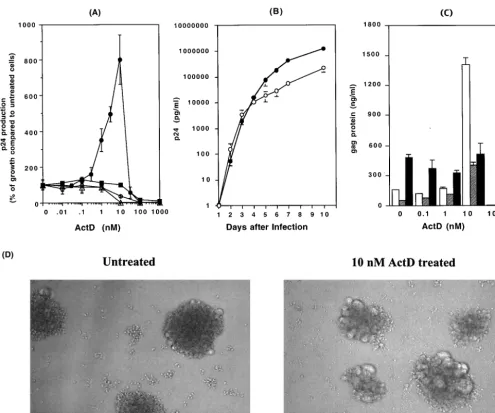
ActD (Fig. 1A). Concentrations of ActD greater than 100 nM
inhibited HIV replication in all tested cells. Trypan blue
stain-ing revealed that these inhibitory effects were likely to be a
consequence of the cellular toxicity of ActD (
⬍
1% viable cells
at more than 100 nM ActD). Interestingly and unexpectedly,
however, low concentrations (1 to 10 nM) of ActD upregulated
HIV-1 replication by 8- to 10-fold in MT-2 cells. This ability of
ActD to enhance HIV-1
NL4.3replication was not seen using
Jurkat, MT-4, or PHA-stimulated PBMCs. To determine if the
increase in p24 production in MT-2 cells was associated with
an increase in viral titers or was merely an increase in the
release of p24 antigens from infected cells, TCID
50titers and
p24 levels of tissue culture supernatants from control and
ActD (10 nM)-treated cultures were compared. The TCID
50titers of day 7 culture supernatants in the absence and presence
of 10 nM ActD were 460
⫾
76 and 329
⫾
63 TCID
50per ng of
p24, respectively (
P
⬎
0.05). Thus, the increase in p24 levels in
ActD-treated cultures was reflective of enhanced HIV-1
rep-lication rather than merely being due to increased release of
p24 protein from MT-2 cells.
To assess if the difference in virus replication was secondary
to a shift in replication kinetics, levels of virus were measured
on a daily basis for 10 days (Fig. 1B). For the first 3 days after
infection, there was no difference between the ActD-treated
culture and the control; however, after day 4, higher levels of
virus were seen in the ActD-treated culture (p24
concentra-tions on day 7 in the absence and presence of ActD were 569
⫾
10 and 4,522
⫾
72 ng/ml, respectively;
P
⬍
0.01). Therefore,
ActD appeared to be acting by prolonging the time of
produc-tive infection rather than by enhancing infectivity. To further
understand this enhancement of HIV-1 replication by ActD,
the impact of ActD on the replication of two other viruses,
HIV-1
DH12(51) and HIV-2
MVP15132, was also investigated.
Replication of HIV1
DH12increased from 53
⫾
2.3 to 403
⫾
50
ng/ml in the presence of 10 nM ActD on day 7 (
P
⬍
0.01).
Replication of HIV-2
MVP15132was not enhanced by ActD (p27
concentrations on day 7 in the absence and presence of 10 nM
ActD were 460
⫾
35 and 513
⫾
119 ng/ml, respectively [
P
⬎
0.05]) (Fig. 1C).
To determine whether these changes were a consequence of
alterations in receptor or coreceptor expression, levels of CD4
and CXCR4 expression on MT-2 cells were also analyzed.
Fresh uninfected MT-2 cells were cultured for 3 days in the
presence or absence of 10 nM ActD and were then analyzed by
flow cytometry for CD4 and CXCR4 expression. Of note, the
expression of CD4 and of CXCR4 on ActD-treated MT-2 cells
was downregulated by 30 and 80%, respectively (Fig. 2).
Therefore, the increase in HIV-1 replication induced by ActD
after 4 days of infection was not due to an increase in receptor
or coreceptor expression leading to enhanced infectibility in
later rounds of infection. To define the cell specificity of this
phenomenon, MT-4 or Jurkat cells and PBMCs were also
treated with 10 nM ActD for 3 days and the expression levels
of CD4 and CXCR4 were monitored. The CD4 expression on
MT-4 cells and Jurkat cells was upregulated by 1.5- and 2-fold,
respectively; however, CXCR4 expression was not influenced
by the treatment. ActD had no effect on the expression of CD4
or CXCR4 on PBMCs (data not shown). To evaluate whether
on November 8, 2019 by guest
http://jvi.asm.org/
or not the prolongation of time of productive infection was due
to decreased syncytium formation and enhanced survival of
infected cells, cultures were monitored for syncytium
produc-tion. As seen in Fig. 1D, ActD treatment led to an increase in
syncytium formation.
MT-2 cells are transformed with human T-cell leukemia
virus type 1 (HTLV-1) and produce infectious HTLV-1 virions
(11, 31). HTLV-1 has been reported to enhance HIV
replica-tion in vitro (4, 9, 34, 36, 62). To determine whether or not the
enhancement of HIV production by ActD was due to
en-hanced HTLV-1 production, p19 levels in culture supernatants
were measured with a p19 antigen capture kit (Zeptometrix
Corp.). No difference in HTLV-1 production was seen in the
absence or presence of 10 nM Act D (the amounts of p19 on
day 4 in the absence and presence of 10 nM ActD were 290
⫾
33 and 227
⫾
23 ng/ml, respectively [
P
⬎
0.05]). Therefore, the
activation of HIV-1 was not associated with HTLV-1
activa-tion.
[image:4.603.47.543.66.479.2]To determine the direct effects of ActD on HIV-1 RT
ac-tivity in MT-2 cells, RT enzyme assays were performed using
lysates of HIV-1-infected MT-2 cells. Various concentrations
of ActD (0 to 100
M) had no direct effects on RT activity
FIG. 1. ActD upregulates HIV-1 replication but not HIV-2 replication. (A) MT-2 (closed circles), MT-4 (open circles), Jurkat (open triangles),
or PHA-stimulated PBMCs (closed squares) were infected with HIV-1
NL4.3for 2 h at 37°C. The infected cells were cultured for 7 days in the
presence of various concentrations of ActD (0 to 1,000 nM). HIV-1 replication was measured by a p24 antigen capture assay. Results show the
percentage of virus growth in ActD-treated cells compared to that in untreated cells. In this experiment, the p24 concentrations in the culture
supernatant of MT-2, MT-4, Jurkat, and PBMCs in the absence of ActD were 159
⫾
33, 56
⫾
11, 9.1
⫾
2.1, and 10
⫾
1.4 ng/ml, respectively.
(B) HIV-1
NL4.3-infected MT-2 cells were cultured in 10 ml of RPMI-10 in T-25 cm
2culture flasks in the presence (closed circles) or absence (open
circles) of 10 nM ActD, and culture supernatants were collected every day. The assay was performed in triplicate, and the p24 levels in culture
supernatants were measured by the p24 antigen capture assay. (C) MT-2 cells were infected with HIV-1
NL4.3(white bar), HIV-1
DH12(gray bar),
or HIV-2
MVP15132(black bar) for 2 h at 37°C. After washing, the MT-2 cells were cultured for an additional 7 days at 37°C in the presence of 0
to 100 nM ActD. The p24 (for HIV-1) or p27 (for HIV-2) antigen levels in the culture supernatants were measured by p24 or p27 antigen capture
assays. (D) MT-2 cells were infected with HIV-1
NL4.3and cultured for 4 days in the absence or presence of 10 nM ActD.
on November 8, 2019 by guest
http://jvi.asm.org/
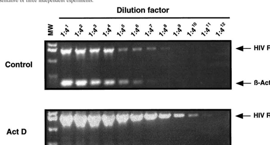
(data not shown). To examine the effect of ActD on HIV-1
gene transcription, a semiquantitative RT-PCR was utilized to
compare the levels of HIV-1 RNA and

-actin in control and
ActD-treated cells (Fig. 3). To confirm the absence of
contam-inating DNA, control cDNA reactions were prepared in which
RT was omitted from each reaction mixture. Amplified
prod-ucts were not seen in these controls (data not shown). In the
presence of 10 nM ActD, HIV-1 RNA transcription increased
16-fold. Therefore, despite its well-known ability to inhibit
transcription in most systems (43, 52), ActD enhanced HIV-1
RNA transcription in MT-2 cells.
Effect of ActD on drug susceptibility.
Given the
enhance-ment of HIV-1 replication in MT-2 cells by ActD, we were
interested in determining whether or not this increase in
rep-lication had an effect on the rate of emergence of
drug-tant variants. To address this question, a series of drug
resis-tance assays was performed to determine the IC
50s for a series
of drugs, utilizing control or ActD-treated cells (Table 1). In
the presence of 10 nM ActD, HIV grown in MT-2 cells showed
a high level of resistance to thymidine analogs (
⬎
1,000-fold
increase in AZT IC
50,
⬎
250-fold for d4T). In contrast, only a
modest increase (two- to fivefold) in the IC
50s of indinavir, ddI,
ddC, 3TC, abacavir (ABC), PMEA, and EFV was observed in
the presence of ActD.
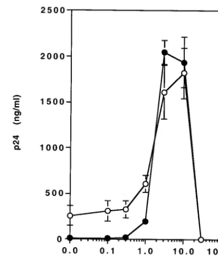
To better evaluate the impact of ActD on resistance to AZT,
MT-2 cells were infected with HIV-1
NL4.3and cultured for 7
[image:5.603.52.276.69.313.2]days in the presence of 1
M AZT with various concentrations
of ActD (Fig. 4). In the absence of ActD, HIV-1 replication
was inhibited 96% by the presence of AZT. The inhibitory
FIG. 2. ActD downregulates expression of CD4 and CXCR4 on
MT-2 cells. MT-2 cells were cultured for 3 days in the presence of 0
(gray) or 10 nM (black) ActD. The treated cells were stained with
fluorescence-conjugated anti-CD4 or -CXCR4. The staining patterns
for the isotype control antibodies are shown in white. Fluorescence
intensity was measured by a Coulter XL flow cytometer. (A) The mean
fluorescence intensities (MFI) for CD4 staining on control and
ActD-treated cells were 16.6 and 11.3, respectively. (B) The MFI for CXCR4
on control and ActD-treated cells were 41.6 and 8.29. Results are
representative of three independent experiments.
FIG. 3. ActD enhances HIV-1 RNA transcription. MT-2 cells were infected with HIV-1
NL4.3for 2 h at 37°C. The infected cells were cultured
for 4 days in the presence or absence of 10 nM ActD. Cells were washed and RNA was isolated. The RNA was treated with DNase and
subsequently used to synthesize cDNA. The cDNA (1
g) was serially diluted 1:4 and then served as template for subsequent PCR to amplify
HIV-1 RT or

-actin. To confirm the absence of contaminating DNA, control cDNA reactions were prepared in which RT was omitted from each
reaction. Amplified products were not seen in these controls (data not shown).

-Actin served as an internal control to monitor the efficiency of
amplification of each of the RNAs. The PCR product sizes of HIV RT and

-actin are 1,540 and 353 bp, respectively. MW refers to a molecular
marker consisting of a 1 kbp ladder (Invitrogen).
on November 8, 2019 by guest
http://jvi.asm.org/
[image:5.603.64.517.415.658.2]effect of AZT on HIV replication was completely suppressed
by the addition of ActD at concentrations of 3 to 10 nM.
To understand the mechanism of this high level of resistance
to thymidine analogs, we first performed proviral DNA
se-quencing of the HIV RT gene to look for known mutations
associated with drug resistance (49). No resistance-associated
mutations were found in the protease and RT regions (data not
shown). These data suggested that ActD led to thymidine
an-alog resistance as a consequence of changes at the cellular
level.
To examine this possibility, a DNA microarray analysis was
performed with MT-2 cells in the presence and absence of
ActD. The results of this analysis revealed that ActD treatment
led to a 27.3-fold downregulation in transcription of the TK-1
gene, a gene that catalyzes the initial phosphorylation of
thy-midine analogs (14). Similar changes were not observed in
other nucleoside kinases, including adenosine (no change),
cytidine (1.3-fold increase), and guanosine (1.4-fold increase)
kinases. To confirm the downregulation of transcription of
TK-1, a semiquantitative RT-PCR analysis was performed.
The cells treated with 10 nM ActD had lower expression of the
TK-1 gene in a setting of no changes in transcription of

-actin
(Fig. 5A).
To define the impact of this decrease in RNA transcription
at the protein level, Western blotting was performed using
anti-TK-1 and anti-

-actin antibodies. MT-2 cells treated with
10 nM ActD showed an 85% decrease in the expression of
TK-1 and no change in

-actin protein expression (Fig. 5B).
TK-1 is a cell cycle-regulated enzyme and its activity fluctuates
FIG. 4. ActD treatment reverses the inhibition of HIV replication
by RT inhibitors. MT-2 cells were infected with HIV-1
NL4.3for 2 h at
37°C. The infected cells were cultured for 7 days in the presence of
various concentrations of ActD (0 to 100 nM) without AZT (open
circles) or with 1
M AZT (closed circles). HIV-1 replication was
measured by a p24 antigen capture assay.
TABLE
1.
Ef
fect
of
ActD
on
drug
sensitivity
in
MT-2
cells
ActD (nM) IC50 (nM) a IDV AZT d4T ddI ddC 3TC ABC PMEA EFV01
5
⫾
5.4
(1.0)
9.3
⫾
3.7
(1.0)
40
⫾
16
(1.0)
251
⫾
60
(1.0)
101
⫾
18
(1.0)
131
⫾
96
(1.0)
95
⫾
20
(1.0)
233
⫾
42
(1.0)
0.7
⫾
0.14
(1.0)
10
51
⫾
20
(3.3)
⬎
10,000
(
⬎
1,075)
⬎
10,000
(
⬎
250)
391
⫾
27
(1.6)
471
⫾
73
(4.7)
262
⫾
85
(2.0)
362
⫾
82
(3.8)
586
⫾
70
(2.5)
2.4
⫾
0.6
(3.4)
aData are means ⫾ standard errors. Each construct was independently studied at least three times. Values in parentheses represent the fold dif ferences in IC50 s compared to that of 0 nM ActD.on November 8, 2019 by guest
http://jvi.asm.org/
[image:6.603.314.527.67.324.2]with DNA synthesis, being high in dividing and malignant cells
and low in quiescent cells (50). To assess the effect of ActD on
cell cycle progress in MT-2 cells, a cell cycle analysis utilizing
propidium iodide was performed (Fig. 6). The percentage of
cells in S phase in control and ActD-treated cells was 30.6 and
1.6%, respectively (
P
⬍
0.01). To assess the impact of HIV-1
infection on cell cycle progression in ActD-treated MT-2 cells,
HIV-infected MT-2 cells were also analyzed. HIV-1 infection
had no effect on the decrease in the percentage of cells in S
phase observed after ActD treatment of uninfected cells (data
not shown).
DISCUSSION
ActD has been reported to be a potential inhibitor of HIV-1
replication due to its inhibitory effect on minus-strand transfer
during the process of reverse transcription (10, 15, 17, 26).
Despite the strong prior data on inhibition of HIV-1 reverse
transcription in vitro, the present study has demonstrated that
low concentrations of ActD (1 to 10 nM) actually enhance
replication of HIV-1 in MT-2 cells. Of note, this effect was
limited to 1. Similar effects were not seen with
HIV-2
MVP15132or with HIV-1 replication in Jurkat, MT-4, or
PBMCs.
In an effort to determine the mechanism(s) underlying this
phenomenon, we examined the effect of ActD on HTLV-1
production, HIV-1 replication kinetics, receptor and
corecep-tor expression, transcription of cellular genes, and effects on
cell cycle. As mentioned above, MT-2 and MT-4 cells are
HTLV-1-transformed cell lines. HTLV-1-transformed cells
produce infectious HTLV-1 virions (11, 31) and a number of
cytokines (21). HTLV-1 has been reported to enhance HIV-1
replication in vitro (4, 9, 34, 36, 62). MT-4 cells produce low or
undetectable levels of HTLV-1, while MT-2 cells are high-level
producers of the virus (31). Since ActD enhanced HIV-1
rep-lication in MT-2 cells but not in MT-4 cells, it was speculated
that the mechanism of HIV-1 activation by ActD might be
mediated via the activation of HTLV-1 production from
ActD-treated MT-2 cells. However, this possibility seems unlikely,
since we were unable to find evidence that ActD enhances
HTLV-1 production
Our evaluation of HIV-1 replication kinetics revealed that
replication was enhanced only after day 4 of infection (Fig.
1B). In addition, ActD treatment downregulated the
expres-sion of CD4 and CXCR4 on MT-2 cells by 30 and 80%,
respectively (Fig. 2). Therefore, ActD does not appear to be
acting by extending the time of productive infection or
enhanc-ing the infectibility of MT-2 cells. The data are more consistent
with the influence of ActD on the magnitude of productive
infection. Since ActD downregulated the expression of
recep-tors for HIV-1 infection, it was speculated that ActD might
result in less syncytium formation and prolonged survival of
infected cells. However, as shown in Fig. 1D, ActD enhanced
syncytium formation in MT-2 cells. As demonstrated in Fig. 3,
ActD leads to an increase in HIV-1 transcription. Thus, the
present data suggest that this effect of ActD on HIV-1
repli-cation is most likely a result of alterations in virus gene
tran-scription. Whether this is due to increased transcription or
increased transcript stability is the focus of ongoing
experi-ments.
[image:7.603.59.526.72.268.2]ActD enhanced HIV-1
NL4.3and HIV-1
DH12replication by
FIG. 5. ActD treatment downregulates transcription of TK-1. MT-2 cells were infected with HIV-1
NL4.3for 2 h at 37°C. The infected cells were
cultured for 4 days in the absence or presence of 10 nM ActD. (A) Cells were washed and RNA was isolated. The RNA was treated with DNase
and subsequently used to synthesize cDNA; the cDNA (1
g) was serially diluted 1:4. The diluted cDNA served as template for subsequent PCRs
to amplify TK-1 and

-actin. To confirm the absence of contaminating DNA, control cDNA reactions were prepared in which RT was omitted from
each reaction. Amplified products were not seen in these controls (data not shown).

-Actin served as an internal control to monitor the efficiency
of amplification. The PCR product sizes of TK-1 and

-actin were 540 and 353 bp, respectively. MW refers to a molecular marker consisting of
a 100-bp ladder (Invitrogen). (B) The infected cells were washed and cellular protein was extracted using RIPA buffer (see Materials and
Methods). For each blot, 50
g of total cellular protein was loaded on a 12% bis-Tris sodium dodecyl sulfate-acrylamide gel, and Western blotting
was performed using an anti-TK-1 antibody. Subsequently, the antibody was stripped and then the blot was reprobed using anti-actin antibody.
Images were quantitated using the Epi chemi II Darkroom and LabWork software (Ultra-violet Products, Ltd., Upland, Calif.).
on November 8, 2019 by guest
http://jvi.asm.org/
8- to 10-fold in MT-2 cells but did not stimulate
HIV-2
MVP15132replication in these cells (Fig. 1C). HIV-1 and
HIV-2 both require the
trans
-activator Tat protein for viral
RNA transcription and viral replication in host cells (46). Since
ActD enhanced HIV-1 replication without modifying HIV-2, it
is unlikely that the effect on HIV-1 is mediated via Tat.
Semiquantitative RT-PCR and cell cycle analyses revealed
that ActD treatment enhanced transcription of HIV-1 DNA by
16-fold (Fig. 3). Thus, this ActD effect in MT-2 cells appears to
be related to an increase in RNA production, despite the
well-described activity of ActD to inhibit transcription. Using a
transient promoter function assay, it has been demonstrated
that ActD activates the HIV-1 promoter in HeLa cells via an
enhancement of phosphorylation of RNA polymerase II (Pol
II) (6). Further studies of the mechanism of this effect have
revealed that ActD leads to a dissociation of the complex
between 7SK RNA (a small nuclear RNA) and P-TEFb
(Tat-specific transcription elongation factor complex) (39, 61).
Once dissociated, the inhibitory effects on this complex on the
phosphorylation of Pol II are removed. Thus, it is possible that
ActD leads to an increase in HIV-1 replication by a similar
mechanism. However, given the fact that ActD enhanced
HIV-1 replication only in MT-2 cells (Fig. 1A), an additional
mechanism may be involved. The precise mechanism is still
unclear and is now the focus of additional work.
An additional unexpected finding was the development of
multidrug resistance in the presence of ActD (Table 1). We
speculate that the broad low-level resistance was not truly
resistance but was a reflection of the high level of viral
repli-cation, requiring a high concentration of drug. In contrast, 10
nM ActD treatment led to high-level resistance to AZT and
d4T without induction of nucleotide substitutions associated
with drug resistance. Western blotting and RT-PCR pointed to
a downregulation of TK-1 as the likely mechanism for this
phenomenon (Fig. 5). TK-1 is a key enzyme in the salvage
synthesis of thymidine monophosphate (TMP) from
thymi-dine. Intracellular TMP is quickly phosphorylated to dTDP
(TDP) and dTTP (TTP). Since TTP is an allosteric effector of
ribonucleotide reductase, imbalances in the TTP pool disturb
the supply of both purines and pyrimidines for DNA synthesis
and repair. Therefore, imbalanced dNTP pools increase the
mutation rate (37, 40). Even though ActD suppressed the
expression of TK1 in MT-2 cells, we have not seen any drug
resistance mutations during short-term culture. However, it is
possible that ActD might facilitate the rate of emergence of
HIV-1 mutations associated with drug resistance during
long-term passage in MT-2 cells.
TK-1 is a cell cycle-regulated enzyme. Its activity fluctuates
with DNA synthesis, being highest in dividing and malignant
cells and lowest in quiescent cells (50). The expression of TK-1
is meticulously controlled at the transcriptional and
posttran-scriptional level (28, 53). Its activity increases markedly after
the G
1/S transition and then declines rapidly in G
2(50).
Rep-lication of retroviruses depends on cycling cells. Passage
through the G
1/S phase of the cell cycle is essential for efficient
reverse transcription (8, 13, 20, 22, 23, 56). ActD is known as
a cell cycle synchronizer that arrests the G
1phase in the cell
cycle (5, 29). However, in MT-2 cells, cell cycle analysis
re-vealed that ActD decreased the number of cells in S phase
(Fig. 6). Therefore, the described decrease in the TK-1 level in
MT-2 cells following ActD treatment may be a consequence of
having fewer cells in S phase.
Here we demonstrated that ActD treatment downregulated
TK-1 expression in MT-2 cells. The initial phosphorylation of
AZT and d4T by TK-1 is a critical step in their metabolism,
leading to chain termination of DNA synthesis during the RT
phase of HIV replication (14). Resistance to nucleoside
ana-logs, e.g., AZT, has been well studied at the level of the virus.
Amino acid substitutions (49), insertions (49), or deletions (24,
25, 30, 60) can lead to high-level resistance to AZT. In
addi-FIG. 6. ActD treatment decreases the percentage of MT-2 cells in S phase. MT-2 cells were cultured for 3 days in the absence (A) or presence
(B) of 10 nM ActD. The treated cells were stained with propidium iodide. The stained cells were analyzed for red fluorescence. The left peaks
constitute cells in G
0/G
1, the right peaks constitute cells in G
2/M; cells in S phase are between the G
0/G
1and G
2/M peaks. The distribution of cells
in the G
0/G
1, S, and G
2/M cell cycle phases was calculated from the resulting DNA histogram by using Multicycle AV software (Phoenix Flow
Systems). Results are representative of three independent experiments.
on November 8, 2019 by guest
http://jvi.asm.org/
[image:8.603.79.502.70.268.2]tion, as reported here, it appears that AZT resistance can also
be seen following chemotherapeutic manipulation of the host
cell by downregulation of TK-1 expression.
ACKNOWLEDGMENTS
We thank Tatsuhiko Igarashi and Malcolm Martin for providing
HIV
DH12and Quan-En Yang and Shizuko Sei for valuable assistance
in taking photomicrographs. We also thank Robin Derwar for critical
reading of the manuscript and Anthony S. Fauci for guidance and
support.
This work was supported by NIAID under contract N01-CO-12400
with SAIC-Frederick, Inc.
REFERENCES
1. Adachi, A., H. E. Gendelman, S. Koenig, T. Folks, R. Willey, A. Rabson, and M. A. Martin.1986. Production of acquired immunodeficiency syndrome-associated retrovirus in human and nonhuman cells transfected with an infectious molecular clone. J. Virol.59:284–291.
2. AIDS Clinical Trials Group.1997. Virology manual for HIV laboratories. Division of AIDS, National Institute of Allergy and Infectious Diseases, Bethesda, Md. [Online.] http://www.niaid.nih.gov./daids/vir_manual. 3. Beyl, W., K. Nehring, L. Gu¨rtler, J. Eberle, and F. Deinhardt.1987. AIDS
Verursacht durch HIV-2. Mu¨nch. Med. Wochenschr.129:895–896. 4. Boehnlein, E., M. Siekevitz, D. W. Ballard, J. W. Lowenthal, L. Rimsky, H.
Bogerd, J. Hoffman, Y. Wano, B. R. Franza, and W. C. Greene. 1988. Stimulation of the HIV-1 enhancer by the HTLV-Itaxgene product involves the action of inducible cellular proteins. J. Virol.63:1578–1586.
5. Bonin, L. R., and J. K. McDougall.1997. Human cytomegalovirus IE2 86-kilodalton protein binds p53 but does not abrogate G1checkpoint
func-tion. J. Virol71:5861–5870.
6. Casse´, C., F. Giannoni, V. T. Nguyen, M.-F. Dubois, and O. Bensaude.1999. The transcriptional inhibitors, actinomycin D and␣-amanitin activate the HIV-1 promoter and favor phosphorylation of the RNA polymerase II C-terminal domain. J. Biol. Chem.274:16097–16106.
7. Chen, F. M.1988. Binding specificities of actinomycin D to self-complemen-tary tetranucleotide sequences -XGCY-. Biochemistry27:6393–6397. 8. Chen, I. S. Y., and H. M. Temin.1982. Establishment of infection by spleen
necrosis virus: inhibition in stationary cells and the role of secondary infec-tion. J. Virol.41:183–191.
9. Cheng, H., J. Tarnok, and W. Parks.1998. Human immunodeficiency virus type 1 genome activation induced by human T-cell leukemia virus type 1 Tax protein is through cooperation of NF-B and Tat. J. Virol.72:6911–6916. 10. Davis, W. R., S. Gabbara, D. Hupe, and J. A. Peliska.1998. Actinomycin D
inhibition of DNA strand transfer reactions catalyzed by HIV-1 reverse transcriptase and nucleocapsid protein. Biochemistry37:14213–14221. 11. Dhib-Jalbut, S., P. M. Hoffman, T. Yamabe, D. Sun, J. Xia, H. Eisenberg, G.
Berger, and F. W. Ruscetti.1994. Extracellular human T-cell lymphotropic virus type I Tax protein induces cytokine production in adult human micro-glial cells. Ann. Neurol.36:787–790.
12. Farber, S.1966. Chemotherapy in the treatment of leukemia and Wilms’ tumor. JAMA198:826–836.
13. Fritsch, E. F., and H. M. Temin.1977. Inhibition of viral DNA synthesis in stationary chicken embryo fibroblasts infected with avian retroviruses. J. Vi-rol.24:461–469.
14. Furman, P. A., J. A. Fyfe, M. H. Clair, Sr., K. Weinhold, J. L. Rideout, G. A. Freeman, S. N. Lehrman, D. P. Bolognesi, S. Broder, H. Mitsuya, and D. W. Barry.1986. Phosphorylation of 3⬘-azido-3⬘-deoxythymidine and selective interaction of the 5⬘-triphosphate with human immunodeficiency virus re-verse transcriptase. Proc. Natl. Acad. Sci. USA83:8333–8337.
15. Gabbara, S., W. R. Davis, L. Hupe, D. Hupe, and J. A. Peliska.1999. Inhibitors of DNA strand transfer reactions catalyzed by HIV-1 reverse transcriptase. Biochemistry38:13070–13076.
16. Goodisman, J., R. Rehfuss, B. Ward, and J. C. Dabrowiak.1992. Site-specific binding constants for actinomycin D on DNA determined from footprinting studies. Biochemistry31:1046–1058.
17. Guo, J., T. Wu, J. Bess, L. E. Henderson, and J. G. Levin.1998. Actinomycin D inhibits human immunodeficiency virus type 1 minus-strand transfer in in vitro and endogenous reverse transcriptase assays. J. Virol.72:6716–6724. 18. Haertle, T., C. J. Carrera, D. B. Wasson, L. C. Sowers, D. D. Richman, and
D. A. Carson.1988. Metabolism and anti-human immunodeficiency virus-1 activity of 2-halo-2⬘,3⬘-dideoxyadenosine derivatives. J. Biol. Chem. 263:
5870–5875.
19. Harada, S., Y. Koyanagi, and N. Yamamoto.1985. Infection of HTLV-III/ LAV in HTLV-carrying cells MT-2 and MT-4 and application in a plaque assay. Science229:563–566.
20. Harel, J., E. Rassart, and P. Jolicoeur.1981. Cell cycle dependence of synthesis of unintegrated viral DNA in mouse cells newly infected with murine leukemia virus. Virology110:202–207.
21. Hollsberg, P., and D. A. Hafler.1993. Pathogenesis of diseases induced by human lymphotropic virus type I infection. N. Engl. J. Med.328:1173–1182. 22. Hsu, T. W., and J. M. Taylor.1982. Effect of aphidicolin on avian sarcoma
virus replication. J. Virol.44:493–498.
23. Humphries, E. H., and H. M. Temin.1974. Requirement for cell division for initiation of transcription of Rous sarcoma virus RNA. J. Virol.14:531–546. 24. Imamichi, T., S. C. Berg, H. Imamichi, J. C. Lopez, J. A. Metcalf, J. Fallon, and H. C. Lane.2000. Relative replication fitness of a high-level 3⬘ -azido-3⬘-deoxythimidine-resistant variant of human immunodeficiency virus type-1 possessing an amino acid deletion at codon 67 and a novel substitution (Thr to Gly) at codon 69. J. Virol.74:10958–10964.
25. Imamichi, T., T. Sinha, H. Imamichi, Y. M. Zhang, J. A. Metcalf, J. Falloon, and H. C. Lane.2000. High-level resistance to 3⬘-azido-3⬘-deoxythimidine due to a deletion in the reverse transcriptase gene of human immunodefi-ciency virus type 1. J. Virol.74:1023–1028.
26. Jeeninga, R. E., H. T. Huthoff, A. P. Gultyaev, and B. Berkhout.1998. The mechanism of actinomycin D-mediated inhibition of HIV-1 reverse tran-scription. Nucleic Acids Res.26:5472–5479.
27. Kamitori, S., and F. Takusagawa.1992. Crystal structure of the 2:1 complex between d(GAAGCTTC) and the anticancer drug actinomycin D. J. Mol. Biol.225:445–456.
28. Kauffman, M. G., and T. J. Kelly.1991. Cell cycle regulation of thymidine kinase: residues near the carboxyl terminus are essential for the specific degradation of the enzyme at mitosis. Mol. Cell. Biol.11:2538–2546. 29. Khan, Q. A., and A. Dipple.2000. Diverse chemical carcinogens fail to
induce G1arrest in MCF-7 cells. Carcinogenesis21:1611–1618.
30. Kim, E.-Y., M. A. Winters, R. M. Kagan, and T. C. Merigan.2001. Func-tional correlates of insertion mutations in the protease gene of human immunodeficiency virus type 1 isolates from patients. J. Virol.75:11227– 11233.
31. Koyanagi, Y., Y. Hinuma, J. Schneider, T. Chosa, G. Hunsmann, N. Koba-yashi, M. Hatanaka, and N. Yamamoto.1984. Expression of HTLV-specific polypeptides in various human T-cell lines. Med. Microbiol. Immunol.173:
127–140.
32. Larder, B. A., G. Darby, and D. D. Richman.1989. HIV with reduced sensitivity to zidovudine (AZT) isolated during prolonged therapy. Science
243:1731–1734.
33. Lewis, J. L., Jr.1972. Chemotherapy of gestational choriocarcinoma. Cancer
30:1517–1521.
34. Lindholm, P. F., S. J. Marriott, S. D. Gitlin, C. A. Bohan, and J. N. Brady.
1990. Induction of nuclear NF-B DNA binding activity after exposure of lymphoid cells to soluble Tax1 protein. New Biol.2:1034–1043.
35. Mann, D. L., S. J. O’Brien, D. A. Gilbert, Y. Reid, M. Popovic, E. Read-Connole, R. C. Gallo, and A. Gazdar.1989. Origin of the HIV-susceptible human CD4⫹cell line H9. AIDS Res. Hum. Retrovir.5:253–255.
36. Marriott, S. J., P. F. Lindholm, R. L. Reid, and J. N. Brady.1991. Soluble HTLV-I Tax1 protein stimulates proliferation of human peripheral blood lymphocytes. New Biol.3:678–686.
37. Meuth, M.1989. The molecular basis of mutations induced by deoxyribo-nucleoside triphosphate pool imbalances in mammalian cells. Exp. Cell. Res.
181:305–316.
38. Myers, G., B. Korber, S. Wain-Hobson, K.-T. Jeang, L. E. Henderson, and G. N. Pavlakis.1994. Human retroviruses and AIDS. Los Alamos National Laboratory, Los Alamos, N.Mex.
39. Nguyen, V. T., T. Kiss, A. A. Michels, and O. Bensaude.2001. 7SK small nuclear RNA binds to and inhibits the activity of CDK9/cyclin T complexes. Nature414:322–325.
40. Oliver, F. J., M. K. Collins, and A. Lopez-Rivas.1997. Overexpression of a heterologous thymidine kinase delays apoptosis induced by factor depriva-tion and inhibitors of deoxynucleotide metabolism. J. Biol. Chem.
272:10624–10630.
41. Pauwels, R., E. De Clercq, J. Desmyter, J. Balzarini, P. Goubau, P. Herd-ewijn, H. Vanderhaeghe, and M. Vandeputte.1987. Sensitive and rapid assay on MT-4 cells for detection of antiviral compounds against the AIDS virus. J. Virol. Methods16:171–185.
42. Perrin, L., and A. Telenti.1998. HIV treatment failure: testing for HIV resistance in clinical practice. Science280:1871–1873.
43. Perry, R. P., and D. E. Kelley.1970. Inhibition of RNA synthesis by actino-mycin D: characteristic dose-response of different RNA species. J. Cell. Physiol.76:127–139.
44. Popovic, M., E. Read-Connole, and R. C. Gallo.1984. T4 positive human neoplastic cell lines susceptible to and permissive for HTLV-III. Lancet
ii:1472–1473.
45. Popovic, M., M. G. Sarngadharan, E. Read, and R. C. Gallo.1984. Detec-tion, isolaDetec-tion, and continuous production of cytopathic retroviruses (HTLV-III) from patients with AIDS and pre-AIDS. Science224:497–500. 46. Rabson, A. B., and B. J. Graves.1997. Synthesis and processing of viral RNA,
p. 205–261.InJ. M. Coffin, S. H. Hughes, and H. E. Varmus (ed.), Retro-viruses. Cold Spring Harbor Laboratory Press, Cold Spring Harbor, N.Y. 47. Rill, R. L., and K. H. Hecker.1996. Sequence-specific actinomycin D binding
to single-stranded DNA inhibits HIV reverse transcriptase and other poly-merases. Biochemistry35:3525–3533.
on November 8, 2019 by guest
http://jvi.asm.org/
48. Sambrook, J., E. F. Fritsch, and T. Maniatis.1989. Molecular cloning: a laboratory manual, 2nd ed., p. 18.38. Cold Spring Harbor Laboratory Press, Cold Spring Harbor, N.Y.
49. Schinazi, R. F., B. A. Larder, and J. W. Mellors.2000. Mutations in retroviral genes associated with drug resistance. Int. Antivir. News8:65–91. 50. Sherley, J. L., and J. K. Thomas.1988. Regulation of human thymidine
kinase during the cell cycle. J. Biol. Chem.263:8350–8358.
51. Shibata, R., M. D. Hoggan, C. Broscius, G. Englund, T. S. Theodore, A. Buckler-White, L. O. Arthur, Z. Israel, A. Schultz, H. C. Lane, and M. A. Martin.1995. Isolation and characterization of a syncytium-inducing, mac-rophage/T-cell line–tropic human immunodeficiency virus type 1 isolate that readily infects chimpanzee cells in vitro and in vivo. J. Virol.69:4453–4462. 52. Sobell, H. M.1985. Actinomycin and DNA transcription. Proc. Natl. Acad.
Sci. USA82:5328–5331.
53. Stewart, C. J., M. Ito, and S. E. Conrad.1987. Evidence for transcriptional and post-transcriptional control of the cellular thymidine kinase gene. Mol. Cell. Biol.7:1156–1163.
54. Takusagawa, F., K. T. Takusagawa, R. G. Carlson, and R. F. Weaver.1997. Selectivity of F8-actinomycin D for RNA:DNA hybrids and its anti-leukemia activity. Bioorg. Med. Chem.5:1197–1207.
55. Telesnitsky, A., and S. P. Goff.1997. Reverse transcriptase and the genera-tion of retroviral DNA, p. 121–160.InJ. M. Coffin, S. H. Hughes, and H. E. Varmus (ed.), Retroviruses. Cold Spring Harbor Laboratory Press, Cold Spring Harbor, N.Y.
56. Varmus, H. E., T. Padgett, S. Heasley, G. Simon, and J. M. Bishop.1977.
Cellular functions are required for synthesis and integration of avian sar-coma virus-specific DNA. Cell11:307–319.
57. Wadkins, R. M., and T. M. Jovin.1991. Actinomycin D and 7-aminoactino-mycin D binding to single-stranded DNA. Biochemistry30:9469–9478. 58. Wadkins, R. M., E. A. Jares-Erijman, R. Klement, A. Rudiger, and T. M.
Jovin.1996. Actinomycin D binding to single-stranded DNA: sequence ificity and hemi-intercalation model from fluorescence and 1H NMR spec-troscopy. J. Mol. Biol.262:53–68.
59. Waring, M. J.1981. DNA modification and cancer. Annu. Rev. Biochem.
50:159–192.
60. Winters, M. A., J. M. Schapiro, J. Lawrence, and T. C. Merigan.1998. Human immunodeficiency virus type 1 protease genotypes and in vitro pro-tease inhibitor susceptibilities of isolates from individuals who were switched to other protease inhibitors after long-term saquinavir treatment. J. Virol.
72:5303–5306.
61. Yang, Z., Q. Zhu, K. Luo, and Q. Zhou.2001. The 7SK small nuclear RNA inhibits the CDK9/cyclin T1 kinase to control transcription. Nature414:317– 322.
62. Zack, J. A., A. J. Cann, J. P. Lugo, and I. S. Y. Chen.1988. HIV-1 production from infected peripheral blood T cells after HTLV-I induced mitogenic stimulation. Science240:1026–1029.
63. Zhang, Y.-M., H. Imamichi, T. Imamichi, H. C. Lane, J. Falloon, M. B. Vasudevachari, and N. P. Salzman.1997. Drug resistance during indinavir therapy is caused by mutations in the protease gene and in its Gag substrate cleavage sites. J. Virol.71:6662–6670.
on November 8, 2019 by guest
http://jvi.asm.org/