A STUDY OF POST-OPERATIVE OUTCOMES OF
OPEN VERSUS CLOSED HEMORRHOIDECTOMY
Dissertation submitted to
THE TAMILNADU Dr. M. G. R. MEDICAL UNIVERSITY
In partial fulfillment of the regulations for the award of the degree of M. S. GENERAL SURGERY (BRANCH I)
CHENGALPATTU MEDICAL COLLEGE
THE TAMILNADU Dr. M. G. R. MEDICAL UNIVERSITY CHENNAI, TAMILNADU
CERTIFICATE
This is to certify that this dissertation titled “A STUDY OF POST-OPERATIVE OUTCOMES OF OPEN VERSUS CLOSED HEMORRHOIDECTOMY” has been prepared by
Dr.R.SURESH KUMAR, under my supervision in the Department of General Surgery, Chengalpattu Medical College , Chengalpattu, during
the academic period 2011 – 2014, and is being submitted to The
Tamilnadu Dr.M.G.R. Medical University, Chennai, in partial fulfillment
of the University regulation for the award of the Degree “Master Of
Surgery” (M.S.,General Surgery) and his dissertation is a bonafide work.
Prof.Dr.P.R.Thenmozhi Valli M.D, Prof.Dr.G.RajaBillyGrahamM.S.
DEAN Unit Chief & HOD
Chengalpattu Medical College & Department of General Surgery
Hospital, Chengalpattu Chengalpattu Medical College &
DECLARATION
I, Dr.R.SURESH KUMAR, solemnly declare that the dissertation “A STUDY OF POST-OPERATIVE OUTCOMES OF OPEN VERSUS CLOSED HEMORRHOIDECTOMY” a bonafide work done by me in the Department of General Surgery, Chengalpattu
Medical College, Chengalpattu, Under the able guidance of Prof. Dr. G.
Raja Billy Graham M.S, Professor & Head of the Department, Department
of General Surgery, Chengalpattu Medical College, Chengalpattu.
Place: Chengalpattu
ACKNOWLEDGEMENT
I wish to express my sincere thanks to Dr.P.R.THENMOZHI VALLI M.D, Dean, Chengalpattu Medical College & Hospital, Chengalpattu, for having kindly permitted me to utilize the hospital
facilities.
I wish to express my grateful thanks to ; Prof. Dr.G.RAJA BILLY GRAHAM M.S, Professor & Head of the Department, Department of General Surgery , Chengalpattu Medical College , Chengalpattu for his
immense help, encouragement and constant supervision.
I Am Greatly Thankful To Our Associate Professors
Dr.V.RAMALAKSHMI M.S, For their Valuable Suggestions.
And I wish to thank my unit Assistant Professors
Dr.K.SATHIYAMOORTHI M.S, Dr.N.LAKSHMIPATHY M.S, Dr.JOYCE PRABAKAR M.S, for their valuable suggestions and guidance and great care and attention to prepare this dissertation.
I owe great debt of gratitude to all the Assistant Professors and
Tutors for their able help and support. They have been a source of great
encouragement throughout my Post graduate course.
And I can never forget theatre personnel for their willing
co-operation and assistance. I thank all the patients who took part in my study
CONTENTS
S.NO CONTENTS PAGE.NO
1 INTRODUCTION 1
2 AIMS & OBJECTIVES OF THE STUDY 2
3 REVIEW OF LITERATURE 3
4 MATERIALS AND METHODOLOGY OF
STUDY 57
5 OBSERVATION AND RESULTS 60
6 DISCUSSION 79
7 CONCLUSION 86
8 BIBLIOGRAPHY 87
9 ANNEXURES
1. Proforma 2. Master chart 3. Abbreviations 4. Consent form
94
96
100
ABSTRACT
Milligan-Morgan excision haemorrhoidectomy remains a very popular treatment modality for third and fourth
degree hemorrhoids due to its cost effectiveness and better long-term results. Ferguson haemorrhoidectomy is
believed to result in less postoperative pain because of a closed wound. The aim of this study was to compare the
effectiveness of Milligan-Morgan excision haemorrhoidectomy with Ferguson haemorrhoidectomy,
Methods:
A prospective clinical trial was conducted. Patients with newly diagnosed hemorrhoids requiring
haemorrhoidectomy were randomized to either Milligan-Morgan excision haemorrhoidectomy or Ferguson
haemorrhoidectomy. Surgical technique and prospective care was standardized. Outcome measures were operative
time and bleeding, postoperative pain (measured on visual analogue scale) and rate of wound healing.
Results:
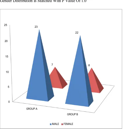
About Sixty Patients Undergoing Hemorrhoidectomy In our Hospital, Who Have Fulfilled The Criteria Have Been Selected And Included In Our Study. Patients Are Randomized In To Two Groups. Group A Will Have 30 Patients Undergoing Open Hemorrhoidectomy. And Group B Will Have 30 Patients Undergoing Closed Hemorrhoidectomy.
The Mean Age Of Group A Sample Was 39.1 Yrs . The Mean Age Of Group B Was 37.77 Yrs
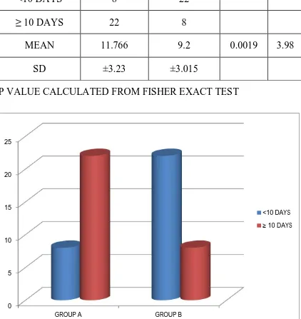
In Group A Mean Operation Time in Minutes Was 40.5. In Group B the Mean Operation Time In Minutes Was 49.33. . The Operating Time for Open Hemorrhoidectomy Was Significantly Less Than Closed Hemorrhoidectomy.
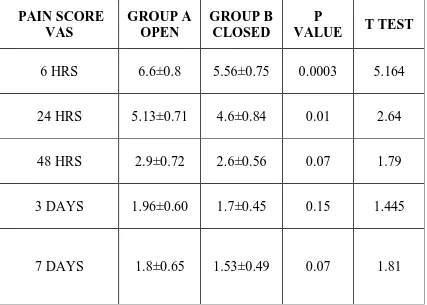
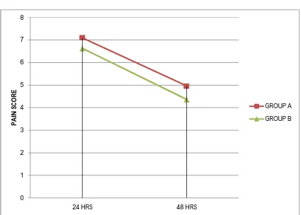
The Post Defecation Pain Score Following Hemorrhoidectomy Is Significantly Low For Closed Hemorrhoidectomy In Comparison To Open Approach.
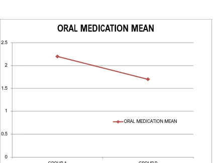
The Mean Analgesic Requirement For Group A Is 2.2 And Group B Is 1.7 With P Value Of 0.004. Thus The Analgesic Requirement Is also Low For The Closed Hemorrhoidectomy Group.
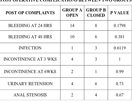
The Post Operative Complication Were Noted In Both The Groups Following Open And Closed Approach Without Any Significant Difference.
Conclusion
There Are Reports Of Better Post Operative Outcome Following Closed Hemorrhoidectomy In Terms Of Pain And Wound Healing. Both Open And Closed Approach Are Less Expensive And Safe, Easy To Perform With Satisfactory Results.
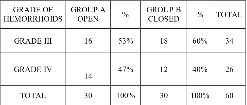
In Short We Think That Patients With Grade III And IV Hemorrhoids Who Are Operated With Fergusons Closed Hemorrhoidectomy Have Better Results.
Key words
Haemorrhoidectomy, bleeding, analgesic , post-operative pain,
INTRODUCTION
Haemorrhoids are one of the most common anorectal diseases for
referral to a surgeon. Haemorrhoidectomy one of the most common
anorectal procedures performed.
Complaints pertaining to haemorrhoids are one of the most common
afflictions of western civilizations. Although the condition is rarely life
threatening the complications of therapy can be.
For haemorrhoids of grade III AND IV the effective treatment even
today remains to be hemorrhoidectomy. Milligan and Morgan described
conventional open hemorrhoidectomy in 1937 and Ferguson in 1959
described closed hemorrhoidectomy. Owing to low expense and technical
ease open hemorrhoidectomy is the procedure of choice, even though
newer modalities have come in to play.
Closed hemorrhoidectomy by Fergusons has got considerable
attention in some parts of the world because of low post operative pain as
presumed due to closure of cut edges of mucosa, faster wound healing and
good patient compliance. However randomized control trials comparing
the above two procedure has been contradictory to assumptions. This study
compares the of outcome open vs. closed hemorrhoidectomy with set
Aims and Objectives of the study
To compare the outcomes following hemorrhoidectomy by the
Milligan-Morgan’s open and Ferguson’s closed technique.
The variables that are being compared are:
a) Post-operative pain,
b) Complications,
c) Length of hospital stay,
REVIEW OF LITERATURE
History of management of haemorrhoidal diseases
Haemorrhoids seem to be the oldest ills known to man. Morgagni in
1749 postulated it to man assuming upright position. In 1700 BC Edwin
smith papyrus, the Egyptians appeared to have infused alum as astringent
for anal disorders. Anal diseases have been coded in Hammurabi in
Babylon in 2250 BC and in papyrus in 1500 BC. Hippocrates in 400 BC
recommended cautery and simple excision. Ancient Greeks practiced anal
dilatation. Celsus in de medicina referred to ligature with flax thread.
Galen too advised surgery by double knotting. Susruta samhita the Ancient
Hindu Sanskrit text describes method of clamp and cautery.
Lorenz heister in his work of chirurgie 1739 advised ligation with
needle and thread. In 1774 jean Louis petit devised sub epithelial
haemorrhoidectomy. Samuel cooper in 1809 took the idea of petit and
described sub mucosal haemorrhoidectomy.
Verneuil in 1855 hypothesized straining at stools as cause for
haemorrhoids which was studied through anatomical advances. Ireland
appears to be the centre for conservative management of haemorrhoids as
practiced by Houston in 1843 using nitric acid. Mitchell 1903 pioneered
Concept of anal sphincter spasm came to existence in 19 th century
Copelan in 1814, fecanier in 1829 nad maisonneuve in 1864 described anal
dilatation through there own methods. Division of sphincter was
mentioned by boyer in 1818 and dupuytren in 1833. Total pile excision
with suturing was devised by whitehead in 1882 which was later
abandoned due to complications but now the concept has been taken in to
endostapling technique.
Allingham and allingham in 1901 from st marks hospital Britain
reported a series of cases of haemorrhoidectomy by salmons method .
During early twentieth century various literatures on operations of
haemorrhoids came from surgeons. These include miles 1919 v shaped
excision of pile with skin tag. Later Milligan and Morgan open
haemorrhoidectomy came to practice. Closed haemorrhoidectomy
stemmed from sub mucosal technique and was described by Fergusons in
1959. Anderson in 1909 and cormie and McNair in 1959 revived clamp
and cautery method.
Fixation by sub mucosal injection practiced by Blanchard and
alright in 1928 using 5 % phenol injection. Next method of rubber band
fixation developed by blaisdell in 1954. This technique was later perfected
by Barron in 1963.cryotherapy was applied by Fraser and gills in 1967 for
management of haemorrhoids. Infrared photocoagulation a recent addition
Anatomy of anal canal Developmental anatomy
Proximal part of anal canal develops form the hindgut but the distal
part of anal canal develops from the anal pit. So the proximal part of anal
canal is endodermal in origin but the distal part is ectodermal in origin as
evident from the epithelial lining of the canal. Perineal body develops from
fusion of anal and cloacal membranes. On either side of the canal anal
tubercles are formed by the somatic mesoderm which results in the
formation of anal sphincters.
Surgical anatomy
Anal canal measures roughly about 2.5 to 5 cm in length1,2. It
extends from the anorectal junction to the anal verge. Anorectal junction is
marked by indention of puborectalis sling which provides an anorectal
angle required for maintaining continence. Anal verge is identified by the
puckering and pigmentation of the anal skin. Anal canal consists of
epithelium, sub epithelial vascular tissue and sphincter along with fibro
muscular supporting structures.
Anal canal is attached to coccyx by a ligament above which lies
median raphae for fusion of levator ani between these structures lies the
post anal space.
Surgical anal canal differs from anatomical and histological anal
but histological anal canal is said to extend from the tip of anal columns
and anatomical canal starts from dentate line.
Anal orifice is an oval slit in anterioposterior plane at rest.
Anteriorly anal canal is separated from the surrounding structures by
perineal body.
Lining of anal canal
Anal canal is divided into upper , middle and lower anal canal based
on the lining epithelium of the canal3. Upper and middle anal canal
measures roughly 1.5 cm and lower anal canal measures 8mm.
Upper anal canal
Upper third of canal is lined by columnar epithelium containing
secretory and absorptive cells along with crypts or tubular glands. Sub
epithelial tissue i loose due to presence of vascular plexuses supplied by
superior rectal artery and these veins form plexuses and drain in through
inter muscular plexuses in to the portal circulation.
There are 6 – 10 vertical folds of mucosa found in the anal canal
which are termed as anal columns of morgagni. These columns have a twig
of superior rectal artery. Anal cushions as described at fixed position
At the end of the column there are cresentric folds called valves.
Between the anal valves there are opening for anal sinuses. Together these
anal valves and sinuses form the dentate line.
At the anal valves the anal glands open, these glands4 are linear
branched one extending up to external anal sphincter.
Middle anal canal
Middle third of canal which is below the dentate line is lined by non
keratinized squamous epithelium devoid of appendages till the inter
sphincteric groove. Middle anal canal is also called as pectin and the sub
epithelium is composed of venous plexus. This part of anal canal is
supplied by peripheral nervous system and hence sensible to pain. Lower
limit of pectin is formed by white line of Hilton.
Lower anal canal
Lower anal canal is lined by keratinized squamous epithelium and
they have appendages.
Anal transitional zone
Transition from columnar to squamous epithelium starts above
dentate line and extend for variable length. They contain thermoreceptors.
Muscles of anal canal
Anal canal is surrounded by internal and external anal sphincter .
They are separated by longitudinal muscle . These are attached to
puborectalis and transverse perinea muscle above.
Internal anal sphincter
Internal sphincter is circular obliquely placed smooth muscle
extending from rectum. Internal sphincter is 1.5 to 3.5 mm in thickness
depending up on the region and gender of the anal canal. Internal sphincter
extends up to the junction of superficial and subcutaneous part of external
sphincter. At the lower end internal anal sphincter is divided by septa
extending from longitudinal muscle layer.
Arterial supply is from the superior and inferior rectal arteries.
Nerve supply of internal anal sphincter
Sympathetic – lower two lumbar segments via inferior hypogastric
plexus.
Parasympathetic is from 2 to 4 sacral segments via inferior
hypogastric plexus.
Reflexes exist between internal sphincter and lower rectum ,
External anal sphincter
External anal sphincter is an oval tube of striated slow twitching
muscle fiber with ability to maintain sustained contraction. They are
divided in to superficial, deep and subcutaneous part though they function
as one unit.6 Superiorly it is attached to puborectalis, transverse perinea
and anococcygeal raphae. Inferiorly it is attached to coccyx via
anococcygeal ligament and perineal body
Arterial supply is by inferior rectal artery and medial sacral artery.
Nerve supply is by pudendal nerve.
Fibro muscular septa
Longitudinal fibro muscular structure is located in intersphincteric
space. It is formed by the fusion of striated and smooth muscle. Striated
muscle is derived from puborectalis and puboanalis. Smooth muscle is
derived from longitudinal muscle of the rectum.7
It Is A Fusion Of Muscle And Fibro Elastic Structure. At The Lower
End Of The Longitudinal Muscle It Divides In To Several Septa To Get
Attached To The Perianal Skin.
The Medial Most Part Of The Septa Is Attaches To The Anal Lining
Marking The Inter Sphincteric Groove. The Lateral Most Septa Attaches
Sub mucosa Of Lower Anal Canal Have Fibro muscular Septa
Which Originate From The Longitudinal Muscle Coat Attaches To The
Anal Verge In Honey Combing Fashion Which Prevents Even Mild
Distension Of Perianal Skin. This Explains The Intense Pain Associated
With External Haemorrhoids.
Anorectal Spaces Perianal Space
Perianal Space Is In Immediate Vicinity Of The Anal Verge
Surrounding The Anal Canal. It Is Continuous With The Intersphincteric
Space. Perianal Space Contains External Haemorrhoidal Plexus, Inferior
Rectal Vessels and Lymphatics.Laterally Extends In To Subcutaneous Fat
of Buttocks And Medially Extends Up To Dentate Line.
Ischioanal Space
Pyramidal Space Bounded By Levator Ani in the Apex, Inferiorly
Bound By Skin.
Anteriorly Bound By Superficial And Deep Transverse Perineal
Muscle And Posterior Perineal Membrane, Posteriorly Bound By Gluteal
Skin. Medial Wall Formed By Levator Ani and External Anal Sphincter.
Lateral Wall Formed By Obturator Fascia with Obturator Internus Muscle
Contents Of Ischioanal Space Is Inferior Rectal Nerve And Vessels
Along With Transverse Perineal Vessels And Perineal Branch Of Fourth
Sacral Nerve.
Intersphincteric Space
It Lies Between the Two Sphincter Muscles and Is Continous Below
With Perianal Space.
Supralevator Space
It is Bounded By Peritoneum Superiorly, Laterally By Pelvic Wall,
Medially By Rectum And Inferiorly By Levator Ani Muscle. It Is Situated
On Either Side Of Rectum.
Sub mucous Space
Lies Below The Mucosa And Extends Distally To Dentate Line .
Proximally Continuous With The Submucous Space Of Rectum. It
Contains The Haemorrhoidal Plexus.
Superficial Postanal Space
Connects the Perianal Space With Each Other Posteriorly
Deep Perianal Space
Retrosphincteric Space Of Courtney Connects Ischioanal Spaces
Retro Rectal Space
Retro rectal Space Lies between Upper Two Third of Rectum and
and Sacrum above Retro rectal Fascia.
Superiorly It Extends In To Retro peritoneum And Inferiorly It Is
Limited by the Retro sacral Fascia. Contents Include The Loose
Connective Tissue. It Is Limited Anteriorly By Fascia Propia Covering
Rectum, Posteriorly By Presacral Fascia And Laterally By Lateral
Ligaments Of Rectum.
Arterial Supply
Superior Rectal Artery
Superior Rectal Artery Is Continuation Of Inferior Mesenteric
Artery After It Crosses The Left Internal Iliac Vessels Near The Base Of
Sigmoid Colon.
Superior Rectal Artery Starts At the Last Branch of Sigmoid Artery.
It Is Located Posterior to the Right of Sigmoid Colon in Close Contact
with the Recto sigmoid Junction.11
Branches from Superior Rectal Arteries Are As Follows:
1. Recto sigmoid Branch
2. Upper Rectal Branch
Terminal Branches Extends Downward Around The Rectum. The
Upper Rectal Branch Has Extramural Anastomosis with Terminal
Branches and Recto sigmoid Branches.
Middle Rectal Arteries
Middle Rectal Artery Supplying the Lower Third of Anal Canal Has
One of the Three Origins Mentioned Below12
1. Internal Pudendal Artery 67 %
2. Inferior Gluteal Artery 17 %
3. Internal Iliac Artery 17%
Inferior Rectal Artery
Inferior Rectal Artery Is a Branch of Pudendal Artery Which In turn
Arises From the Iliac Artery. It Arise In Alocks Canal And Traverses The
Ischioanal Space To Supply The External Anal Sphincter And The Anal
Canal. Arteriographic Studies Show Abundant Anastomosis Between The
Inferior , Meddle And Superior Rectal Arteries At A Deeper Planes In The
Walls Of The Anal Canal And The Rectum.
Venous System
Blood Returns From Anal Canal via Two Systems Portal And
Systemic. The Superior Rectal Vain Drains the Upper Anal Canal, Where
The Internal Haemorrhoidal Plexus Is Situated In To The Portal System
Middle Rectal Vein Drains In To Systemic Circulation via Internal
Iliac Vein. Inferior Rectal Vein Drains The Site Where External
Haemorrhoidal Plexus Is Situated, To the Systemic Circulation Via The
Internal Iliac Vein. There Is Free Communication Between Main Veins Of
The Anal Canal. There Is No Relation Between Occurrence Of
Haemorrhoids And Portal Hypertension.
Lymphatic Drainage
Lymphatic’s From Anal Canal above Dentate Line Drain into
Inferior Mesenteric Nodes via Superior Rectal Lymphatic’s. Lymph Also
Drains Via Middle and Inferior Rectal Lymphatics In To Internal Iliac
Nodes. Below Dentate Line Lymph Drains into Inguinal Nodes.
Innervations
Anal Canal Above Dentate Line Is Supplied By Sympathetic System
And Parasympathetic Via Inferior Hypogastric Plexus , Pain Sensation Is
Carried By Both, Internal Sphincter Is Supplied By The Nerves. Below
Dentate Line Anal Canal Is Supplied By Somatic Nerve The Pudendal
Nerve From Inferior Rectal Nerve Which Also Supplies The External Anal
Sphincter.(14)
Physiology of anorectal canal
Anorectal physiology is understood by studying the dynamics
Physiological tests
1. Anorectal manometry
2. defecography
3. Electromyography of pelvic floor muscle and sphincters
4. Nerve stimulation tests
5. Continence tests
Combination of proctograhy with presuure and electromyography
provides a dynamic understanding of the physiology of anorectal canal.
Mechanism of anal continence
Anal continenece is a complex and highly intergrated process where
in both the conscious will and local reflexes come into play.
The following are factors responsible for anal continence; 1. Stool volume and consistency
Stool volume and consistency is one of the important physical
factors for Anal continence. Based on the consistency and volume of the
stool its transit time in colon varies which has poor reservoir function for
liquids thus influencing the anal continence. This is an important factor to
be noted in patients complaining to anal incontinence (15)
2. Reservoir function
Reservoir function of the distal large bowel is due to lateral
gives a mechanical barrier to the progression of stools. Difference in
electrical activity between rectum and sigmoid delays the progression of
stools from the sigmoid colon. Reservoir function of rectum is due to
following factors (16)
adaptive compliance of the rectum
pressure difference between anal canal and rectum with resultant
force vector towards rectum
anorectal angulation maintained by tonic activity of the
puborectalis.
3. Sphincteric factors
Anal canal is a high pressure zone due to sphincteric factor.
Internal sphincter
Anal basal pressure is contributed by the following;
30% by external anal sphincter
45% by the internal anal sphincter nerve induced
10% by the internal anal sphincter myogenic
15% by the anal cushions (17-21)
So the internal anal sphincters contributes most to the anal basal
External sphincter
Only striated muscle to be tonically active at rest is the external anal
sphincter. Although activity is continually present its basal tone varies up
on the posture. Even at the loss of nerve supply it gains tonic activity and
does not degenerate.(22)
Infant and elderly people the external sphincter is made up of type 2
fibers which exhibits reflex continence. But in adults it is made up of type
1 fibers.
4. Sensory components Rectal sensory perception
Rectal sensory perception is mediated by the extrinsic afferent
neuron. Mechanoreceptors are present in the rectal wall which are sensitive
to the mechanical deformability stretch and tension. The receptors monitor
the filling and contractile state of the rectum. Superficial receptors are
involved in slow ramp distension and the deep receptors are involve in
rapid phase distension. Mechareceptors once activated medicate the
reflexes through both intrinsic and extrinsic pathways which play a key
role in defecation. (23)
Anal sensory perception
Precise perception of contents of anal canal is mediated by the
The following are the sensory receptors identified
Nerve endings that denote
a. Pain (free intraepithelial),
b. Touch (meissner’s corpuscles),
c. Cold (bulbs of krause),
d. Pressure or tension (pacini corpuscles,golgi-mazzoni corpuscles),
e. Friction (genitalcorpuscles).
Though the sensory receptors role in continence has been
controversial , recent studies on temperature sensation in anal canal has
supported the concept of sampling response . This reinforces the role of
sensory receptors in anal continence.
5. NEURAL pathways
Sympathetic nervous system has dual effect on the sphincters. It
arises from the Fifth lumbar segments and mediate contractions through
activity a-adrenoreceptors, whereas the b-adrenoreceptors mediate
relaxation. Parasympathetic nervous system is inhibitory to the sphincter
causing relaxation, emerges from the second, Third, and fourth sacral
spinal segments .(25)
6. Reflexes
Reflex response of both the sphincters are important for
nerve which is elicited by pricking the perianal skin results in skin
dimpling. (26)
Rectal distension causes transient internal anal sphincter relaxation
and contraction of external sphincter. This transient internal sphincter
relaxation allows contents of the rectum to come down to the anal canal ,
resulting in sampling response that differentiated the consistency of the
stools whether solid, liquid or gas. Contraction of external sphincter gains
rime for the impulse to reach the conscious awareness and extended
contraction allows time for adaptive compliance of the colon where the
stretch receptors get deactivated. And the desire for urgency disappears.
Further rectal distension results in relaxation of external sphincter
resulting in defecation.
This relaxation of sphincter up on rectal distension is mediate by the
intramural neural anastomosis between rectum and anal canal for which
nitric oxide seems to be the major neurotransmitter.
Rectal motor activity is associated with high anal pressure this
temporal relationship is an important mechanism for preservation of anal
7. Mechanical factors
Angulation between rectum and anal canal
Tonic activity of the puborectalis muscle maintains the angle of 90
degrees at the anorectal junction this is an important component of anal
continence.
Flutter valve
It has been postulated that increased intraabdominal pressure is
transmitted laterally to the side walls of the anal canal at the ano rectal
junction forming the flutter valve. But it has been controversial because the
high pressure is at the middle of anal canal rather than the upper third of
anal canal.(27)
Flap valve
Parks Et Al Hypothesized This Flap Valve Theory . They Stated
That When Increased Intraabdominal Pressure Is Transmitted To The
Anorectal Angle , Forces The Rectum To Lie On The Upper Anal Canal
Resulting In Flap Valve.
Studies Have Questioned Its Existence And It Remains
Controversial.(28)
8. Corpus Cavernosum Of Anus
Subcutaneous And Submucosal Part Of The Anal Canal Contains A
And Elastic Connective Tissues Known As Corpus Cavernosum Of
Anus. (29)
These Vascular Cushions Have The Ability To Expand And
Contract Which Maintains Finest Degree Of Anal Continence. This Can
Be Understood From The Fact That Patients Who Have Undergone
Haemorrhoidectomy Have Some Alterations In Continence.
Haemorrhoids
Haemorrhoids Are The Most Common Ailments Of Mankind.
Haemorrhoids Are Derived From Greek Which Means Flowing Of Blood,
Haem Stands For Blood And Rhoos For Flowing.
The Following Are The Terminology Used In Various Language For
Haemorrhoids;
Pile Is Derived From Latin Which Means Ball Or Pill.
Profluvio Di Sangue As Written By Galen, Italian
Terminology.
In Ancient French It Is Referred As Flux D Or (Flow Of
Gold)
In Ancient Germans Referred It As Golden Ader
Haemorrhoids Refer To Pathological Presentation Of Anal
Anal Cushions Are Normal Structure Located In The Ano Rectal
Canal. They Are Composed Of Blood Vessels, Smooth Muscle And
Elastic Connective Tissue In The Sub Mucosa. Three Cushions Lie In The
Following Constant Sites. Right Anterolateral, Left Lateral And Right
Poster Lateral. Owing To Their Rich Vascular Supply, Sensitive Location,
And Engorgement Tendency And Prolapse, Hemorrhoidal Venous
Cushions Are One Of The Commonest Anal Pathology.
Etiology And Pathophysiology
Cause Of Haemorrhoids Remains Elusive Despite Several Theories
Being Proposed.
Varicose Vein Theory
Dilatation Of Internal Rectal Venous Plexus As Result From
Pathological Change Has Been Shown To Be Invalid. The Fact That
Persons With Portal Hypertension Have No Variation In Incidence Of
Haemorrhoids Is And Additional Evidence Against Varicose Veins
Theory. Anal Varices Occurring In Patients With Portal Hypertension Are
Quite Different From Haemorrhoids In Terms Of Both The Appearance
And Management. Varicose Veins Theory Fails To Account For
Haemorrhoids Presenting In A Single Anal Cushion. Most Common
Vascular Hyperplasia Theory
Vascular Hyperplasia Theory Has Also Been Obsolete. The
Hypothesis That Haemorrhoids Occurs Due To Congestion And
Hyperplasia Of Anal Cushions Has Been Disproved By Histological
Studies.
Sliding Mucosa Theory
This Theory Proposes That Internal Haemorrhoids Arises Due To
Degeneration Of Smooth Muscle In The Microstructure Of Anal Cushion
Known As Trietzs Muscle , It Is Fibro Muscular Scaffolding Supporting
The Cushions. Anal Cushions Prolapse And Engorge Due To Pressure
From Anal Canal And Interruption Of Venous Return Observations To
Support The Sliding Mucosa(29,30,31) Theory:
1. Anal Cushions Are Present In Both Normal Individual And Fetus.
These Cushions Are Similar In Structure When Compared To
Symptomatic Patients With Hemorrhoids.
2. Excisional Specimen Shows Smooth Muscles.
3. Degenration Of Smooth Muscle Occurs In Persons Having Poor
Fiber Diet And Straining At Stools.
4. An Inherited Connective Tissue Weakness Explains Genetic
Predisposition And Association Of Haemorrhoids With Genital
5. Fragmentation Of Supportive Scaffold Of Anal Cushions In Aging
Population Correlates With Increase In Haemorrhoidal Symptoms
6. Mucosal Fixation Procedure Stems From The Above Theory.
Injection Sclerotherapy, Stapled Haemorrhoidopexy Utilizes The
Advantage Of Fixing The Mucosa To Underlying Anal Wall, Thus
Restoring The Function Of Trietz Muscle. The Rationale For
Stapled Hemorrhoidopexy Fits Well With The Sliding Mucosa
Theory, As It Aims To Restore The Prolapsed “Hemorrhoids” To
Their Original Anatomical Position.
Internal Sphincter Dysfunction Theory
Patients With Haemorrhoids Have Increased Internal Sphincter
Activity And Ultraslow Anal Pressure Wave. Anorectal Ambulatory
Physiology Has Shown Increased Sampling Response In Patients With
Haemorrhoids Possibly An Attempt To Expel The Irritating
Haemorrhoidal Plexus. Increased Internal Sphincter Activity Creates
Further Venous Obstruction And Pain During Defecation. These Findings
Seem To Point Internal Sphincter Dysrhythmia As A Cause Of
Hemorrhoids. But Direct Pressures Studies By Inserting Needle In To
Haemorrhoidal Plexus Have Shown Increased Pressure In The Vessels
Possibly Indicating Vascular Origin. And Decrease In Anal Pressures In
Sphincter Dysfunction Is Secondary Phenomenon In Presence Of
Hemorrhoids Rather Than A Cause.
Predisposing And Associated Factors
Heredity
Anatomical Features
Nutrition
Occupation
Senility
Endocrine Changes
Food And Drugs
Pregnancy
Exercise
Obesity- It Is Because Of The Pressure Due To Heavy Weight.
Sedentary Lifestyle- Immobility Can Lead To Constipation Which
Can cause Increased Abdominal Pressure During Bowel Movement.
C on s t i p a t i on - S t r a i n i n g Du r i n g C h r on i c C on s t i p a t i on
C a n C a u s e Internal Hemorrhoids To Bulge.
Chronic Diarrhea- Repeated Pressure And Straining Can Irritate The
Poor Bathroom Habits- Overly Aggressive Wiping Of The
Anus Canworsen Hemorrhoids.
Postponing Bowel Movement- Re-Absorption Of Water In The
Coloncan Lead To Constipation And Possible Fecal Impaction.
I n t a k e O f F i b e r - D e p r i v e d D i e t - N o B u l k I n T h e
F o o d C a n L e a d T o Constipation.
Cirrhosis Of The Liver- It Can Cause Pooling Of Blood In The
Vessels Around The Rectum.
CLASSIFICATION OF HAEMORRHOIDS
Classification Of Hemorrhoids Is Necessary Not Just To Choose
Between Treatments But For The Comparision Of Outcomes Between
Different Therapies. Based On The Degree Of Prolapsed And Its Location
It Has Been Classified. Haemorrhoids Originating Above The Dentate
Line Is Know As Internal Haemorrhoids And They Are Covered By
Mucosa. Those Originating Below The Detate Line Are Called As External
Hemorroids covered By Squamous Epithelium. Sometime A Patient May
Present With Both Internal And External Haemorrhoids Which Are
The Following Golighers Classification Of Internal Hemorrhoids Is
Being Used:
(1) Grade I: BLEDDING CUSHIONS WITH OUT PROLAPSE
(2) Grade II: The Anal Cushions Gets Prolapsed On Straining
Through Anus But Reduces
(3) Grade III: PROLAPSED CUSHIONS THAT ARE REPLACED
MANUALLY
(4) Grade IV: Prolapsed Cushion And Are Irreducible. Also Include
Thrombosed And Incarcerated Internal Hemorrhoids Even Those
Involving The Circumferential Rectal Mucosal Proplapse.
Hemorrhoids Are Also Classified As Primary And Secondary.
Primary Hemorrhoids Are Those Occurring In 3 7 11 Clock Positions
Above Dentate Line And Secondary Hemorrhoids Are Those That Occur
In Between The Primary Anal Cushion Location. Included In The Above
Classification Is The Prolapsed And Non Prolapsed One. But It Is Not A
Widespread Classification .
Disadvantage Of Golighers Classification Is Exclusion Of Skin Tags
Which Can Become Sypmtomatic and Present As Swelling ,Chronic
Inflammation Lead To Fibrosis Of The Skin Tag That Cannot Be Reduced
Epidemology
The Age Distribution Of Hemorrhoids Demonstrated A Hyperbolic
Pattern, With A Peak Between Age 45 And 65 Years And A Subsequent
Decline After Age 65 Years. The Presence Of Hemorrhoids In Patients
Younger Than 20 Years Old Dwas Unusual . Hemorrhoidectomies Are
Performed 1.3 Times More Commonly In Males Than In Females. Most
Clinical Presentation
Patients with hemorrhoidal pathology have varying degrees of
presentation from bleeding to sweeling, pain, discharge, pruritus.
individuals with large cushions have more prolapsed component and
present with more troublesome picture.
But younger individual who have tight anus have severe discomfort
and bleeding show minimal abnormality on examination on contrary
elderly with large cushions have no symptoms at all.
Most Common Symptom Reported In Literature Is Bleeding Besides
Prolapsed. Symptoms Of Hemorrhoids Seems To Weekly Correlate With
The Degree Of Prolapsed But They Vary Over Time With Regard To
Bleeding.
Anal Pain
Uncomplicated hemorrhoids are not painful. Pain In Hemorrhoids Is
Caused By Thrombosis , Ulceration, Gangrene Of Hemorrhoids Following
Prolapse. Pain Is Present In About A Half Of Patient With Hemorrhoids
And It Is Related To The Prolapsed And Gets Relieved On Manual
Reduction of Prolapsed Hemorrhoids.
So patients with severe pain in absence of external component
Bleeding
Most Common Symptom Of Hemorrhoids Is bleeding. It Occurs
During Or After Defecation And Gets Excacerbated On Straining. Blood Is
Seen As Stains In Tissue Paper Or Toilet Bowl. Prolapse Cushion Id Not
Reduced Will Have Impaired Venous Return With Resultant Venous Stasis
And Erosion Of Epithelium During Defecation Results In Bleeding.
Anemia Rarely Occurs In Patients With Hemorrhoids. If Patient Has
Anemia And Is Aged Above 50 Yrs Then Thorough GI Investigation
should Be Done To Exclude Other Causes.
Soiling
Prolapsed Of Anal Cushions Disrupts The Normal Closure Of The
Anal Canal. This Results In Mucus Soiling. There Is Difference Between
Soiling In Hemorrhoids And Fecal Soiling Due To Lax Sphincter. The
Soiling occurs Between Defecation And During Daily Activities.
Pruritus
Irritation of Perianal Skin Resulata From Chronic Exposure To
Moisture Of Mucus Discharge From The Prolapsed Cushion Resulting In
Anal Pruritus. Anal Priritus Can Also Be Idiopathic .
Prolapse
Descend Of Anal Cushions Occur Due To Fragmentation Of
Or Requires Manual Reduction. Prolapsed Needs Reduction Because Its
Persistence Predisposes To Thrombosis And Necrosis Of Hemorrhoids. If
It Is Irreducible Or An External Component Is Present It Requires
Excision. Either A Single Cushion Of Circumferential Mucosa Can
Prolapse.
Anal Incontinence
Patients With Hemorrhoids Have Symptoms Of Fecal Incontinence
Due To Prolapsed Hemorrhoids Because The Anal Cushions Have Become
Defective And There Is No Proper Closure Of Anal Canal. Even Patients
After Hemorrhoidectomy Present With Incontinence As The Function Of
Normal Anal Cushion Has Been Compromised Is Their Absence.
Differential Diagnosis Of Hemorrhoids
Thrombosed External Hemorrhoids
Fistula In Ano
Anal Fissure
Hypertrophied Anal Papilla
Perianal Abscess
Rectal Prolapse
Condyloma Acuminate
Skin Tags
Polyp
Perianal Crohns Diseases
Anorectal Carcinoma
EXAMINATION History
From a very clear and careful history taking a definitive diagnosis
can be arrived based on the symptoms such as pain, bleeding and
discomfort. Especially there relationship to defecation increases the
likelihood of the diagnosis. Imperative need for endoscopic examination
can not be excluded as it is required to rule out other potential life
threatening causes for rectal bleeding.
Inspection
Proper inspection is carried out either in jack knife position or sims
position.
Parting the gluteus fold will reveal the presence of discharge in
perianal region which occurs in 3 rd degree hemorrhoids. It is important to
differentiate between rectal prolapsed and cushion prolapsed. As their
managements differ. Proper inspection with palpation is necessary to
differentiate hemorrhoids from other disorders in that region.
Palpation
After inspection it is palpation that is necessary. As it is required to
exclude other causes of anal region. Hemorrhoids unless thrombosed are
not painful and also not palpable. Palpation should be gentle with
Endoscopy Proctoscopy
Proctoscopy will demonstrate presence of pathological cushions
along the bleeding. It is imperative to rule out other causes of bleeding
before diagnosing hemorrhoids as the cause. Even presence of pathological
cushions does not mean that it is the cause of bleeding.
Sigmoidoscopy
If there is any doubt in the diagnosis complete colonic examination
should be carried out. Patients above 50 yrs with high suspicion for cancer
should undergo colonoscopy . it is better to use flexible sigmoidoscopy to
prevent iatrogenic bleeding.
Complications Of Hemorrhoids
Haemorrhage
Strangulation
Gangrene
Fibrosis
Suppuration
Pyelephebitis (Portal Pyemia)
Management Of Hemorrhoids
Management Of Internal Symptomatic Hemorrhoids Varies From
Simple Reassurance To Operative Hemorrhoidectomy. Treatment Options
1. Dietary And Life Style Modifications
2. Nonoperative /Ambulatory Procedures
3. Operative Hemorrhoidectomy.
Generally Less Symptomatic Patients Are Managed With Dietary
Modifications , Change Of Defecation Habits, Or Ambulatory Procedures.
More Symptomatic Patients Are Managed With Operative Intervention.
Suggested Plan Of Management S.
No Condition Treatment
1. Grade 1 Hemorrhoids Exclusion Of Other Causes Of Bleeding, Diet, Psyllium Seed, Or Bran,
Rubber Band Ligation Electrocoagulation
2. Grade 2 Hemorrhoids Rubber Band Ligation Electrocoagulation
3. Grade 3 Hemorrhoids Rubber Band Ligation Electrocoagulation
Closed Hemorrhoidectomy
Stapled Hemorrhoidopexy
4. Grade 4 Hemorrhoids Rubber Band Ligation
Closed Hemorrhoidectomy
Stapled Hemorrhoidopexy
5. Prolapsed Strangulated Hemorrhoids
Emergency Closed Hemorrhoidectomy
Rubber Band Ligation
Stapled Hemorrhoidopexy
6. Thrombosed External Hemorrhoids
7. Hypertrophied Papillae Symptomatic
Excision
Conservative management Medical management Advice
Advice is best suited for those with minor symptoms having
incorrect diet and improper defecation habits. It best to start with perianal
lavage along with high fiber diet and omitting diarrhoeagenic foods for
patients recently diagnosed with pathological cushions. Those having
contraindications to any ambulatory or surgical procedure should follow
conservative line of management.
Changing defecation habits
There are three most prevalent defecation errors prevalent among
the patients with hemorrhoids
1. Insistence to pass stools atleast once daily
2. Neglect the first urge to defecate
3. Insistence to pass stools in large portions
Thorough history should be elicited and advice should be given
regarding unwise defecation habits .
Diet manipulation
Pharmaceutical fiber agents
3. Methyl cellulose
4. Sterculia
Logical first line therapy is diet manipulation and adding bulking
agents.
Simplest way to achieve bulky stools is to add high fiber diet .
failure to adhere diet plan necessitates pharmaceutical fiber
agents.(32,33,34)
Reducing The Straining at stools is the primary goal in diet
manipulation as prolonged defecation attempt has been associated with
development of pathological cushions
Medical Therapy Vasotopic drugs
Following are some of the drugs in market
1. Hydroxyethylrutosides
2. Calcium dobesilate
3. Micronized purified flavonodic fraction known as daflon
Oral administration of these drugs has been suggested in treating
venous ulcer and oedema. Calcium dobesilate has been been suggested to
reduce blood viscosity. Daflon has been suggested to work through
strengthening of vein wall by prolonging the duration of action of
Topical treatment with sitz bath by immersing the perineum in warm
water at 40 c has resulted in reduced anal pressure and decrease in oedema
of the anal canal along with reduction in symptom of anorectal diseases.
Various topical creams and lotion are available. Topical agents
contain anaesthetic drug or steroids and analgesics.
Preparation H is being implicating in causing reduction in oedema of
perianal region. Studies show that it contains skin respirator factor that
causes early wound healing when compared to placebo
Invasive Therapy principles
History Of Treatment Of Hemorrhoids Parallels Development Of
Three Broad Methods Of Invasive Treatment, Each One Related To A
Hypothesis About The Causes Of Symptoms
1. Prevention Of Prolapsed By Fixation
2. Prevention Of Congestion Or Impedence On Venous Return By
Stretching Of Internal Sphincter
3. Excision Of Engorged Internal Cushions
Ambulatory Procedures Sclerotherapy
Sclerotherapy causes mucosal fixation by creating a aspectic
Using 23 guage syringe the submucosal layer is entered and
sclerosant agents are injected .(34)
Following are some of the sclerosant agents available
1. 5 % phenol in almond oil
2. urea
3. quinine
4. hypertonic saline solution
Technique; an oblique ended proctoscopy is passed in the anal canal
and the inspection of lower end of the canal is done. Once the mucosa is
visible the proctoscopy is removed completely until the mucosa closes the
anal canal indicating the site of the cushions.
On with drawing the proctoscopy the junction of reddish an purplish
mucosa will be visible. this is the base at the cushion. Its approximate
distance from the pectin line is noted. The proctoscpy is position as to treat
the 8 clock hemorrhoids first. The syringe containing sclerosant Is taken
introduced obliquely through mucosa for a distance of 1 cm above the
dentate line.
A very small quantity of sclerosant roughly 3-5ml is injected in to
the cushion. Any inadvertent injection in to mucosa that is more
superficially will cause mucosal sloughing and ulceration. Any deep
injection could damage the sphincter or could cause pain.
wrong place. As the procedure is carried out in insensate line of the anal
canal it is deemed painless.
Following the treatment of 7 clock cushion other cushions are
simultaneously injected. Any bleeding could necessitate pack with cotton
ball or rubber band ligation.
Complications of sclerosant injection
1. Lower urinary tact sepsis
2. Local sepsis
3. Chronic prostatitis
4. Hematuria
5. Hemospermia
6. Oleogranuloma
7. Fibrosis causing stenosis of anal canal a long term complication
Rubber band ligation Principle
It works in the same principle as sclerotherapy. It causes fixation of
hemorrhoidal tissue not by inflammation but by ulceration following
strangulation of the mucosa.
Equipment
Different types of instruments
1. Barron rubber o ring ligator
4. Suction ligator include mcgown and luz goltner ligator
First three instruments require an assistant to hold the grooved
proctoscopy during the procedure. Suction ligator has the advantage of not
requiring any assistant .but the disadvantage with suction ligator is that
small tissue is strangulated . earlier band ligators had barrel on through
which the grasped tissue is brought through.
Position of the patient (35-39)
No special bowel preparation is required and patient is positioned in
left lateral .
Technique:
PROCTOSCOPY is passed in to the anal canal usually grooved
proctoscopy are preferred . base of the cushions and he dentate line are
observed. The pathological cushion is grasped and brought in to the barrel
of the instrument using allis forceps. Once the position has been checked
the band is applied at the neck of the anal cushion. Although there are no
sensory innervations in the upper anal canal robber band ligated with in a
centimeter from the dentate line causes discongort. Hence it it
recommended to position the band 1.5 centimeters from the line.. once the
position of cushion is confirmed it is pulled through the barrel and the
handel is squeezed to release the band. Usually two rubber bands are
Multiple banding in single sitting is being recommended now
provided patients tolerate the procedure .
Recently a technique of video endoscopy banding has been
introduced . though it does not produce substantial benefit from the rest
Complications of band ligation
1. Pain ; This is avoided by placing the band higher up in the anal canal
well away from the dentate line. Moderate degrees of discomfort are
being complained by patient this generally requires analgesics and
observation. Persistent pain necessitated removal of the band
immediately as it could be difficult once oedema sets in.
occasionally bupivacaine is injected to reduce pain.
2. Bleeding ; sometimes this could occur from the ulcer bed as the
tissue sloughs of from the granulation tissue.
3. Pelvic cellulitis; measures suggested to prevent such complication
are to give preoperative antibiotics , rule out immunodeficiency in
the patient, and meticulous technique.
Results and comparison with other procedures
Band ligation has been associated with good satisfaction among the
patients and it is being recommended for grade I & II haemorrhoids.
randomized controlled trial has shown that rubber band ligation is
comparable similar in efficacy to sclerotherapy and photocoagulation. But
Cryotherapy
Cryotherapy works through freezing the tissue using liquid nitrogen
or nitrous oxide . but as it is associated with problems such as pain and
mucous discharge the procedure has been abandoned.(42)
Photocoagulation Principles
Nath et al in 1977 used the concept of coagulation of proteins in
treating haemorrhoids. The infer red radiations produced by tungsten halo
lamp casues coagulation of tissues. This is given in pulsed manner.
Infrared radiation raised the temperature of local tissue to 100 c which
produces an area of coagulated protein. It works approximately at 3mm
depth and 3mm width. It burnt the tissue is same way as cryotherapy. Dead
tissue separates and the resulting ulcer heals by 4 weeks.(40)
Equipment
It consists of
1. 15 v wolfram halogen lamp produces radiation
2. Gold plated reflector for focus
3. Radiation passes through quartz light shaft
It has a timing piece for precise pulsed application. And the tip of
the probe is covered with polymer cap,helps in prevention of adherence to
Technique
Usually left lateral of knee elbow position is preferred. The grooved
proctoscopy would show the pathological cushion. The tip of the probe is
firmly placed over the pile mass and pulsed radiations are automatically
timed once the trigger is pulled. As the brightness of light causes lack of
accommodation for operators eye. It is prudent to close the eyes
momentarily. The contact of probe with the mucosa is most important as
any tissue in between will be burnt. Approximately 6 pulsed irradiation are
given per cushion.
Complications
1. Pain : of the ambulatory procedures photocoagulation has the least
amount of pain. The cause of patients mild discomfort could be due
to application of photocoagulation close to the anal verge or the
dentate line.
2. Bleeding : secondary bleeding is least compared to other procedures
because the damaged cause is more superficial. As the depth of
penetration is 3mm and diameter of effect is just 3mm.
3. No stenosis or urinary retention has been documented
Comparison with other forms of treatments
In comparison with cryotherapy and rubber band ligation the
a. Higher satisfaction rate
b. Fewer side effects
c. Good control of bleeding
d. Simple fast and effective mode
Thus photocoagulation is the most useful office procedure in dealing
with grade I and II haemorrhoids.
Bipolar diathermy (bicap or ligasure) Principle
Bipolar RF current causes coagulation of vessels with tissue
destruction, ulceration, and fibrosis by the local application of heat. The
pathway of current is short so the depth of penetration is less.(41)
Technique
Using a disposable non conductive anoscope the side probe is
applied directly on the cushion. The generator is set at infinity and
activated by foot switch. A white coagulum forms approximately of 3mm
depth. All the cushions are treated in one session. Originally this procedure
was done under local anethesia but now it is done under general anesthesia
and hemorrhoidectomy is performed with help of this instrument which is
popularly known as bloddless haemorrhoidectomy.
Comparison with other procedures
Direct current therapy
The technique utilizes monopolar low voltage electrical current that
is applied to the haemorrhoidal tissue for over a period of ten minutes
causes the tissue to coagulate and heal by fibrosis. The disadvantage of this
technique is that it requires 10 minutes of application per cushion, which is
substantially more than that required by other ambulatory procedures.
Hence this has lost popularity but still practiced.
Operative treatment of haemorrhoidal disease
Those who fail to get symptomatically improve with ambulatory
procedure for haemorrhoids are considered for operative intervention.
Indications for operative intervention include:
1. III degree haemorhhoids
2. IV degree haemorrhoids
3. Prolapsed haemorrhoids
4. Thrombosed haemorrhoids
5. Strangulated hemorrhoids
6. Haemorrhoids with external component
7. Complicated haemorrhoids
8. Haemorrhoids associated with other anorectal disorders
Operative interventions could be categorized in to few types
1. Excision of haemorrhoids – haemorrhoidectomy
Named procedures in haemorrhoidectomy
1. Millian Morgan open haemorrhoidectomy
2. Fergusons closed haemorrhoidectomy
3. Submucosal haemorrhoidectomy by parks
4. White head haemorrhoidectomy
Closed Ferguson haemorrhoidectomy
In 1959 Ferguson developed this technique .the prime reason for
development of this technique wast o reduce the disadvantages of open
haemorrhoidectomy.(43)
Three principles objectives were placed
a. To remove as much vascular tissue possible with out sacrificing on
the anoderm
b. Minimize postoperative serous discharge by prompt healing of anal
canal with epithelium
c. To prevent stenosis that may complicate healing of large raw wound by granulation tissue
Indications
Almost 90 % of haemorrhoidal disease can be managed with out
operation. However when operation is indicated closed
patients who develop complicated haemorrhoids can be managed with
closed approach immediately.
Relative contraindications
Patients who have associated following conditions
a. Crohn’s disease as complication rates are high
b. Portal hypertension – banding preferred if required
c. Lymphoma and leukemia- are immunocompromised and hence
infection rates are high . So open haemorrhoidectomy is preferred.
d. Bleeding diathesis
Preparation
Like in any other operative haemorrhoidectomy patient should
receive enema on the night before surgery and on the day of surgery at
early hours. Complications should be explained to the patients. Regional,
general, local anesthesia can be given.
Technique
Patient in jack knife position with two rolls of towel under the chest
and two rolls under the iliac crest. patient is anesthetized and positioned as
required. The gluteus is parted and after painting speculum examination
with pratt bivalve speculum if available. This helps surgeon to analyze
which quadrants would require haemorrhoidectomy and presence of
haemorrhoidal tissues are only excised. The aim is to preserve as much as
unique skin in ala canal as possible. It is also necessary to inspect the rectal
mucosa above as it may also be prolapsed along with the haemorrhoids.
Submucosal plane is infiltrated with bupivacaine with epinephrine
solution to create a bloodless field. After examination fansler proctoscope
is introduced it is of contant diameter so prevents excessive stretching
while operating. It is positioned in fashion that the excessive
haemorrhoidal tissue is positioned in the operating channel of the
anoscope.the skin tag or haemorrhoidal tissue is grasped with allis forceps
and using metzenbaum scissors the mucosa is incised below the instrument
with curvature towards anal canal along the haemorrhoidal tissue while
pressing firmly to buttress against the internal sphincter with the belly of
the scissors. Most prominent region of haemorrhoidal tissue along with
submucosal plexus is excised. bleeding submucosal vessels are cauterized.
Following initial excision additional vascular plexus underneath the
mucosa are filleted leaving the sphincter bare. These additional vascular
tissue beneath the mucosa are excised to decrease the chance of recurrence.
After complete excision of the haemorrhoidal tissue the redundant cushion
tissues are excised to a point above the internal sphincter . the cut ends of
the mucosa are closed with absorbable catgut sutures. Apex bite is placed
to include the mucosa along with submucosa and muscle to prevent
including the external wound. After closure of the wound the anal canal is
inspected to judge additional cushions requiring haemorrhoidectomy.
Skin tags if present should be excised along with this procedure. It is
better to start with largest cushion and to proceed to smallest . after
completing the procedures in required areas the anal canal is inspected for
bleeding if present it should be controlled with figure of eight stitch.
Adjuvant sphincherotomy is not recommended.
Submucosal haemorrhoidectomy
This procedure was developed by parks . the prime aim of the
technique is to excise the haemorrhoidal plexus by incising the mucosa.
Submucosal plane is infiltrated with epinephrine containing anesthetic
solution and inverted racket incision is made over the haemorrhoidal
tissue. The handle portion of the incision is made over the rectal mucosa
and the circular portion of the incision is made over the perianal region and
the skin of anal canal. Mucocutaneous flaps are raised on either side
paying particular attention to the mucosal suspensory ligament, starting
from below to the pile mass. The pedicle is identified and ligated, flaps fall
back over the raw area. The edges are approximated by absorbable sutures.
Postoperative care
1. Intravenous fluids as required and kept minimum
2. Regular high fiber diet
3. Narcotic analgesics as required
4. Sitz bath
Open haemorrhoidectomy
In 1917 Milligan and Morgan from uk practiced open
haemorrhoidectomy. Preoperative preparations are same as any other
haemorrhoidectomy.
Technique
The patient is placed in lithotomy position after giving regional
anesthesia, the buttocks are placed well beyond the edge of the table. The
perianal region is painted and drapped. Submucosal plane is infiltrated
with diluted adrenaline solution to minimize bleeding. The dermal
component of haemorrhoids are seized and pulled out this exposes the
mucosal component of the haemorrhoids. Depending up on its size the
cushions will prolpase outside the canal. The purple anal mucosal
component of the haemorrhoids are now grasped using an artery forceps.
And drawn outwards this causes the pile to prolapsed well beyond the anal
verge to expose the pink rectal mucosa at its upper pole.(44-46)
that the pile mass has been exposed to maximum and so doing helps to
place the transfixation suture at the apex under vision. The procedure is
first done at 3 o clock position. Left hand of the operator is passed in to the
anal canal and the cushion is grasped and fixed with the index finger of the
left hand.
The operator then makes a v shaped incision at the mucocutaneous
junction with blunt scissors. The apex of v incision points towards the anal
verge. If the tip of surgeons finger presses firmly against the scissors as it
dissects the lower end of the internal sphincter gets exposed thus
preserving it from the injury. The instrument holding the skin component
is displaced towards the lumen. The raw area of the wound is exposed. The
longitudinal strands of fiber entering the venous plexus is being cut. The
anal cushion is well separated from the internal sphincter. Mucosa above
and below the cushion is incised to narrow the pedicle.
Transfixation done at the apex of the pedicle. The cushion is then
cut leaving behind the apex with ligature in situ and raw area. The same
procedure is first repeated in the 7 o clock position then the 11 o clock
position. While making incision for the three cushions it should be taken
care to preserve as much as possible the normal tissue bridge in between
the cushion to prevent anal stenosis later on. At the end of the procedure a
nice clover leaf shaped anal margin should be visible with intact skin
impregnated with lubricant, as a hemostatic pack. If accessory piles are
present it should be included in any of the three clock position.
Postoperative care is the same as for closed haemorrhoidectomy,
with adequate analgesia as required.
White head haemorrhoidectomy
It is circumferential excision of the haemorrhoids along with
anoderm. The skin is then sutured to the rectal mucosa. This procedure
resulted in mucosal ectropion and has been abandonded. It also caused
stenosis and incontinence.
The new approach of endostapling technique seems to utilize this
concept.
Laser haemorrhoidectomy
Laser has been used instead of scissors to perform
haemorrhoidectomy. Both ND yag and co2 lasers have been in use. The
laser beam is focused on to the haemorrhoidal tissue until it is covered by a
white membrane. It causes minimal damage to surrounding structures.(47)
Diathermy haemorrhoidectomy and ligasure haemorrhoidectomy
Diathermy is being used instead of the scissors in performing open
Bipolar diathermy has also been used which is called as ligasure it
combine both the cutting and coagulation along with minimal thermal
spread instead of scissors in the open technique.
Harmonic scalpel haemorrhoidectomy
Harmonic scalpel uses low temperatures when compared to other
procedures. It divides tissues using high frequency ultrasound energy
which disrupts the protein hydrogen bond with in tissues. Blood vessels
are coapted and sealed by the denatures protein.
Stapled haemorrhoidectomy
In 1993 longo described the endostapling technique. He used a
standard circular gun stapler. His aim was to;
a. Restore the normal topography of the anal canal to improve the
venous return and prevent obstruction
b. Interruption to terminal arterial branches would reduce the blood
flow in to the subepithelial plexus and reduce the mucosal prolapse
there by reducing the trauma caused by fecal impaction
Mucomucosal anastomosis in a insensitive area could reduce the
post operative pain.(48)
Equipment
b. Suture threader st100
c. Circular anal dilator cad33
d. Purse string suture anoscope psa33
Technique
After anesthetizing the patient is placed in jack knife position. Anal
varge is held by three forceps and the anoderm is everted. The dilator is
introduced and it causes the reduction of the prolapsed mucosa. The
obturator is being removed and the cushions fall in to the lumen of the
dilator. Purse string suture is taken about 5 cm from the dentate line using
anoscope. Then the circular stapler is introduce and the purse string suture
is brought through the stapler and care must be taken to include only the
mucosa. As the mucosa is brought in to the casing of the stapler the gun is
fired . and it is kept for 20 seconds to maintain hemostasis. The stapler is
then removed and the stapled line is being inspected for the doughnut.
At the end of the procedure the prolapsed mucosa has been removed
leaving behind the mucomucosal staple line. Which should be 2cm above
the dentate line. The tissue is sent for HPE.
Comparison to other procedures
It is less painful, easier to perform and shorter hospital stay. But the
dreaded complications include pelvis sepsis, stricture.
1. Postoperative pain – anal spasm and raw are has been implicated
2. Urinary retention – postop pain and fluid overload has been
implicated
3. Reactionary haemorrhage – ligature slippage and bleeding from raw
area has been implicated
4. Secondary haemorrhage – due to sepsis
5. Anal fissure – unhealed wounds
6. Abscess
7. Fistula – due to infection
8. Formation of skin tags – oedema adjacent to the
haemorrhoidectomy results in skin tag
9. Pseudopolyp and epidermal cysts – foreign body reaction
10.Incontinence- due to sphincter injury. Anal leakage and soiling are
common in initial operative days which gets cured by 6 wks. Any
frank incontinence though rare Is dangerous
11.Anal stenosis – scar retraction
Management of prolapsed thrombosed internal haemorrhoids
Conservative management has limited effect in relieving the
symptoms. Hence now haemorrhoidectomy is advocated .the difference is
that on table anal dilatation form 5 minutes reduces the oedema and the
cushions are reduced there by facilitating safe haemorrhoidectomy as in
emergency and elective haemorrhoidectomy. Only that antibiotics would
be recommended.
Management of haemorrhoids in special circumstances
a. Pregnancy: initially conservative management is being planned. But when it gets complicated as it occurs during the post partum it
can be excised.
b. Immunocompromised: better to follow conservative approach as infection is worrisome.
c. Inflammatory bowel disease: crohns disease is a contraindication to operative management.
Management of thrombosed external haemorrhoids
Patient is advice conservative management with sitz bath and
analgesics.
If conservative management fails then the clot is let out under
MATERIALS AND METHODS
Source of dataStudy Design
Prospective study. Sample Size
60 patients who fulfill the criteria will be included in the study. The
patients who fit in to the criteria will be randomized in to two groups A
for open hemorrhoidectomy and group B closed hemorrhoidectomy.
Simple random sampling will be utilized
Sampling Procedure
Simple random sampling will be employed.
Study Methods
The patient who have fitted in to the criteria will be given the
consent form for their enrolment in to the study. Patients in group A will
have open approach of hemorrhoidectomy describe by Millian Morgan.
Patients in group B will have closed approach of hemorrhoidectomy
described by Ferguson’s.
Both the procedures will be conducted under Spinal Anesthesia.
All the patients will be administered with analgesics as per their