Copyright © 2004, American Society for Microbiology. All Rights Reserved.
EBNA2 Amino Acids 3 to 30 Are Required for Induction of LMP-1
and Immortalization Maintenance
Alexey V. Gordadze,
1Chisaroka W. Onunwor,
1RongSheng Peng,
1David Poston,
1Elisabeth Kremmer,
2and Paul D. Ling
1*
Department of Molecular Virology and Microbiology, Baylor College of Medicine, Houston, Texas 77030,1and Institut
fu¨r Molekulare Immunologie, GSF Forschungszentrum fu¨r Umwelt und Gesundheit, Munich, Germany2
Received 18 September 2002/Accepted 7 January 2004
Epstein-Barr virus (EBV) nuclear antigen 2 (EBNA2), a direct transcriptional activator of viral and cellular genes, is required for EBV-induced B-cell transformation. The functional role of conserved regions within the amino terminus of the protein preceding the poly-proline region has yet to be fully characterized. Thus, we tested whether the EBNA2 amino-terminal 30 amino acid residues, containing evolutionarily conserved region 1, are required for stimulating viral and cellular gene expression necessary for B-cell transformation in a viral transcomplementation assay. We found that these residues are required for its ability to induce LMP-1 expression in lymphoblastoid cell lines (LCLs), to stimulate LMP-1 promoter reporter plasmids in transient-cotransfection assays, and to rescue LCL growth following inactivation of endogenous wild-type EBNA2 protein. Deletion of amino acid residues 3 to 30 also impaired its ability to self-associate in coimmunopre-cipitation assays. These data indicate that EBNA2 residues 3 to 30 comprise an essential domain required for induction of LMP-1 expression and, consequently, for maintenance of the immortalized phenotype of LCLs. The ability to self-associate into dimers or multimers conferred by this domain may be an important mech-anism for these effects.
Epstein-Barr virus (EBV) is a ubiquitous human lymphoc-ryptovirus associated with both lymphoid and epithelial cell malignancies as well as lymphoproliferative diseases in immu-nocompromised patients (27, 41). In vitro infection of primary B cells with EBV drives these cells to become activated pro-liferating lymphoblasts (27). This process is mediated by the latent proteins EBV nuclear antigen 2 (EBNA2), EBNA3A and -C, EBNA1, and LMP-1 (3, 27). Our laboratory is partic-ularly interested in defining the role of EBNA2 in EBV-medi-ated B-cell transformation. Understanding the mechanisms by which latent proteins mediate B-cell immortalization will pro-vide insight into general mechanisms of viral persistence and may lead to advances in therapeutics aimed at EBV-associated malignances that would bypass the toxic sequelae of currently available chemotherapeutic agents.
After initial infection of B cells by EBV, the W promoter (Wp) directs transcription of the EBNA-LP and EBNA2 genes that are produced from both bicistronic and alternatively spliced transcripts (43, 60). Following synthesis of EBNA2, transcription switches from Wp to an upstream promoter known as the C promoter (Cp), which is also coincident with expression of the other EBNAs and LMPs (1, 63). EBNA2 is a transcriptional activating protein that stimulates Cp, the LMP2A promoter, and the LMP-1/LMP2B divergent pro-moter, making it a central regulator of its own expression and of other latency gene products (12, 23, 32, 50, 55, 63, 72). Interestingly, although evolutionarily well-conserved, Cp is not required for EBNA expression during B-cell immortalization
in vitro, since EBNA expression can be maintained through usage of the Wp (13, 52, 67, 68). EBNA1 can also be expressed from the latency Q promoter (Qp), while LMP2A is known to be dispensable for B-cell immortalization (29, 35–37, 45). Ge-netic experiments have demonstrated the importance of LMP-1 for EBV-driven B-cell immortalization, and EBNA2 appears to be a potent regulator of LMP-1 in transient-trans-fection assays as well as in lymphoblastoid cell lines (LCLs) (11, 12, 23, 24, 28, 61). Thus, of the viral latency proteins required for B-cell immortalization, expression of LMP-1 ap-pears to be the only one highly dependent on EBNA2 for its expression.
EBNA2 has also been shown to induce cellular genes, which include the proto-oncogene c-mycas well as CD21, Hes-1, EBI 1 and 2, and Runx3, but the spectrum of cellular genes directly activated by EBNA2 has not yet been fully elucidated (4, 5, 9, 31, 44, 48, 49). This problem is further complicated by the fact that many cellular genes induced during EBV infection may be the result of cooperative activities between one or more latent proteins (18, 38, 46, 59). It is assumed that activation and maintenance of cellular gene expression by EBNA2 is impor-tant for growth proliferation of B cells, since LCLs constitu-tively expressing LMP-1 from a promoter independent of EBNA2 control still required EBNA2 for continued growth (70). However, it has not yet been demonstrated which cellular gene(s), whose expression is highly dependent on EBNA2, is critical for EBV-driven immortalization.
The EBNA2 protein does not bear any significant sequence homology with cellular proteins. This circumstance has posed a significant obstacle in determination of its functions. Identifi-cation of EBNA2 functional domains has been aided by the identification of nine evolutionarily conserved regions (CR1 to -9) among EBNA2 alleles isolated from different primate
lym-* Corresponding author. Mailing address: Department of Molecular Virology and Microbiology, Baylor College of Medicine, Mail Stop BCM-385, One Baylor Plaza, Houston, TX 77030. Phone: (713) 798-8474. Fax: (713) 798-3586. E-mail: pling@bcm.tmc.edu.
3919
on November 8, 2019 by guest
http://jvi.asm.org/
phocryptoviruses (34). Their evolutionary conservation sug-gests that these regions might mediate protein-protein inter-actions, which are important for the viral life cycle. The CRs are not evenly distributed throughout the EBNA2 sequence. They form two clusters, one at the amino terminus (CRs 1 to 4) and the other near the carboxy terminus (CRs 5 to 9) (34). Residues between CRs 4 and 5, referred to as the divergent region, vary considerably in length and sequence between dif-ferent EBNA2s (34). Despite its divergence, this region has been implicated in interactions with cellular ligands potentially involved in mediation of EBNA2 function in immortalization (17, 57, 64, 65).
Biochemical and genetic studies have established that sev-eral of the CRs in EBNA2 mediate interactions with cellular proteins required for its function. EBNA2 interacts via CR6 with target promoters through an interaction with a cellular DNA binding protein known as CBF1 (RBPJk) (16, 21, 33, 58, 71). This interaction is facilitated by CR5-mediated binding to SKIP, which likewise interacts with CBF1 (69). EBNA2 pro-teins recruited to promoters through these interactions acti-vate gene transcription through an acidic activation domain containing CR8 (6–8, 34). The activation domain interacts with several components of the cellular transcription machinery, including TFIIH, TAF40, TFIIB, the coactivator p100 that interacts with TFIIE, p300/CBP, and PCAF (53, 54, 62). The activation domain appears to be somewhat generic, since it can be functionally replaced by another acidic activation domain encoded by the herpes simplex virus VP16 protein (6). EBNA2 also contains two nuclear localization signals encoded within CR9 and a conserved glycine-arginine-rich region (8, 34).
In contrast to the carboxy-terminal half of EBNA2, the roles of CRs 1, 2, 3, and 4 and a long stretch of prolines located near the amino-terminal half of the protein are more poorly under-stood. The major questions concerning this region are the following. (i) What domains (e.g., CRs) are required for B-cell transformation? (ii) What role does this region play in medi-ating induction of viral or cellular genes? (iii) Could it mediate important transforming activities independent from its tran-scriptional functions? (iv) What interactions with cellular pro-teins occur within this region (if any), and how might they mediate EBNA2 functions?
Genetic studies using a marker rescue approach showed that deletion of amino acids 2 to 280 containing CRs 1 to 4 resulted in viruses unable to transform B cells (20, 66). This suggested that one or more of these regions may mediate interactions required for EBNA2 function and consequently for B-cell im-mortalization. Recent studies have indicated that EBNA2 may contain two regions mediating homotypic interaction, one lo-cated in the first 58 amino acids amino terminal to the poly-proline region (PPR) and the other following the PPR between amino acids 97 and 121 (19, 56). A model has been proposed to explain the genetic and biochemical data (19). The model suggests that the amino-terminal half of EBNA2 comprises two independent homotypic association domains, either of which is sufficient for activity in transient reporter gene acti-vation or B-cell transformation (19). At the present time, how-ever, the ability to self-associate (dimerize or oligomerize) or activate the LMP-1 promoter in reporter gene assays is not always predictive of EBNA2 transforming capability (19, 20, 66). For example, an EBNA2 with amino acids 2 to 88 deleted
is severely impaired in its ability to self-associate and activate the LMP-1 promoter, yet it is still functional in B-cell trans-forming activity as measured in marker rescue experiments (66). In contrast, another mutant with amino acids 2 to 95 deleted is also impaired for its ability to self-associate but can stimulate the LMP-1 promoter to levels similar to wild-type EBNA2, yet it is transformation defective (66). Finally, a mu-tant lacking the PPR was reported to activate the LMP-1 promoter similar to wild-type EBNA2 and is fully functional for self-association, yet it is transformation defective (18, 65). The inconsistency of these results led us to hypothesize that the first 95 amino acids in EBNA2 might provide important func-tions required for EBV-mediated transformation in addition to self-association or stimulation of LMP-1 expression.
We have recently developed a genetic system that can be used for analysis of EBNA2 regulation of viral and cellular gene expression during immortalization (14). This assay uses the EBV-immortalized EREB cell line expressing a condi-tional estrogen receptor-EBNA2 fusion protein (EREBNA2) (23, 25, 26). In the absence of estrogen, EREBNA2 function is inactivated and EREB cells undergo growth arrest and die (26). We have previously shown that transduction of EREB cells with a lentivirus expressing wild-type EBNA2 was able to rescue cellular growth when the endogenous EREBNA2 pro-tein was inactivated by removing estrogen from the growth medium (14). In contrast, expression of an immortalization-incompetent mutant EBNA2 gene encoding a protein unable to interact with CBF1 (WW326SR) failed to rescue the EREB cells after estrogen withdrawal (14). Thus, the transcomple-mentation approach can be used to study the effect of muta-tions in EBNA2 on cellular growth and/or phenotype. We have previously used this approach to examine the effects of acti-vated Notch, the functional cellular homolog of EBNA2, and a mutant EBNA2 lacking the PPR to support B-cell immortal-ization (14, 15). The transcomplementation approach offers an advantage over the previously used marker rescue approach, as it allows analysis of EBNA2 target gene expression in the context of EBV-immortalized cell lines. By correlating effects of EBNA2 mutations on immortalization phenotype with changes in cellular or viral EBNA2 target gene expression, it should be possible to determine the functional significance of the amino-terminal CRs.
The goal of this study was to express a mutant EBNA2 lacking CRs 1 and 2 and the PPR (amino acid residues 1 to 103) in our transcomplementation assay. This region was of particular interest, since it apparently fails to affect EBNA2’s ability to stimulate the LMP-1 promoter but is absolutely re-quired for the ability of EBNA2 to support immortalization independently of LMP-1 induction (66). Based on these re-sults, we hypothesized that this region of EBNA2 was neces-sary for induction of cellular genes or pathways critical for immortalization. Unexpectedly, in transcomplementation as-says, the results indicated that in addition to residues 3 to 103, amino acids 3 to 30 of EBNA2 are critical for its ability to transactivate LMP-1 and immortalize B cells.
MATERIALS AND METHODS
Cell culture.EREB2.5, DG75, and Eli-BL cells were maintained in RPMI 1640 medium supplemented with 10% fetal bovine serum (HyClone) and 15 mM
HEPES, pH 7.4, and grown in 5% CO2at 37°C. 293T cells were maintained in
on November 8, 2019 by guest
http://jvi.asm.org/
Dulbecco’s modified Eagle’s medium (high glucose) supplemented with 10%
fetal bovine serum (HyClone) and grown in 5% CO2at 37°C. For EREB2.5 cells,
the culture medium contained-estradiol (Sigma) unless otherwise specified.
Expression plasmids.EBNA2 and EBNA-LP expression plasmids pPDL176A and pSG5EBNA-LP have been described previously (18, 39). pPDL176A was further modified by PCR so that the EBNA2 protein contained an epitope tag on its carboxy-terminal end (pAG155). pPDL176A was used as template to intro-duce the hemagglutinin (HA) affinity tag (YPYDVPDYAS) at the carboxy
ter-minus of EBNA2 using the following oligonucleotide primers: 5⬘-CGAGATC
TTTAGCTAGCGTAATCTGGAACATCGTATGGGTACTGGATGGAGGG
GCAGGTCTT-3⬘ and 5⬘-CCAGGGGTCATCAACGACCAAC-3⬘. The
PCR-derived fragment was digested with AvaI and BglII restriction enzymes and cloned into pPDL176A replacing the wild-type AvaI/BglII fragment, resulting in plasmid pAG155. Nested amino-terminal deletions were generated by PCR using pPDL176A as a template. Initially, all PCR-generated fragments were subcloned into the T-easy vector (Promega), and sequences were verified. The PCR products in T-easy plasmids were digested with EcoRI and BamHI and cloned into the same sites in pAG155, replacing the wild-type sequences. The
plasmids containing the amino-terminal deletions were as follows: pAG190 (⌬
3-30), pAG191 (⌬3-52), pAG177 (⌬3-97), pAG167 (⌬3-103), pAG163 (⌬3-115),
and pAG164 (⌬3-156). Sequences upstream of initiation methionine codons in
the mutant EBNA2 expression plasmids were identical to that in pPDL176A.
The 5⬘primer used for PCR with the⌬3-30 and⌬3-52 deletions was 5⬘-CATC
CAAAGCATTCGCATAGCAGATGC-3⬘, while the 3⬘primers were 5⬘-CGGA
ATTCCATCATGCCTATTCCCTCGAATCCCTACCAGGAAC-3⬘ (⌬3-30) and
5⬘-CGGAATTCCATCATGCCTGGGGAAAACACGGGGGTGCC-3⬘ (⌬3-52).
The 5⬘primer used for PCR with the⌬3-115,⌬3-156,⌬3-103, and 3-97 deletions was
5⬘-GTCGCAGATGGGTGGCCACCAT-3⬘, while the 3⬘primers were 5⬘-CGGAA
TTCCATCATGCCTCAGCGCAGGGATGCCTGGAC (⌬3-103), 5⬘-CGGAATT
CCATCATGCCTCTTGATAGGGATCCGCTAG (⌬3-115), 5⬘-CGGAATTCCA
TCATGCCACAAGGCCCACAAACAGCC (⌬3-156), and 5⬘-CGGAATTCCAT
CATGCCTCCACCACCCCCACCACCTCAGCGCAGGGATGC (⌬3-97).
The expression plasmid F199 expressing EBNA2 amino acid residues 1 to 199 with a carboxy-terminal Flag epitope tag was made by PCR amplification and cloning of the appropriate sequence into the vector pSG5. This plasmid is similar to one previously described (19), except that it was derived from EBV strain B95-8, which contains several extra proline residues in the PPR relative to strain W91.
Transient transfections, electroporations, and reporter gene assays.For
re-porter gene assays, 0.5 g of a reporter plasmid with indicated amounts of
effector plasmids encoding the wild-type or mutant EBNA2 proteins were tran-siently transfected into DG75 cells by a DEAE-dextran method as described previously (39). pJT123A and LMPLUC0 firefly luciferase reporter plasmids have been described previously (32, 39). Luciferase activity was determined using the Dual-Luciferase reporter assay system (Promega) according to the manufac-turer’s recommendations. For LMP-1 protein induction in EBV-positive type I
Burkitt’s lymphoma cells, 107Eli-BL cells were electroporated with 10g of each
effector plasmid encoding EBNA-LP and wild-type or mutant EBNA2 proteins
at 200 V and 975F using the Genepulser II (Bio-Rad) and allowed to recover
in 10 ml of culture medium. Cell lysates were made 48 h after electroporation and processed for LMP-1 expression analysis by Western blotting.
Lentiviral vector construction and production.The lentivirus vector pLIG has been described previously (14). To generate a lentiviral vector expressing wild-type or mutant EBNA2 proteins, pAG155 or the mutant derivatives (pAG163, pAG164, pAG167, pAG177, pAG190, and pAG191) were digested with EcoRI and BglII to release fragments containing their coding sequences. The fragments were gel purified, blunt ended with Klenow fragment (Gibco BRL), and ligated into XbaI-cleaved and blunt-ended pLIG proviral vector. Lentiviruses were pro-duced by transient transfection of 293T cells as described previously (14, 51). Briefly, 50% confluent 293T cells were transfected with a mixture of plasmid DNAs, including proviral plasmid, a human immunodeficiency virus Tat-encod-ing plasmid, pBC12-Tat, and a vesicular stomatitis virus G protein-encodTat-encod-ing plasmid, pME-VSVG, by the Ca-phosphate coprecipitation method. The cells were incubated with the DNA precipitate for at least 8 h, shocked with 10% dimethyl sulfoxide, and further incubated in Dulbecco’s modified Eagle’s me-dium. Forty-eight hours later, lentivirus-containing supernatants were collected,
cleared of cellular debris by centrifugation at 3,000⫻gfor 10 min, and stored at
⫺70°C until use.
Lentiviral transduction, flow cytometry, and enrichment for GFP-positive cells.Lentiviral transduction of EREB cells was performed as described
previ-ously (14). Briefly, 5⫻105log-phase EREB2.5 cells were transduced with 2 ml
of lentivirus-containing supernatant and further incubated in estrogen-contain-ing medium. Five days posttransduction, cells were analyzed for green
fluores-cent protein (GFP) expression by fluorescence-activated cell sorting (FACS). Enriched GFP-positive populations were established by positively sorting for the brightest 17.5% of all positive cells in the transduced cell pools. GFP-expressing cells were sorted or analyzed using a Becton Dickinson or Cytomation
flow cytometer. Generally, 3.5⫻105to 5⫻105output cells were collected by
FACS followed by expansion under standard culture conditions in the presence of antibiotic-antimycotic (Gibco BRL). Once sufficient numbers of cells were obtained, they were then maintained under standard cell culture conditions. The mean fluorescence intensity of GFP and EBNA2 levels expressed in the trans-duced cells was stable for at least 6 weeks in all cell lines, and this was the time period during which analysis of the cell pools was performed (14).
Immunoblot assays.Transiently transfected, transduced, or nontransduced cells were lysed in Laemmli sample buffer, and proteins were resolved by dis-continuous sodium dodecyl sulfate-polyacrylamide gel electrophoresis (SDS-PAGE) on a 7.5% gel as described previously (30). The protein load per lane was equalized by quantification using Bradford’s method (Bio-Rad DC protein assay) according to the manufacturer’s recommendations. After gel separation, the proteins were transferred to nitrocellulose membranes. The membranes were blocked with 5% nonfat dried milk in phosphate-buffered saline. Wild-type and mutant EBNA2 proteins were detected by incubation of the blots with either EBNA2-specific rat monoclonal antibody R3 or anti-HA mouse monoclonal antibody (Covance) followed by horseradish peroxidase-conjugated anti-rat (Jackson Laboratories) or anti-mouse (Amersham) antibodies, respectively. De-tection of LMP-1 using monoclonal antibody S12 has been described previously
(40). c-myc was detected by a monoclonal anti-c-mycantibody (Santa Cruz)
followed by the horseradish peroxidase-conjugated mouse secondary anti-body. After secondary antibody incubation, the proteins were visualized by using an enhanced chemiluminescence kit (Pierce).
Proliferation assays.To measure proliferation of transduced and
nontrans-duced EREB2.5 cells in the absence of estrogen, 5⫻106log-phase cells were
washed twice with 10 ml of estrogen-free medium and resuspended in the
estrogen-free culture medium at 5⫻105/ml concentration. After 24 h, the cells
were recounted, washed once again, and seeded in 96-well plates in 200l of
estrogen-free culture medium/well at the indicated concentrations. Proliferation was monitored as described before by a 3-(4,5-dimethylthiazol-2-yl)-2,5-diphe-nyltetrazolium bromide (MTT) conversion assay using the MTT cell prolifera-tion kit I (Molecular Roche) as described by the manufacturer.
Immunoprecipitation assays.Cell lysis and immunoprecipitations were car-ried out as described by Harada et al. (19).
RESULTS
EBNA2 lacking amino acids 3 to 103 (⌬3-103) fails to rescue EREB cell proliferation or maintain expression of LMP-1 and c-myc.It has been previously reported that an EBNA2 protein with amino acids 2 to 95 deleted, containing CRs 1 and 2 and all but 2 of the 37 prolines in the PPR from a W91 strain of EBV, results in an EBNA2 mutant capable of stimulating the LMP-1 promoter with wild-type efficiency yet incapable of supporting EBV-mediated immortalization of primary B cells in vitro (66). LMP-1 is the only viral gene essential for immor-talization whose expression in immortalized cells requires EBNA2 (25–27, 69). Thus, we hypothesized that perhaps the portion of the protein encompassing CRs 1 and 2 and the PPR may be required to stimulate cellular genes or to modulate cellular pathways critical for B-cell proliferation and/or sur-vival in B cells. To test this hypothesis, we introduced a similar mutation into EBNA2 from the prototype B95.8 strain of EBV by deleting amino acids 3 to 103. The larger size of the deletion was due to the fact that the B95.8 EBNA2 PPR described by Dambaugh et al. (10) possesses eight additional proline resi-dues compared to the W91 EBNA2 and three more than the B95.8 EBNA2 sequence published by Baer et al. (2, 8, 66). Sequence analysis of our EBNA2 cDNA revealed that it was identical to the one described by Dambaugh et al. (10).
Following transduction of EREB cells with lentiviruses ex-pressing either wild-type or mutant EBNA2 proteins and
on November 8, 2019 by guest
http://jvi.asm.org/
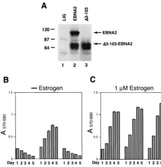
richment for GFP-positive cells by FACS, we showed that 90% of the cells expressed either wild-type or mutant EBNA2 pro-teins (Fig. 1A and data not shown). The transduced cell pools were all sorted using a similar gating strategy for GFP fluores-cence that resulted in similar mean fluoresfluores-cence intensities of GFP expression (reference 13 and data not shown). In these transduced cell pools, several minor polypeptides migrating at 64,000 to 70,000 were invariably detected by immunoblot anal-ysis with either the anti-HA or EBNA2-specific monoclonal antibodies (Fig. 1A). These bands have been observed previ-ously and do not appear to interfere with EBNA2 function in immortalization maintenance (15). The ⌬3-103EBNA2 pro-tein, which migrated closely with some of these polypeptides, was expressed at wild-type levels in the transduced cells (Fig. 1A).
We next tested the transduced cells for their ability to liferate after inactivation of the endogenous EREBNA2
pro-tein by withdrawal of estrogen from the culture medium. Briefly, cell pools expressing either wild-type EBNA2 or⌬ 3-103EBNA2 were expanded for 2 weeks after initial sorting for GFP expression and then subjected to estrogen withdrawal. While the wild-type EBNA2-expressing EREB cells continued to proliferate, the ⌬3-103EBNA2-expressing cells ceased to proliferate and died out at a rate similar to control cell pools transduced with an empty lentiviral vector (Fig. 1B). There-fore, the ⌬3-103EBNA2 mutant protein failed to support maintenance of EBV-mediated immortalization. In contrast, all three transduced cell pools proliferated with similar effi-ciency when cultured in the presence of estrogen (Fig. 1C). In an attempt to explain the failure of⌬3-103EBNA2-expressing cells to proliferate in the absence of estrogen, we analyzed expression of the viral LMP-1 and the cellular proto-oncopro-tein c-myc in these cells following estrogen withdrawal for 3 days. The results showed that, whereas cells expressing
wild-FIG. 1. ⌬3-103–EBNA2 fails to support survival and proliferation of EREB cells upon EREBNA2 inactivation. (A) Western blot analysis of pLIG (lane 1), pLIG.EBNA2 (lane 2), and pLIG.⌬3-103-EBNA2 (lane 3) transduced cell pools after enrichment by FACS were grown in the presence of 1M estrogen for 2 weeks prior to Western blotting. EBNA2 expression was analyzed using an anti-HA monoclonal antibody. Equal amounts of cellular protein for each cell pool were analyzed. Molecular mass markers are shown in kilodaltons on the left. (B) EREB cell pools transduced with the pLIG vector alone (LIG), pLIG.EBNA2 (LIG.EBNA2), or pLIG.⌬3-103–EBNA2 (LIG.⌬3-103EBNA2) lentiviruses after enrichment by FACS were grown in the presence of 1M estrogen for 2 weeks prior to the experiment. At day 0, the cells were washed of estrogen and placed in the estrogen-free medium. Twenty-four hours later, the cell pools were plated in 200-l aliquots of estrogen-free medium in 96-well plates at 2⫻104cells/well and monitored for survival and proliferation by MTT assay daily over the course of 5 days, starting with day 1 (day of
plating) after estrogen withdrawal. Optical density at 570 nm (A570) and 690 nm (A690) wavelengths of completed MTT reactions were determined
to calculate the A570-690values by subtractingA690fromA570. (C) The same experiment as that shown in panel B, except that cells were grown in
medium containing estrogen.
on November 8, 2019 by guest
http://jvi.asm.org/
[image:4.603.131.455.70.405.2]type EBNA2 maintained LMP-1 and c-myc expression, cells expressing⌬3-103EBNA2 lacked detectable LMP-1 and c-myc
(Fig. 2). Thus, ⌬3-103EBNA2 was significantly impaired at maintaining LMP-1 and c-myc expression in transformed B cells.
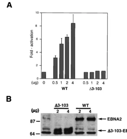
⌬3-103EBNA2 fails to stimulate LMP-1 expression in tran-sient-cotransfection assays.Because a similar EBNA2 mutant was previously shown to transactivate the LMP-1 promoter similar to the wild-type protein in transient-cotransfection as-says (66), we also evaluated the ability of ⌬3-103EBNA2 to transactivate an LMP-1 promoter luciferase reporter plasmid in transient-cotransfection assays. Consistent with our results presented in Fig. 2,⌬3-103EBNA2 also failed to appreciably stimulate LMP-1 promoter activity, even at the highest con-centration of EBNA2 expression plasmid (Fig. 3A). Western blot analysis of wild-type and mutant EBNA2 expression in this assay showed that both proteins were expressed at comparable levels (Fig. 3B). Therefore, ⌬3-103EBNA2 is also severely defective in stimulating the LMP-1 promoter in transient-ex-pression assays.
Amino acids 3 to 30 of EBNA2 are required for transacti-vating the LMP-1 promoter but not a promoter with multim-erized CBF1-binding sites.We have previously demonstrated that the PPR is a modulatory domain dispensable for both EBNA2-mediated activation of the LMP-1 promoter and for immortalization maintenance. The ⌬3-103 deletion removes CRs 1 and 2, and we hypothesized that perhaps at least one of these regions might be required for these EBNA2 activities. In a previous study, a mutant EBNA2 lacking CRs 1 and 2 (⌬2-88 [W91] EBNA2) could still support immortalization, albeit with a significantly lower-than-wild-type efficiency, possibly due to its impaired ability to transactivate LMP-1 expression (66).
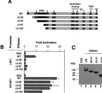
Therefore, we examined a similar mutant EBNA2 in our ex-periments as well as three additional deletion mutants missing CR1 (⌬3-30), CR1 and most of CR2 (⌬3-52), and CRs 1 and 2 and all but 7 of the 45 prolines (⌬3-97; equivalent to⌬2-88 W91 EBNA2) (Fig. 4A). In transient-cotransfection assays the wild type, but none of the mutants, was able to stimulate an LMP-1 reporter plasmid (Fig. 4B). With the exception of⌬ 3-103EBNA2, however, the mutants were capable of stimulating a reporter plasmid containing multiple (eight) CBF1-binding sites similar to wild-type levels, indicating that the mutant proteins were not globally defective for transactivating func-tion (Fig. 4B).⌬3-103EBNA2 was consistently able to stimu-late the CBF1-responsive promoter to within 60% of the wild-type protein level (Fig. 4B). The expression levels of the wild-type and mutant EBNA2s were similar (Fig. 4C). These results demonstrated that amino acids 3 to 30 of EBNA2 specifically drive expression of LMP-1 in B cells.
[image:5.603.51.272.69.216.2]EBNA2 amino acids 3 to 30 are critical for induction of LMP-1 in type I Burkitt’s lymphoma cells. To exclude the
[image:5.603.311.529.70.309.2]FIG. 2. ⌬3-103–EBNA2 fails to support expression of key viral and cellular EBNA2 target genes in EREB cells following EREBNA2 inactivation. EREB cell pools transduced with the pLIG vector alone (LIG), pLIG.EBNA2 (WT), or pLIG.⌬3-103–EBNA2 (⌬3-103.1 and ⌬3-103.2) lentiviruses after enrichment by FACS were grown in the presence of estrogen for 2 weeks prior to the experiment.⌬3-103.1 and ⌬3-103.2 represent two independently generated cell lines. At day 0, the cells were washed of estrogen and incubated in either estrogen-free (lanes 1 to 4) or estrogen-containing (lanes 5 to 8) medium for 4 days. Cell lysates were prepared, and equal amounts of cellular proteins were separated by SDS electrophoresis. The separated proteins were transferred to nitrocellulose membranes and probed with monoclonal antibodies against LMP-1 (A) and c-myc(B). Migration of LMP-1 and c-mycis indicated by arrows on the right.
FIG. 3. ⌬3-103–EBNA2 cannot transactivate an LMP-1 promoter reporter plasmid. (A) A 0.5-g aliquot of an LMP-1 promoter-lucif-erase reporter plasmid was cotransfected into DG75 cells with the indicated amounts of wild-type EBNA2 (WT) or⌬3-103–EBNA2 (⌬ 3-103) expression plasmids. The amount of expression plasmid in each transfection reaction was equilibrated with the empty SG5 expression plasmid. The results are presented as fold activation of the promoter in the presence of an effector. Average transactivation efficiencies repre-senting three independent experiments are presented. Standard error bars are shown. (B) Representative Western blot analysis of wild-type and⌬3-103–EBNA2 expression in samples from the transient-trans-fection experiments described in the legend for panel A. At 48 h after transfection, cells transfected with the SG5 plasmid alone (lane 1), the indicated amounts of ⌬3-103–EBNA2 (⌬3-103; lanes 2 and 3), or wild-type EBNA2 (WT; lanes 4 and 5) expression plasmids were sub-jected to SDS-PAGE and Western blot analysis using an anti-HA monoclonal antibody. Cell lysate aliquots containing equal amounts of total cellular protein were analyzed in each lane. Molecular mass markers are shown in kilodaltons on the left. Arrows on the right indicate migrations of WT and⌬3-103 proteins.
on November 8, 2019 by guest
http://jvi.asm.org/
possibility that the EBNA2 mutants were unable to transacti-vate the LMP-1 promoter-reporter plasmid because it lacks essential EBNA2-responsive cis-acting elements, we also tested the EBNA2 mutants for their ability to induce LMP-1 expression in type I Burkitt’s lymphoma cells. The type I cell line Eli-BL does not express LMPs or EBNAs except EBNA-1. Ectopic expression of EBNA2 has been shown to result in a modest induction of LMP-1 that is dramatically enhanced by coexpression with the EBNA-LP protein (38, 39). We showed that wild-type EBNA2, but not the mutant EBNA2s, induced LMP-1 expression in Eli-BL cells (Fig. 5A), despite the fact that all EBNA2 proteins were expressed at similar levels in these cells (Fig. 5B).
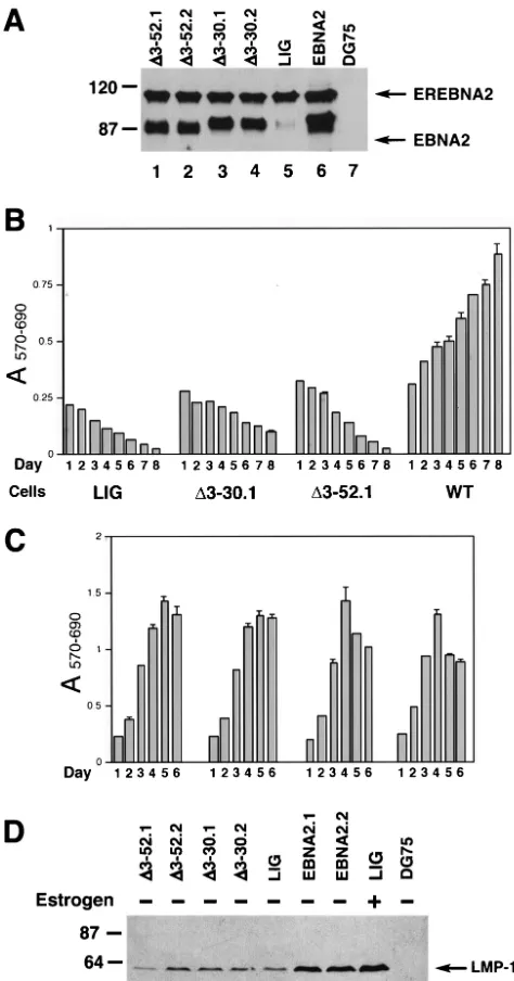
EBNA2 amino acids 3 to 30 are required for immortaliza-tion maintenance.We next performed transcomplementation assays to determine whether the deletion mutants were defec-tive in activating LMP-1 in EBV-infected cells and also to address the ability of the mutants to support cellular prolifer-ation. Two independently generated EREB cell pools express-ing each mutant protein were generated, and each expressed
[image:6.603.127.457.74.374.2]an EBNA2 protein that exhibited the expected electrophoretic mobility (Fig. 6A and data not shown). The cell pools were then grown in medium without estrogen and subsequently monitored for their proliferative capacity. Whereas wild-type EBNA2 efficiently supported cell proliferation in the absence of estrogen, deletion mutants⌬3-30 and⌬3-52 failed to do so (Fig. 6B). In addition, neither the ⌬3-30 nor ⌬3-52 EBNA2 mutant could appreciably maintain LMP-1 expression at day 4 after estrogen withdrawal compared to WTEBNA2 in EREB cells (Fig. 6D). ⌬3-30EBNA2 appeared to promote limited survival of EREB cells, as judged by the MTT assay (Fig. 6B), and prolonged persistence of GFP-positive cells under estro-gen-free conditions (data not shown). However, even after 2 weeks of culture in the estrogen-free medium, these cell pools failed to produce a detectable outgrowth (data not shown). As a control, we showed that, in the presence of estrogen, all of the cell pools proliferated with compatible efficiency (Fig. 6C). Mutant EBNA2s containing the larger deletion⌬3-97 or de-letions from amino acids 3 to 115 and 3 to 156 likewise were unable to complement EBNA2 function upon inactivation of
FIG. 4. LMP-1 promoter transactivation by EBNA2 mutants. (A) Schematic of EBNA2 deletion mutants generated for this study. CRs are shown as gray boxes. Nuclear localization signals (NLS), the transcriptional activation domain (TAD), and promoter targeting domain (Skip/CBF1) are indicated. The PPR is shown in black. (B) A 0.5-g aliquot of an LMP-1 promoter-luciferase reporter plasmid (LMP-1) or JT123A reporter plasmid (8XCBF1) was cotransfected into DG75 cells with 4-g amounts of expression plasmids encoding wild-type EBNA2 (WT),⌬3-30–EBNA2 (⌬3-30),⌬3-52–EBNA2 (⌬3-52),⌬3-97–EBNA2 (⌬3-97), and⌬3-103–EBNA2 (⌬3-103). The amount of expression plasmid in each transfection reaction was equilibrated with the empty SG5 expression plasmid. The results are presented as fold activation of the promoter in the presence of the effector. Average transactivation efficiencies representing three independent experiments are presented. Standard errors are also shown. (C) A representative Western blot analysis of wild-type and mutant EBNA2 expression in samples from the transient-transfection experiments described in the legend for panel B. At 48 h after transfection, cell lysates were prepared and equivalent amounts of proteins for each sample were subjected to SDS-PAGE and Western blot analysis using the anti-EBNA2 monoclonal antibody R3. Molecular mass markers are shown in kilodaltons on the left.
on November 8, 2019 by guest
http://jvi.asm.org/
endogenous EREBNA2 by estrogen withdrawal (data not shown). The data from the transcomplementation and LMP-1 promoter transactivation assays using the mutant EBNA2s are summarized in Table 1.
EBNA2 amino acids 3 to 30 are impaired for self-associa-tion.Recent studies have suggested that EBNA2 possesses two redundant self-association domains located in the amino-ter-minal 210 amino acid residues (19). To determine whether amino acid residues 3 to 30 contribute to self-association, we performed coimmunoprecipitation assays from transfected cell extracts expressing a smaller EBNA2 polypeptide truncated for the carboxy-terminal 291 amino acid residues (F199) and either wild-type EBNA2 or EBNA2 deletion proteins. The results indicated that the⌬3-30 mutant EBNA2 was much less efficient at associating with the truncated F199 polypeptide than wild-type EBNA2 (Fig. 7). As expected, a larger deletion in EBNA2 removing amino acid residues 1 to 156 resulted in a polypeptide that was completely unable to coprecipitate with F199. Further studies will be required to determine whether
residues 3 to 30 mediate direct EBNA2-EBNA2 interactions or if a cellular intermediate is required.
DISCUSSION
Our results define a novel domain between amino acid res-idues 3 and 30 of EBNA2 that is required for stimulation of LMP-1 expression and proliferation of EBV-immortalized cells. This region, which includes CR1, is distinct from other domains required for B-cell immortalization that include the Skip/CBF1 interaction domains located in CRs 5 and 6 and the transactivation domain.
The requirement of an EBNA2 region within amino acids 3 to 30 for transactivation of LMP-1 has been suggested previ-ously in other studies (8, 47). Cohen et al. introduced a linker insertion at EBNA2 amino acid 19 and also deleted amino acids 19 to 34, which lie just outside of CR1 (8). Both muta-tions resulted in a decreased ability to transactivate LMP-1 and reduced the overall efficiency of B-cell immortalization by re-combinant viruses expressing the mutant proteins (8). Sjoblom et al. introduced a deletion in amino acids 2 to 19 and tested the mutant protein in transient-cotransfection assays (47). In-terestingly, they observed a marked reduction in the protein’s ability to stimulate the LMP-1 promoter that had little overall effect on stimulation of the viral latency C promoter. Our results are the first to fully characterize the role of the first 30 amino acid residues containing CR1 in transient-reporter gene assays for LMP-1 promoter induction, to correlate this activity to LMP-1 expression in LCLs, and to determine that this domain is required for LCL proliferation.
The failure of⌬3-103EBNA2 to upregulate LMP-1 was un-expected, as a similar mutant was reported to transactivate the LMP-1 gene comparable to wild-type EBNA2. The discrep-ancy between our results and those previously published could be due to differences in the EBNA2 allele used (B95.8 versus strain W91). The W91 EBNA2 PPR is eight proline residues shorter than the B95.8 EBNA2 PPR (hence, ⌬2-95 versus
⌬3-103) (8, 10). Although our deletion closely matches the
⌬2-95EBNA2 mutant described before, it is unclear whether the other single 16 amino acid differences between W91 and B95.8 EBNA2 also contributed to our disparate results. In addition, Yalamanchili et al. used an LMP-1 reporter contain-ing a significantly shorter promoter sequence extendcontain-ing from
⫹40 to⫺234 (as opposed to⫹40 to⫺327, used in our exper-iments) that may lack an element required for transactivation by wild-type EBNA2 (66). However, this does not seem likely, as various LMP-1 promoter-reporter gene constructs extend-ing from⫹40 to between⫺234 and⫺512 in the LMP-1 pro-moter sequence exhibit similar EBNA2 responsiveness (32, 55). Our finding that⌬3-103EBNA2 was severely defective in transactivation of the LMP-1 promoter was further strength-ened by⌬3-103EBNA2’s inability to maintain LMP-1 expres-sion in LCLs (Fig. 2) or to stimulate LMP-1 expresexpres-sion in type I Burkitt’s lymphoma cells (Fig. 5), where the entire LMP-1 promoter is present. In addition, all other deletion mutant EBNA2s tested in the present study were unable to stimulate LMP-1 expression (Fig. 4, 5, and 6). While the reporter gene assay results differed between our study and that of Yalaman-chili et al., it is quite possible that within the context of primary B-cell infection that the⌬2-95 (W91) EBNA2, like our⌬3-103
FIG. 5. Induction of LMP-1 gene expression in type I Burkitt’s lymphoma cells. (A) A 10-g aliquot of SG5 plasmid, SG5–EBNA-LP expression plasmid, SG5–wild-type EBNA2 (WT), or expression plas-mids encoding ⌬3-30–EBNA2 (⌬3-30),⌬3-52–EBNA2 (⌬3-52),⌬ 3-97–EBNA2 (⌬3-97), or⌬3-103–EBNA2 (⌬3-103) were introduced into Eli-BL cells by electroporation. The amount of expression plasmid in each transfection reaction mixture was equilibrated with SG5 plasmid (empty expression vector). At 48 h after electroporation, cell lysates were prepared and equivalent amounts of proteins for each sample were subjected to SDS-PAGE and Western blot analysis using anti-LMP-1 monoclonal antibody. Results from a representative experi-ment are shown. Molecular mass markers are shown in kilodaltons on the left. (B) A representative Western blot analysis of wild-type and mutant EBNA2 expression in samples from the transient-transfection experiments described in the legend for panel A. At 48 h after elec-troporation, cell lysates were prepared and equivalent amounts of proteins for each sample were subjected to SDS-PAGE and Western blot analysis using an anti-EBNA2 R3 monoclonal antibody. Molecu-lar mass markers are shown in kilodaltons on the left.
on November 8, 2019 by guest
http://jvi.asm.org/
(B95-8) EBNA2, may be unable to stimulate LMP-1 expres-sion, giving rise to the null immortalization phenotype ob-served in the marker rescue assays (66).
Another unexpected finding of our study was that the smaller deletion, mutant ⌬3-97EBNA2, which retains seven prolines of the PPR, was unable to complement loss of EBNA2 function in the EREB cells. In marker rescue assays, a similar W91 EBNA2 mutant (⌬2-88) has previously been shown to be immortalization competent, even though it was unable to effi-ciently stimulate the LMP-1 promoter in reporter gene assays (66). One possible reason for the different outcomes may be due to inherent differences in the transcomplementation and marker rescue assays. Induction of various growth-promoting activities, such as autocrine loops due to epigenetic changes in cellular gene expression, appears to contribute to overall effi-ciency of immortalization and growth of EBV-infected cells (22, 42). EREB cells may be already epigenetically pro-grammed and restricted in their potential to develop compen-satory changes in gene expression, which would not allow sur-vival and outgrowth of mutant EBNA2-expressing cell pools after estrogen withdrawal. It is possible that primary B cells possess an inherently higher potential for epigenetically driven compensatory events, which could make up for the deficiency in the function of the EBNA2 deletion mutant. Since EREBNA2 possesses an intact amino terminus, the estab-lished EREB cells may not have been under selective pressure during initial stages of immortalization to develop
compensa-FIG. 6. ⌬3-30EBNA2 and⌬3-52EBNA2 are defective in support-ing proliferation of EREB cells followsupport-ing EREBNA2 inactivation. EREB cell pools transduced with the pLIG vector alone (LIG) or pLIG.EBNA2 (WT), as well as two independently generated EREB cell pools transduced with the pLIG.⌬3-30EBNA2 (⌬3-30.1 and⌬ 3-30.2) or pLIG.⌬3-52EBNA2 (⌬3-52.1 and⌬3-52.2) lentiviruses after enrichment by FACS were grown in the presence of estrogen for 2 weeks prior to the experiment. (A) Western blot analysis of pLIG (LIG), pLIG.EBNA2 (WT), and pLIG.⌬3-30EBNA2 (⌬3-30.1 and ⌬3-30.2) and pLIG.⌬3-30EBNA2 (⌬3-52.1 and ⌬3-52.2) transduced EREB cell pools using an EBNA2 R3 monoclonal antibody. Equal amounts of cellular protein for each cell pool were analyzed. Molec-ular mass markers are shown in kilodaltons on the left. (B) At day 0, logarithmically growing cell pools expressing pLIG, pLIG.⌬ 3-30EBNA2 (⌬3-30.1), or pLIG.⌬3-52EBNA2 (⌬3-52.1) were washed of estrogen and placed in the estrogen-free medium. Twenty-four hours later, the cell pools were plated in 200-l aliquots of estrogen-free medium in 96-well plates at 2 ⫻ 104 cells/well and monitored for
survival and proliferation by MTT assay daily over the course of 8 days starting with day 1 (day of plating) after estrogen withdrawal. Optical densities at 570 nm (A570) and 690 nm (A690) of completed MTT
[image:8.603.45.282.71.523.2]reaction mixtures were determined to calculate the A570-690values by
TABLE 1. Stimulation of LMP1 expression and LCL proliferation by mutant EBNA2 proteins
Mutation Proliferationa LMP1 transactivationb
⌬3–30 ⫺ ⫺
⌬3–52 ⫺ ⫺
⌬3–97 ⫺ ⫺
⌬3–103 ⫺ ⫺
⌬3–115 NSc NSc
⌬3–156 NSc NSc
Wild type ⫹⫹ ⫹⫹
⌬59–103 (⌬PPR)d ⫹⫹ ⫹⫹⫹⫹⫹⫹
aAs determined by the transcomplementation assay.
bAs determined by transient-cotransfection assays in DG75 and Eli-BL cells.
cNS, data not shown.
dFrom reference 14.
subtractingA690fromA570. (C) Same experiment as for panel B except
that cells were grown in medium containing estrogen and proliferation was monitored over the course of 6 days. (D) ⌬3-30– and⌬3-52– EBNA2s fail to support expression of the LMP1 gene in EREB cells following EREBNA2 inactivation. EREB cell pools transduced with the pLIG vector alone (LIG), pLIG.EBNA2 (EBNA2.1 and EBNA2.2), pLIG.⌬3-30–EBNA2 (⌬3-30.1 and⌬3-30.2), or pLIG.⌬ 3-30–EBNA2 (⌬3-52.1 and ⌬3-52.2) lentiviruses after enrichment by FACS were grown in the presence of estrogen for 2 weeks prior to the experiment. At day 0, the cells were washed of estrogen and incubated in either estrogen-free (lanes 1 to 7 and 9) or estrogen-containing (lane 8) medium for 4 days. DG75 is an EBV-negative Burkitt’s lymphoma cell line. Cell lysates were prepared, and equal amounts of cellular proteins were separated by SDS-PAGE. The separated proteins were transferred to nitrocellulose membranes and probed with monoclonal antibodies against LMP-1. Molecular mass markers are shown in kilo-daltons on the left.
on November 8, 2019 by guest
http://jvi.asm.org/
[image:8.603.300.543.92.184.2]tory epigenetic changes similar to those which may have been required for cells infected with EBVs expressing⌬2-88EBNA2 (W91 strain) in marker rescue assays. Thus, the differences in outcomes between those results reported here versus earlier marker rescue assay results may be due to different physiolog-ical and developmental states of the B cells at the initial start-ing points of the assays.
Based on a coimmunoprecipitation assay, amino acid resi-dues 3 to 30 appear to function as a self-association domain, suggesting that EBNA2 dimerization or multimerization is crit-ical for LMP-1 induction and B-cell immortalization. However, further studies will be required to determine whether this do-main facilitates direct EBNA2-EBNA2 interactions or if a cel-lular cofactor is involved. Given that distinct domains confer-ring dimerization and multimerization often exist within a polypeptide, additional amino-terminal mutants need to be
tested to determine how the domain of amino acids 3 to 30 contributes to these activities.
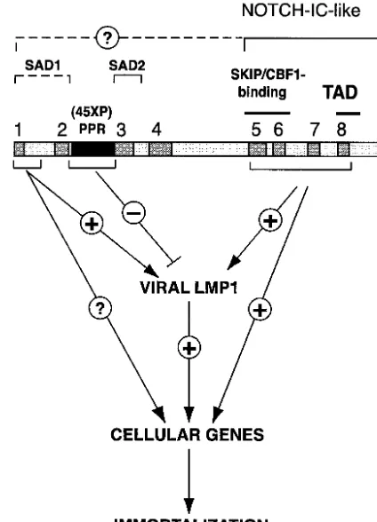
Based on data presented here, we have refined identification of a domain within the amino terminus of the EBNA2 mole-cule that is important for induction of LMP-1 expression and LCL proliferation (Fig. 8). The model provides a foundation from which more refined mutagenesis of the amino acid 3 to 30 region can be pursued to determine the precise mechanism by which it contributes to transactivation of viral or cellular genes essential for EBV-mediated transformation.
ACKNOWLEDGMENTS
We thank Ron Javier for critical reading of the manuscript. This work was supported by a National Space Biomedical Research Institute grant NCC (9-58) (cooperative agreement) and ACS grant RPG-00-099-01.
REFERENCES
1. Alfieri, C., M. Birkenbach, and E. Kieff.1991. Early events in Epstein-Barr
virus infection of human B lymphocytes. Virology181:595–608. (Erratum,
185:946, 1991.)
[image:9.603.49.277.72.325.2]2. Baer, R., A. T. Bankier, M. D. Biggin, P. L. Deininger, P. J. Farrell, T. J.
FIG. 7. ⌬3-30EBNA2 is impaired in its ability to self-associate. EBV-negative DG75 cells were cotransfected with 5 g each of a plasmid expressing a truncated EBNA2 (F199; amino acids 1 to 199 with a carboxy-terminal Flag epitope tag) and a plasmid expressing wild-type or amino-terminal deletion variants of full-length EBNA2 proteins. (A) Western blots indicating equivalent expression of EBNA2 and F199 in cotransfected cells. The panel on the left was probed with a monoclonal antibody to EBNA2, and the one on the right was probed with a monoclonal antibody to the Flag epitope tag. Equal amounts of transfected cell lysates containing wild-type (lane 1) and amino-terminal deletions⌬3-30EBNA2 and⌬3-156EBNA2 (lanes 2 and 3, respectively) were compared. (B) Cell lysates were precipi-tated using the M2 anti-Flag antibody (⫹), divided into two equal parts, resolved by SDS-PAGE, and transferred to nitrocellulose. One blot was probed using the R3 anti-EBNA2 antibody, while the other was probed using an anti-Flag antibody. Migration of EBNA2 and F199 proteins is indicated. Migration of molecular mass standards is shown on the left. Lysates expressing wild-type EBNA2 and F199 (lane 1), expressing ⌬3-30EBNA2 and F199 (lanes 2 and 3), and ⌬ 3-156EBNA2 and F199 were tested. Lane 2 was precipitated using 50% of the lysate used in lanes 1, 3, and 4.
FIG. 8. Functional roles of EBNA2 regions critical for immortal-ization. The carboxy-terminal CRs 5, 6, and 8 function to mimic an activated Notch protein (Notch-IC). The amino-terminal CRs of EBNA2 contribute to EBV-induced immortalization in several ways. The PPR modulates the EBNA2’s ability to stimulate LMP-1 expres-sion (14). Residues 3 to 30 containing CR1 comprise a genetic deter-minant critical for EBNA2 transactivation of the LMP1 gene and for proliferation of EBV-immortalized cells. It remains unknown whether this region is required for transactivation of EBNA2 cellular target genes. The two self-association domains (SAD1 and 2) have previously been mapped to residues 1 to 58 and 97 to 121 in EBNA2.
on November 8, 2019 by guest
http://jvi.asm.org/
[image:9.603.315.527.73.367.2]Gibson, G. Hatfull, G. S. Hudson, S. C. Satchwell, C. Seguin, et al.1984. DNA sequence and expression of the B95–8 Epstein-Barr virus genome.
Nature310:207–211.
3. Bornkamm, G. W., and W. Hammerschmidt.2001. Molecular virology of
Epstein-Barr virus. Philos. Trans. R. Soc. London B356:437–459.
4. Burgstahler, R., B. Kempkes, K. Steube, and M. Lipp.1995. Expression of the chemokine receptor BLR2/EBI1 is specifically transactivated by
Epstein-Barr virus nuclear antigen 2. Biochem. Biophys. Res. Commun.215:737–743.
5. Callahan, J., J. Aster, J. Sklar, E. Kieff, and E. S. Robertson.2000. Intra-cellular forms of human NOTCH1 interact at distinctly different levels with
RBP-jin human B and T cells. Leukemia14:84–92.
6. Cohen, J. I.1992. A region of herpes simplex virus VP16 can substitute for a transforming domain of Epstein-Barr virus nuclear protein 2. Proc. Natl.
Acad. Sci. USA89:8030–8034.
7. Cohen, J. I., and E. Kieff.1991. An Epstein-Barr virus nuclear protein 2 domain essential for transformation is a direct transcriptional activator.
J. Virol.65:5880–5885.
8. Cohen, J. I., F. Wang, and E. Kieff.1991. Epstein-Barr virus nuclear protein 2 mutations define essential domains for transformation and transactivation.
J. Virol.65:2545–2554.
9. Cordier-Bussat, M., M. Billaud, A. Calender, and G. M. Lenoir.1993. Ep-stein-Barr virus (EBV) nuclear-antigen-2-induced up-regulation of CD21 and CD23 molecules is dependent on a permissive cellular context. Int. J.
Cancer53:153–160.
10. Dambaugh, T., K. Hennessy, L. Chamnankit, and E. Kieff.1984. U2 region of Epstein-Barr virus DNA may encode Epstein-Barr nuclear antigen 2.
Proc. Natl. Acad. Sci. USA81:7632–7636.
11. Dirmeier, U., B. Neuhierl, E. Kilger, G. Reisbach, M. L. Sandberg, and W. Hammerschmidt.2003. Latent membrane protein 1 is critical for efficient growth transformation of human B cells by Epstein-Barr virus. Cancer Res.
63:2982–2989.
12. Fahraeus, R., A. Jansson, A. Ricksten, A. Sjoblom, and L. Rymo.1990. Epstein-Barr virus-encoded nuclear antigen 2 activates the viral latent mem-brane protein promoter by modulating the activity of a negative regulatory
element. Proc. Natl. Acad. Sci. USA87:7390–7394.
13. Fuentes-Panana, E. M., S. Swaminathan, and P. D. Ling.1999. Transcrip-tional activation signals found in the Epstein-Barr virus (EBV) latency C promoter are conserved in the latency C promoter sequences from baboon and rhesus monkey EBV-like lymphocryptoviruses (cercopithicine
herpesvi-ruses 12 and 15). J. Virol.73:826–833.
14. Gordadze, A. V., R. Peng, J. Tan, G. Liu, R. Sutton, B. Kempkes, G. W. Bornkamm, and P. D. Ling.2001. Notch1IC partially replaces EBNA2
func-tion in B cells immortalized by Epstein-Barr virus. J. Virol.75:5899–5912.
15. Gordadze, A. V., D. Poston, and P. D. Ling.2002. The EBNA2 polyproline region is dispensable for Epstein-Barr virus-mediated immortalization
main-tenance. J. Virol.76:7349–7355.
16. Grossman, S. R., E. Johannsen, X. Tong, R. Yalamanchili, and E. Kieff.
1994. The Epstein-Barr virus nuclear antigen 2 transactivator is directed to
response elements by the Jrecombination signal binding protein. Proc.
Natl. Acad. Sci. USA91:7568–7572.
17. Grundhoff, A. T., E. Kremmer, O. Tureci, A. Glieden, C. Gindorf, J. Atz, N. Mueller-Lantzsch, W. H. Schubach, and F. A. Grasser.1999. Characteriza-tion of DP103, a novel DEAD box protein that binds to the Epstein-Barr
virus nuclear proteins EBNA2 and EBNA3C. J. Biol. Chem.274:19136–
19144.
18. Harada, S., and E. Kieff.1997. Epstein-Barr virus nuclear protein LP stim-ulates EBNA-2 acidic domain-mediated transcriptional activation. J. Virol.
71:6611–6618.
19. Harada, S., R. Yalamanchili, and E. Kieff.2001. Epstein-Barr virus nuclear protein 2 has at least two N-terminal domains that mediate self-association.
J. Virol.75:2482–2487.
20. Harada, S., R. Yalamanchili, and E. Kieff.1998. Residues 231 to 280 of the Epstein-Barr virus nuclear protein 2 are not essential for primary
B-lympho-cyte growth transformation. J. Virol.72:9948–9954.
21. Henkel, T., P. D. Ling, S. D. Hayward, and M. G. Peterson.1994. Mediation of Epstein-Barr virus EBNA2 transactivation by recombination
signal-bind-ing protein J. Science265:92–95.
22. Jochems, G. J., M. R. Klein, R. Jordens, D. Pascual-Salcedo, F. van Boxtel-Oosterhof, R. A. van Lier, and W. P. Zeijlemaker.1991. Heterogeneity in both cytokine production and responsiveness of a panel of monoclonal human Epstein-Barr virus-transformed B-cell lines. Hum. Antibodies
Hy-bridomas2:57–64.
23. Kaiser, C., G. Laux, D. Eick, N. Jochner, G. W. Bornkamm, and B. Kempkes.
1999. The proto-oncogene c-mycis a direct target gene of Epstein-Barr virus
nuclear antigen 2. J. Virol.73:4481–4484.
24. Kaye, K. M., K. M. Izumi, and E. Kieff.1993. Epstein-Barr virus latent membrane protein 1 is essential for B-lymphocyte growth transformation.
Proc. Natl. Acad. Sci. USA90:9150–9154.
25. Kempkes, B., M. Pawlita, U. Zimber-Strobl, G. Eissner, G. Laux, and G. W. Bornkamm.1995. Epstein-Barr virus nuclear antigen 2-estrogen receptor fusion proteins transactivate viral and cellular genes and interact with
RBP-Jin a conditional fashion. Virology214:675–679.
26. Kempkes, B., D. Spitkovsky, P. Jansen-Durr, J. W. Ellwart, E. Kremmer, H. J. Delecluse, C. Rottenberger, G. W. Bornkamm, and W. Hammer-schmidt.1995. B-cell proliferation and induction of early G1-regulating
proteins by Epstein-Barr virus mutants conditional for EBNA2. EMBO J.
14:88–96.
27. Kieff, E., and A. B. Rickinson.2001. Epstein-Barr virus and its replication, p.
2511–2574.InD. M. Knipe and P. M. Howley (ed.), Field’s virology, 4th ed.,
vol. 2. Lippincott-Williams & Wilkins, Philadelphia, Pa.
28. Kilger, E., A. Kieser, M. Baumann, and W. Hammerschmidt.1998. Epstein-Barr virus-mediated B-cell proliferation is dependent upon latent membrane
protein 1, which simulates an activated CD40 receptor. EMBO J.17:1700–
1709.
29. Kim, O. J., and J. L. Yates.1993. Mutants of Epstein-Barr virus with a selective marker disrupting the TP gene transform B cells and replicate
normally in culture. J. Virol.67:7634–7640.
30. Laemmli, U. K.1970. Cleavage of structural proteins during the assembly of
the head of bacteriophage T4. Nature227:680–685.
31. Larcher, C., B. Kempkes, E. Kremmer, W. M. Prodinger, M. Pawlita, G. W. Bornkamm, and M. P. Dierich.1995. Expression of Epstein-Barr virus nu-clear antigen-2 (EBNA2) induces CD21/CR2 on B and T cell lines and
shedding of soluble CD21. Eur. J. Immunol.25:1713–1719.
32. Laux, G., F. Dugrillon, C. Eckert, B. Adam, S. U. Zimber, and G. W. Bornkamm.1994. Identification and characterization of an Epstein-Barr
virus nuclear antigen 2-responsiveciselement in the bidirectional promoter
region of latent membrane protein and terminal protein 2 genes. J. Virol.
68:6947–6958.
33. Ling, P. D., D. R. Rawlins, and S. D. Hayward.1993. The Epstein-Barr virus immortalizing protein EBNA-2 is targeted to DNA by a cellular
enhancer-binding protein. Proc. Natl. Acad. Sci. USA90:9237–9241.
34. Ling, P. D., J. J. Ryon, and S. D. Hayward.1993. EBNA-2 of herpesvirus papio diverges significantly from the type A and type B EBNA-2 proteins of Epstein-Barr virus but retains an efficient transactivation domain with a
conserved hydrophobic motif. J. Virol.67:2990–3003.
35. Longnecker, R., C. L. Miller, X. Q. Miao, A. Marchini, and E. Kieff.1992. The only domain which distinguishes Epstein-Barr virus latent membrane protein 2A (LMP2A) from LMP2B is dispensable for lymphocyte infection and growth transformation in vitro; LMP2A is therefore nonessential. J.
Vi-rol.66:6461–6469.
36. Longnecker, R., C. L. Miller, X. Q. Miao, B. Tomkinson, and E. Kieff.1993. The last seven transmembrane and carboxy-terminal cytoplasmic domains of Epstein-Barr virus latent membrane protein 2 (LMP2) are dispensable for
lymphocyte infection and growth transformation in vitro. J. Virol.67:2006–
2013.
37. Longnecker, R., C. L. Miller, B. Tomkinson, X. Q. Miao, and E. Kieff.1993. Deletion of DNA encoding the first five transmembrane domains of
Epstein-Barr virus latent membrane proteins 2A and 2B. J. Virol.67:5068–5074.
38. Nitsche, F., A. Bell, and A. Rickinson.1997. Epstein-Barr virus leader pro-tein enhances EBNA-2-mediated transactivation of latent membrane propro-tein
1 expression: a role for the W1W2 repeat domain. J. Virol.71:6619–6628.
39. Peng, R., A. V. Gordadze, E. M. Fuentes Panana, F. Wang, J. Zong, G. S. Hayward, J. Tan, and P. D. Ling.2000. Sequence and functional analysis of EBNA-LP and EBNA2 proteins from nonhuman primate
lymphocryptovi-ruses. J. Virol.74:379–389.
40. Peng, R., J. Tan, and P. D. Ling.2000. Conserved regions in the Epstein-Barr virus leader protein define distinct domains required for nuclear localization
and transcriptional cooperation with EBNA2. J. Virol.74:9953–9963.
41. Rickinson, A. B., and E. Kieff.2001. Epstein-Barr virus, p. 2575–2627.In
D. M. Knipe and P. M. Howley (ed.), Field’s virology, 4th ed., vol. 2. Lippincott-Williams & Wilkins, Philadelphia, Pa.
42. Rochford, R., M. J. Cannon, R. E. Sabbe, K. Adusumilli, G. Picchio, J. M. Glynn, D. J. Noonan, D. E. Mosier, and M. V. Hobbs.1997. Common and idiosyncratic patterns of cytokine gene expression by Epstein-Barr virus
transformed human B cell lines. Viral Immunol.10:183–195.
43. Rogers, R. P., M. Woisetschlaeger, and S. H. Speck.1990. Alternative splic-ing dictates translational start in Epstein-Barr virus transcripts. EMBO J.
9:2273–2277.
44. Sakai, T., Y. Taniguchi, K. Tamura, S. Minoguchi, T. Fukuhara, L. J. Strobl, U. Zimber-Strobl, G. W. Bornkamm, and T. Honjo.1998. Functional re-placement of the intracellular region of the Notch1 receptor by Epstein-Barr
virus nuclear antigen 2. J. Virol.72:6034–6039.
45. Schaefer, B. C., J. L. Strominger, and S. H. Speck.1995. Redefining the Epstein-Barr virus-encoded nuclear antigen EBNA-1 gene promoter and transcription initiation site in group I Burkitt lymphoma cell lines. Proc. Natl.
Acad. Sci. USA92:10565–10569.
46. Sinclair, A. J., I. Palmero, G. Peters, and P. J. Farrell.1994. EBNA-2 and
EBNA-LP cooperate to cause G0to G1transition during immortalization of
resting human B lymphocytes by Epstein-Barr virus. EMBO J.13:3321–3328.
47. Sjoblom, A., A. Nerstedt, A. Jansson, and L. Rymo.1995. Domains of the Epstein-Barr virus nuclear antigen 2 (EBNA2) involved in the transactiva-tion of the latent membrane protein 1 and the EBNA Cp promoters. J. Gen.
Virol.76:2669–2678.
48. Spender, L. C., G. H. Cornish, A. Sullivan, and P. J. Farrell.2002.
on November 8, 2019 by guest
http://jvi.asm.org/
sion of transcription factor AML-2 (RUNX3, CBF␣-3) is induced by Ep-stein-Barr virus EBNA-2 and correlates with the B-cell activation phenotype.
J. Virol.76:4919–4927.
49. Strobl, L. J., H. Hofelmayr, G. Marschall, M. Brielmeier, G. W. Bornkamm, and U. Zimber-Strobl.2000. Activated Notch1 modulates gene expression in
B cells similarly to Epstein-Barr viral nuclear antigen 2. J. Virol.74:1727–
1735.
50. Sung, N. S., S. Kenney, D. Gutsch, and J. S. Pagano.1991. EBNA-2
trans-activates a lymphoid-specific enhancer in theBamHI C promoter of
Epstein-Barr virus. J. Virol.65:2164–2169.
51. Sutton, R. E., H. T. M. Wu, R. Rigg, E. Bohnlein, and P. O. Brown.1998. Human immunodeficiency virus type I vectors efficiently transduce human
hematopoietic stem cells. J. Virol.72:5781–5788.
52. Swaminathan, S.1996. Characterization of Epstein-Barr virus recombinants
with deletions of theBamHI C promoter. Virology217:532–541.
53. Tong, X., R. Drapkin, D. Reinberg, and E. Kieff.1995. The 62- and 80-kDa subunits of transcription factor IIH mediate the interaction with
Epstein-Barr virus nuclear protein 2. Proc. Natl. Acad. Sci. USA92:3259–3263.
54. Tong, X., R. Drapkin, R. Yalamanchili, G. Mosialos, and E. Kieff.1995. The Epstein-Barr virus nuclear protein 2 acidic domain forms a complex with a novel cellular coactivator that can interact with TFIIE. Mol. Cell. Biol.
15:4735–4744.
55. Tsang, S. F., F. Wang, K. M. Izumi, and E. Kieff.1991. Delineation of the
cis-acting element mediating EBNA-2 transactivation of latent infection
membrane protein expression. J. Virol.65:6765–6771.
56. Tsui, S., and W. H. Schubach.1994. Epstein-Barr virus nuclear protein 2A forms oligomers in vitro and in vivo through a region required for B-cell
transformation. J. Virol.68:4287–4294.
57. Voss, M. D., A. Hille, S. Barth, A. Spurk, F. Hennrich, D. Holzer, N. Mueller-Lantzsch, E. Kremmer, and F. A. Grasser.2001. Functional coop-eration of Epstein-Barr virus nuclear antigen 2 and the survival motor
neu-ron protein in transactivation of the viral LMP1 promoter. J. Virol.75:
11781–11790.
58. Waltzer, L., F. Logeat, C. Brou, A. Israel, A. Sergeant, and E. Manet.1994.
The human Jrecombination signal sequence binding protein (RBP-J)
targets the Epstein-Barr virus EBNA2 protein to its DNA responsive
ele-ments. EMBO J.13:5633–5638.
59. Wang, F., C. Gregory, C. Sample, M. Rowe, D. Liebowitz, R. Murray, A. Rickinson, and E. Kieff.1990. Epstein-Barr virus latent membrane protein (LMP1) and nuclear proteins 2 and 3C are effectors of phenotypic changes in B lymphocytes: EBNA-2 and LMP1 cooperatively induce CD23. J. Virol.
64:2309–2318.
60. Wang, F., L. Petti, D. Braun, S. Seung, and E. Kieff.1987. A bicistronic Epstein-Barr virus mRNA encodes two nuclear proteins in latently infected,
growth-transformed lymphocytes. J. Virol.61:945–954.
61. Wang, F., S. F. Tsang, M. G. Kurilla, J. I. Cohen, and E. Kieff.1990. Epstein-Barr virus nuclear antigen 2 transactivates latent membrane protein
LMP1. J. Virol.64:3407–3416.
62. Wang, L., S. R. Grossman, and E. Kieff.2000. Epstein-Barr virus nuclear protein 2 interacts with p300, CBP, and PCAF histone acetyltransferases in
activation of the LMP1 promoter. Proc. Natl. Acad. Sci. USA97:430–435.
63. Woisetschlaeger, M., X. W. Jin, C. N. Yandava, L. A. Furmanski, J. L. Strominger, and S. H. Speck.1991. Role for the Epstein-Barr virus nuclear antigen 2 in viral promoter switching during initial stages of infection. Proc.
Natl. Acad. Sci. USA88:3942–3946.
64. Wu, D. Y., G. V. Kalpana, S. P. Goff, and W. H. Schubach.1996. Epstein-Barr virus nuclear protein 2 (EBNA2) binds to a component of the human
SNF-SWI complex, hSNF5/Ini1. J. Virol.70:6020–6028.
65. Wu, D. Y., A. Krumm, and W. H. Schubach.2000. Promoter-specific target-ing of human SWI-SNF complex by Epstein-Barr virus nuclear protein 2.
J. Virol.74:8893–8903.
66. Yalamanchili, R., S. Harada, and E. Kieff.1996. The N-terminal half of EBNA2, except for seven prolines, is not essential for primary B-lymphocyte
growth transformation. J. Virol.70:2468–2473.
67. Yandava, C. N., and S. H. Speck.1992. Characterization of the deletion and
rearrangement in theBamHI C region of the X50–7 Epstein-Barr virus
genome, a mutant viral strain which exhibits constitutiveBamHI W
pro-moter activity. J. Virol.66:5646–5650.
68. Yoo, L. I., M. Mooney, M. T. Puglielli, and S. H. Speck.1997. B-cell lines immortalized with an Epstein-Barr virus mutant lacking the Cp EBNA2
enhancer are biased toward utilization of theoriP-proximal EBNA gene
promoter Wp1. J. Virol.71:9134–9142.
69. Zhou, S., M. Fujimuro, J. J. Hsieh, L. Chen, and S. D. Hayward.2000. A role
for SKIP in EBNA2 activation of CBF1-repressed promoters. J. Virol.74:
1939–1947.
70. Zimber-Strobl, U., B. Kempkes, G. Marschall, R. Zeidler, C. Van Kooten, J. Banchereau, G. W. Bornkamm, and W. Hammerschmidt.1996. Epstein-Barr virus latent membrane protein (LMP1) is not sufficient to maintain prolif-eration of B cells but both it and activated CD40 can prolong their survival.
EMBO J.15:7070–7078.
71. Zimber-Strobl, U., L. J. Strobl, C. Meitinger, R. Hinrichs, T. Sakai, T. Furukawa, T. Honjo, and G. W. Bornkamm.1994. Epstein-Barr virus nuclear antigen 2 exerts its transactivating function through interaction with recom-bination signal binding protein RBP-J kappa, the homologue of Drosophila
Suppressor of Hairless. EMBO J.13:4973–4982.
72. Zimber-Strobl, U., K. O. Suentzenich, G. Laux, D. Eick, M. Cordier, A. Calender, M. Billaud, G. M. Lenoir, and G. W. Bornkamm.1991. Epstein-Barr virus nuclear antigen 2 activates transcription of the terminal protein
gene. J. Virol.65:415–423.