CANCER IN RELATION TO HISTOLOGICAL
GRADING, AGE AND LYMPH NODE
INVOLVEMENT
A DISSERTATION SUBMITTED TO
THE TAMILNADU DR.M.G.R MEDICAL UNIVERSITY
In partial fulfillment of the regulations for the award of the
Degree of M.S., (GENERAL SURGERY) BRANCH – I
DEPARTMENT OF GENERAL SURGERY STANLEY MEDICAL COLLEGE AND HOSPITAL THE TAMILNADU DR.M.G.R MEDICAL UNIVERSITY
CHENNAI
This is to certify that the dissertation entitled
“HORMONE RECEPTOR STATUS IN BREAST CANCER IN RELATION
TO HISTOLOGICAL GRADING, AGE AND LYMPH NODE
INVOLVEMENT” is the bonafide work done byDr. R.Rani Suganya,Post Graduate student (2011 – 2014) in the Department of General Surgery, Government Stanley Medical College and Hospital, Chennai under my direct guidance and supervision, in partial fulfillment of the regulations of The Tamil Nadu Dr. M.G.R Medical University, Chennai for the award of M.S., Degree (General Surgery) Branch - I, Examination to be held in April 2014.
Prof. R.V. SURESH, M.S., Prof. KAMARAJ M.S., Professor of Surgery, Professor and Head of surgery, Dept. of General Surgery, Dept. of General Surgery, Stanley Medical College, Stanley Medical College,
Chennai-600001. Chennai-600001.
PROF. S. GEETHA LAKSHMI, M.D., PhD, The Dean,
I, Dr. R.Rani Suganya solemnly declare that this dissertation entitled “ HORMONE RECEPTOR STATUS IN BREAST CANCER IN RELATION TO HISTOLOGICAL GRADING, AGE AND LYMPH NODE INVOLVEMENT “ is a bonafide work done by me in the Department of General Surgery, Government. Stanley Medical college Hospital, Chennai under the supervision of my unit chief Prof. R.V. SURESH. M.S., with the guidance ofProf P.DARWIN. M.S., and my Head of the DepartmentProf. KAMARAJ.M.S.
This dissertation is submitted to The Tamilnadu Dr. M.G.R. Medical University, Chennai in partial fulfillment of the university regulations for the award of M.S., Degree (General Surgery) Branch - I, Examination to be held in April 2014.
Place: Chennai.
I am highly indebted to my guide Prof. R.V. Suresh, Professor of Surgery for his constant help, inspiration and valuable advice in preparing this dissertation.
I express my deepest sense of thankfulness to my Assistant Professors Dr. C. Arun Babu, Dr. D.S. Kumaresan And Dr.R.Vijayalakshmi for their valuable inputs and constant encouragement without which this dissertation could not have been completed.
I consider it a privilege to have done this study under the supervision of my beloved Professor and Head of the Department Prof.Kamaraj, who has been a source of constant inspiration and encouragement to accomplish this work.
I express my sincere gratitude to my mentor Prof. P. Darwin, former Head of Department of General Surgery. I thank him for the constant support, able guidance, inspiring words and valuable help he rendered to me during my course.
for hormone receptors and HER 2/neu oncoprotein.
I am grateful to the Dean Prof. S. Geethalakshmi for permitting me to conduct the study and use the resources of the College.
S. NO.
CHAPTER
PAGE NO
1. INTRODUCTION 01
2. AIMS AND OBJECTIVES 04
3. REVIEW OF LITERATURE 05
4. MATERIALS & METHODS 50
5. OBSERVATION AND RESULTS 53
6. DISCUSSION 67
7. CONCLUSION & SUMMARY 84
8. BIBLIOGRAPHY 88
9. ANNEXURE
(i) INSTITUTIONAL ETHICAL COMMITTEE APPROVAL CERTIFICATE
(ii) PROFORMA (iii) MASTER CHART
(iv) TURNITIN SCREEN SHOT
TITLE:
Hormone receptor status in Breast cancer in relation to Histological grading, age and lymph node involvement.
AUTHOR:
Dr.Rani Suganya, M.S., General Surgery Post Graduate III Year KEYWORDS:
Estrogen receptor, Progesterone receptor, HER 2/neu oncoprotein, Grading, Lymphnode , Age.
BACKGROUND:
Aim of the study is to evaluate hormone receptor status and HER 2/neu in breast carcinoma by using immunohistochemical method and to correlate with histological grade, age and lymphnode involvement in breast cancer patients attending General Surgery OPD, Govt. Stanley Medical College Hospital , Chennai.
RESULTS:
in nodal negative patienrs.HER 2/neu overexpression is found in about 64% of nodal positive patients. Higher the histological grade lower the receptor positivity and greater the HER 2/neu overexpression. There is an inverse relation between the receptor and HER 2/neu overexpresssion. CONCLUSION:
Estrogen, Progesterone receptor positive tumours are more common in the post menopausal women, tumours of more than 2cm in size,
Histological grade I and in nodal negative patients. Oncoprotein
INTRODUCTION
Breast cancer is the major health problem for the women throughout the world. It accounts for 33% of all female cancers and 20% of cancer related deaths in women. Every year 9,00,000 new cases are diagnosed and causes approximately 3,76,000 deaths annually worldwide.
In Chennai breast cancer accounts for 26.8% of all cancers in women. A few decades back breast carcinoma is more common in women above 50 years comprising about 65% to 70% with 30% to 35% of women were below 50 years of age. But at present the scenario has changed with increasing incidence below 50 years of age comprising of about 49%. Breast cancer scenario in India also shows a significant trend of increased incidence of breast cancer in much younger age than earlier.
Prognosis and management of breast carcinoma depends upon the histological type, grade, tumour size, nodal status, hormonal receptor status and HER-2/neu overexpression.
Identification of biomarkers plays an important role in the prognosis and management of breast carcinoma. At the time of diagnosis, determination of hormonal status forms an important step in primary assessment. Identification of Estrogen and Progesterone receptor at the time of diagnosis plays a crucial role to plan for optimal treatment of breast carcinoma.
HER2 amplification or protein overexpression is associated with accelerated cell growth and proliferation. It is also associated with an increased risk of recurrence and shortened overall patient survival. The prognosis of HER-2/neu positive tumours are worse than HER2/neu negative tumours. It serves as a marker of aggressive disease and a biologic target for treatment. It is sensitive to treatment with monoclonal antibody Trastuzumab (Herceptin).
Tumour grading is done on the basis of Bloom Richardson grading system. It grades breast carcinoma by adding up scores for tubule formation, nuclear pleomorphism and mitotic count each of which is given a score of 1 to 3.Receptor status together with tumour grade helps to categorize breast cancer into several molecular classes that have different prognosis and may have different responses to specific therapies.
AIMS AND OBJECTIVES OF THE STUDY
1. To evaluate ER, PR receptor status and HER 2/neu oncoprotein expression in carcinoma breast using immunohistochemical method.
2. To evaluate tumour grading, lymph node status in histopathological specimen
3. To correlate ER, PR and HER 2/neu status with lymph node, tumour grading and age of the patient.
REVIEW OF LITERATURE
EMBRYOLOGY OF THE BREAST`
A. TO D : Stages In The Formation Of Duct System And Potential Glandular Tissue From The Epidermis.
E : Eversion of the Nipple Near Birth.
Ingrowth of ectoderm in each breast forms the primary bud. This primary bud inturn forms 15 to 20 secondary buds. Epithelial cords develops from the secondary bud and extends into the surrounding mesenchyme forming lactiferous ducts. These lactiferous ducts opens into the shallow mammary pits. In infancy proliferation of mesenchyme transforms the mammary pit into a nipple. At birth, breasts of both males and females are identical.
The female breast extends from the level of second or third rib superiorly to the level of sixth or seventh rib inferiorly. It extends transversely from the lateral border of sternum medially to the anterior axillary line laterally. The posterior surface of the breast lies on the fascia of the pectoralis major, serratus anterior and external oblique muscles and the upper extent of the rectus sheath. The retromammary bursa is present in between the investing fascia of the breast and the fascia of the pectoralis major muscles. The portion of the breast that lie across the anterior axillary fold is called the axillary tail of Spence
NIPPLE AND AREOLA
sensory innervation is responsible for the initiation of chain of neurohumoral events when the infant sucks that results in milk letdown.
BLOOD SUPPLY
A - Direct mammary branches of the axillary artery. B - Branches of lateral thoracic artery.
C - Perforating branches of internal thoracic artery.
The principle blood supply are from
3. Branches from the axillary artey including lateral thoracic artery and pectoral branch of thoracoacromial artery.
4. Medial mammary artery is formed by the arborising branches of the internal mammary artery and second, third and fourth anterior intercostal perforators.
5. The pectoralis major, pectoralis minor, serratus anterior and subscapularis muscle receive branches from the lateral thoracic artery.
The principle group of veins are
1. Perforating branches of the internal thoracic vein. 2. Perforating branches of the posterior intercostal veins. 3. Tribuitaries of the axillary vein.
4. Vertebral plexus of Batson invests the vertebra and extends from the base of the skull to the sacrum is responsible for the breast cancer metastases to the vertebrae, skull, pelvic bones and the brain.
LYMPHATIC DRAINAGE
1. Axillary group or lateral group consists of 4 to 6 lymph nodes which lie on the medial or posterior aspect of the vein and receives lymphatic drainage from the upper extremity.
2. External mammary or anterior or pectoral group consists of 5 to 6 lymph nodes and lie along the lower border of pectoralis minor muscle and receive lymphatic drainage from the lateral aspect of the breast.
3. The scapular or subscapular or posterior group consists of 5 to 7 lymph nodes lies along the lateral border of the scapula and receives lymphatic from the lower posterior neck, posterior shoulder and posterior trunk.
4. The central group consists of 3 to 4 lymph nodes and lie along the posterior border of the pectoralis minor muscle and receives lymphatics from the axillary, external mammary and the scapular group and also directly from the breast.
6. The interpectoral group or Rotter’s nodes consists of 1 to 4 lymph nodes lies between the pectoralis major and minor muscle and receive lymphatics from the breast.
The lymph nodes are grouped into levels in relation to the pectoralis minor
1. Level I lymph nodes are located lateral to the lower border of pectoralis minor muscle and includes axillary, external mammary and scapular groups.
3. Level III lymph nodes are located superior to medial border of pectoralis minor muscle and includes subclavicular group.
NERVE SUPPLY
1. Lateral cutaneous branches of the third to sixth intercostals nerves provide sensory supply to the breast and the anterolateral chest wall.
2. Cutaneous branches of the cervical plexus specifically the anterior branches of the supraclavicular nerves supply a limited area of the skin of the breast.
MICROSCOPIC ANATOMY
The mature breasts composed of
1. Glandular epithelium
2. Fibrous stroma and supporting structures
3. Fat and infiltrating cells including lymphocytes and macrophages.
efferent ductules form the lobules. These are the milk forming glands. The lactiferous ducts in the subareolar region widens to form the lactiferous sinuses which opens through 10 to 15 orifices in the nipple. These ducts are lined by low columnar or cuboidal epithelium.
The branching ductal system is surrounded by myoepithelial cells which have contractile properties that propel the milk formed in the lobules towards the nipple. The basement membrane surrounds the epithelial and myoepithelial cells of the ducts and is made up of laminin, type IV collagen and proteoglycans. The basement membrane is important in differentiating between carcinoma in situ and invasive cancer.
Hormones like estrogen, progesterone, prolactin, thyroid hormone, oxytocin, cortisol and growth hormone are essential for the normal development and function of the breast. Development of ductal epithelium is initiated by estrogen and differentiation of the ductal epithelium and the lobular development by the progesterone hormone. The hormonal stimulus for lactogenesis is prolactin.The release of estrogen and progesterone from the ovaries are stimulated by gonadotropic hormones like luteinizing hormone (LH) and follicle stimulating hormone (FSH). Basophilic cells of anterior pituitary secrete LH and FSH, which is regulated by the release of gonadotropin releasing hormone (GnRH) from the hypothalamus, which in turn is controlled by the positive and negative feedback effects of the circulating estrogen and progesterone.
During prepuberty the breast is composed of fibrous stroma and few ducts lined with epithelium. With the onset of menstrual cycle, there is commencement of breast engorgement and duct epithelium proliferation. Breast development are initiated by low amplitude of pituitary gonadotrophins, which increase estradiol level. This causes increase in deposition of fat, branching system of ducts and lobular units.
During pregnancy, the circulating ovarian and placental estrogen and progesterone causes the breast to enlarge with the proliferation of ductal and lobular epithelium. The accessory glands of Montgomery becomes prominent and the areolar skin darkens. The minor ducts develop and branch during the first and second trimester. During the third trimester colostrums fills the ductal spaces and alveolar epithelium.
INCIDENCE AND EPIDEMOLOGY
Breast cancer is the major health problem for the women throughout the world. It accounts for 33% of all female cancers and 20% of cancer related deaths in women. Every year 9,00,000 new cases are diagnosed and causes approximately 3,76,000 deaths annually worldwide.
In Chennai breast cancer accounts for 26.8% of all cancers in women. A few decades back breast carcinoma is more common in women above 50 years comprising about 65% to 70% with 30% to 35% of women were below 50 years of age. But at present the scenario has changed with increasing incidence below 50 years of age comprising of about 49%. Breast cancer scenario in India also shows a significant trend of increased incidence of breast cancer in much younger age than earlier.
RISK FACTORS
Multiple factors are associated with the increased risk of developing breast cancer.
FAMILIAL FACTORS
True hereditary predisposition exists in about 5% to 10% of the cases. The risk of developing breast cancer is 1.5 to threefold if the woman has first degree relative with breast cancer.
GENETIC FACTORS
Mutations in BRCA 1 and BRCA 2 gene is associated with increased risk of developing breast and ovarian carcinoma. It accounts for 5% to 10% of breast carcinoma. It is inherited in autosomal dominant fashion.
HORMONAL FACTORS
1. Increased exposure to endogenous estrogen – Early age at menarche, late menopause, nulliparity, delayed age at full term pregnancy increase the risk of breast carcinoma.
2. Obesity and postmenopausal hormone replacement therapy increases the risk of breast carcinoma.
DIETARY AND LIFESTYLE FACTORS
1. Diet high in fat is associated with increased risk of breast carcinoma.
2. Risk increases linearly with increased alcohol consumption.
3. Decreased intake of vitamin c, beta carotene and folate is associated with increased risk.
BENIGN BREAST DISEASE
ENVIRONMENTAL FACTORS
Exposure to ionising radiation, electromagnetic fields, organochlorine pesticides at younger age before the age of 15 is associated with increased risk of breast carcinoma.
ETIOLOGY AND PATHOGENESIS
Breast carcinoma can be divided based on the risk factor into
1. Sporadic breast carcinoma.
2. Hereditary breast carcinoma.
HEREDITARY BREAST CARCINOMA
SPORADIC BREAST CARCINOMA
Sporadic breast carcinoma is associated with major risk factors related to early menarche, late menopause, duration of exposure to estrogens, exogenous exposure to estrogens. These carcinomas overexpress estrogen receptors.
CLASSIFICATION OF PRIMARY BREAST CANCER
NONINVASIVE EPITHELIAL CANCER
1. Lobular carcinoma insitu (LCIS)
2. Ductal carcinoma insitu or intraductal carcinoma.(DCIS) Papillary, cribriform, solid and comedeo types.
A-LCIS
INVASIVE EPITHELIAL CANCER
Invasive lobular carcinoma
Invasive ductal carcinoma
Invasive ductal carcinoma, NOS
Tubular carcinoma
Mucinous or colloid carcinoma
Medullary carcinoma
Invasive cribriform carcinoma
Invasive papillary carcinoma
Adenoid cystic carcinoma
Metaplastic carcinoma
MIXED CONNECTIVE AND EPITHELIAL TUMOURS
Phylloides tumours, benign and malignant
Carcinosarcoma
Angiosarcoma
MICROSCOPIC TYPES OF BREAST CARCINOMA
NONINVASIVE BREAST CANCER
Noninvasive breast cancers are lobular carcinoma in situ (LCIS) and ductal carcinoma in situ (DCIS).Lobular carcinoma in situ is considered as risk factor for breast cancer and is characterised by normal outline of the lobule , with expanded filled acini. Ductal carcinoma in situ is a heterogenous lesion with four types namely papillary, solid, cribriform and comedo. DCIS is characterised by discrete spaces with normal basement membrane filled with malignant cells surrounded by normal epithelial cells. Papillary and cribriform types are of lower grade. The solid and comedo types are high grade lesions. The malignant cells within the ductal membrane proliferate and undergoes central necrosis, because blood supply to these cells are outside the basement membrane. These necrotic debris undergoes calcification and can be detected in mammogram as segmental calcification. DCIS coexists with invasive cancers.
INVASIVE BREAST CARCINOMA
INVASIVE DUCTAL CARCINOMA – NOS TYPE
It accounts for 80% of breast cancers. It presents with microscopic or macroscopic lymph node metastases. Most common in perimenopausal and postmenopausal women. According to WHO classification, invasive ductal carcinoma is most frequent and it should not fall into other types of invasive breast carcinoma.
Hormone receptor studies of invasive ductal carcinoma NOS type shows 80% of ER/PR positivity and 30% of HER 2 neu positivity.
INVASIVE LOBULAR CARCINOMA (ILC)
The term infiltrating lobular carcinoma was introduced by Foot and Stewart. It accounts for 10% of breast carcinomas. Microscopically it is composed of small cells with round nuclei, small amount of cytoplasm and inconspicuous nucleoli. Intracytoplasmic mucin displace the nucleus peripherally , signet-ring cell carcinoma. It is multicentric , multifocal and often bilateral.
MUCINOUS CARCINOMA
Mucinous carcinoma also called as colloid carcinoma , accounts for about 2% of all invasive beast carcinoma. More common in elderly females. Presents as bulky tumour. Microscopically, it is characterised by the presence of extracellular pools of mucin surrounded by aggregates of low grade malignant cells. The cut surface of the tumour shows glistening and gelatinous surface, with areas of fibrosis. The degree of fibrosis imparts a firm consistency to the tumour. About 66% of mucinous carcinomas exhibit hormone receptors. 33% of cases exhibit lymph node metastases. Survival rates at 5 and 10 years are 73% and 59% respectively. Mucinous carcinoma shows HER 2/neu negativity.
MEDULLARY CARCINOMA
IHC studies shows negativity to estrogen expression and they exhibit immunophenotype p53 positive and HER-2/neu negative.
NEUROENDOCRINE CARCINOMA
Nuroendocrine carcinoma was first described by Coombes RC in the year 1975. In the year 1977 Cubilla AL at al described in eight patients a new entity called primary carcinoid tumour. WHO defines neuroendocrine carcinoma when the tumour exhibits neuroendocrine marker positivity in more than 50% of the tumour cell population. It accounts for 10% to 18% of all invasive breast carcinoma.
Microscopically it has infiltrative pattern composed of cells arranged in sheets, nests or trabecular formation with peripheral pallisading of cells.
IHC shows 67% of the tumour exhibits estrogen positivity and 56% of the tumour exhibits progesterone positivity. HER-2/neu overexpression is negative in this type of carcinoma.
PAPPILARY CARCINOMA
IHC shows Estrogen receptor and progesterone receptor positivity and HER-2/neu negativity.
CARCINOMA WITH METAPLASIA
WHO publication in the year 2003 defined metaplastic carcinoma as heterogenous neoplasm composed of spindle cells, mesenchymal cells with mesenchymal differentiation.
IHC studies shows hormone receptors and HER-2/neu negativity and they are positive for keratin, EMA and S100.
TUBULAR CARCINOMA
PROGNOSTIC AND PREDICTIVE FACTORS FOR
INVASIVE CARCINOMA
Prognostic and predictive factors are grouped into tumour factors and host factors.
Tumour factors include nodal status, tumour status, histologic grade, lymphovascular invasion, pathologic stage, hormone receptor status, DNA content, extensive intraductal component.
Host factors include age, menopausal status, family history, previous breast cancer, immunosupression, nutrition, prior chemotherapy, prior radiation therapy.
1. LYMPH NODE STATUS
In invasive breast carcinoma axillary lymph node status is the most important prognostic factor. Macrometastases greater than 0.2 cm has an important prognostic significance.
2. TUMOUR SIZE
3. HISTOLOGIC GRADE
The degree of tumour differentiation in invasive breast carcinoma is assessed using Bloom Richardson grading system. It includes tubule formation, nuclear grade and mitotic rate. Each element is given a score of 1 to 3 with 1 for best prognosis and 3 for the worst prognosis.
Tubule formation
More than 75% of the tumour 1
10% to 75% of the tumour 2
<10% of the tumour 3
Mitotic count
0 to 9 mitoses/10 hpf 1
10 to 19 mitoses/10 hpf 2
20 or > mitoses/10 hpf 3
Nuclear Pleomorphism
Moderate nuclear size 2
Variation
Marked nuclear size variation 3
COMBINED HISTOLOGIC GRADE
Low Grade I 3-5 Intermediate Grade II 6-7 High Grade III 8-9
4. LYMPHOVASCULAR INVASION
Tumour cells may present within the lymphatics or within the capillaries surrounding the tumour. Presence of lymphovascular invasion is the strong predictor of survival outcome and is a poor prognostic indicator.
5. PATHOLOGICAL STAGE
6. ESTROGEN AND PROGESTERONE RECEPTORS
Immunohistochemistry assays are used to study the hormone receptor status. Women with hormone receptor positive have better prognosis than hormone receptor negative.
7. HER-2/neu
HER-2/neu or human epidermal growth factor receptor 2 is a transmembrane glycoprotein. It’s main function is control of cell growth. Overexpression of HER-2/neu is a bad prognostic factor.
8. DNA CONTENT
Those tumours with aneuploidy have abnormal DNA indices and are associated with bad prognosis. The amount of DNA present in each individual cell can be assessed by flow cytometric analysis. It can also be done by image analysis of tissue sections.
9. MICROMETASTASIS
10. TUMOUR ANGIOGENESIS
Growth of new blood vessels adjacent to the tumour determines the risk of metastasis. More the number of blood vessels, more the risk of distant metastasis.
IMMUNOHISTOCHEMISTRY
Immunohistochemistry over the past two decades has evoloved into a revolutionary diagnostic tool in the practice of pathology. The identification of highly specific epitopes in paraffin wax embedded tissues with an antibody and labelling system is the procedure usually followed in cellular pathology. Immunohistochemistry is useful in a number of cases where morphology and clinical data alone are not sufficient for a firm diagnosis of the type of disease in tissue section.
Later in the year 1966 the indirect technique was introduced by Nakane and Pierce, in that unlabelled antibody is followed by a second substrate or antibody. Immunohistochemistry developed through stages from peroxidase-antiperoxidase (1970), Labelling with Alkaline Phosphatase (1971), Avidin – biotin technique (1977, 1979) and to double layer dextrin polymer technique (1993) which carries both disadvantages and advantages for each techniques.
DEFINITIONS
IMMUNOHISTOCHEMISTRY
It is a technique for identifying tissue or cellular constituents (antigen) by means of antigen – antibody interactions. The antibody site being identified either by use of a secondary labelling method or by direct labelling of the antibody.
ANTIGENS
ANTIBODIES
Antibodies are a class of serum proteins and they are called as immunoglobulins. They are formed in the humoral immune system after recognition of a foreign antigen by plasma cells, the end cell of B cell transformation. IgA, IgD, IgE, IgG and IgM are the five types of antibodies found in the blood. IgG is the most commonest antibody and is the most commonly used antibody in immunohistochemistry.
DETECTION SYSTEM
Two methods are employed for the detection of antigen – antibody reaction complex
1. Direct method. 2. Indirect method.
DIRECT METHOD
ADVANTAGE
1. Simple to use.
2. Require one application of regeant, followed by application of the appropriate chromogen substrate.
DISADVANTAGE
1. Sensitivity is low
2. Unable to detect small amounts of antigen that is essential for the diagnosis.
INDIRECT METHOD
This technique involves application of unconjugated primary antibody followed by the application of a labelled antibody. Horse radish peroxidase is the most commonly used label. It is a more sensitive technique, rapid and relatively less expensive.
CURRENT TECHNIQUES
molecules along with peroxidase enzymes are attached to a dextran polymer “backbone”. This method is rapid, especially for frozen sections, able to detect small amount of antigen present in the tissue and reproducibility present.
A new indirect technique called as Dextran polymer conjugate Two – Step visualisation technique described by Dako A/S, is based on the dextran technology used in the EPOS system. It is less time consuming.
ANTIGEN RETRIVAL IN IMMUNOHISTOCHEMISTRY
The following technique can unmask antigen in routinely processed tissues.
1. Proteolytic enzyme digestion
2. Microwave antigen retrival.
3. Pressure cooker antigen retrival
PROTEOLYTIC ENZYME DIGESTION
Huang et al. (1976), Curran & Gregory (1977) and Mepham et al. (1976) described a technique of pre-treating formalin fixed paraffin sections with proteolytic enzymes to unmask the antigenic determinants. The most popular enzymes used are protease and trypsin. It is used for demonstrating immunoglobulins and complement in renal biopsies. It pitfalls include over digestion, antigen destruction and under digestion.
MICROWAVE ANTIGEN RETRIVAL
It involves boiling of dewaxed formalin fixed paraffin sections with a number of solutions and allows rapid and uniform heating. Proliferation markers such as anti-Ki67 and MIB1, which was previously used only on frozen sections, now can be used after heat pre-treatment on paraffin wax sections.
PRESSURE COOKER ANTIGEN RETRIVAL
methods no such inconsistencies are encountered and is less time consuming.
MICROWAVE AND TRYPSIN ANTIGEN RETRIVAL
Sandison et al. (1994) reported that a combination of microwave antigen retrival followed by trypsin digestion enables more reliable identification. Pitfalls include pre-treatment of the tissues by microwaving makes the tissue very sensitive to proteolytic enzyme digestion.
USES OF IMMUNOHISTOCHEMISTRY IN BREAST
PATHOLOGY
1. Evaluation of estrogen and progesterone receptor status by using specific antibodies to the estrogen and progesterone receptor proteins.
2. Evaluation of HER-2/neu protein overexpression by using specific antibodies to the HER-2/neu receptor protein.
4. Evaluation of metastatic lesions of possible origin from the breast by using antibodies to ER, GCDFP, CK 7/CK 20 and other markers.
5. Assessment of spindle cell lesions (mesenchymal lesions vs metaplastic carcinoma).
6. Differentiating lobular in situ carcinoma from ductal carcinoma in situ by using antibodies to E-cadherin.
HORMONE RECEPTORS
Elwood V.Jensen was the first person to identify the estrogen receptors in the year 1958, at the University of Chicago. Later in the year 1996, the gene for the second estrogen receptor (ERß) was identified by Kupier et al. ER/PR receptors are nuclear receptors and are in about 7% of epithelial cells of the normal breast tissue. They are more abundant in the lobular cells than the ductal cells.
stromal cells and in males in efferent ducts. Beta receptor encoded by 14q is found in ovarian granulosa cells, kidney, brain, bone, heart, lungs, prostate and endothelial cells. Progesterone receptor is regulated by the estrogen receptor, thereby it is an indicator for intact ER functional pathway.
Without stimulation the estrogen receptors are located in the cytosol. Only upon stimulation by the estrogen hormone the receptor migrate from the cytosol into the nucleus. Dimerization of the receptor complex occurs and bind to the DNA. This DNA- receptor complex causes transcription of DNA to mRNA and activates MAPK/P13K pathway and causes change in cell function. Moreover estrogen is a steroid hormone and does not require a membrane bound receptors for them to bind with the estrogen receptor.
Hormone receptors helps in predicting the response to endocrine therapy and they are well established markers for carcinoma breast. Receptors can be assessed on either frozen section or on paraffin blocks by immunohistochemistry. Immunohistochemistry is found superior to Ligand Binding Assay (LBA).
PROGESTERONE RECEPTOR
It is an intracellular steroid receptor that binds to the hormone progesterone. It is controlled by the estrogen receptor and is encoded by the gene PGR 11q22. It is dependent in the estrogen functional pathway. Progesterone hormone stimulates the progesterone receptor by binding to it. Estrogen and progesterone receptor share a common functional and structural organisation. Expression of progesterone receptor is a predictive marker and PR positive breast carcinoma is ER positive also.
SCORING SYSTEM
Estrogen and Progesterone receptor express positivity to nuclear staining. They are read by the proportion of the tumour cells expressing positivity and the intensity of the reaction. Both are added to obtain a score.
H – SCORE SYSTEM
It is a semiquantitative assay and was proposed by Goulding et al (1995). It is based on the different degree of relativity as shown by tumour cells. A score of 0 – 3 was given.
0 – No reaction 1 – Weak reaction 2 – Moderate reaction 3 – Strong reaction
Score calculation = % weakly positive cells x 1 + % moderately positive cells x 2 + % strongly positive cells x 3
QUICK SCORE SYSTEM
This score is based on the proportion of the stained cell and the intensity of the staining. It was proposed by Barnes et al.(1998).
PROPORTION OF STAINING INTENSITY OF STAINING
0 = No nuclear staining 0 =No staining 1 = < 1% nuclei staining 1 = Weak staining 2 = 1 – 10% nuclei staining 2 = Moderate staining 3 = 10 – 33% nuclei staining 3 = Strong staining 4 = 33 – 66% nuclei staining
5 = 66 – 100% nuclei staining
The total score is 8. When the score is more than 2 it is considered as positive. Advantage of the score is it predicts the probability of the response to hormone therapy
Experience to date suggests that using a simple scoring system will predict the values for treatment.
Score of 0 indicates that endocrine therapy will not work. Score of 2 – 3 indicates 20% chance of response to therapy. Score of 4 – 6 indicates 50% chance of response.
Where progesterone receptors content has been determined, endocrine therapy is deemed worthwhile with low ER but high PR.
HER-2/neu RECEPTORS
HER-2 /neu or c-erb-2 is the second member of the type 1 tyrosine kinase family. It encodes a transmembrane glycoprotein that is overexpressed in tumour cells. This proto oncogene is encoded by 17q11.2-q12. It belongs to EGFR family. Gene amplification is associated with poor prognosis. It can be detected by Florescent In Situ Hybridisation (FISH) and by immunohistochemistry for the protein overexpression. IHC is easy to perform and is cost effective than FISH.
SCORING SYSTEM
Staining pattern Score HER-2/neu expression
No staining or
membrane staining in <10% of cells
A faint staining only a part of membrane >10% of cells
0
1 +
Negative
A weak to moderate complete staining in >10% of cells
A strong complete membrane staining >10% of tumour cells
2 +
3 +
Weakly positive
Strongly positive
ADVANTAGES OF ASSESSMENT OF ER / PR RECEPTORS IN
BREAST CARCINOMA
1. For tumour behaviour in breast carcinoma Estrogen and Progesterone receptors are strong predictive and weak prognostic factors.
2. ER / PR positive tumours show response to hormone therapy in 50 – 60% of cases.
3. ER / PR positive tumours are associated with reduced risk of mortality and recurrence.
MATERIALS AND METHODS
This is an analytical study done during the period from January 2013 to October 2013. It is conducted to female patients presented with a breast lump in the female OPD, The department of General surgery, Govt. Stanley Hospital, Chennai.
Female patients with palpable lump are admitted and are subjected to detailed history regarding age, parity, family history, socio economic status, menstrual history, lactational history and any previous biopsy reports if any.
Newly diagnosed patients and with unilateral breast malignancies, with no history of neoadjuvant chemotherapy are included in this study. Patients with bilateral breast malignancies and has a history of neoadjuvant chemotheroy are excluded from this study.
Based on the results of triple assessment if proven to be malignant, staging work-up done with X-Ray chest, Ultrasonogram of abdomen and pelvis and Bone scan (locally advanced breast carcinoma).
Based on the above findings patients are categorised as
1. Early Breast carcinoma (T1, T2, N1, N0).
2. Locally Advanced Breast Carcinoma (TxN2, T3Nx, T4Nx)
In Early breast cancer patient is subjected to MRM. The specimen is sent for histopathological study and hormone receptor study. Based on the histopathological report and hormone receptor status, patient is followed up with adjuvant chemotherapy and radiotherapy.
In Locally advanced breast carcinoma patient is subjected to trucut biopsy and the specimen is sent for histopathological study and hormone receptor status.
Based on the report patient is started on neoadjuvant chemotherapy followed by MRM. Postoperatively patient is started with chemotherapy/ radiotherapy.
OBSERVATION AND RESULTS
TABLE 1.
DISTRIBUTION OF BENIGN AND MALIGNANT BREAST
TUMOURS.
S.NO PERIOD
BENIGN
TUMOURS
MALIGNANT
TUMOURS
[image:62.595.95.509.286.641.2]1 Jan 2013 – Oct 2013 73 50
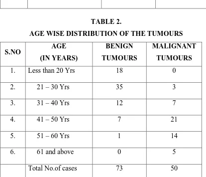
TABLE 2.
AGE WISE DISTRIBUTION OF THE TUMOURS
S.NO AGE
(IN YEARS)
BENIGN TUMOURS
MALIGNANT TUMOURS
1. Less than 20 Yrs 18 0
2. 21 – 30 Yrs 35 3
3. 31 – 40 Yrs 12 7
4. 41 – 50 Yrs 7 21
5. 51 – 60 Yrs 1 14
6. 61 and above 0 5
Total No.of cases 73 50
Table 2 shows the distribution of breast tumours according to age.
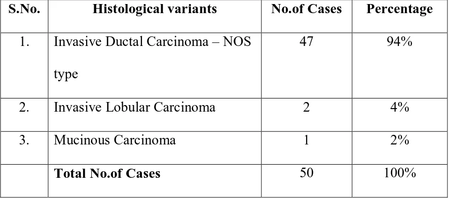
TABLE 3.
DISTRIBUTION OF HISTOLOGICAL VARIANTS IN BREAST CARCINOMA
S.No. Histological variants No.of Cases Percentage
1. Invasive Ductal Carcinoma – NOS type
47 94%
2. Invasive Lobular Carcinoma 2 4%
3. Mucinous Carcinoma 1 2%
Total No.of Cases 50 100%
Table 3 shows the distribution of histological variants in breast
carcinoma. Among the 50 cases, 47 cases (94%) were Invasive Ductal Carcinoma Nos type, 2(4%) were Invasive Lobular carcinoma and 1 (2%) was Mucinous carcinoma.
94% 4%2%
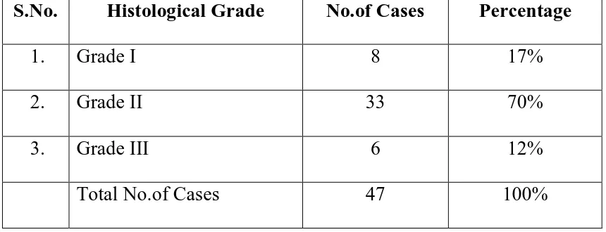
[image:63.595.99.516.512.733.2]TABLE 4.
DISTRIBUTION OF HISTOLOGICAL GRADE IN INVASIVE DUCTAL CARCINOMA - NOS TYPE.
S.No. Histological Grade No.of Cases Percentage
1. Grade I 8 17%
2. Grade II 33 70%
3. Grade III 6 12%
Total No.of Cases 47 100%
Table 4 shows the distribution of histological grading in breast
carcinoma according to Modified Bloom Richardson scoring system. Only 47 cases were included for grading, in that 8 (17%) cases were in grade I , 33 cases (70%) were in Grade II, 6 cases (13%) were in Grade III.
17%
70% 13%
Chart 2. Distribution of Histological Grade
in IDC - NOS Type
[image:64.595.97.514.400.721.2]TABLE 5.
CORRELATION OF ESTROGEN RECEPTOR WITH PROGESTERONE RECEPTOR
S.No Group No.of Cases Percentage
1. ER+/PR+ 22 44% 2. ER-/PR+ 9 18% 3. ER+/PR- 4 8% 4. ER-/PR- 15 30%
Total No.of Cases 50 100%
Table 5. shows correlation of Estrogen receptors and Progesterone
receptors. Among the 50 cases 22 cases were positive for both Estrogen and Progesterone receptors, 9 cases positive for progesterone receptor, 4 cases positive for estrogen receptor, while 15 cases were negative for both estrogen and progesterone receptors.
44%
18% 8%
30%
Chart 3. Correlation of Estrogen
Receptor With Progesterone Receptor
[image:65.595.126.495.497.732.2]ER-/PR-TABLE 6.
EXPRESSION OF HER-2/neu IN BREAST CARCINOMA
S.No HER-2/neu expression Total No. of
cases Percentage 1. Positive 18
50
[image:66.595.94.524.102.245.2]36% 2. Negative 32 64%
Table 6. shows HER-2/neu overexpression in 50 cases of breast
carcinoma, among them 18 cases (36%) were found to be positive, while 32 cases (64%) were found to be negative.
`
POSITIVE 36%
NEGATIVE 64%
[image:66.595.103.534.389.717.2]TABLE 7.
CORRELATION OF TUMOUR SIZE WITH HORMONE RECEPTORS
S.No
Tumour Size Total ER/PR
Positive Percentage
1. T1 < 2 cm 7 6 85%
2. T2 2 – 5 cm 17 16 94%
3. T3 > 5 cm 26 13 50%
[image:67.595.96.524.175.356.2]Total No.of Cases 50 35
Table 7. shows correlation of hormone receptors with tumour size.
Chart 5.
CORRELATION OF TUMOUR SIZE WITH HORMONE RECEPTORS
0% 10% 20% 30% 40% 50% 60% 70% 80% 90% 100%
T1 < 2 cm T2 2 – 5 cm T3 > 5 cm
TABLE 8.
CORRELATION OF HER-2/neu WITH TUMOUR SIZE
S.No. Tumour size HER-2/neu
positive
Total cases Percentage
1. <2 cm 1 7 14% 2. 2 – 5 cm 5 17 23% 3. > 5 cm 14 26 53%
Table 8. shows correlation of HER-2/neu with tumour size. HER-2/neu
overexpression was noted in 14% of T1 sized tumour, 23% in T2 sized tumour and 53% in T3 sized tumour. HER-2/neu overexpression was found in increasing size of the tumour.
Chart 6.
CORRELATION OF HER-2/NEU EXPRESSION WITH TUMOUR SIZE
0 5 10 15 20 25 30
<2 cm 2 - 5 cm > 5 cm
TABLE 9.
CORRELATION OF RECEPTOR STATUS WITH HISTOLOGICAL GRADING
S.No Histological
grade
Total No. of cases
ER/PR Positive
Percentage
1. Grade I 8 8 100%
2. Grade II 33 23 69%
Grade III 6 2 33%
Table 9. shows correlation of hormone receptor status with tumour grade.
CHART 7.
CORRELATION OF RECEPTOR STATUS WITH
HISTOLOGICAL GRADE
0 5 10 15 20 25 30 35
Grade I Grade II Grade III
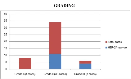
TABLE 10.
CORRELATION OF HER-2/neu WITH HISTOLOGICAL GRADING
S.No Histological grade HER-2/neu
positive
Percentage
1. Grade I (8 cases) 0 0
2. Grade II (33 cases) 11 33%
3. Grade III (6 cases) 4 66%
Table 10. shows correlation of HER-2/neu with histological grade. It was
found that HER-2/neu overexpression was not seen in grade I tumours, where as it was expressed in 33% of grade II tumours and 66% of grade III tumours. Stastistically found to be significant (p=0.001).
CHART 8.
CORRELATION OF HER-2/neu WITH HISTOLOGICAL GRADING 0 5 10 15 20 25 30 35 40
Grade I (8 cases) Grade II (33 cases) Grade III (6 cases)
TABLE 11.
CORRELATION OF RECEPTOR STATUS WITH NODAL STATUS
S.No Nodal metastasis ER/PR positive Percentage
1. Present (17 cases) 9 52%
2. Negative (29 cases) 21 72%
Table 11. shows correlation of hormone receptors with nodal status. Out
of 46 patients 17 had nodal metastasis, among whom 9 showed receptor positivity. Out of 29 nodal negative patients, receptor status was positive in 21 patients. This explains higher receptor expression in nodal negative patients. Statistically found to be significant (p=0.001).
CHART 9.
CORRELATION OF RECEPTOR STATUS WITH NODAL
STATUS
0 5 10 15 20 25 30
PRESENT NEGATIVE
[image:73.595.102.538.468.723.2]TABLE 12.
CORRELATION OF HER-2/neu WITH NODAL STATUS
S.No Nodal metastases HER-2/neu positive Percentage
1. Present (17 cases) 11 64%
2. Negative (29 cases) 2 6%
Table 12. shows correlation of HER-2/neu with nodal status. Out of 46
cases, HER-2/neu overexpression was seen in 64% of nodal positive patients as opposed to 6% of nodal negative patients. Statistically found to be significant (p=0.001).
CHART 10.
CORRELATION OF HER-2/neu WITH NODAL STATUS
0 5 10 15 20 25 30 35
PRESENT NEGATIVE
[image:74.595.100.535.404.678.2]TABLE 13.
CORRELATION OF RECEPTORS WITH ONCOPROTEIN EXPRESSION
ER/PR Status HER-2/neu
Positive Negative
Total No. of cases
Positive 9 26 35
Negative 9 6 15
Total No. of cases 18 32 50
Table 13. shows an inverse relationship of estrogen, Progesterone
receptor status with the HER-2/neu status.
Statistical analysis was performed and found to significant (p=0.001).
CHART 11.
[image:75.595.101.537.442.748.2]DISCUSSION
INCIDENCE AND AGE OF OCCURRENCE:
In Indian scenario, breast carcinoma accounts for 33% of all female cancers and 20% of cancer related deaths in women. In Chennai breast cancer accounts for 26.8% of all cancers in women. A few decades back breast carcinoma is more common in women above 50 years comprising about 65% to 70% with 30% to 35% of women were below 50 years of age. But at present the scenario has changed with increasing incidence between 41- 50 years of age comprising of about 49%. Breast cancer scenario in India also shows a significant trend of increased incidence of breast cancer in much younger age than earlier.
TABLE 14.
COMPARATIVE ANALYSIS OF DISTRIBUTION OF HISTOLOGICAL VARIANTS OF BREAST CARCINOMA
Histological
Types Dixon JM et al. Omar Hameed Current study
IDC – NOS type 79% >70% >70%
Lobular carcinoma
10% 5 – 15% 4%
Mucinous carcinoma
2% 1 – 5% 2%
Medullary carcinoma
2% 1 – 7% 0
Papillary carcinoma
1% 2% 0
Solid
neuroendocrine
<1% Rare 0
Metaplastic ca 0 2-5% 0
Table14. shows the comparative analysis of the distribution of
THE IDENTIFIABLE HISTOLOGICAL VARIANTS ARE AS
FOLLOWS
1. INVASIVE DUCTAL CARCINOMA –NOS TYPE:
This is the largest group of malignant tumours accounting to 65-80% of breast carcinoma . Our study shows 94% distribution. Grossly the tumour size varied from 2cm to 12 cm. Cut section of most of the tumour showed gray white, ill-defined tumour mass, firm to hard in consistency. Occasionally tumour showed areas of hemorrhage and necrosis. Microscopically the neoplastic cells were arranged in diffuse sheets,nests and cords along with glandular and tubular differentiation. In few cases comedo - pattern of necrosis was seen. About 40 percent of the cases (17/43) showed nodal metastasis. Most of the tumours were found to be in histological grade II. Resected margin involvement was seen in two patients and was confirmed by microscopic examination.
2. INVASIVE LOBULAR CARCINOMA:
small to medium sized with uniform, hyperchromatic nuclei, and mild nuclear pleomorphism. Microscopically, metastasis was found in 23 lymph nodes.
3. MUCINOUS CARCINOMA:
In Omar Hameed et al study incidence of Mucinous carcinomas was 5-15% and the mean age of occurrence was 58-68 years. The present study data represents 2% of distribution and the age of occurrence was 73years. This is parallel with above mentioned study. Grossly the tumour mass was about 6cm -10cm, well circumscribed with gelatinous appearance. On cut section soft, gel like material was noted. Microscopically clusters of bland appearing epithelial cells with abundant extracellular mucin were noted .
HORMONE RECEPTOR STATUS AND HER-2/NEU IN BREAST
CARCINOMAS
breast carcinoma. In his study Estrogen, Progesterone receptors were positive in 70-80% of the tumours and HER-2/ neu expression was positive in 15-20% of the breast carcinoma specimen.
In the year 1993, Wilbur D et al. did a study about hormone receptor status in 30 patients by immunohistochemical method on paraffin wax embedded blocks. He described Estrogen receptors positivity in 73% (22/30) of patients, Progesterone receptors positivity in 63% (19/30) of patients, and HER-2/neu overexpression in 37% (11/30) of patients.
In the year 2003 Lici et al. have reported about the incidence of invasive carcinoma by hormone receptor status from the year 1992 to 1998, in a population based study. He found that there is a increase in prevalence over the years with increase in hormone receptor positivity from 75.4% to 77.5% in United States.
32.6% were Estrogen receptor positive and Progesterone receptor positive were 46.1%. He reported a high incidence of hormone receptors non-reactivity in breast carcinoma patients in India.
In the year 2008 , Col.V. Dutta et al. did a study in Armed Forces Medical College, Pune. He analyzed the hormone receptors and HER-2/neu overexpression in breast carcinoma. In total of 75 tumours which was studied, 33% (25/75) of cases expressed Estrogen receptor positivity, Progesterone receptor or both where found to be negative in as 67% (50/75). HER-2/neu overexpression was seen in 58 %( 43/75) of cases. This study revealed that receptor negativity is higher in this population of tumours when compared with the western communities.
In the year 2009, Lakmini K.B Mudduwa studied the hormone receptor status of breast carcinoma by using the Quick score method. She reviewed a total of 151 cases and documented the prevalence of Estrogen receptor positivity in 45.7% of the cases, Progesterone receptor positivity in 48.3% of the cases and both receptors negativity in 54.3% of the total cases. According to her study HER-2/neu overexpression was seen in 19.1% (26/136) of the total cases.
cancer referral institute in India. A total of 11,780 cases were reviewed for this period. The percentage of hormone receptor positive expression varied from 52% to 57%.
In the year 2007, Vikash Kumar et al. did a study on HER-2/neu overexpression which has a much higher incidence among Indian breast cancer patients which is 46.3% in comparison to 25-30% in Western world.
In the present study Estrogen, Progesterone receptor or both were positive in 70% of cases and both receptors were negative in 30% of cases (Tab.5).
HER-2/neu overexpression was positive in 36% of cases (Tab.6). Hence this study is comparable with the studies conducted in the Asian communities. There appears to be a minimal variation in receptor expression;because technically this could be explained by the differences in the technique of evaluation and inter laboratory variations.
ESTROGEN, PROGESTERONE RECEPTOR AND HER-2/NEU
STATUS IN HISTOLOGICAL VARIANTS
whereas Medullary carcinoma and Metaplastic carcinoma were both found to be hormone receptor negative. He has also found that a small group of Invasive ductal carcinoma-NOS type and Lobular carcinomas expressed receptor negativity.
In the year 2000, Desai et al. reported that Invasive lobular carcinoma, Mucinous carcinoma and mixed tumours were Estrogen and Progesterone receptor positive, where as high grade infiltrating ductal carcinomas, in situ comedo-ductal carcinomas ,Medullary carcinomas were found to be receptor negative.
The author Diab SG et al did a study on the tumour characteristics and clinical outcome of Mucinous and Tubular breast carcinomas and found Estrogen receptor positivity in 92% of cases and reported Progesterone receptor positivity in 68% of cases. HER-2/neu overexpression is found to be less than 5% of tumours. Similar results were also reported in studies conducted by Shousha S, et al. in the year 1989.
In the year 1995, Rosen et al. did a study on HER-2/neu expression in nodal negative patients. He has also reported low incidence of oncogene expression in Papillary carcinomas.
In the year 1991, Soomro S et al. did a study about oncogene expression in different histological variants of invasive breast carcinomas and reported low expression in Neuroendocrine carcinomas.
Immunohistochemical studies done by LeeAK revealed Neuroendocrine marker positivity in 82% of the cases. However 67% expressed Estrogen receptor positivity and 56% showed Progesterone receptor positivity. However ,HER-2/neu over expression was found to be negative.
The present study also shows Estrogen and Progesterone receptor positivity and HER-2/neu expression negativity. This is in correlation with the above mentioned studies.
In the year 1987, Oberman HA et al. did a study which showed that Metaplastic carcinomas were hormone receptor and oncoprotein negative called as Triple negative.
Medullary carcinoma has a higher nuclear grade with receptor negativity.
In the year 1995, a study conducted by Rosen PP et al. and Soomro S.et al. in the year 1991 showed a low incidence of HER-2/neu overexpression in Metaplastic and Medullary carcinoma.
In the year 1992,Kuenen–Boumeester V et al did a study on the immunohistochemistry of Androgen receptors in relation to the Estrogen and Progesterone receptors. He also reported that a small group of lobular carcinomas were found to express both receptors negativity and Androgen receptor positivity.
In the year 1995, a study conducted by Rosen PP et al. showed Estrogen receptor positivity in 87.55% of the cases and Progesterone receptor positivity in 75% of the cases in Invasive lobular carcinomas. A small group of these tumours also expressed both receptor negativity and Androgen receptor positivity.
In the year 2005, Riva et al studied about the
immunhistochemical analysisof Androgen receptor in carcinoma breast
Lobular carcinomas along with Estrogen and Progesterone receptors negativity.
In correlation with the above mentioned studies the Invasive lobular carcinoma in the current study also expressed both receptors and HER-2/neu negativity. Clinicopathologically this can be explained by the aggressive nature of the tumour with higher incidence of nodal metastases.
PR POSITIVE
CORRELATION OF AGE AND RECEPTORS EXPRESSION
TABLE 15.
CORRELATION OF AGE AND RECEPTORS EXPRESSION.
Age group( years ) Total No. of cases ER/PR positive
31 – 40 7 3
41 -50 21 14
51 – 60 14 11
60 and > 5 4
In the year 2008, Col.V. Dutta et al. studied about the Estrogen, Progesterone receptor expression with the age. Out of the total 75 cases, 35% of cases were in 51-60 years age group. The results showed that the receptor positivity increases with advancing age. Young patients tend to have a higher level of circulating estrogen and hence correspondingly low expression of receptors.
The present study shows receptor status positivity of 78% in patients older than 50years of age group. The Increased immunoreactivity with advancing age is parallel to above the mentioned studies.
ESTROGEN, PROGESTERONE RECEPTOR, HER-2/NEU WITH
OTHER VARIABLES
In the year 1995, Rosen PP et al. in his study correlated Estrogen, Progesterone receptor positivity with histological grade and tumour size. He concluded that Estrogen and Progesterone receptors are expressed more in the low grade and tumours which are of lesser diameter. He also reported that HER-2/ neu overexpression was increased among the nodal positive patients and tumours which are more than 2cm in diameter.
J.Buon et al. reported that HER-2/neu receptor overexpression is increased in higher grade tumours. Hormone receptor positivity was seen 100% in grade I tumours, 76.30% in grade II tumours and 41.18% in grade III tumours. Their receptor positivity tends to have an inverse relationship with the tumour grade.
receptor and oncoprotein expression does not necessarily correlate with advanced grade tumours in our population.
In contrast with S.Goyle et al. in the year 2008 the present study showed the Estrogen, Progesterone receptor positivity of 100% in grade I, 69% in grade II, 33% in grade III tumours. HER-2 /neu overexpression showed 66% in grade III, 33% in grade II, none in grade I tumours. This explains the overexpression of oncoprotein with higher histological grade tumours. Thus, it reflected a direct relationship with higher nuclear grade, which was comparable with J.Buon et al & Rosen PP et.
Current study shows correlation of hormone receptors and HER-2/neu with tumour size.The tumours are categorized into three according to T in TNM staging. T1- < 2cm, T2=2-5cm, T3 = > 5 cm. Receptor positivity is expressed in 85% of T1, 94% of T2 and 50% of T3 tumours. This explains that receptor positivity has an inverse relationship with the tumour size.
CORRELATION WITH NODAL STATUS
In the year 2008, Col.V. Dutta et al. expressed a strong correlation between HER-2/neu and nodal metastases. He reported that 70% of nodal positive tumors overexpressed HER-2/neu oncoprotein.
H.J. Huang et al. did a study in 1362 women with primary breast tumour. He found that Estrogen receptor positivity was expressed less in nodal positive tumours.
In the present study out of 46 patients 17 cases showed metastasis while 29 cases has no metastases. Receptor positivity was found to be higher among the nodal metastasis negative patients which was about 72% (21/29).HER-2/neu overexpression was seen in 64% of nodal positive cases than the nodal negative patients which was found to be 6 %.
CORRELATION OF ESTROGEN, PROGESTERONE
RECEPTOR WITH HER-2/NEU
The present study showed an inverse relationship between these hormone receptors and oncoprotein expression.Hence it is comparable with the above mentioned studies.
In the year 2006, Francis G et al. did a study of 591 tumours and concluded that more than 20% of HER-2/neu positive tumours showed moderate or strong staining for Estrogen receptors.
In the year 2009, Bhargava R et al. has reviewed about 205 cases and concluded that 15% (32/205) of the cases were triple negative, 4% (8/205) of cases were positive for Estrogen receptor and HER-2/neu hybrid oncoprotein expression.
SUMMARY AND CONCLUSION
The benign lesions had a peak occurrence in the age group 21 to 30 years, whereas malignant tumours had a peak in the age group 41 to 50 years.
Among the various histological variants in breast carcinoma, Invasive Ductal carcinoma – NOS type constituted about 94% of cases.
Estrogen, Progesterone receptor positivity and HER-2/neu negativity in Mucinous carcinomas.
Triple negativity in small group of Invasive lobular carcinoma.
Regarding the histological grade of breast carcinoma, Grade II tumours were common accounting for 70 %.
Estrogen and Progesterone receptor or both was found in 70% while 30% were found to be receptor negative.
Out of the total 50 cases, 26 cases were T3 tumours of more than 5cms in diameter.
Larger the tumour size lesser is the expression of hormone receptor, whereas smaller sized tumours expressed more receptor positivity. This inverse correlation was statistically significant (P=0.003). HER-2/neu overexpression was found in tumours of more than 2 cm in size.
Among the 46 cases, nodal metastasis were found in 17 cases and negative in 29 cases.
Among the 29 nodal negative patients Estrogen, Progesterone receptor were positive in 21 cases. Thus, there is higher receptor expression in nodal negative patients. This was found to have a significant correlation (P=0.001).
HER-2/neu overexpression was observed in 64% of nodal positive patients, which is statistically significant (P=0.001).
Higher the histological grade of breast carcinoma, greater the HER-2/neu overexpression, which was found to have a significant correlation (P=0.001).
CONCLUSION
1. Dowsett M, Hanna W M, Kockx M et al. Standarisation of HER-2/neu testing: Result of an international proficiency –testing study. Mod. Pathol 2007: 20: 584-591.
2. Gown A M: Current issues in Estrogen receptor and HER-2/neu testing by immunohistochemical method in breast cancer. Mod. Pathol 2008; 21: S8-S15.
3. Schwartz’s Principle of Surgery; F Charles Brunicardi 8th ed. 2005.Mc Graw –Hill Companies .Inc.The Breast. P 453-500.
4. Park’s Textbook of Preventive and Social Medicine19th ed, 2007.P 378-380.
5. Bancroft JD, Marilyn Gamble (Ed) Theory and practice of histological techniques. Churchill Livingstone 2002. P
6. Sondik.E.J.Breast cancer trends, Incidence, Mortality and Survival. Cancer: 1994; 74; 995-999.
2001-2002.
9. Moore DH, Breast carcinoma Etiological Factors, Adv. Cancer Res; 1983:40;189-253.
10. Pathak DR et al. Breast carcinoma Etiological Factors, Reproductive and Hormonal Factors.Cancer 2000; 88: 1230-1238.
11. Priti Lal MD, Lee K. Tan MD, et al .Correlation of HER-2/neu status with Estrogen, Progestrone receptor and histological features in 3,655 Invasive breast carcinoma. Am J Clin. Pathol 2005.
12. Susan c. Lester, The Breast . Robbins and Cotran Pathological basis of Disease , 7th Ed(2004) P.1120-1153.
13. M willams Audeg, The new era of cancer risk assessment in Surgical Oncology P.261-265.
14. Gavin Harris, Sarah E Pinder , et al Invasive carcinoma special types. In Foundation in diagnostic pathology (2006). P. 201-220.
2005; Elsevier. Breast P. 1763-1876.
17. WHO Classification of tumors. Pathology and Genetics of Breast and Female genital organs.Lyon: IARC press 2003; 13-59.
18. Rosen PP, Menendez Botet C J, et al Pathological review of Breast lesions analyzed for Estrogen receptor protein. Cancer Res 1975; 35; 3187-3194.
19. Fisher ER ,Andersons, Dean , et al .Solving the dilemma of IHC and other methods. Cancer: 103:164-173.
20. Foote FW Jr, Stewart FW et al. A Histological Classification Of Carcinoma of Breast. Surger 1946;19: 74-79.
21. Tavassoli FA, Pathology of Breast. Infiltrating and Familiar sub-types. Norwalk, Appleton and Lange.1992; 293-294.
22. Dixon JM et al Long term Survivors after Breast cancer. Br. J. Surg. 1985; 72:445.
cancer Virchow Archive: 1995; 426; 107-115.
25. Suo Z, Risberg B, et al The expression of EGFR family Ligands in breast cancer I Int J .Surg pathol 2002 ; 10: 91-99.
26. Diab SG, Clark GM, Osborne C K, et al . Tumor characteristics and clinical out come of Tubular and Mucinous breast carcinoma. J Clin .Oncol 1999; 17:1442-1448.
27. Jensen ML, Kiaer A, Melsan F, et al . Medullary Carcinoma Vs poorly differentiated carcinoma on IHC study with keratin 19, and ER staining Histopathology 1996;29:241-245.
28. Ponsky JL, Gliga L, Reynolds S, et al. Medullary carcinoma of breast as association with negative hormone receptors J Surg Oncol 1984;25:76-78.
29. Horsfall DJ. Tilley WD, Orell SR, Marshall VR, Cant EL et al.Relationship between ploidy and Steroid Hormone Receptor in Primary Invasive Breast Cancer.
in carcinoma of breast and the relationship to prognosis an IHC study. Am J Clin Pathol 1985; 84:687-692.
32. Niremudi A,Pinder SE, Lee AHS et al. Neuroendocrine differentiation and prognosis in Breast carcinoma . Histopathology 2000 ;40: 215-222.
33. Papootti M, Gugliottu P, Eusebi V et al. IHC analysis of benign and malignant papillary lesions of breast. . Am J Surg. Pathol 1983;7 :451-463.
34. ZekiogluO,Erhan Y,Ciris M et al. Invasive micropapillary carcinoma of breast. IHC profile compared with Invasive Ductal Carcinoma. Histopathology 2004; 44:18-23.
35. Kahfmann MW ,Marti JR, Gallenger HS et al . Carcinoma of Breast with Pseudosarcomatous Metaplasia. Cancer 1984; 53:1908-1917.
Cancer 1989;64: 1490-1499.
38. Rosen PP,Lesser ML, Arroya CD et al. IHC detection of HER-2 in patients with axillary lymphnode negative breast cancer. A study of epidemiological risk factors and prognosis.Cancer 1995;75:01320-1326.
39. Brader B, Graf D, Padovan R etal ER, PR as prognostic marker Tumori 1988; 74 ; 45-52.
40. Mary US born, Wenaejusz, Domagula (Eds) in Comprehensive cytopathology by Bibbo 2nd ed 1997.
41. Jay H.B Histochemistry in Ivan D James L, (Eds) Anderson pathology, Mosby St louis , 1996.
42. Pluzek, K.Y Sweeney , E,Miller, K.D Isaacson .P. A major advance for IHC E pos J. Pathol 169 ( Suppl) abstract 220
immunofluorosent staining in paraffin sections improved by trypsin digestion Laboratory Investigation 35: 383-391
45 Miller K,Auld . J, Jessup. E, Rhodes A et al. Antigen unmarking by pressure cooker method . A comparison with microwave oven heating and traditional methodsAdvances of anatomical pathology, 2 : 60-64
46. Langols, N.E.T , King G., Herricot R et al .Non-enzymatic retrieval of antigen staining of PGP 9.5 J.Pathol 173 ; 249-253
47. Jensen EV, Jacobson HI, Basic guides to mechanism of action of hormone receptors. Recent Prog Horm Res 1962; 18 ; 387-414.
48. Suzanne A.W Fuqua Rachel Schiff Disease of breast ed : The biology of ERs – 585 -590.
49. Shoker et al ER expression in normal and precancerous breast J Pathol 1999; 188: 237-244