JOURNAL OF VIROLOGY, Sept. 1988, p. 3388-3398 0022-538X/88/093388-11$02.00/0
Copyright © 1988,American Society for Microbiology
Binding of
Cellular Proteins
to
the
Regulatory Region of
BK
Virus DNA
RHEA-BETH MARKOWITZ* AND WILLIAMS. DYNAN
Departmentof Chemistry and Biochemistry, CampusBox215, University of Colorado, Boulder, Colorado 80309-0215 Received 15 March 1988/Accepted 7 June 1988
The humanpapovavirusBKhasanoncoding regulatory region locatedbetween thedivergentlytranscribed early and late coding regions. Many strains of BK virus (BKV) have direct DNA sequence repeats in the regulatory region, althoughthenumberandextentofthese repeatsvaries widely between independent isolates. Untilrecently, littlewasknownabout the individual functional elements within the BKVregulatory region,and the biological significance of the variable repeat structure has been unclear. To characterize the interaction betweensequencesin the BKVregulatory region and hostcelltranscription factors,wehavecarriedoutDNase Ifootprinting and competitive binding experimentsonthreestrainsof BKV, includingonestrain that doesnot contain directsequencerepeats.We have usedrelatively crude fractionsfrom HeLa cell nuclear extracts,as
well asDNA affinity-purified preparations of proteins. Ourresults demonstrate thatBK(Dunlop), BK(WW),
andBK(MM) each contain multiple binding sites forafactor, NF-BK,that isamember of the nuclear factor
1family of transcription factors. We predict thepresenceofthreetoeightbindingsitesforNF-BK in the other strains of BKV for whichaDNAsequenceisavailable. Thissuggeststhatthebindingof thisproteinislikely toberequired for biological activity of the virus. In addition toNF-BK sites, BK(WW) and BK(MM) each contain a single binding site for transcription factor Spl, and BK(Dunlop) contains two binding sites for transcription factor AP-1. The AP-1 sites in BK(Dunlop) span the junction of adjacent direct repeats, suggestingthat repeatformationmaybeanimportant mechanism for denovoformation of binding sitesnot presentina parentalstrain.
BK virus (BKV) isahumanpapovavirus first isolated in 1971 from the urine of a renal allograft patient (19). A number of different strains of BKV have subsequently been isolated, in most cases from the urine of renal allograft recipients or patients suffering from Wiskott-Aldrich syn-drome, an X-linked recessive disorder characterized by defects incellular and humoral immunity (4). Comparative DNA sequenceanalysis shows that the BKV isolates have
extensivehomologiesin theirprotein-coding region,bothto each other and to simian virus 40 (SV40) (25, 27, 56, 71). However, these studies also indicate that the noncoding regulatory regions of BKV and SV40 are quite different in
sequence.
In both BKVand SV40,thisnoncoding regulatory region islocatedbetween the early and late transcription units,near
the origin ofviral DNAreplication. In SV40, this region is about 350 base pairs (bp)in size andcontains mostorall of
the cis-acting elements required for initiation of viral RNA synthesis. There are several repeated sequence motifs, in-cluding the 21-bprepeats and the72-bp repeats. The latter have been showntopossessenhancerfunction, that is, they
increase the levelof transcription from the early promoter regardless of orientation and position (3, 18, 34, 44).
In BKV, the noncoding regulatory region varies in size between isolates. Repeated sequences are present in this region ofmost viral strains. For example, the widely used BK(Dunlop) strain contains a triplication ofa 68-bp block
withan 18-bp deletion within the middle repeat (56). Using the chloramphenicol acetyltransferase assay, Rosenthal et al.(54) demonstrated that theseBK(Dunlop)repeatspossess transcriptional enhancer function. Studies using deletion mutants constructed in vitrodemonstrate that one copyof
the BK(Dunlop) 68-bp block is not sufficient for early
*Correspondingauthor.
transcription, but that activity is restored when certain
sequenceswithin the68-bpblockarerepeated (64). Arecent comprehensivedeletionanalysisofBK(prototype),which is similar to BK(Dunlop), showed a progressive stepwise
de-cline inearlygeneexpression,assequenceswithin the68-bp blocksweredeleted (11).
The repeat structure varieswidely between BKV strains and in some cases is quite complex. Recently, however, a newstrain, BK(WW),was obtainedbyisolation and
molec-ular cloning ofviral DNA directly from patienturine (55). This differs from theprocedures used toisolate most other BKV strains, which generally involved passageof the virus in cell culture prior to cloning. BK(WW) is distinguished from most other BKV strains by the absence of repeated DNA sequences. The workers who isolated this strain have pointedoutthattheregulatory regions ofmanyother BKV strainscanbederived fromBK(WW) by postulatingasmall
number of duplication and deletion events. Such events mighthaveoccurred either innatureorduringpassageof the virusin culture. Specific examples of how this mighthave occurred with BK(prototype), BK(Dunlop), and BK(MM) will bepresented inthe Results section.
The transcriptional enhanceractivity associated with the BKVregulatory regionis almostcertainly dependentonthe bindingofspecifichost celltranscription factors,ashasbeen shown with other papovaviruses. Aprior report described tworecognitionsites in theregulatory regionofBK(Dunlop) for theTGGCA-binding protein,nowthoughttobe function-ally the same as nuclear factor 1 (NF1) (39, 46). Little is known about other proteins that may bind to this region, however, and comparative studies of different strains have notbeencarried out. There has been considerable specula-tion in the literatureabout the DNAsequencessimilartothe adenovirus Ela and SV40 enhancer core motifs which are
presentinBKV(23, 67),but it isnotknown if theseactually 3388
Vol. 62,No. 9
on November 10, 2019 by guest
http://jvi.asm.org/
correspond to protein-binding sites or constitute functional units of the enhancer.
We have screened protein fractions derived from HeLa cell nuclear extracts for the presence of proteins that recog-nize specific DNA sequences in BKV. This approach has been used in SV40 to identify many different proteins that bind the enhancer region (68). Initially, we carried out DNase I footprinting experiments with relatively crude heparin-agarose step-gradient fractions. Subsequently, the identity of specific proteins was confirmed by footprinting with DNA affinity-purified protein preparations and by com-petitive binding experiments.
A surprisingly simple picture emerged from these studies. The enhancer region of each of the three strains BK(Dun-lop), BK(WW), and BK(MM) contained three to six binding sites for a protein we refer to as NF-BK, which is amember of theNF1 family of transcription factors. This was the only protein we detected that recognized sites in all three strains, suggesting that it is essential for biological activity of the enhancer region. An additional recognition site for the transcription factor Spl was detected in two strains, and two sites for the transcription factor AP-1 were detected in a third strain.
MATERIALS AND METHODS
Preparation of radiolabeled DNA probes. (i) BK(Dunlop) strain. The 216-bp HaeIII DNA fragment containing the BK(Dunlop) regulatory region was inserted via BamHI linkers into the BamHI site of pUC19 to create the plasmid pDun/Bam. To prepare the probe for DNase I footprinting, the EcoRI-HindIII fragment of pDun/Bam containing the BK(Dunlop) sequences was excised, treated with calf intes-tinal phosphatase (Boehringer Mannheim Biochemicals), and purified by preparative gel electrophoresis. This frag-ment was then radiolabeled with T4 polynucleotide kinase and
[y-32P]ATP
(crude grade, 7,000Ci/mmol; ICN Pharma-ceuticals Inc.). To prepare a late-strand probe that was singly end labeled at theHindIIIsite, theDNA wasdigested with KpnI to remove a smallfragment of labeled DNA from the opposite end. Similarly, toprepare anearly-strand probe labeled at theEcoRI site, the DNA was digested with PstI. After the final restriction endonuclease digestion, the DNA was extracted with PCIA (phenol-chloroform-isoamyl alco-hol, 25:24:1 [vol/vol]), extracted with CIA (chloroform-isoamyl alcohol, 24:1[vol/vol]),
andprecipitated from etha-nol.(ii) BK(WW) strain. Theplasmid pHS Bam (55) contained a 426-bp HhaI-SstI fragment encompassing the BK(WW) regulatory region. This fragment, which as the result of the cloning procedure was flanked by BamHI sites, wasexcised withBamHI and inserted into pUC19 at the BamHI site to create pWW/Bam-9 and pWW/Bam-1. These plasmids con-tain the WW regulatory sequences in theorientations anal-ogous to pDun/Bam and pMM/Bam, respectively. Early-strand probes were generated from theEcoRI-end-labeled, PstI-digested fragment from pWW/Bam-9 or the HindIll-end-labeled, KpnI-digested fragment frompWW/Bam-1.
(iii) BK(MM) strain. The291-bpHaeIII fragment contain-ing the BK(MM) regulatory region was inserted viaBamHI linkers into the BamHI site ofpUC19 to create the plasmid pMM/Bam. Radiolabeled DNA probes were prepared as described above. The BK(MM) fragment was inserted in an orientation opposite to that of the BK(Dunlop) fragment. Therefore, a late-strand probe that was singly end labeled at the EcoRI site was prepared by PstI digestion, and an
early-strandprobethatwas
singly
endlabeledattheHindIII
sitewas preparedby KpnIdigestion.
DNase Ifootprinting. DNase I
footprinting
wasperformed
essentially as described (16, 48), except that allbinding
reactions contained 40
p.g
ofpoly(dI-dC)
alternating
co-polymer(Pharmacia, Inc.)perml and 40 ,ugofpentadeoxy-nucleotide mixture [pd(N)5]
oligonucleotide
mixture(Phar-macia) per ml. Binding reactions were carried out on ice.
Sampleswerethen warmedto
20°C
and treated with DNase I. Reactions were terminated asdescribed,
extracted with PCIA and CIA, precipitated withethanol,
andelectropho-resed on 8%denaturing
polyacrylamide gels
(41).
Sequence
markerswerepreparedbythe methodofMaxamandGilbert (41). In competitive
binding
experiments,
thecompetitor
DNA wasmixed with theradiolabeled
probe DNA,
poly(dI-dC) copolymer, and
pd(N)5
oligonucleotide
prior
to the addition ofprotein.Preparation of DNA-binding proteins. Nuclei were pre-pared asdescribed
previously
(16)using
HeLacells from12 to24litersofsuspension culture(approximately
5 x10⁹
to1 x1010
cellstotal). The nuclei were suspendedin 2packed-cell volumes ofextraction
buffer,
and aftercentrifugation,
theresulting supernatantwas
applied
directly
to acolumn of heparin-agarosepreparedasdescribedpreviously (10)
(1-ml
bed volume per liter of
original
culture).
The column waseluted with a0.15to0.4 M KCl step
gradient,
andprotein-containing fractions were
pooled.
Protein concentrationsweredetermined bythe methodof Bradford
(5).
DNA affinity columns were
prepared
as describedby
Kadonaga and
Tjian (33).
Twocomplementary
oligonucleo-tidescontaining thedesired consensus sequences were syn-thesizedby the
,-cyanoethyl
phosphoramidite
method with an AppliedBiosystems
model 380B DNAsynthesizer
and werepurified bypreparativeelectrophoresis
ona20%poly-acrylamide gel
containing
8 M urea. The two oligonucleo-tidesweredissolved inasolution of50mMTrischloride[pH
7.6], 10mMMgCl2,5 mM
dithiothreitol,
0.1 mMspermidine,
and 0.1 mM EDTA and were annealed and radiolabeled as
described
previously (33).
Theligation
reaction wascarried out byincubating
440p.g
of annealedoligonucleotide
in a1-mlreaction
containing
66 mMTris chloride(pH
7.5),
5 mMMgCl2,
5 mMdithiothreitol,
and4 mM ATPfor24hat25°C.
The extent ofligation
wasanalyzed
by
polyacrylamide
gel
electrophoresis.
Following
purification
by
extraction with PCIA andCIA,andprecipitation
fromethanol,
theoligonu-cleotide
ligation
products
were dissolved in waterandcou-pled to cyanogen bromide-activated
Sepharose
CL2B300 (Sigma ChemicalCo.)
asdescribed. For NF-BKpurification,
the complementary
oligonucleotides
GATCTGGAATGCAGCCAA and
GATCTTGGCTGCATTCCA
were used. For AP-1purification,
thecomplementary
oligonucleotides
GATCATGGTTGCTGACTAATTGAGA
and GATCTCTCAATTAGTCAGCAACCAT were used. For
Spl
purification,
the complementary
oligonucleotides
GATCGGGGCGGG GC and GATCGCCCCGCCCC were used.Heparin-agarose
0.15to0.4 MKCl-step
gradient
fractionsweremixedwith 200 ,ugof
poly(dI-dC),
dilutedtoa conduc-tivity equivalent to column buffercontaining
0.15 MKCl,
and subjected to
chromatography
on the DNAaffinity
col-umns. The DNAaffinity
column(1-ml
bed volume per 8 liters oforiginalculture)
wasequilibrated
with buffer Z(25
mM HEPES(K+)(N-2-hydroxyethylpiperazine-N'-2-eth-anesulfonic
acid)
[pH7.8],
12.5 mMMgCl2,
1 mMdithio-threitol, 20%[vol/vol]
glycerol,
0.1%[vol/vol]
NonidetP-40)
containing 0.15 M KCl. The extract wascycled
over the columnfourtimesby
gravity
ataflowrateofapproximately
on November 10, 2019 by guest
http://jvi.asm.org/
3390 MARKOWITZ AND DYNAN
BK(WW)
BK(Dunlop)
BK(MM)
OQRI
cEz
GD
cgx;
(
1DHha
I4-
~~~~~~~~~~~~~~~~~~~~AUG
P,earyRNA
ORI Hae
'II
H®e
IIIae
inearl R-AUG
earlyRNA
ORI Hae III
<D (i)
(2D
9:w
Oi3
He IIIearlyRNA Key:
Pblock block ;*i.i;* Rblock
I-100bp
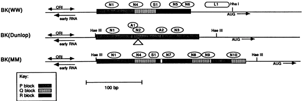
FIG. 1. Sequence relationship ofBK(WW), BK(Dunlop), and BK(MM). Stippledboxes representthe regulatory region of BK(WW), which hasbeen divided arbitrarily into three sequence blocks, P, Q, and R (see Key). Singlelinesrepresentearly-regionDNA totheleft and late-regionDNA to the right of the regulatory region. Bindingsites inBK(WW)forNF-BK
(Ni,
N4,N5,andN6),Spl(Si),
andanunknown factor (L1)areindicated. The AUGcodonfor the agnoprotein,thepresumptiveinitiation sitefor earlyRNA, andthe replication originregion aremarked. Restrictionsites usedinsubcloning proceduresareindicated(seeMaterials andMethods). BK(Dunlop)couldhave arisenfrom BK(WW) byadeletionof boththeQ and Rblocksand 4bp offlankingDNA,triplicationof thePblock,andsubsequentdeletion of 18bp withinthe secondofthe threerepeated P blocks. BindingsitesinBK(Dunlop)forNF-BK(Ni,N2,andN3) andAP-1(Ai andA2)andthe 18-bp deletion within the middle P block in BK(Dunlop) (A) are indicated. BK(MM) could have arisen from BK(WW) by a series of duplication and deletionevents asdescribed in thetext. Binding sites in BK(MM) forNF-BK(Ni,N4, N7, N8, N9, andN10)and Spl(Si) areindicated. The small openboxes withinthe BK(MM)sequenceindicatetheinsertions ofuniqueDNA.5columnvolumes per h. Fractions were eluted with 5 ml of
bufferZ containing 0.15 M KCI at aflow rate of 2 column volumes per h, followed by a 30-ml gradient of 0.15 to 1.75 M
KCIat aflow rate of 3 column volumes per h. Fractions were
collected and assayed for binding activity by DNase I
footprinting. Typically, heparin-agarose fractions werefirst
passed over the AP-1 DNA affinity column. The
AP-1-depletedflowthrough from thiscolumn was passed over the
Spl affinity column, and the AP-1- and Spl-depleted
flow-through from this column was passed over the NF-BK
column. Specific DNA binding activity was detected in
fractions eluting from the respective affinity columns at
approximately 0.25 to 0.4 M KCI. NF-BK and AP-1
prepa-rationswerepurified approximately 100-fold, and Spl
prep-arationswerepurified approximately 30-fold. RESULTS
Sequence relationship of BK(WW), BK(Dunlop),
BK(pro-totype), and BK(MM). Asdiscussed previously, a compari-sonof the BK(WW)regulatoryregionwith thoseof
BK(pro-totype), BK(Dunlop), BK(MM), and other BKV isolates suggeststhatthe latterstrains could have been derived from BK(WW) by a small number ofsequenceduplications and deletions (55, 56, 71). To illustrate how this might have occurred and to facilitate subsequent discussion of the
binding sitesfor cellularfactors,wehavearbitrarily divided the WW sequence into three blocks, labeled P, Q, and R in
Fig. 1. These blocks contain 68, 39, and 63 bp of DNA, respectively. BK(Dunlop) could have arisen from BK(WW)
bydeletion of the Q and R blocks and4bp offlanking DNA,
triplicationof the Pblock, andsubsequent deletion of 18 bp within the second of the three repeated Pblocks. BK(pro-totype) (not shown) could have arisen in a similar way but thedeletion contains only theRblock; the Q block remains. BK(WW) reportedly will not grow in cultured cells (55);
BK(prototype) grows but is unstable (56); BK(Dunlop) growsrelatively well. This suggests that duplication of the P
block favors viral growth in culture and that the Q and R blocksequences are nonessential.
Thegeneration ofBK(MM) from BK(WW) ismore com-plicated butcan be readily visualized. The first stepcould have involved the deletion of the rightmost 55 bp of the R block, together with12 bp of the adjacent uniquesequence. Aunitencompassing 53 bp of the P blockand34bp oftheQ block may then have been duplicated, with 4 bp ofunique sequenceinserted between therepeats. Another duplication encompassing 36bp of the P block and 25 bp of the Q block may then have occurred, with 2 bp inserted between the duplicated segments, togive the final structure asshown.
Binding of cellular proteins to the regulatory region of BK(Dunlop) DNA.Todevelopabetterunderstanding of the function ofthe differentsequence blocks, weconstructed a map of the binding sitesfor various host cell proteins that bindtothisregion using the technique of DNase I footprint-ing. Because BK(Dunlop) is the best-characterized ofthe BKV laboratory strains, we began our studies using this DNA. DNase Ifootprinting probes were prepared for both strands of DNA as described in Materials and Methods. Heparin-agarose fractions of HeLa cell nuclear extracts were used as a source ofDNA-binding proteins. Previous studies haveshown thatfractionspreparedasdescribed(see Materials andMethods)areenrichedfor abroadspectrum of DNA-binding proteins and transcriptionfactors(16,48).
We identifiedfive sites withintheBK(Dunlop) regulatory DNA that were protected by heparin-agarose fractions of HeLacellnuclear extract (Fig. 2A, lanes 2 through4; Fig. 2B,lanes3through 5).Threeof thesesites, Ni, N2, and N3, are located within the triplicated P blocks. Ni and N3 are identical in sequence and show complete protection. N2 differs slightlyin sequence from Ni andN3 because of the previouslymentioneddeletioninternaltothe middleP block and showsonly partial protection. Ni and N3 coincidewith sites identifiedbyNowocketal. (46)asbindingregionsfora
partially purified TGGCA-binding protein. It has been sug-J. VIROL.
on November 10, 2019 by guest
http://jvi.asm.org/
[image:3.612.63.562.74.241.2]B
NF-H-A AP-1 BK
GO O 0 OM
N3 ._
N 2
Ali
N I
FIG. 2. DNase Ifootprint of the regulatory region of BK(Dun-lop). DNA probes were singly end labeled and incubated with
heparin-agarose fractions of HeLa cell nuclear extracts (H-A) or
with DNAaffinity-purified protein preparations (AP-1 and NF-BK)
asindicated. DNase Icleavage wasperformed andproducts were
analyzedonsequencing gelsasdescribedin Materials and Methods.
Protected regions are indicated by brackets and numbered as
describedinthetext.(A) The DNA probewas5'end labeledonthe
early strand. Lanes 1, 5, and 10, No proteinextractadded;lanes2, 3, and 4, heparin-agarose fractions, 20, 30, and 30 ,ug of protein, respectively; lanes 6 and 7, DNA affinity-purified AP-1 fractions, 0.2
and 0.3 ,ug of protein, respectively; lanes 8 and 9, DNA affinity-purified NF-BK fractions, 0.3 and 0.4 ,ug of protein, respectively;
lane 11, marker (M), HpaII digestion products of pBR322 DNA (largest fragment shown is 217 bp). (B) The DNA probewas5'end
labeled on the late strand. Lane 1, Products of the G-specific
sequencingreactionusingthetechniqueof Maxam and Gilbert(41);
lanes2, 3, 7, and12, no protein extractadded; lanes 4, 5, and 6,
heparin-agarose fractions, 20, 30,and 30,ugofprotein, respectively; lanes 8 and9, DNA affinity-purified AP-1 fractions, 0.2 and 0.3 pgof protein, respectively; lanes 10and11, DNA affinity-purifiedNF-BK
fractions, 0.3 and 0.4 ,ugofprotein, respectively; lane 13, marker
(M), HpaII digestion products ofpBR322 DNA (largest fragment
shown is 217bp).
gestedthat this proteinmay be the sameas NF1,a protein
isolatedfrom HeLa cell nuclei which is requiredfor adeno-virus replication (39, 45). All three sites, Ni, N2, and N3, containsequencessimilaroridenticaltotheNF1consensus, TGG(N6-7)T/GCCAA(13),and becauseof thissimilarity,it is likelythat they are protected bythe same protein.
The two other protected sites, Al and A2, span the junctions between the tandemly repeated P blocks in the BK(Dunlop) regulatory region.Thesejunctions contain the
sequence TGACTCA, corresponding to theconsensus rec-ognition sequenceforAP-L,atranscriptionfactor thatbinds to thecontrol regions ofSV40 DNA and the human
metal-lothioneinIIAgene(37, 38).This suggeststhatAP-1 maybe
responsible forthe protectionseen atsites Aland A2. To better determine the identity of the proteins responsi-ble for theprotectionof the five sites inBK(Dunlop) DNA, we isolated these proteins using sequence-specific DNA affinity column chromatography. Columns were prepared
essentially as described by Kadonaga and Tjian (33). The oligonucleotide used to isolate the protein that binds sites
Ni, N2, and N3 contained the sequence
TGGAATGCAGC
CAA, whichoccursin sitesNi and N3 in the BKV P block. Asdescribed above,this containsapartialmatchtotheNF1 consensus recognition sequence (underlined). Recent re-portsfrom several laboratories suggest thatNF1, TGGCA-binding protein, and other proteins are part ofa family of related factors (13, 29, 39, 42, 46). In view of the present confusion ofnomenclature,wewillprovisionallyrefertothe
protein isolated from our column as NF-BK, until it is demonstrated whether ornot it isidentical to otherspecific members of thisfamily. Theoligonucleotide usedto isolate AP-1 contained the sequence ATGGTTGCTGACTAATT
GAGA from the SV40 AP-1-binding region. The AP-1
rec-ognition sequence (underlined) in this oligonucleotide matchessequencesinBK(Dunlop)atsixofsevenpositions,
but the flanking sequences are different from the flanking
sequences in BK(Dunlop). Heparin-agarose fractions of a HeLacell nuclearextract werepassedoverthe NF-BK and AP-1affinity columns,andpurifiedfactorswereelutedwith KCl gradients as described in Materials and Methods. The
DNA-affinity purified preparations are purified 100-fold or morerelativetotheheparin-agarose fractions, asjudged by
theamountofprotein requiredtogiveafootprint (see figure legends).
These
purified protein preparations
wereused in DNase Ifootprinting experiments, using
both strands ofBK(Dunlop)as
probes.
Resultsareshown inFig.
2.Theaffinity-purified
AP-1fractions showedcomplete
protection
of sitesAl and A2 of BK(Dunlop). In other experiments (results notshown), these same fractions gave full
protection
of theknownAP-1sites in SV40.These results confirmthat AP-1
recognizes the Al and A2 sites spanning the junction be-tweenadjacentPblocks ofBK(Dunlop).Theaffinity-purified
NF-BK fractions showed complete
protection
of sitesNi,
N2, and N3 ofBK(Dunlop),
confirming
that all three sites arerecognized by
the same orclosely
related proteins. It is evident from the results withpurified proteinsthat sites Al and N2overlap.
Steric hindrancebetweenproteins
boundat thesesites mayaccountfortherelatively
weakprotection
atsiteN2 insome
experiments using heparin-agarose
fractions(for example, Fig. 2A,
lanes 2through
4).Binding of cellular proteins to the regulatory region of
BK(WW) DNA. We next examined the
binding
of cellularprotein
fractions to BK(WW) DNA. DNase Ifootprinting
probes
wereprepared
for theearly
strand of cloned viral DNA as described in Materials and Methods.Heparin-agarose fractions ofHeLa cell nuclear extracts and DNA
affinity
columnpreparations
of AP-1 and NF-BK wereprepared as
previously
described. Inaddition,
fractionscontaining transcription
factor Splwere preparedby
DNAaffinity chromatography, by using
anoligonucleotide
con-taining
the Spl consensusrecognition
sequence GGGGCGGGGC.
The results of these experiments are shown in
Fig.
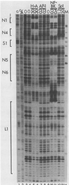
3. A number ofprotectedregions
were seenusing
theheparin-agarosefractions
(lanes
5 and 6). Atthe topof thegel,
siteNi
corresponds
to the same P blockfootprint
seen withBK(Dunlop).
SitesAl, N2, A2,
and N3 are not present inA
NF-H-A AP-1 BK
o Z OM
Nl
33,
1
N4,
'235
:z
N3 *
I
stwU
*s_w isiSi i
on November 10, 2019 by guest
http://jvi.asm.org/
[image:4.612.64.298.76.379.2]3392 MARKOWITZ AND DYNAN
TABLE 1.
Recognition
sequences for
NF-BKinBK(Dunlop),
BK(WW), andBK(MM)aSite Sequence
N .TGGAATGCAGCCAAA
N2.GGGAATGCAGCCAAA
N4.TGGGCAGC C/A GCCAGT
N5.TGGAAACTGGCCAAA
N6.TGGCTGCTTTCCACT
N9.TGAAACCATGCCAAA
L .TGGCCTTGTCCCCAG
Consensus... TGGAAT/A G/C C/T A/T GCCAAA
N5
|GXLi
X
X
~~-4h.
1 2 3 4 5 6 7 8 910 1112 1314
FIG. 3. DNase Ifootprintof theregulatory regionofBK(WW).
The DNA probe was 5' end labeled on the early strand. For
experimental details, see the legend to Fig. 2 and Materials and Methods. Protectedregionsareindicatedbybrackets and numbered
asdescribed in the text. Asterisks indicateapossible Spi
recogni-tion sequence wherenobindingwasdetected. Lane i,Productsof theMaxam-GilbertG-specific sequencing reaction;lane2,products
of the Maxam-Gilbert G- andA-specific sequencing reaction;lanes
3, 4,and9,noproteinextractadded;lanes 5 and6,heparin-agarose fractions,30 ~Lgofprotein;lanes 7 and8,DNAaffinity-purifiedAP-i
fractions,0.2 j±gofprotein; lanes and ii,DNAaffinity-purified NF-BKfractions,0.3 and 0.4 ~±gofprotein, respectively;lanes i2 and i3, DNA affinity-purified Spi fractions, i.2 and 2.0 p.g of
protein,respectively;lane i4,marker(M),HpaIIdigestion products
ofpBR322DNA(largest fragmentshown is 242bp).
BK(WW); these sites arose in BK(Dunlop) as the result of the Pblock tniplication. The nextthree protectedsites that
are seen in BK(WW) have therefore been labeled N4, N5,
and N6. N4spansthejunctionbetween the P andQblocks,
N5 is within the R block, and N6 spans the righthand junction of the R block. All three sites contain significant
matches to the P block NF-BK recognition site (Table 1), suggestingthat all are recognized bythe same protein. The finalprotectedsite inBK(WW) DNA,labeledLi, is located
to therightof the R block, immediately upstreamfrom the
start codon for the BKV agnoprotein. This site has some
aThefollowingNF-BKrecognitionsites have beenomitted becausetheir
sequenceis identicaltositesalready listed: N3, N7, N8 and N10.
similaritytothe NF-BKrecognition site in the P block but is
moredivergent than thesequencesin sitesN2, N4, N5, and
N6 (Table 1).
Affinity-purified NF-BK fractions gave full protection of sitesNi, N4,N5, and N6 of BK(WW), confirming that these sitesarerecognized by the sameorclosely related proteins.
By contrast, site Li showed only limited protection with purified NF-BKfractions, suggesting that NF-BK hassome
affinity for thesesequencesbutmaynot accountfor thefull protection seenwithheparin-agarose fractions.
We noticed that theBK(WW) regulatory region contained a sequence GGGGCGGGGT that is a good match to the consensusrecognition sequencefor the transcription factor
Spl. Thissequenceis locatedatthejunction between the Q and R blocks. Although this region was not significantly protected by heparin-agarose fractions, we did observe complete protection with affinity-purified Spl, and have labeled the site Si (Fig. 3, lanes 12 and 13). Because there is bothamatchtotheconsensusand observedprotection with purifiedprotein, we believe thisisa bona fideSpl
recogni-tion site. The relative lack of protection with
heparin-agarosefractions mayreflecta lowconcentration ofSpl in this particular preparation. Thiswas theonly site observed
for Spl; we did notdetect protection ofaputative Spl site (AGGGAGGGAGC) in the Pblock (Fig.3, asterisks) using eitherheparin-agaroseorDNA affinity-purified fractions.
Wealso testedBK(WW)DNAwithaffinity-purified AP-1. Asexpected,noprotection of BK(WW) DNAwasobserved with AP-1fractions (Fig. 3, lanes 7 and 8). The AP-1 sites thatwere seenwithBK(Dunlop) DNA spanned the junction between thetriplicatedPblocks, and this triplication is not presentin BK(WW).
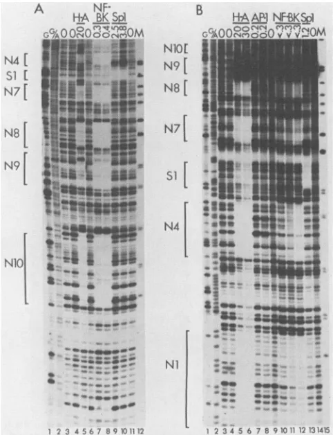
Binding of cellular proteins to the regulatory region of BK(MM) DNA. DNase I footprinting experiments with BK(MM) DNA are shown in Fig. 4. As in the previous
experiments,weusedheparin-agarose fractions of HeLa cell
nuclearextracts,aswellasNF-BK, AP-1, and Spl fractions purifiedby DNA affinity columnchromatography.
Six regions were protected by heparin-agarose fractions (Fig. 4A, lane 5; Fig. 4B, lanes 5 and 6). Site Ni (Fig. 4B only; not shown in Fig. 4A) is the same P blockfootprint seenin the other strains. SiteN4 isatthejunctionof the P and Q blocks and corresponds to the N4 site in BK(WW) DNA. Sites N7, N8, and N10 arise as the results of dupli-cations ofportions ofthe Pand Q blocks. N9, however, is unique to BK(MM) andarises as the result ofa novel Q-P junctioncontaininga2-bp insert.This unusualcreationofa
protein-binding siteacross arepeatjunction is analogous to the situation in BK(Dunlop), where novel AP-1 sites were
createdat the P-Pjunctions.
Ni [
N4 [
Si
[
J. VIROL.
on November 10, 2019 by guest
http://jvi.asm.org/
[image:5.612.315.559.92.191.2]B
N1O[ N9
[
N8[
N7
N47
at..,
NIl
1 '2 3 4 5 6 7 B 910 11 12131415
FIG. 4. DNase 1 footprint of the regulatory region of BK(MM). Forexperimental details, see the legend to Fig. 2 and Materials and Methods. (A) The DNA probe was 5' end labeled on the early strand. Lane 1, Products of the Maxam-Gilbert G-specific sequenc-ing reaction; lane 2, products of the Maxam-Gilbert G- and A-specific sequencing reaction; lanes 3, 4, 6, and 11, no protein extract added; lane 5, heparin-agarose fractions, 20 jig ofprotein; lanes 7 and 8, DNA affinity-purified NF-BK fractions, 0.3 and 0.4 ,ug of protein, respectively; lanes 9 and 10, DNA affinity-purified Spl fractions, 2.5 and 3.8 ,ug of protein, respectively; lane 12, marker (M), HpaII digestion products of pBR322 DNA (largest fragment shown is 242 bp). (B) The DNA probe was 5' end labeled on the late strand. Lane 1, Products of the Maxam-Gilbert G-specific sequenc-ing reaction; lane 2, products of the Maxam-Gilbert G- and A-specific sequencing reaction; lanes 3, 4, 9, and 14, no protein extract added; lanes 5 and 6, heparin-agarose fractions, 20 and 30 ,ug of protein, respectively; lanes 7 and 8, DNA affinity-purified AP-1 fractions, 0.2 jig of protein; lanes 10, 11, and 12, DNA affinity-purifiedNF-BK fractions, 3, 5, and 10,ul, respectively; the highest amount tested (10,ul) contained less than 0.3 ,ug of protein; lane 13, DNA affinity-purified Spl fractions, 1.2 ,ug of protein; lane 15, marker (M), HpaII digestion products of pBR322 DNA (largest fragment shown is 309 bp).
All six of the sitesprotected by heparin-agarose fractions in BK(MM) DNA contain close matches to the NF-BK consensus recognition sequence (Table 1). We tested the BK(MM) probes in DNase Ifootprinting experiments using affinity-purified NF-BK and found, asexpected, full protec-tion of sitesNi, N4, N7, N8, N9 and N10, with essentially the sameboundaries as seen with the heparin-agarose frac-tions.
BK(MM) contains anSpl consensus recognition sequence coinciding with the single Q-Rjunction present inthis strain. This sequence is protectedbypurified Sp-1 (Fig.4A,lanes9
and 10; Fig. 4B, lane 13). This site is analogoustothesingle Spl site in BK(WW) and therefore has been labeled Si.
Competitive binding experiments. Experiments with frac-tions purified by DNA sequence affinity chromatography, togetherwithsequence comparisons, strongly suggests that the observedfootprints at sitesNi through N10 are attrib-utableto asinglefactor, NF-BK. To confirm this,wecarried outcompetitive binding experiments using double-stranded oligonucleotides containing either the NF-BK recognition sequencefrom site Nior aheterologous sequencefrom the promoterregion ofanunrelated virus. In these experiments
competitor oligonucleotide was mixed with probe DNA, heparin-agarose fractions were added, complexes were al-lowed toform, and DNase Ifootprintingwascarried out as
before.
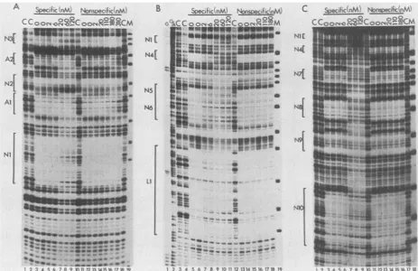
Figure 5A shows acompetitive binding experiment using BK(Dunlop) probe. Significantinhibition of NF-BK binding atsites Ni and N3is evident with 20nMspecificcompetitor, whereas no inhibition is seenwith the heterologous oligonu-cleotide at concentrations as high as 310 nM. Inhibition of bindingtositeN2ismoredifficulttosee,becauseprotection is only partial even in the absence of competitor. However, it appears that inhibition occurs with 6 to 20 nM of the specific competitor, and that there is no inhibition with the heterologous competitor. These results suggest that, as expected,sites Ni, N2, and N3 of BK(Dunlop)areprotected by the same or closely related protein species.
In contrast, the binding of AP-1 at sites Al and A2 of BK(Dunlop) DNA is not inhibited by any of the concentra-tions of the NF-BK oligonucleotide that we tested. Some inhibition ofAP-1 binding was seen at the highest concen-tration tested of the heterologous oligonucleotide, probably because this oligonucleotide contains a sequence
(TGiACGTG)
thatpartially
matches the AP-1 consensus recognition sequence. This result confirms that sites Ai and A2 are protected by aprotein that is different from the one that binds to sites Ni, N2, and N3.Competitive binding studies were extended to BK(WW) DNA (Fig. 5B). In this case, binding to four protected regions, Ni, N4, N5, and N6, was inhibited by the NF-BK oligonucleotide. There was noinhibition with the heterolo-gous oligonucleotide. As with BK(Dunlop), these results suggest that the same protein is binding to all sites. There was some inhibition of binding to site Li with the NF-BK oligonucleotide, butthe inhibition was notcomplete, evenat 120 nM. This is consistent with the results of the binding experiments usingpurified factors and suggests that protec-tion inthe Li region arises predominantly from the binding
ofa different factor.
Competitive binding studies with BK(MM) DNA are shown inFig. SC. Here,there aresixprotectedregions, Ni, N4, N7, N8,N9, andN10. Binding to sites Ni,N4, N7, and N8 is inhibited by 20 nM NF-BK oligonucleotide, whereas there is no inhibition by 310 nM heterologous
oligonucleo-tide. Again, this suggests that these sites are protected by a singlefactor, NF-BK. Some inhibition ofbinding is evident at sitesN9 and N10. These are more difficult to see because protection of these sites withheparin-agarose fractions in the absence ofcompetitor is not complete(Fig. 5, lanes 3 and 4; Fig. 4A, lane 5). However, someinhibition occurs at20nM specific competitor at site N9 (note thedisappearance of the hypersensitive band just above the protectedregion)and site N10. No inhibition of protection is seen with the heterolo-gous oligonucleotide at 310 nM. Similar results were ob-tained in several independent experiments, suggesting that
A
N4
[
Si [N7[
NB[
N9
- ew-*
2356 B901
on November 10, 2019 by guest
http://jvi.asm.org/
[image:6.612.55.296.78.392.2]3394 MARKOWITZ AND DYNAN
Specific .nM Nnsi>ecific'nM) C C o
o:> -4
88C
oc2
ooCmw
--~~~~-~
lb l.
lb.Ilb
d23 4 5 6 -78 ' )017112U31415161718S19
B Specific:%M) NwsoecIficKnM
0 02
GGcCoonC, 04°2>! oo°_---m
N4[ I6* -S . _
[
N5
stss
""§
I 2 34A 6789 10111213A141161718 19
C
NU
N44
NI
[image:7.612.77.540.75.375.2]IM
FIG. 5. Competitive bindingtoNF-BK sites in the regulatoryregion of BK virus. Competitor DNAwasadded in theamountsindicated toamixture containing singly end-labeled DNA probes, poly(dI-dC) copolymer, and pd(N)5 oligonucleotides asdescribed inthetextand Materials and Methods. The double-stranded oligonucleotide containing the NF-BK recognition sequence from site Ni (5' TGGAAT
GCAGCCAA3')wasusedasthe specific competitor. Adouble-stranded oligonucleotide containingasequencepresentin the human T-cell lymphotropic virustype1promoter(5'TGGGCTAGGCCCTGACGTGTCCCCCTGAAGACAAA3')wasusedasthenonspecific competitor.
Heparin-agarose fractions (30 ,ug) of HeLa cellnuclearextracts wasaddedtothe DNA mixture, DNase Icleavage wasperformed, and
products were analyzed on sequencing gels as described in Materials and Methods. Protected regions are indicated by brackets and
enumeratedasdescribedinthetext.(A) The DNA probewas5'end labeledonthelate strand ofBK(Dunlop). Lanes 1, 2, 10, and 18, No proteinextractadded(control [C]); lanes 3 through 9, NF-BK-specific competitor DNAwasadded in the following concentrations: 0, 0, 2,
6, 20, 60, and 120 nM, respectively; lanes 11 through17, nonspecific competitor DNAwasadded in thefollowing concentrations: 0, 0, 2, 20, 110, 180, and 310 nM, respectively; lane 19, marker(M), HpaII digestion products of pBR322 DNA (largest fragment shown is 201 bp). (B) The DNAprobewas5'end labeledonthe early strand of BK(WW). Lane 1, Products of the Maxam-GilbertG-specificsequencing reaction;
lane2, products of theMaxam-GilbertG-andA-specific sequencing reaction; lanes 3, 4, and 12,noproteinextractadded (control [C]); lanes
5through 11,NF-BK-specific competitor DNAwasadded in the following concentrations: 0, 0, 2, 6, 20, 60, and 120 nM, respectively; lanes
13through 18, nonspecific competitor DNAwasadded inthefollowing concentrations: 0, 0, 2, 20, 110, and 180 nM, respectively; lane19, marker(M),HpaII digestion products of pBR322 DNA (largest fragment shown is 309 bp). (C) The DNA probewas5'endlabeledonthe early
strandofBK(MM). Lanes 1, 2, 10, and 17, No proteinextractadded(control [C]); lanes 3 through 9,NF-BK-specific competitor DNAwas
added inthefollowing concentrations: 0, 0, 2, 6, 20,60, and 120 nM, respectively; lanes 11 through 16, nonspecific competitor DNAwas
added in thefollowing concentrations: 0, 0, 2, 20, 110, and 180 nM, respectively; lane 18, marker (M), HpaII digestion products of pBR322 DNA(largest fragment shown is 309 bp).
protection at N9 and N10 is due, in part, tothebinding of NF-BK, butthatother proteinsmay also contribute.
DISCUSSION
One of theongoing questionswithBKV istowhatdegree the enhancer region is similar to that of the related and better-characterized virus SV40. The present results show thedifferencesbetweenthetwo systems.Thedistinguishing feature of the enhancer region in BKV is the presence of multiple binding sites for NF-BK; these were the only protein recognition sites thatwe detected in all three BKV strainstested. Theubiquity of NF-BK binding sites in BKV stands in contrast to SV40, where NFl-related protein-bindingsiteshave notbeen reported in the enhancer region. Although different strains of BK virus have undergone complexrearrangements, multiple binding sites for NF-BK
are always present. Where sites have been lost through deletions,newsites have beenformedby duplications and in one case, by a duplication coupled with a small insertion. BK(WW),which doesnotcontainrepeatedsequencesinthe regulatory region, has four NF-BK sites: Ni within the P block, N4attheP-Qjunction, N5withintheRblock, and N6 at the R block-late region junction. BK(Dunlop) has a
deletion that results in the loss of sites N4, N5, and N6. However, the Pblocktriplication hasresulted in the
gener-ation oftwo additional NF-BK binding sites, N2 and N3. BK(MM)hasadeletion,relativetoBK(WW),that resultsin the loss of sites N5 andN6,butduplicationshave resultedin theformation of four additionalsites, N7, N8, N9, andN10. Wecanpredictthe location ofNF-BKsites inanumberof
other BKV strainsonthe basisofourresultswithBK(WW), BK(Dunlop), andBK(MM). Thus, we expect that BK(pro-A
Ai
N[
N2[
Al
Ni
J. VIROL.
on November 10, 2019 by guest
http://jvi.asm.org/
totype) contains the sameNF-BK sitesasBK(Dunlop) plus
an N4-like siteattheP-Q
junction.
BK(IR), isolated fromapancreaticadenoma of beta islet cells(7, 49),contains a total ofeight NF-BK binding sites: three Ni-like binding sites
withinthe Pblock, three N4-like sitesatP-Q junctions, one
N5-like site withinthe Rblock, andoneN6-like siteatthe R
block-late
region
junction. BK(pm526),a small-plaque vari-antisolatedfromaBK(prototype)-inducedhamsterpineocy-toma, contains three potential NF-BK binding sites, one
Nl-like site within thePblock andtwoN4-like sitesatP-Q junctions. BK(pm527),adifferentsmall-plaque variant,
con-tains five potential NF-BK sites, including three N4-like
sitesand two
Ni-like
sites (61-65).Thereare anumberof linesof evidence that suggest that
the NF-BK sites are important for growth in culture. All
strains of BKV
apparently
containmultiple
sites.Often,
there are many sites within relatively short stretches of DNA, for example, eight sites within 320 bpin BK(IR)andsix sites within 370 bp in BK(MM). In enhancer-defective
mutants containing only a single Pblock, duplication ofa
region
encompassingtheNi
sitewasobserved (64).AroleofNF-BKbindinginearly transcription ofBKVisalso
consis-tent with recent findings showing that the binding of the same or a related
protein
is essentialfor mouse mammary tumorviruspromoterfunction (6, 35, 42).A
systematic
linker scanmutationalanalysis
ofthe BKVearly
enhancer is found in thecompanion
article (12). Toeliminatethe effect ofsequenceredundancy, theseworkers introduced their mutations into a viral
background
that hasonly
asingle
P block. Thebackground
is identical toBK(WW),
exceptthatthe Rblock ismissing.
Thisanalysis
shows a correlation betweenearly transcriptional activity
and the presence ofsequences in theNi
and N4 sites (12 [note that the N4 site overlaps the genetically defined celement]).
This is further evidence that the NF-BK sites definedby footprinting
arefunctional elements ofthe BKVenhancer.
The
recognition
sequenceforNF-BK(Table 1)isasubsetofthe
recognition
sequenceforNF1,TGG(N6-7)
T/G CCAA(10,
13, 40). NF1 wasoriginally
describedas ahostprotein
required
for adenovirusreplication
(45).
NF1 appears to befunctionally equivalent
totheTGGCA-binding protein
ofthelaboratory
ofNowock andSippel
(39, 46, 47). Otherworkhas
suggested
thatNF1is identicaltotheCCAAT transcrip-tion factor (29, 31) that binds in the upstream region of several promoters,although
this remainscontroversial,
and a recentstudy
showed that there are a"multiplicity
of CCAAT boxbinding proteins"
thatbindtothesesequencesin different promoters (14, 42). In viewof this
complexity,
we have chosen to refer to the BKV-binding protein as
NF-BK,
until itsidentity
is betterestablished.The presence of two
binding
sites for thetranscription
factor AP-1 that cross thejunction
betweenadjacent
Pblocks in
BK(Dunlop)
wassurprising. Apparently,
thesebinding
siteswerecreatedatthetimethe Pblocktriplication
was formed. The
possibility
thatfunctionally
important elementsspan repeatjunctions
has beenlargely
overlookedin prior studies of the BKV regulatory region, perhaps
because thisphenomenonisnotknownto occurin the SV40 enhancer.
The three AP-1
recognition
sites in SV40, noneof which arelocatedatrepeatjunctions,
arebelievedtocontributetoinduction ofSV40 RNAsynthesis by tetradecanoyl phorbol
acetate (1, 38). The AP-1-binding sites in BKV may have similar functions. Studies with deletionmutantsof BK(pro-totype)indicate that there issomeloss ofearlygene
expres-sion when an AP-1 recognition sequence (site A2 in our nomenclature) is removed (11). AP-1 recognition sites are
clearly notessentialfor viral viability, however, sincethey are notpresentinall strains.
BK(WW) and BK(MM) have asingle binding site for the transcription factor Spl (17) atthejunction of the Q and R blocks. This site iscompletelyprotected by affinity-purified Spl, and hasa10 of 10bp matchto aknownSpl site in the IE3 promoter of herpes simplex virus (30). This site is not presentin BK(Dunlop) and is partially deleted in
BK(pro-totype). In additiontotheSpl siteattheQ-R junction, there isasequenceAGGGiAGGAGCattheearly-proximalend of the P block that hasan8 of 10 match(underlined)totheSpl
recognition consensus (32). Although there is genetic evi-dence that theregion containing this potential Spl site may be importantforearly promoterfunction(11, 12), we have never detected binding at this site by affinity-purified Spl
fractions in any ofthe strains tested. This suggests either thatit is averyweak Spl siteorthatit isarecognition site
foradifferentprotein.
We have also observed protection of a late-proximal regionin BK(WW) that overlaps the start codon for
agno-protein.Thissite, whichwecallLi,isprotected by
heparin-agarosefractions. Althoughthere is some protectionat Li
withpurifiedNF-BK, competitive binding experiments sug-gestthat protection at Li arises primarilyfrom a different factor. The Li sequence is also present in the viral DNA of
BK(Dunlop)andBK(MM), althoughitwas notpresentin the subcloned probes used in our binding experiments. In a
study usingdeletion mutants, deletion of Li sequences had no effectonearlygene expression (11), suggestingthat the
function ofthis site, if any, might be related to late gene
expression. InSV40, there is evidence that sequencesnear
andwithin the late
coding
regionaffect late geneexpression (2, 20, 52, 53, 57, 60).There has beenconsiderable
speculation
about theroleofmotifs homologous tothe SV40enhancer core(67)and the Elaenhancercore(23)thatarepresentwithintheregulatory region of BKV. The SV40 enhancer core sequence, TG
GAAAGT, was
originally
defined by correlating loss ofenhancer function with
specific point
mutations locatedwithinthe72-bprepeatsand isnowknowntobethebinding site for one or more cellular proteins (28, 43, 70). On the basis ofthe presence of similarsequences within Moloney
murine sarcomavirus, BK(Dunlop), andpolyomavirus, the
SV40 enhancer core consensus (G)TGG A/T A/i A/T (G)
was proposed (67). Inthe light ofcurrent knowledge,
how-ever,thisfairly degenerateconsensus may notbe
meaning-ful. It has beenpointedoutbyWeberetal.(66)that theTGG A/TA/T A/T Gmotifoccursinatleast 38placeswithin the
SV40 genome. Moreover, the sequence in BK(Dunlop)
matches
(underlined)
SV40inonly
four oftheeight positions
(ATGGTTTG).
Our
experiments
show no evidence that the SV40 en-hancer core homology in BKV is recognized by cellularproteins. Althoughthehomology overlapsthe late endofthe NF-BK
binding
sites within thePblock(sites
Ni,
N2, N3,N7, and N9), only part of the sequence is protected. In
addition,identicalpatternsofprotectionare seenwith crude
heparin-agarose
fractions and DNAaffinity-purified
NF-BK, suggesting that NF-BK is theonly
factorbinding
in thisregion.
Asecond enhancer motifthoughttobe present in BKV,
the Ela enhancer core, was
originally
definedby
deletion mutants affectingexpression of the adenovirus type 5early
region1A(Ela)transcription
unit(23).Aconsensussequenceon November 10, 2019 by guest
http://jvi.asm.org/
3396 MARKOWITZ AND DYNAN
(A/C GGAAGTGA A/C) was proposed on the basis of
se-quence homologies with 13 other transcription units, BK
(Dunlop) among them. This sequence
(AGGAAAGTGCA)
(matches are underlined) occurs in BK(Dunlop) at the late end ofthe P blocks and overlaps binding sites Al and A2.However, not all the sequence is protected at these sites, and identical footprints are seen with both heparin-agarose
fractionsand DNA affinity-purified AP-1. The Ela enhancer coresequenceis also present in BK(MM) at the early side of sites N4, N8, and N10 and in BK(WW) at the early side of sites N4, N5, and N6. Although the complete sequence is
protectedat sitesN5 and N6, protection at these other sites does notcoverthe entire sequence, and identical footprints are seen with both heparin-agarose fractions and DNA
affinity-purified
NF-BK. These results suggest that the pres-ence of Ela enhancer core homologies in BKV is actually due to the similarity between this consensus and the AP-1 and NF-BKrecognition sequences and does not reflect thepresenceof anadditionalcellular Ela enhancer core-binding
protein.
The companion article (12) reaches similar conclusions
abouttheSV40and adenovirus Ela homologies on the basis
ofindependent evidence. For the most part, mutations that
specifically affectthese sequences have a less than twofold
effectonearly-direction transcription.
One question that may be raised about the studies
pre-sented here is whether the map of factor-binding sites is
completeorwhethersome sites may have been overlooked. Itispossible that some factors are present in the HeLa cell
nucleusin verylimiting amounts. In addition, some factors
might not be recovered efficiently in our extraction or
preliminary fractionation steps or might not bind to DNA under theconditionsused for footprinting. Genetic evidence
suggests that aGC-rich sequence of the early proximal end
ofthe Pblockisrequiredfor early promoter function, but we have not detected protein binding in this region (11, 12). With this exception, however, the results of our binding
studies aregenerallyconsistent with prior mutational analy-sesof the BKVregulatoryregion (11, 64) and with the linker scananalysisinthe companion article (12). More than half of the DNA sequence in the regulatory region is protected by thefactors defined here.
Asnotedpreviously,a hypothesis has been put forward by
Rubinsteinet al. (55) that BK(WW), which does not contain
sequence repeats and does not grow in culture, represents the authentic virusas it exists in the human population. If
this is correct, the occurrence of sequence repeats and
deletions indifferent strains of BKV may be attributable to
selectivepressure in culture. The spontaneous generation of
sequencerepeatsinresponse to selective pressure is consis-tentwiththeknowntendencyof enhancer mutants of BKV and SV40 to revert by sequence duplication (24, 64). It is alsoconsistentwith theobservation that primary isolates of BKV usually require an extended period of incubation in culturebeforethefirstcytopathiceffects are observed (7, 8, 15, 19,21, 22, 26, 36, 50, 51, 58, 59, 69). This would allow timeforrepeatsto begenerated and fixed in the population. Itisunlikelythat rearrangements during passage in culture are the only mechanism for repeat formation in BKV.
BK(prototype) and BK(Dunlop), which were isolated
inde-pendently, haveidentical repeatstructures and differ only in theendpoint of adeletiononthe late side of the regulatory
region. Moreover, athird strain of BKV, BK-17, that was isolated bymolecular cloning directlyfrom the kidney tissue of an accident victim is identical to BK(Dunlop) in the
regulatory region (R. Frisque, personal communication).
These findings demonstrate that sequence repeats are present in BKV in vivo in at least some individuals.
ACKNOWLEDGMENTS
We thankNadia Rosenthal for BK(Dunlop) plasmid DNA, Ray Wu for BK(MM) viral DNA, Riva Rubinstein for BK(WW) plasmid DNA, John Letovsky for preparations of purified AP-1 and Spl, Jeffrey Schneringer for expert technical assistance, Jennifer Nyborg for the gift ofhuman T-cell lymphotropic virus type I oligonucleo-tides used in competition experiments, and Cheryl Grosshans for the synthesis of DNA oligonucleotides.
This work was supported by Public Health Service grant CA 44958 from the National Institutes of Health to W.S.D.
LITERATURE CITED
1. Angel, P., M. Imagawa, R. Chiu, B. Stein, R. J. Imbra, H. J. Rahmsdorf, C. Jonat, P. Herrlick, and M. Karin. 1987. Phorbol ester-inducible genes contain a common cis element recognized by a TPA-modulated transacting factor. Cell 49:729-739. 2. Ayer, D., and W. S. Dynan. 1988. Simian virus 40 major late
promoter: a novel tripartite structure that includes intragenic sequences. Mol. Cell. Biol. 8:2021-2033.
3. Banerji, J., S. Rusconi, and W.Schaffner.1981. Expression of a
P-globin
gene is enhanced by remoteSV40sequences. Cell 27: 299-308.4. Blaese, R. M., W. Strober, R. S. Brown, and T. A. Waldman. 1968. The Wiskott-Aldrich syndrome. A disorder with a possi-ble defect in antigen processing or recognition. Lancet i:1056-1061.
5. Bradford, M. 1976. A rapid and sensitive method for the quantitation of microgram quantities of protein utilizing the principle of protein-dye binding. Anal. Biochem. 72:248-254. 6. Buetti, E., and B. Kuhnel. 1986. Distinct sequence elements
involved in the glucocorticoid regulation of the mouse mam-marytumor virus promoter identified by linker scanning muta-genesis. J. Mol. Biol. 190:379-389.
7. Caputo, A., A. Corallini, M. P. Grossi, L. Carra, P. G. Balboni, M. Negrini, G. Milanesi, G. Federspil, and G. Barbanti-Brodano. 1983. Episomal DNA of a BK virus variant in a human insuli-noma.J. Med. Virol. 12:37-49.
8. Coleman,D. V., M. R. Wolfendale, R. A. Daniel, N. K. Dhanjal, S. D. Gardner, P. E. Gibson, and A. M. Field. 1980. A prospec-tive study of human polyomavirus infection in pregnancy. J. Infect. Dis. 142:1-8.
9. Davison, B. L., T. Leighton, and J. C. Rabinowitz. 1979. Purification of Bacillus subtilis RNA polymerase with heparin-agarose. J. Biol. Chem. 254:9220-9226.
10. deVries, E., W. vanDriel, S. J. L. van den Heuvel, and P. C. van der Vliet. 1987. Contactpoint analysis of the HeLa nuclear factorI recognition site reveals symmetrical binding at one side ofthe DNA helix. EMBO J. 6:161-168.
11. Deyerle, K. L., J. A. Cassill, and S. Subramani. 1987. Analysis ofthe early regulatory region of the human papovavirus BK. Virology 158:181-193.
12. Deyerle,K. L., and S. Subramani. 1988. Linker scan analysis of theearly regulatory region of human papovavirus BK. J. Virol. 62:3378-3387.
13. Diffley,J. F. X., and B. Stillman. 1986. Purification of a cellular, double-stranded DNA-binding protein required for initiation of adenovirus DNA replication by using a rapid filter-binding assay. Mol. Cell. Biol. 6:1363-1373.
14. Dorn, A., J. Bollekens, A. Staub, C. Benoist, and D. Mathis. 1987. A multiplicity of CCAAT box-binding proteins. Cell 50: 863-872.
15. Dougherty, R. M., and H. S. DiStefano. 1974. Isolation and characterization ofa papovavirus from human urine (38131). Proc. Soc. Exp. Biol. Med. 146:481-487.
16. Dynan, W. S. 1987. DNase I footprinting as an assay for mammalian gene regulatory proteins, p. 75-87. In J. K. Setlow (ed.), Genetic engineering. Plenum Publishing Corp., New York.
17. Dynan,W.,and R. Tjian. 1983. The promoter-specific transcrip-J. VIROL.
on November 10, 2019 by guest
http://jvi.asm.org/
tion factorSpl binds toupstream sequences intheSV40early promoter.Cell 35:79-87.
18. Fromm, M., and P. Berg. 1983. Simian virus 40 early- and late-regionpromoterfunctionsareenhancedby the 72-bp repeat insertedatdistant locations and inverted orientations. Mol.Cell Biol. 3:991-999.
19. Gardner, S. D., A. M. Field, D. V. Coleman, and B. Hulme. 1971. Newhuman papovavirus (BK) isolated from urine after renaltransplantation. Lancet i:1253-1257.
20. Ghosh,P. K., M. Piatak, J. E. Mertz, S. M. Weisman, and P. Lebowitz. 1982. Alteredutilization ofsplice sites and5'termini inlate RNAsproduced by leader regionmutantsof simian virus 40.J. Virol.44:610-624.
21. Gibson,P. E.,andS. D. Gardner. 1983. Strain differences and some serological observations on several isolates of human polyomaviruses. Prog. Clin. Biol.Res. 105:119-132.
22. Goudsmit, J., M. L. Baak, K. W. Slaterus, and J. van der Noordaa. 1981. Human papovavirus isolated from urine ofa child withacutetonsillitis. Br. Med.J. 293:1363-1364. 23. Hearing,P., and T. Shenk. 1983. The adenovirus type 5 ElA
transcriptional control region contains a duplicated enhancer element. Cell 33:695-703.
24. Herr, W.,andJ. Clarke. 1986. TheSV40 enhancer iscomposed ofmultiple functional elements that can compensate for one another. Cell45:461-470.
25. Howley, P. M. 1980.Molecularbiology of SV40andthehuman polyomavirusesBKandJC,p.489-550. InG. Klein(ed.), Viral oncology. Raven Press,New York.
26. Howley, P.M., G. Khoury, J. C.Byrne, K. K. Takemoto,and M. M.Martin. 1975.Physicalmapof theBKvirusgenome. J. Virol. 16:959-973.
27. Howley, P. M., M. F. Mullarkey,K. K. Takemoto,and M. A. Martin. 1975. Characterization of human papovavirus BK DNA. J. Virol. 15:173-181.
28. Johnson, P. F., W. H. Landschulz, B. J. Graves, and S. L. McKnight.1987.Identification ofa ratliver nuclearproteinthat binds to the enhancer core element of three animal viruses. GenesDev. 1:133-146.
29. Jones,K.A., J.T.Kadonaga,P.J. Rosenfeld,T.J. Kelly,andR. Tjian. 1987. A cellular DNA-binding protein that activates eukaryotic transcriptionand DNA replication.Cell 48:79-89. 30. Jones, K. A., and R. Tjian. 1985. Spl binds to promoter
sequences and activated HSV "immediate-early" gene
tran-scriptionin vitro. Nature(London)317:179-182.
31. Jones,K.A.,K. R.Yamamoto,and R.Tjian.1985. Twodistinct transcription factors bind to the HSV thymidine kinase
pro-moterin vitro. Cell 42:559-572.
32. Kadonaga, J. T., K. A. Jones, and R. Tjian. 1986. Promoter-specificactivation ofRNApolymeraseIItranscription by Spl. Trends Biochem. Sci. 11:20-23.
33. Kadonaga, J. T., and R. Tjian. 1986. Affinity purification of sequence-specificDNAbinding proteins.Proc.Natl. Acad. Sci. USA 83:5889-5893.
34. Khoury, G., and P. Gruss. 1983. Enhancer elements. Cell 33: 313-314.
35. Kuhnel, B., E. Buetti, and H. Diggelman. 1986. Functional analysisof theglucocorticoidregulatoryelements present inthe
mousemammarytumorviruslongterminalrepeat. J.Mol. Biol.
190:367-378.
36. Lecatsas, G.,andJ.A. L. VanWyk.1978. DNAviruses in urine after renaltransplantation. S. Afr. Med. J. 53:787-788. 37. Lee, W.,A.Haslinger,M.Karin,and R.Tjian.1987.Activation
oftranscription bytwofactors that bindpromoterand enhancer sequencesof the human metallothionein gene and SV40. Nature (London)325:368-372.
38. Lee, W., P.Mitchell, and R.Tjian. 1987.Purifiedtranscription factorAP-1bindstoTPA-inducibleenhancer elements. Cell 49: 741-752.
39. Leegwater, P. A. J., P. C. van der Vliet, R. A. W. Rupp, J. Nowock,and A. E.Sippel. 1986. Functionalhomologybetween the sequence-specific DNA-binding proteins nuclear factor I fromHeLa cells and the TGGCA protein fromchicken liver. EMBOJ. 5:381-386.
40. Leegwater, P. A. J., W. van Driel, and P.C.vanderVliet. 1985. Recognition site of nuclear factor I, asequence-specific DNA-binding protein from HeLa cells that stimulates adenovirus DNAreplication. EMBOJ. 4:1515-1521.
41. Maxam, A., and W.Gilbert. 1980.Sequencing end-labeled DNA withbase-specific chemical cleavages. Methods Enzymol. 65: 499-560.
42. Miksicek, R., U. Borgmeyer, and J. Nowock.1987. Interaction of the TGGCA-binding protein with upstream sequences is re-quired for efficient transcription of mouse mammary tumor virus. EMBOJ. 6:1355-1360.
43. Mitchell, P. J., C. Wang, and R. Tjian. 1987. Positive and negative regulation of transcription in vitro: enhancer-binding protein AP-2 is inhibited by SV40 T antigen. Cell 50:847-861. 44. Moreau, P., R.Hen,B.Wasylyk,R.Everett,M. P.Gaub,andP.
Chambon. 1981. The SV40 72 basepair repeathas astriking effect on gene expression both in SV40 and other chimeric recombinants. Nucleic AcidsRes.9:6047-6068.
45. Nagata,K.,R. A.Guggenheimer,T.Enomoto, J.H.Lichy,and J. Hurwitz. 1982.Adenovirus DNAreplication in vitro: identi-fication ofahostfactor that stimulates synthesis of the preter-minal protein-dCMP complex. Proc. Natl. Acad. Sci. USA79: 6438-6442.
46. Nowock, J.,U.Borgmeyer,A. W.Puschel,R. A. W.Rupp, and A.E. Sippel. 1985. The TGGCAprotein bindstothe MMTV-LTR, the adenovirus origin of replication, and the BK virus enhancer. Nucleic AcidsRes. 13:2045-2061.
47. Nowock, J., and A. E. Sippel. 1982. Specific protein-DNA interactionatfour sitesflanking the chickenlysozymegene.Cell 30:607-615.
48. Nyborg, J., W. S.Dynan, I. S. Y. Chen, and W. Wachsman. 1988. Binding of host cell factors to DNA sequences in the HTLV-I LTR: implications for viral gene expression. Proc. Natl. Acad.Sci. USA 85:1457-1461.
49. Pagnani, M., M. Negrini, P. Reschiglian, A. Corailini, P. G. Balboni,S.Scherneck,G.Macino,G.Milanesi,and G. Barbanti-Brodano. 1986. Molecular and biological properties of BK virus-IR, aBKvirus variant isolated from ahumantumor.J. Virol. 59:500-505.
50. Pater, M. M., A. Pater, and G. Di Mayorca. 1981. Genome analysis of MG virus, ahumanpapovavirus. J. Virol. 39:968-972.
51. Pauw, W., andJ.Choufoer. 1978. Isolation ofavariantofBK virus with altered restrictionendonucleasepattern.Arch.Virol. 57:35-42.
52. Piatak, M., P. K. Ghosh,L. C. Norkin, and S. M. Weissman. 1983. Sequences locatingthe 5' endsofthemajor simian virus 40 mRNAlateforms. J. Virol. 48:503-520.
53. Piatak, M.,K. N. Subramanian, P.Roy, andS. M. Weissman. 1981.Latemessenger RNAproduction by viable simian virus40
mutantswithdeletionsinthe leaderregion. J. Mol. Biol. 153:
589-618.
54. Rosenthal, N.,M. Dress,P. Gruss, andG. Khoury. 1983. BK viral enhancerelement andahumancellularhomolog. Science 222:749-755.
55. Rubinstein, R.,N.Pare,and E. H.Harley. 1987.Structure and function ofthe transcriptional control region ofnonpassaged BKvirus. J.Virol. 61:1747-1750.
56. Seif, I.,G.Khoury, and R. Dhar. 1979.The genomeofhuman papovavirusBKV.Cell 18:963-977.
57. Somasekhar,M.B.,andJ.E. Mertz.1985. Sequencesinvolved indeterminingthe locations of the5'endsof the lateRNAsof simian virus40. J. Virol. 56:1002-1013.
58. Takemoto,K.K.,A.S.Rabson,M. F.Mularkey,R. M.Blaese, C. F.Garon,andD.Nelson.1974.Isolation ofpapovavirusfrom braintumorand urine ofapatient with Wiskott-Aldrich syn-drome. J. Natl. CancerInst.53:1205-1207.
59. Ter Schegget, J., C. J. A. Sol, E. Wouters Baan, J. Van Der Noordaa,and H. VanOrmondt. 1985. Naturally occurringBK virus variants(JLandDik)withdeletionsin theputative early enhancer-promotersequences. J.Virol. 53:302-305.
60. Viliarreal, L. P., R. T. White, and P. Berg. 1979. Mutational alterationswithin the simian virus40leadersegment generate
on November 10, 2019 by guest
http://jvi.asm.org/
3398 MARKOWITZ AND DYNAN
altered16S and19SmRNAs. J. Virol. 29:209-219.
61. Watanabe, S., S. Kotake, A. Nozawa, T. Muto,andS. Uchida. 1982.Tumorigenicity ofhuman BKpapovavirusplaque isolates, wild-typeandplaque morphology mutant, in hamsters. Int. J. Cancer29:583-586.
62. Watanabe, S., and K. Yoshiike. 1982.Changeof DNAnearthe origin of replication enhances the transforming capacity of human papovirus BK. J. Virol.42:973-985.
63. Watanabe, S., and K. Yoshiike. 1985.Decreasing the number of 68-base-pair tandem repeats in the BK virus transcriptional control region reduces plaque size and enhancestransforming capacity.J. Virol. 55:823-825.
64. Watanabe, S., and K. Yoshiike. 1986. Evolutionary changesof transcriptionalcontrolregion inaminute-plaque viable deletion
mutantofBKvirus. J. Virol.59:260-266.
65. Watanabe, S., K. Yoshiike, A. Nozawa,Y.Yuasa, andS. Uchida. 1979. Viable deletion mutant ofhuman papovavirus BK that inducesinsulinomas in hamsters. J.Virol.32:934-942. 66. Weber, F., J. de Villiers, and W. Schaffner. 1984. An SV40
"enhancertrap" incorporates exogenous enhancers or
gener-atesenhancers from itsownsequences.Cell36:938-992. 67. Weiher, H., M. Konig, and P. Gruss. 1983. Multiple point
mutationsaffecting the simian virus 40 enhancer. Science219: 626-631.
68. Wildeman, A. G., M. Zenke, C. Schatz, M. Wintzerith, T. Gundstrom,H. Matthes, K.Takahashi,andP. Chambon.1986. Specific protein bindingtothesimian virus 40 enhancer in vitro. Mol.Cell. Biol. 6:2098-2105.
69. Wright,P.J.,G. Bernhardt, E.0. Major,andG.diMayorca.
1976. Comparison of the serology, transforming ability, and polypeptide composition of human papovaviruses isolated from urine.J.Virol. 17:762-775.
70. Xiao, J.-H.,I.Davidson, M. Macchi,R.Rosales,M.Vigneron,
A. Staub, and P. Chambon. 1987. In vitro binding of several cell-specific and ubiquitous nuclear proteinstotheGT-I motif of theSV40 enhancer. Genes Dev. 1:794-807.
71. Yang, R. C. A., and R. Wu. 1979. BKvirus DNA: complete nucleotidesequenceofahumantumorvirus. Science 206:456-462.
J. VIROL.