JOURNALOFVIROLOGY, Mar. 1991,p. 1304-1309 0022-538X/91/031304-06$02.00/0
CopyrightC) 1991,American Society forMicrobiology
Sendai Virus Protein-Protein
Interactions
Studied
by
a
Protein-Blotting
Protein-Overlay
Technique: Mapping
of Domains
on
NP
Protein Required for
Binding
to
P
Protein
H. E. HOMANN,* W. WILLENBRINK, C. J. BUCHHOLZ, AND W. J. NEUBERT
Abteilung fur Virusforschung, Max-Planck-Institut fur Biochemie, 8033 Martinsried, FederalRepublic of Germany Received 8 October 1990/Accepted 3 December 1990
Proteins from Sendai virus particles and from infected cells were analyzed in a blotting
protein-overlayassayfor theirinteraction with invitro-synthesized, [35S]methionine-labeledviralproteins NP, P, and M.After separation by sodium dodecyl sulfate-polyacrylamide gel electrophoresis,transferontopolyvinylidene difluoride membranes, and renaturation, the immobilized proteins were found to interact specifically with radiolabeled proteins. NPproteinsfrom virusparticlesand from infected cells retained35S-P protein equally well.Conversely,Pprotein from virusparticlesand from infected cells retained35S-NP protein.3.S-Mprotein
was retained mainlyby NP protein butalso by several cellular proteins. To determine the domains on NP
protein required for binding to immobilized Pprotein, a series of truncated and internally deleted 35S-NP proteinswasconstructed. Theonlydeletion that did not affectbinding resides between residues 426 and497. The carboxyl-terminal 27 residues (positions 498 to 524) contribute significantly to the binding affinity. Removal of 20 residues (positions 225 to 244) in the hydrophobic middle part of NP protein completely abolisheditsbindingtoPprotein.
Thecurrentviewof paramyxovirus replicationis basedon
studies of different members of the paramyxovirus family andby analogy to another negative-strand virus, vesicular stomatitis virus (see references 1 and 22 for reviews). We used Sendai virus to gain insight into the mechanisms involvedin theregulation of transcriptionandreplicationof paramyxoviruses. Itcontainsasingle-stranded 15.4-kb RNA
genome ofnegative strand polarity which directs
transcrip-tion of six mRNAs in the order NP (nucleocapsid protein), P/C (polymerase-associated protein), M (matrix protein), F (fusionprotein),HN(hemagglutinin-neuraminidaseprotein), and L (large protein) (18, 39, 40). The genomic RNA is tightly associatedwith thenucleocapsid proteinNP(60kDa) into a helical nucleocapsid core. Nucleocapsids are tran-scriptionallyactiveonlywhen the L(220kDa)and the P(79 kDa) proteins are present (8, 13, 15). While L protein is regardedasthecore of the viral RNA polymerasecomplex,
little isknown aboutthefunctions ofPprotein.
Viraltranscription and replication take place in the cyto-plasm of the infected cell. During transcription, monocis-tronic mRNAs aresynthesized. Duringreplication, full-size
antigenome RNA isproduced, which concurrently becomes encapsidatedbyNPprotein. Itisnotcompletely understood how the switch between transcription and replication is achieved. This derives mainly from the lack of knowledge abouthowproteins of thepolymerasecomplex interactwith each other, with the nucleocapsid template, and possibly with tubulin and microtubule-associated proteins, which havebeenfound to stimulate transcription invitro (19, 30). Nucleocapsids isolated from infectedcellswereshown
pre-viouslyto bind in vitro-synthesized Pprotein (36), butit is stillnot exactly known whichcomponentof the nucleocap-sid mediates this association. Furthermore, M protein also binds to nucleocapsids (27) and is regarded as a negative
regulator for transcription. It is therefore of importance to
*Corresponding author.
learn which factors affect and modulate the interaction between thenucleocapsid (i.e., NP protein) and P proteinas wellas Mprotein.
We analyzed the interactions between these proteins by using renaturing protein blots. We applied cDNA-derived protein NP, P, orM,labeledwith[35S]methionine duringin
vitro translation, toblots containing proteinsfromegg-grown
Sendai virusparticles and from infected cells separated by sodium dodecyl sulfate-polyacrylamide gel electrophoresis (SDS-PAGE). All three in vitro-synthesized proteins were specificallyretainedbyimmobilized viralproteins.We com-bined this protein-binding assay and mutagenesis to map
domains on NP protein responsible for its binding to P protein.
(This work was presented in part at the VIIth Interna-tional Congress of Virology, Berlin, Federal Republic of Germany, 1990.)
MATERIALSANDMETHODS
Virus, cell cultures, infection, and preparationof cellular extracts. Seed stocks of theFushimi (D52) strain of Sendai viruswere grown inembryonated eggs, and virus
purifica-tion, Vero cellculture, and cellinfection (0.1 infectiousunit
per cell) were performed as previously described (20). Ex-tracts were prepared 12 h after infection. Cells were
de-tachedfromculture dishesbytreatmentwith 20 mM EDTA inphosphate-buffered saline (PBS). Theywerepelleted for 5
minat2,000 xgandresuspendedin lysis buffer (50 mM Tris
hydrochloride [pH 8.0], 150mMNaCl, 1.5 mM MgCl2, 1% NonidetP-40, 0.1% SDS [BDH], 1 Rgof aprotinin perml,
200,ugofphenylmethylsulfonyl fluorideperml). Nucleiwere
removed by centrifugation at 12,000 x g for 2 min. The
supernatantwas stored at -20°C.
Synthesisof cDNAandcloningof theNP, P, and Mgenes.
Sendai virus Fushimi strain genomic RNA, purified by the guanidinium-isothiocyanate lysis procedure (4), was
tran-scribed into cDNA by using avian myeloblastosis virus
1304
Vol.65,No. 3
on November 10, 2019 by guest
http://jvi.asm.org/
reverse transcriptase (Beard, Life Sciences) and synthetic oligonucleotides as gene-specific primers, as previously de-scribed (20). Primers used for first-strand cDNA synthesis were
NP1728-1704,
P3555-3538,
andM4748-4730.
The cDNA was amplified by 30 cycles of polymerase chain reaction using oligonucleotidesNP54-75,
NP17051729,
andP317,3191
as sec-ond-strand primers. The polymerase chain reaction-ampli-fied M gene cDNA was cut with CfoII. Thus, the NP gene cDNA ranges from positions 54 to 1728, the P/C gene cDNA ranges from positions 1705 to 3555, and the M gene cDNA ranges from positions 3666 to 4748. DNA was filled in with Klenow polymerase, phosphorylated, and ligated intoSmaI-cut, dephosphorylated plasmids pBluescribe
M13+
orpBlue-scriptIISK- bearing promoters for T3 orT7 RNA
polymer-ase on both sides of the polylinker. The DNA was transformed into XLI-Blue cells (Stratagene). All clones were sequenced (32, 33, 45). All numbering refers to the Sendai virus Z strain sequence (39, 40), irrespective of a one-nucleotide insertion detected at position 1598 in the NP gene (33).
Synthesis of NP gene deletion clones and in vitro transcrip-tion and translatranscrip-tion. Constructs 1, 2, and 9 (see Fig. 3A) were generated from
pBS-NPrev,
which carries the NP gene start codon next to the T7 promoter. Internal deletion constructs 3 to 8 were generated from clone pBS-NP, which carries the insert in the opposite orientation, by using the restriction enzymes depicted in Fig. 3. When necessary, we filled in the ends and subsequently cut at the single NdeI site in the vector. The resulting fragments were purified from agarose gels by electroelution. Construct 9 was generated frompBS-NPrev.
Theidentity
ofevery recombinant was verified by nucleotide sequencing to ensure that all deletions were in frame. Plasmid pBS-NP and NP gene deletion clones, lin-earized with PvuII, and plasmids pBS-P and pBS-M, linear-ized with HindIII, were transcribed with T3 RNA polymer-ase (Boehringer Mannheim Biochemicals). Truncated NP gene RNA was generated frompBS-NPreV
and linearized withBallor XbaI by transcription with T7 RNA polymerase in a20-,ul
reaction (28). After removal of template DNA by treatment with DNase I, 3,ul
of the reaction mix was translated in vitro by using 35,ul
of rabbit reticulocyte lysate (Promega) and 50,uCi
of [35S]methionine (1,000 Ci/mmol;Amersham) in a volume of 50 pl. The incorporation of
[35S]methionine
was determined by trichloroacetic acid pre-cipitation. A2.5-,u
sample of the translation reaction was analyzed by SDS-PAGE. For detection ofradiolabeled pro-teins, gels were acid fixed, dried, and placed on X-ray film. Protein binding assay. Purified virus particles and extracts from infected or mock-infected cells were denatured with SDS (BDH) in the presence of,-mercaptoethanol at room temperature. Protein(1.2 mg) wasapplied to a 12-cm slot of a preparative SDS-polyacrylamide gel (24). After electro-phoresis, the proteins were blotted to polyvinylidene diflu-oride (PVDF) membranes (Immobilon; Millipore) (42). Transfer buffer was 25 mM Tris-192 mM glycin (pH 8.3). Blots were then incubated in standard bindingbuffer(SB) (10 mM HEPES[N-2-hydroxyethylpiperazine-N'-2-ethane-sulfonic acid] [pH7.4], 10 mMMgCl2, 50 mM NaCl, 0.1 mM EDTA, 1 mM dithiothreitol, 10% glycerol) for 12 h at 4°C.
They were cut into 5-mm strips, which were thenincubated for 3 h in SB-5% bovine serum albumin (BSA) to saturate free binding sites. For binding of [35S]methionine-labeled
proteins, individual strips were incubated for 24 h at4°C in 1.4 ml of SB-5% BSA containing 35,000cpmof trichloroace-tic acid-precipitable activity of in vitro translation products
per ml. Subsequently, the strips were washed five to six
timesfor30mineachin SB-0.25%BSA, dried, treatedwith
a
fluorographic
reagent (Amplify-Spray; Amersham), and exposed to Kodak X-Omat R films. When signals were to be quantitated by liquid scintillation counting, fluorography reagent was omitted.RESULTS
In vitro-synthesized P protein is retained by immobilized Sendai virus NP protein. P protein was shown earlier to attach to purified nucleocapsids, and the P protein domains requiredfor attachment were thoroughly mapped (36, 37). In an attempt to understand which component of the nucleo-capsid mediates binding of P protein, we separated Sendai virus proteins by SDS-PAGE, blotted them on PVDF
mem-branes, renatured the immobilized proteins, and then tested
which of them would bind in vitro-synthesized P protein. However, P RNA also encodes the nonstructural proteins
C', C, Y, and X. The X protein, which is identical to the
carboxyl-terminal 95 amino acids of P protein (5, 11), was
previously shown to be unable to attach to nucleocapsids
(37). Therefore, X protein should have no effect in subse-quent binding assays. Whether the proteins C', C, and Y, which are encoded in a different reading frame, would also bind to viral proteins remains to be investigated.
The P/C gene cDNA was transcribed in vitro, and the RNAobtained was subsequently translated in a rabbit retic-ulocyte lysate in the presence of[35S]methionine. Separation of the translation products by SDS-PAGE revealed that in vitro-synthesized "S-P protein comigrates exactly with P protein from in vivo-labeled virus particles (Fig. 1A, laneP). The products of low molecular weight represent the non-structural proteins C', C, Y, and X. In the unprogrammed control translation reaction no proteins were detected (lane -). Blots were incubated with the radiolabeled in vitro translation products in a buffersolution containing BSAas a nonspecific competitor (see Materials and Methods for de-tails). The negative control contained unprogrammed trans-lation reaction. Transtrans-lation products of P RNA were re-tained by immobilized NP protein (Fig. 1B, lane P). The identity of NP protein was proven byimmunostaining witha monoclonal anti-NPantibody (data not shown). To analyze
which of the translation products of PRNA wereretainedby
immobilized NP protein, we generatedtruncated RNA lack-ing the last 30carboxyl-terminal codons fromNdeI-cleaved Sendai virus P/C gene cDNA. The resulting
35S-Plr
proteinmoved in SDS-polyacrylamide gels slightly faster than the 568-amino-acid full-size P protein. The bands representing
the proteins C', C, and Y remained unchanged, and the X protein band disappeared (Fig. 1A, lane
POr).
When we applied the truncated translation products to ablot contain-ing immobilized Sendai virus protein, no radioactivity was retained (Fig. 1B, lane Ptr). This result demonstrates that"5S-P protein is the only translation product of P/C gene
RNA that binds to NP protein. Upon removal of the car-boxyl-terminal 30 amino acids, thebinding is abolished.
The carboxyl-terminal 30 amino acids ofP protein were found to be required for binding of P protein to
nucleocap-sids (36). As the same region of P protein is
required
for binding toimmobilized NP protein, thisNPprotein seemsto behave identically to NPprotein assembled within a nucleo-capsid. Our results therefore show that the methodappliedis well suited to analyze interactions between proteins. The protein binding assay we performed not only confirms the earlier results ofRyan and Kingsbury (36) butbeyond
that shows that NPprotein is the component of thenucleocapsid
on November 10, 2019 by guest
http://jvi.asm.org/
1306 HOMANN ET AL.
A
NIPP NA - V
HN to;NF
-2w1 As
B
_ v'rus mock-' inf. cells
V NP P M NP P M- NP P M
V
HN~~~~~~~~~H
1F
! !~~~~~~
FIG. 1. 35S-P protein bindstoNPproteinvia itscarboxyl
termi-nus. (A)Analysis of in vitro-synthesized full-size and truncated P
protein. ThecDNAclonepBS-PwaslinearizedwitheitherHindlIl
or NdeI and transcribed in vitro. Aliquots of
[35S]methionine-labeledinvitro translation productswereseparatedby12.5%
SDS-PAGE.Lanes P, Ptr,and- containthefull-size and the truncated
product and unprogrammed control translation reaction,
respec-tively. Lane Vcontainsinvivo-labeledvirusparticle proteins. The gelwasacid-fixed, dried,andautoradiographed. (B)Protein-binding
assay.Proteins from purifiedSendai virusparticleswereseparated bypreparative 10% SDS-PAGE, blottedonto aPVDFmembrane, andrenatured in SB. The membranewascutinto 5-mmstrips,and free protein-binding sites were blocked by 3 h of incubation in
SB-5%BSA. Forbindingof[35S]methionine-labeled proteins, indi-vidualstripswereincubated for 24 hat4°Cin 1.4 ml ofSB-5% BSA containing 35,000cpmof trichloroaceticacid-precipitable activityof
full-size "S-P protein (lane P)ortruncated "S-Pprotein (lanePtr)
perml.Theywerethen washed for 3 h in SB-0.25%BSA, dried,and
exposedtoanX-rayfilmfor 17 h.
to which P protein binds and that NP protein interacts directlywith Pprotein.
Analysis of interactionsof invitro-synthesized proteinsNP, P, and M with viral and cellular proteins. To test for the specificity of the viral protein-protein interaction and to reveal whether viral proteins would also bind to cellular proteins, we performed comparative binding assays.
Pro-teinsfrom egg-grown virus particles and from infectedand
mock-infected cells were separated by SDS-PAGE and
blottedtoPVDFmembranes. The blotted proteinswerethen tested for their ability to retain 35S-P protein, 35S-NP
pro-tein, or35S-Mprotein.
All radiolabeled in vitro translation products (Fig. 2A, lanes NP, P, and M) in SDS-polyacrylamide gels move
identicallytoinvivo-labeled virus particle proteins (lane V).
35S-Mprotein, like virus-particle M protein, separatesinto
twobands which have been described previously as
repre-sentingdifferent phosphorylated forms (25).
Intheprotein-bindingassaywith blots containingproteins from egg-grownvirus particles, 35S-NP was predominantly
retainedby P protein, and conversely, "5S-P was
predomi-nantly retained by NP protein (Fig. 2B). The bands at the positionof NPand Pproteinappearbroader than thoseseen
in the aniline blue stain (lane V) because the film was
overexposedto show thatasubset ofadditional light bands
appears. Neither 35S-NP nor 35S-P protein is retained by
mock-infected cell proteins (Fig. 2B, middle panel). There-fore, the additional bands probably represent degradation
FIG. 2. Binding of35S-NP, 35S-P,and35S-Mproteinstoproteins from Sendai virusparticles and infected and mock-infected cells.(A) Comparison of cell-free synthesized Sendai virus proteins with proteins derived from purified in vivo-labeled virus particles.2.5-pd aliquots of thetranslation reactions containingNP, P, orMRNA (lanes NP, P,andM) wereanalyzed by 10% SDS-PAGE. Thegel
was acid-fixed, dried, and autoradiographed. Lane - contains
unprogrammed translation reaction. Lane V contains in vivo [35S]methionine-labeled proteins from virus particles, purified from thesupernatantof infected cells.(B)Protein-bindingassay.Proteins frompurified virus particles (virus)wereseparatedbypreparative 10% SDS-PAGE, andextractsfrom mock-infected (mock)or lyti-cally infected cells (inf. cells)wereseparated by preparative12.5% SDS-PAGE. Proteinswereblottedontomembranesandrenatured. 5-mm strips were either stained with aniline blue (lane V) or
incubated with35S-NP(lanesNP),3"S-P(lanesP),3"S-M(lanesM),
or 15 pul of unprogrammed translation reaction (lanes -) as de-scribedin thelegendtoFig.1. Membraneswerewashed, dried,and
exposedtoX-rayfilms.
products of viral proteins or egg proteins that have been packaged into virions.
35S-M
waspredominantlyretainedby NP protein. This is consistent with results ofcross-linking experiments performedby Markwell and Fox(27).With blots containing immobilizedinfected cellproteins,
"5S-NPand
35S-P
were also retainedbyP and NPproteins,respectively (Fig. 2B, lanesNP andP).The signalintensity with
35S-NP,
relative to thatof "S-P protein, was weakerthan on blots containing virus-particle proteins. This was
duetothe fact thatVero cells containonlysmallamountsof P protein,asrevealedbyimmunostaining (datanotshown).
35S-M protein binding was less specific than binding of
35S-NP and 35S-P proteins. Although
35S-M
waspredomi-nantly retained by NP protein, it also bound to several nonviralproteins (Fig. 2,lanesM).However,theidentityof theseproteinsand their effectonvirusmultiplicationremain tobe clarified.
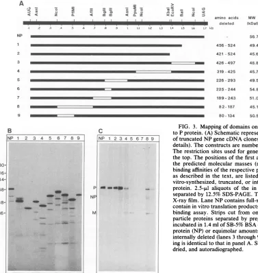
Characterization ofdomainsonNPprotein responsiblefor
bindingtoimmobilized renatured Sendai virus Pprotein.Our
assaysystemallowedmappingoffunctional domainsonNP
protein,which sofar had notbeenpossible. To identify the domainson NPprotein thatwererequired forbindingtoP protein, a series of nine truncated or internally deleted
"5S-NP
proteinswassynthesized.The mutatedregionscoverabout 80% of the coding region. Restriction enzyme sites used, positionsof thedeletedaminoacids, and the deduced B
tr. P
A
IPtr P V
-4.
U NP
p
HN F+NP
M
IC
Y
x
J. VIROL.
on November 10, 2019 by guest
http://jvi.asm.org/
[image:3.612.125.239.74.254.2] [image:3.612.326.555.76.266.2]SENDAI VIRUS PROTEIN-PROTEIN INTERACTION 1307
3) CE
-i, . 2 = =
-1) a Ci 'a=a(A=L
< < z C.L ammto S CL Z
_1 I
m
z6
0 n0 u In ; <:amino acids
li
~~~~~~~deleted
I de.eteI.: .2 .3 .4 .5 4 .7 a1 .9 Ll 12 L3 L4 L5 L6 L7 kb
MW relative binding
(kDal) affinity (%)
56.7 100
456-524 49.4 47
421 -524 45.6 45
426-497 48.8 100 319-425 45.7 19
226-293 49.5 5
225-244 54.8 3
189- 243 51.0 2 8 2- 187 45.1 18
80-134 50.5 5
B
NP 1 2 3 4 5 6 7 8 9
.4 _
_ _ t
C
molecularmassesof translation productsaredepictedinFig.
3A. The deletion junctions weresequenced toconfirm that thedeletionswereinframe. Themigrationof truncated and
internally deleted [35S]methionine-labeled in vitro transla-tion products in SDS-polyacrylamide gels corresponded to thereduction in theirsizes, withtheexceptionofconstruct 8 which has a higher apparent molecular mass than that
predicted from the sequence (Fig. 3B). Minor amounts of smaller radiolabeled products presumably represent poly-peptides whose synthesis has started at internal AUG codons. This has also been observeduponcell-freesynthesis of measles virus NPprotein (16).
We tested these mutated 35S-NP proteinsfor theirability tobind toPprotein. The mutatedproteinscontain 7to28% (i.e., 1 to 4) fewer methionine residues than the full-size 35S-NP protein. By using 7% less trichloroacetic acid-pre-cipitable activity forevery missing methionine, we applied
equimolar amounts of the mutated 35S-NP proteins to indi-vidualstripsofonepreparative blotcontaining Sendaivirus particle proteins. The signals obtainedin the binding assay
(Fig. 3C) were quantified by cutting the respective bands from the blot andby subsequent liquidscintillationcounting. The radioactivity retained by adjacent pieces ofequal size
FIG. 3. Mapping of domains on NP protein required for binding to Pprotein.(A) Schematic representation ofinternallydeleted and of truncatedNP gene cDNA clones (see Materials and Methods for details). Theconstructs are numbered from 1 through 9 at the left. The restriction sites used forgenerating deletions are depicted on thetop. Thepositionsof the first and last deleted amino acids and the predicted molecular masses (mw) are indicated. The relative binding affinities ofthe respective proteins to P protein, determined asdescribed in the text, are listed at the right. (B) Analysis of in vitro-synthesized, truncated, orinternallydeleted forms of35S-NP protein. 2.5-,ul aliquots of the in vitro translation reaction were separated by12.5% SDS-PAGE. The gel was dried and exposed to X-rayfilm.Lane NPcontains full-size35S-NPprotein. Lanes 1 to 9 contain in vitro translationproducts ofconstructs 1 to 9. (C)Protein binding assay. Strips cut from one blot, containing Sendai virus particle proteins separated by preparative 10% SDS-PAGE, were incubatedin 1.4ml ofSB-5% BSA containing50,000cpm of35S-NP protein(NP) orequimolaramountsoftruncated (lanes 1 and 2) or internallydeleted(lanes3through9)35S-NPproteins. The number-ing is identicaltothatinpanel A.Stripsweresubsequently washed, dried, and autoradiographed.
was subtracted as background. The percentage of activity retainedby immobilized P protein versus the total
radioac-tivity employed was determined. The values obtained for
mutatedNPproteinswere setinrelation to that of full-size 35S-NPprotein,whichwasconsidered100%. The resultsare
depictedin Fig. 3Aasrelative binding affinity.
The NP gene of Sendai virus Fushimi strain contains 524 amino acids (34). Deletion of amino acids 426to 497 (con-struct 3) did not affect binding of NP to immobilized P
protein. Removal ofcarboxyl-terminal amino acids 456 to
524 (construct 1) or 421 to 524 (construct 2) reduced the
bindingaffinitytoabout 50%.Withconstructs4,8, and9,in
which thedeletions reside between amino acidpositions319 and425,82 and187,and 80 and134,respectively,theaffinity was even lower. Inrepeated experiments, the least
binding
wasobservedwithconstructs5, 6,and7,lacking 67, 20,and
55 overlapping amino acids betweenpositions226 and 293,
225 and244,and 189and243,
respectively,
inahydrophobic
domainof the NPprotein. Our data thus indicate that
large
partsofNPproteinseem tobeinvolved in
binding
ofNPtoPprotein.The
hydrophilic
carboxyl
terminus(positions
498to524)playsanimportantrole,as
comparison
ofconstructs1 and 2 to construct 3 shows, but the
hydrophobic
middlepartofNPprotein provedtobemore
important.
The smallregionbetween
positions
225and 244 is essential forbinding.
A
NP
2 3
4 5
6
7 8 9
INP 1 2 3 4 5 6 7 8 9
180- 116-
84-
58-
48-
36-P
NP
ml
MONO
m
I
VOL.65, 1991
on November 10, 2019 by guest
http://jvi.asm.org/
[image:4.612.66.443.71.469.2]1308 HOMANN ET AL.
DISCUSSION
This report demonstrates that Sendai virusproteins,
sep-arated by SDS-PAGE and blotted to immobilizing
mem-branes,
canbeused to study their interactionswithproteinssynthesized
in vitro from recombinant cDNA clones.Immo-bilizing
electrophoretically separated proteins notonlyusesthe high resolving power of SDS-PAGE but also allows
renaturation ofenzymatic activity through removal of the
detergent(14, 23, 41, 44). Renaturation of proteins blotted to
animmobilizingmatrix (42) proved to be useful for studying
their interactions with DNA and RNA (2, 3, 12, 21, 35, 38),
with virusparticles (6, 29),with growthfactors (31), andwith
iodinated purified proteins (2, 26).
Incombination with in vitro systems for the synthesis of
proteins
from cloned cDNA,theserenaturingblotsrepresent apowerful
tool for studying protein-proteininteractions. Weapplied
in vitro-synthesized radiolabeled Sendai viruspro-teinsNP,P,and M to renaturingblotsand observed specific
retention by immobilized viral proteins in the absence of RNA. The sensitivity of the method probably derives from
the high specific activity of the protein probes that will be
retained, even if only a small percentage of immobilized
proteins on the membranes is renatured. The facts that
35S-NP is retained by immobilized P protein and,
con-versely, that 35S-P is retained by immobilized NP protein
indicatethat the interaction detected with our assay system
is indeed specific. Neither 35S-NP nor 35S-P protein is
retained by nonviral proteins. Additionally, the domain
responsible
for P protein binding to nucleocapsids (37) isidentical to that required for binding to immobilized
rena-tured NP protein. Our results therefore confirm that NP
protein
is the component of the nucleocapsid to which Pprotein
binds.We observed that NP proteins from virus particles and
from infected cells retain35S-P protein equally well.
How-ever,the signals obtained could very well be caused by only asmallsubpopulation oftheimmobilized proteins, since the molar amounts ofradiolabeled proteins applied in the
bind-ing
assay are less than 0.1% of those blotted to the PVDF matrix. NP andP are both phosphoproteins (25, 43) whichare phosphorylated, at least in vitro, by L protein (9).
Phosphorylation may definedifferent NP or P protein
popu-lations between which we could not discriminate in our
assay system. Further work is now in progress to analyze
isoforms of NP and P proteins, separated by
two-dimen-sional electrophoresis, fortheir binding affinities.
The assay system developed now allows definition of domains on NP protein required for interaction with other
proteins.
This will help elucidate the role(s) of NP protein, the mostabundant viral protein, in virus multiplication. Thedeletion mutant analysis with NP protein revealed that very
large parts of this protein are involved in the binding to P
protein. From the seven internally deleted NP proteins
tested, only the deletion of amino acids 426 to 497 did not
affect the affinity to Pprotein. These results do not
neces-sarily show that all of theresidues from positions 80 to 426
are directly involved in complex formation, but they may
rather reflect that the NP-P protein interaction is very sensitive toconformational changes in NP protein.
However, the effect of internal deletions does not
corre-latewith the size of the regionremoved. With the exception of amino acids 80 and 81, the deletion of construct 9 is
includedin thedeletion ofconstruct 8. Although the deletion
in construct 8 is nearly twice that inconstruct 9, its effect on
the binding affinity is weaker than that observed with
con-struct 9. Therefore, amino acids 80 and 81 may indeed be
involved in
binding
to Pprotein. Additionally,
the smallestdeletion (construct6)causesthemost
pronounced
reductionin binding to P protein. The
carboxyl-terminal
27 aminoacidsofNP
protein
also contributesignificantly
toitsaffinity
to Pprotein,butbinding is stillobserved whenthis
region
isremoved. The carboxyl terminus does not represent an
independentbinding domain forP protein, sincemost
inter-nal deletions abolish binding to P protein even though the
carboxyl terminus is present.
Theresolution ofourdeletion analysis maybe toolowto
state thatamino acids 80 and 81, 225 to244, and 498to524 indeed cooperate toform thebinding site forPprotein,
but,
evenif these aminoacidsmay notbeinphysicalcontactwith P protein, ourresults show that theseregions are
important
forbinding.With NPprotein assembled intoa
nucleocapsid,
only its
carboxyl
terminus isexposed
tothesurface(17).
Butinternal regions ofNPprotein arealso likelytobind
directly
to P protein. Transcriptionally active
nucleocapsids
areresistant to RNases, and additionally, P protein was shown
by cross-linking experiments to contact the RNA
(34).
Therefore, it seemsthat NPproteins are notremoved at the sites of polymerase action but that thepolymerase
complex
stays in close contact to NP proteins. The
hydrophobic
middle half ofparamyxovirus NPproteins, unlike the
hydro-philic and highly charged carboxyl termini, is
highly
con-served (33). This also indicates that regionsrequired
foressential functions ofNP protein reside in its middle half.
This is further supported by the finding that a monoclonal
antibody which recognizes a determinant between
positions
290and 295 mosteffectivelyinhibits in vitro transcription (7, 10).
More elaborate analysis ofNPprotein by linker-insertion
and site-directed mutagenesis will provide further
informa-tion about which residues are required for interaction with viral polymerase proteins.
ACKNOWLEDGMENTS
We thank P. H. Hofschneider for critical discussions and helpful advice, R. Koshy, J. Wells, and V. Radwitz for
critical
reading of the manuscript, and K. Wiedemann and C. Baumann for excellent technical assistance.This study was supported by a grant from the Deutsche
Forschungs-gesellschaft,
Schwerpunkt "Persistierende Virusinfektionen." REFERENCES1. Banerjee, A. K. 1987. Transcription and replication of rhabdo-viruses. Microbiol. Rev. 51:66-87.
2. Bowen, B., J. Steinberg, U. K. Laemmli, and H. Weintraub. 1980. The detection of DNA-binding proteins by protein blot-ting. Nucleic Acids Res. 8:1-21.
3. Boyle, J. F., and K. V. Holmes. 1986. RNA-binding proteins of bovine rotavirus. J. Virol. 58:561-568.
4. Chirgwin, J. M., A. E. Przybyla, R. J. MacDonald, and W. J. Rutter. 1979.
Isolation
of biologically active ribonucleic acid from sources enriched in ribonuclease. Biochemistry 18:5294-5299.5. Curran, J., and D. Kolakofsky. 1989. Scanning independent ribosomal initiation of the Sendai virus Y proteins in vitro and in vivo. EMBO J. 8:521-526.
6. Defer, C., M. T. Belin, M. L. Caillet-Boudin, and P. Boulanger. 1990. Human adenovirus-host cell interactions: comparative study with members of subgroups B and C. J. Virol. 64:3661-3673.
7. Deshpande, K. L., and A. Portner. 1984. Structural and func-tional analysis of Sendai virus nucleocapsid protein NP with monoclonal antibodies. Virology 139:32-42.
J. VIROL.
on November 10, 2019 by guest
http://jvi.asm.org/
8. Deshpande, K. L., and A. Portner. 1985. Monoclonal antibodies to the P protein of Sendai virus define its structure and role in transcription. Virology 140:125-134.
9. Einberger, H., R. Mertz, P. H.Hofschneider, and W. J. Neubert. 1990. Purification, renaturation, and reconstituted protein ki-nase activity of the Sendai virus large (L) protein: L protein phosphorylates the NP and P proteins in vitro. J. Virol. 64:4274-4280.
10. Gill, P. D., S. Takai, A. Portner, and D. W. Kingsbury. 1988. Mapping of antigenic domains of Sendai virus nucleocapsid protein expressed in Escherichiacoli. J. Virol. 62:4805-4808. 11. Giorgi, C., B. M. Blumberg, and D. Kolakofsky. 1983. Sendai
virus contains overlapping genes expressed from a single mRNA. Cell 35:829-836.
12. Gorlach, M., M. Hermann, M.Schwemmle, and K. Hilse. 1989. Binding of globin mRNA, ,-globin mRNA segments and RNA homopolymers by immobilized protein of polysomal globin messenger ribonucleoprotein. Eur. J. Biochem. 184:589-596. 13. Gotoh, H., T. Shioda,Y.Sakai, K. Mizumoto, and H. Shibuta.
1989. Rescue of Sendai virus from viral ribonucleoprotein-transfected cells by infection with recombinant vacciniaviruses carrying Sendai virus L and P/C genes. Virology 171:434 443. 14. Hager, D. A., and R. R. Burgess. 1980. Elution ofproteins from
sodium dodecyl sulfate-polyacrylamidegels, removal of sodium dodecyl sulfate, and renaturation ofenzymatic activity: results with sigma subunit of Escherichiacoli RNApolymerase, wheat germ DNA topoisomerase,andother enzymes. Anal. Biochem. 109:76-86.
15. Hamaguchi, M., T.Yoshida, K. Nishikawa, H. Naruse, and Y. Nagai. 1983. Transcriptive complex of Newcastle disease virus. I. Both L and P proteins are required to constitute an active complex. Virology 128:105-117.
16. Hasel, K. W., S. Day, S. Millward, C. D. Richardson, W. J. Bellini, and P. A. Greer. 1987. Characterization of cloned measles virus mRNAs by invitrotranscription, translation, and immunoprecipitation. Intervirology28:26-39.
17. Heggeness, M. H., A. Scheid, and P. W. Choppin. 1981. The relationship of conformational changes in the Sendai virus nucleocapsid to proteolytic cleavage of the NPprotein. Virol-ogy 114:555-562.
18. Hidaka, Y.,T. Kanda, K. Iwasaki, A. Nomoto, T. Shioda, and H. Shibuta. 1984. Nucleotide sequence of a Sendai virus genome region covering the entire M gene and the 3' proximal 1013 nucleotides ofthe F gene. Nucleic Acids Res. 12:7965-7973. 19. Hill, V. M., and D. F. Summers. 1990. A minor
microtubule-associated protein isresponsible for thestimulation of vesicular stomatitis virus transcriptionin vitro. J. Gen. Virol.71:289-298. 20. Homann, H. E., W. J. Neubert, and P. H. Hofschneider. 1990. Sendai virus gene expression in lytically and persistently in-fected cells.Virology177:131-140.
21. Hubscher, U. 1987. Doublereplicasouthwestern. Nucleic Acids Res. 15:5486.
22. Kolakofsky, D., and L. Roux. 1987. The molecular biology of paramyxoviruses, p. 277-297. In P. Bercoff(ed.), The molecular basis of viral replication. PlenumPublishing Corp., New York. 23. Lacks, S. A., and S. S. Springhorn. 1980. Renaturation of enzymes after polyacrylamide gelelectrophoresis in the pres-ence of sodium dodecyl sulfate. J. Biol. Chem. 255:7467-7473. 24. Laemmli, U. K. 1970. Cleavage of structural proteins during assembly of the head ofbacteriophage T4. Nature (London) 227:307-310.
25. Lamb, R. A., and P. W. Choppin. 1977. The synthesisof Sendai virus polypeptides in infected cells. III. Phosphorylation of polypeptides. Virology81:382-397.
26. Lesot, H., U. KuhI, and K. von der Mark. 1983. Isolation ofa
laminin-bindingprotein from muscle cell membranes. EMBOJ.
2:861-865.
27. Markwell, M. A. K., and C. F. Fox. 1980. Protein-protein interactions withinparamyxoviruses identifiedbynative disul-fide bonding or reversible chemical cross-linking. J. Virol. 33:152-166.
28. Melton, D. A., P. A. Krieg, M. R. Rebagliati,T. Maniatis, K. Zinn, and M. R. Green. 1984. Efficient in vitro synthesis of biologically active RNA and RNAhybridization probes from plasmids containing a bacteriophage SP6 promoter. Nucleic AcidsRes. 12:7035-7056.
29. Mischak,H.,C.Neubauer,B.Berger,E.Kuechler,and D. Blaas. 1988. Detection of the human rhinovirus minorgroup receptor onrenaturingwesternblots.J. Gen. Virol. 69:2653-2656. 30. Moyer, S.A.,S. C.Baker,andJ. L. Lessard. 1986.Tubulin, a
factor necessary for the synthesis of both Sendai virus and vesicularstomatitis virus.Proc.Natl. Acad.Sci. USA 83:5405-5409.
31. Nakamura, T.,K. Takio,Y. Eto,H. Shibai,K.Titani, and H. Sugino. 1990. Activin-bindingproteinfromratovaryis follista-tin. Science 247:836-838.
32. Neubert,W.J. 1989.Cloningandsequencingof thepolymerase
gene (P) of Sendai virus(strain Fushimi). Nucleic Acids Res. 17:10101.
33. Neubert, W. J., C. Eckerskorn, and H. E. Homann. Virus Genes,in press.
34. Raghow,R.,and D. W.Kingsbury. 1979. Protein-RNAcontacts
in Sendai virus nucleocapsids revealed byphoto-crosslinking. Virology98:267-271.
35. Roy, P.,A.Adachi,T. F.Urakawa,T. F.Booth,and P. Thomas. 1990.Identification ofbluetonguevirusVP6proteinas anucleic acid-binding protein and the localization ofVP6 in virus-in-fectedvertebratecells. J. Virol.64:1-8.
36. Ryan,K.,and D. W.Kingsbury. 1988.Carboxyl-terminal
region
of Sendai virus P protein is required for binding to viral nucleocapsids. Virology167:106-112.
37. Ryan, K., and A. Portner. 1990. Separate domains of Sendai virus Pprotein arerequiredforbindingtoviral nucleocapsids. Virology174:515-521.
38. Schenkel,J.,C. E.Sekeris,A.Alonso,and E. K. F. Bautz.1988. RNA-bindingproperties ofhnRNPproteins. Eur. J. Biochem. 171:565-569.
39. Shioda, T.,K.Hidaka, H.Shibuta,A.Nomoto, and K.Iwasaki. 1983. Sequence of3,687 nucleotides from the 3'end ofSendai virus genome RNA and thepredictedamino acid sequencesof viralNP,Pand Cproteins. Nucleic AcidsRes. 11:7317-7330. 40. Shioda, T.,K. Iwasaki,and H. Shibuta. 1986. Determination of
the complete nucleotide sequence of the Sendai virus genome RNAand thepredictedamino acidsequencesof theF, HNand L proteins. Nucleic AcidsRes. 14:1545-1563.
41. Szewczyk, B.,W. G.Laver, and D. F.Summers. 1988. Purifica-tion, thioredoxinrenaturation, andreconstituted
activity
ofthe threesubunitsof the influenzaAvirusRNApolymerase. Proc. Natl. Acad. Sci. USA 85:7907-7911.42. Towbin,H.,T.Staehelin,andJ.Gordon. 1979.
Electrophoretic
transfer ofproteins from polyacrylamidegels tonitrocellulose sheets. Proc. Natl.Acad. Sci. USA76:4350-4354.
43. Vidal, S., J. Curran, C. Orvell, and D.
Kolakofsky.
1988. Mapping of monoclonal antibodiestotheSendai virusPprotein
and thelocalization of itsphosphates. J.Virol.62:2200-2203. 44. Weber, K., and D. Kuter. 1971. Reversible denaturation of
enzymesby sodium dodecylsulfate. J. Biol. Chem. 246:4504-4509.
45. Willenbrink,W.,andW.J.Neubert.1990.
Cloning
andsequenc-ing of the matrix protein gene (M) of Sendai virus (strain
Fushimi). Nucleic AcidsRes. 18:3993.
![FIG. 2.fromComparisonaliquotsfromwasunprogrammedproteinsthe(lanes5-mmorcallyincubatedexposedSDS-PAGE.scribed[35S]methionine-labeled10% 15 Binding of 35S-NP, 35S-P, and 35S-M proteins to proteins Sendai virus particles and infected and mock-infected cells](https://thumb-us.123doks.com/thumbv2/123dok_us/1316603.85155/3.612.326.555.76.266/fromcomparisonaliquotsfromwasunprogrammedproteinsthe-mmorcallyincubatedexposedsds-methionine-proteins-proteins-particles-infected-infected.webp)