JOURNAL OFVIROLOGY, Aug. 1992,p. 5114-5118 0022-538X/92/085114-05$02.00/0
Copyright )1992,AmericanSociety forMicrobiology
Functional Tolerance of the Human Immunodeficiency Virus
Type
1
Envelope Signal Peptide
to
Mutations in the
Amino-Terminal
and
Hydrophobic Regions
HEINZ
ELLERBROK,1*
LUCD'AURIOL,2 CATHERINE VAQUERO,'ANDMARCSITBON' Laboratoire d'ImmunologieetOncologie des Maladies Rtrovirales, INSERM U152, Institut Cochin deGenetique Moleculaire, UniversitedeParis V, 27, rueduFaubourgSt-Jacques, F-75014 Paris, and Genset
SA,
F-75011Paris,2
FranceReceived 14October 1991/Accepted 1 May 1992
Wedemonstrated that the leadersequenceof the humanimmunodeficiencyvirustype 1envelopefunctions
assignal peptide (SP) despite low scoringinapredictionprogram.AsexpectedforSP,thehydrophobiccore (HC) is essential, and noother sequence could compensate for HC deletion. Contraryto otherSPs, major
substitutionsintheHC,suchasintroductionofbasic, polar,ora-helix-breaking residues,stillallowed efficient
translocation and glycosylation. Also, extensive deletions or substitutions of the chargedresidues at the N terminus hadlittle ifanyinhibitory effect.This report, which is the firststudyofhuman
immunodeficiency
virusSP,describes theexceptionaltolerance of thispeptidetomutations.
According to the signal hypothesis (3), targeting and translocation into the endoplasmic reticulum of retroviral envelope glycoproteins (Env) dependson anamino-terminal signal peptide (SP) (39). In general, SP contain approxi-mately15to30 aminoacids organizedinatripartitestructure comprising an amino-terminal
(Ntem)
region and a central hydrophobiccore(HC)followedby small polaraminoacids (35-37, 39). MostretroviralSPs,identifiedonly bytheirHC located immediately upstream from the mature protein,score poorly in prediction programs such as PSIGNAL
(Intelligenetics) (not shown). Exceptforafewmutations in
the avianRoussarcomavirus SP(41), noretroviral SPhas
been specifically studied. Because of the importance of human immunodeficiency virus type 1 (HIV-1) as a
patho-gen,westudied theHIV-lBRU SP(38).Since Envexpression issubmittedtocomplex regulations (42),weusedaninvitro
systemwith rabbitreticulocyte lysate,T7 RNApolymerase, and dog pancreatic microsomal membranes (Promega), as previously described for the study of SP functions (2, 3, 15, 23, 24, 34, 39).
Tolerance oftheHIV-1Envhydrophobiccoretomutations. TheT7-transcribedmRNAcorrespondingtotheentire
HIV-1BRU
envcoding region
(Fig.
1A)
translated in rabbitretic-ulocyte lysate in presence of microsomal membranes (6) yieldedacompletegpl60 envelopeprecursor(Fig.1B)anda
120-kDaprotein,gpl2O*, resultingfromatranslationalarrest occurringintheRev-responsive element(6). Bothproducts weresensitivetoendoglycosidasesFandH, confirmingtheir extensive glycosylation (not shown). Oligonucleotide-di-rectedmutagenesisofthe wild-type envwas performed by usingahighly efficientmethodadapted from that of Kunkel (20) to Bluescript phagemid (Stratagene) (6). Mutated and wild-typeenvgenes weretranscribed in vitro,mRNAswere quantitated, identicalamountsof mRNAsweretranslatedin parallel, and aliquotswere run on sodium dodecyl sulfate-gels. Allmutantswere transcribedwith similar efficiencies,
and translation in the absence of microsomal membranes yielded similaramountsof the unglycosylated Pr9OenV,
indi-*Correspondingauthor.
catingthat the overall transcriptional andtranslational effi-ciencies were not altered by the different mutations. Al-though the prediction score for the potential SP of HIV-1
Env was very low (PSIGNAL, PC/Gene; Intelligenetics),
deletion of the entire SPsequencewiththeexceptionof the AUG initiation codon (pEASP) yielded only the nonglyco-sylated precursor Pr9Oenv, similar to the wild-type pE160 translated in absence of microsomal membranes (Fig. 1B, lane e). Therefore,nomechanism independentofan
amino-terminal SP was used for translocation of Env, and as expected for an SP, deletion of the HC abolished protein glycosylation (pEAWhc and pEAhc;Fig. 2A).
GeneralrequirementsforfunctionalHC identified in vitro and in vivo seem to include overall hydrophobicity and a-helicalstructure(15, 23, 27, 37). TheHC of the HIV-1 SP is flanked at its N terminus by a unique Trp motif that
includeshydrophobicandchargedresidues.Usingadeletion
mutant, we found that this Trp motif (pEAW) was not essential for translocation (Fig. 2A). Introduction in the center of theHC of eitheran a-helix-breaking proline or a charged aminoacid blocks translocation of several SPs (2, 15, 23, 24,27,33, 37).Such mutationsanalyzedinvitroorin
vivo result in complete or dramatic inhibition of
glycosyl-ation. We derived four mutants with single mutations at position -8or -9, thecenterof theHC of the HIV-1 Env SP. Glycosylation was unaltered after either a charged (arginineinpER-8)or apolar (serineinpES-8)residuewas
substituted for Gly-8 (Fig. 2B). Substitutionofargininefor Leu-9 (pER-9) resulted in substantial accumulation of the nonglycosylated
Pr9Menv;
however, glycosylation was not completely abolished in this mutant (Fig. 2B). Thus, the effect of the positive charge introduced into this HCap-pearedto be position dependent. Itwas not clear whether the alterationwasduetothe introductionofachargeperse ortothesubsequentreduction of theoverallhydrophobicity due to replacement of Leu-9. Alteration of glycosylation similartothat observed withpER-9wasalsoobservedwith pEP-9, in which Leu-9 was replaced by the hydrophobic proline. Therefore,bothsecondarystructureand hydropho-bicityatthisposition mightbeimportantforafunctional SP. Accordingly,introductionofaprolineinthe middle ofthe SP
5114
Vol. 66, No. 8
on November 9, 2019 by guest
http://jvi.asm.org/
cleavage
N-terminal charges
Ihydrophobic
coreI
siteB)
ab
cd
e-30 -20 -10 -1
+ + -+ ± + +
M R V K E K Y Q H L W R W G W K W G T M L L G I L M I C S A tryptophan not;if
5803 8388
SP gp
CTGCAGTGACAL
Pst /
gpl6O
_
_
gp
120
*
-PrgoEEnV--
iENV
[image:2.612.88.535.79.268.2]Pst I
FIG. 1. The HIV-lBRU envelope signal peptide. (A) Schematic representation of the HIV-1 envelope construct used for in vitro transcription and translation.Only the HIV-derived insert is shown. Numbering is according to reference 38. By using the two PstI sites, this regionwasintroduced into the Bluescriptvector. Theregion coding for thegpl60 envelope precursor is drawn as a box. The surface protein SUgpl20 and the transmembraneprotein TMgp41areindicated. The surface-transmembranecleavage site between residues 516 and 517 is indicated withanarrow above the box. The SP is shaded, and thecorresponding amino acid sequence is shown above. The three distinct SP regions are indicated above thesequence, and the tryptophan motif is underlined. The amino acid sequence is according to the one-letter code, andnumbering indicates positionsstarting from the carboxy-terminal amino acid of the SP as -1. (B) Translation of an SP deletion mutant ofHIV-1 env.Wild-type pE160(lanesaandd)andpEASP (lanes b and e)weretranslated in vitrofor 90minin the absence(lanesaandb) orpresence(lanes d ande) of microsomal rough membranes. Translation intheabsenceofspecificmRNAis shown in lane c.Glycosylated andunglycosylatedEnvproductsareindicated.
A)
I)E160 M RVK EKYQHLW RW GW KW GT ML L GI LM IC SA-I
pEASP *
-)EAWilC * * * * * * * * * * * * *
Co M
-3
o ro
0o
nx *
_<1
aIj
+1 T
*
*
pEAhe
p)EAW * * * * * * * * *- -** * * *** * *** * *
M R V K E K Y Q HI L W R W G W K W G T M
-9-8
L L G
-1 +1
I L M I C S A T
pEAllie * * * * * * * * ** * * **** * - * * * *
I)E
R8
* * * * * * * * * * * * * * * * * * * * * * R * * * * * * * *pE S-8 * * * * * * * * * * * * * * * * * * * * * * S * * * * * * * *
pEIR_9 * * ** * * * ** * ** * ** * ** * * R * * ** * ** * pEtR8R-9 * * * * * * * * * * * * * * * * * * * * * R R * * * * * ** *
pE P9 * * * * * * * * * * * * * * * * * * * **p * * * * * ** *
1,
A
FIG. 2. Mutations in theHCofthe SP.Translatedproteinsareshownontheright. Main productsareindicated. X indicatesanonspecific
proteinalsoproducedwithoutaddition of mRNA intheratreticulocytelysate.Thesequenceof SP of pE160 and schematic representations
of themutationsareshownonthe left.Dashed linesrepresentdeletions;asterisksrepresentunchanged amino acids. Mutated residuesare
indicated. -1 and +1 indicate thecarboxyterminus ofthe SP and the aminoterminusofthemature protein, respectively.(A)Deletion
mutants. Forcomparison, productsofpE160 translatedintheabsence(a)orpresence(b) of microsomal rough membranesareshown. (B)
Point mutations intheHC. pE160andpEAHCareincludedascontrols.
A)
B)
I)E160
on November 9, 2019 by guest
http://jvi.asm.org/
[image:2.612.86.541.412.667.2]5116 NOTES
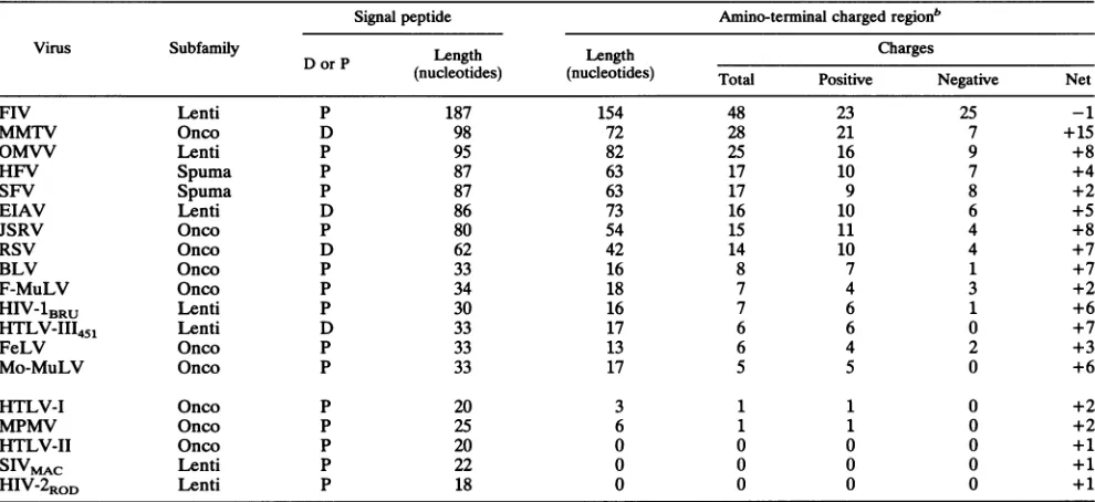
TABLE 1. Amino-terminal charges of retroviral envelope signal peptidesa
Signal peptide Amino-terminal chargedregionb
Virus Subfamily D orP Length Length
Charges
(nucleotides) (nucleotides) Total Positive Negative Net
FIV Lenti P 187 154 48 23 25 -1
MMTV Onco D 98 72 28 21 7 +15
OMVV Lenti P 95 82 25 16 9 +8
HFV Spuma P 87 63 17 10 7 +4
SFV Spuma P 87 63 17 9 8 +2
EIAV Lenti D 86 73 16 10 6 +5
JSRV Onco P 80 54 15 11 4 +8
RSV Onco D 62 42 14 10 4 +7
BLV Onco P 33 16 8 7 1 +7
F-MuLV Onco P 34 18 7 4 3 +2
HIV-lBRU Lenti P 30 16 7 6 1 +6
HTLV-III451 Lenti D 33 17 6 6 0 +7
FeLV Onco P 33 13 6 4 2 +3
Mo-MuLV Onco P 33 17 5 5 0 +6
HTLV-I Onco P 20 3 1 1 0 +2
MPMV Onco P 25 6 1 1 0 +2
HTLV-II Onco P 20 0 0 0 0 +1
SIVMAC
Lenti P 22 0 0 0 0 +1HIV-2RoD Lenti P 18 0 0 0 0 +1
aRetroviruses (references):FIV(feline immunodeficiency virus,PPRisolate) (25);MMTV(mousemammary tumorvirus) (13, 21); OMVV (ovine maedi-visna virusstrainSA) (26);HFV(human foamy virus)(7); SFV (simian foamy virus) (22);EIAV(equine infectious anemiavirus) (18); JSRV(jaagsiekte sheep retrovirus) (43); RSV (Roussarcomavirus) (14);BLV(bovine leukemiavirus) (28);F-MuLV(Friend murine leukemiavirus) (19);HIV-1BRU(38);HTLV-III451(17);FeLV (feline leukemia virus strain B)(5); Mo-MuLV (Moloney murine leukemiavirus) (31);HTLV-I(29);MPMV(Mason-Pfizer monkey virus) (32);HTLV-II(30); SIVmAc(simian immunodeficiency virus frommacaque) (4);HIV-2ROD (10).Thecorrespondingsubfamilies,Lentivirinae(Lenti),Oncovirinae(Onco),and Spumavirinae(Spuma)areindicated. Also indicatedis whether thecleavage site has been determinedbyamino acidsequencing (d)orpredictedfrom the nucleotidesequence(p).
bThe amino-terminalcharged region is located betweentheinitiation methionineand thelastcharged residue before the HC. Thenetchargeincludes the positively charged NH2groupofthe amino-terminal methionine.
HC of an Eschenichia coli ribose-binding protein inhibits translocation, and apseudorevertantwithincreased hydro-phobicity of theHC hasbeen isolated (16). In thatmodel, translocation is completely blocked, whereas we observed only partial inhibitionofglycosylationaftersubstitution ofa proline.Anotherexampleof the remarkable tolerance of the HIVHCtomutation isthatasignificantamountof glycosy-lated Envwas detected evenwhen arginines were substi-tuted forbothGly-8 and Leu-9 (pER-8R_9) (Fig. 2B).The Trp motif includes hydrophobic residues alternating with charged orpolar aminoacids and seemstobe acomposite
structurebetween thecharged Nterm and theHC.This motif mightthen add sufficient hydrophobicityto thecentral part of the SPtoovercomethepotentiallydeleteriousmutations
such as those in pER_8,
pER-8R_9,
or pEP-9. Since weobserved that deletionof this motif alone hadno detectable effectonproteinglycosylation, usefurther combined muta-tions willhelpto testthis hypothesis.
Accumulation of a nonglycosylated precursor observed
withcertainmutants(Fig. 2) mightbe duetoeitherinhibition of translocationorsubsequentglycosylation. Todistinguish between these possibilities, we monitored accessibility of
theproductstoproteinase K. Addition ofprotease to wild-type products resulted in a size reduction of gpl60 to approximately 135 kDa, most likely due to the expected proteolysis of 150 amino acids of the cytoplasmic domain (11) (not shown). The gpl2O* was protected fromprotease because of the absence of thecytoplasmicdomain duetoa
translationalarrestin theRev-responsiveelement(6). Addi-tion of 0.5% Nonidet P-40 prior to proteolysis allowed degradationof bothproteins, indicatingthatprotectionwas
indeed due to translocation and not to
intrinsic
proteaseresistance.
In contrast, addition of protease in absence ofdetergent
tothePr9Oenv
producedby pEASP
orpER-8R_9
resulted in itscomplete
degradation
(not
shown),
demon-strating
lack of translocation of thisproduct.
Toleranceof the HIV-1 SPtomutationsin the Nterminus. The HIV SP
Nterm
region
hasstriking
accumulation
ofcharges:
seveninthe 16 amino acidspreceding
theHC
(Fig.
1A).
Wefound thatonly
1 of 185eukaryotic
nonretroviralSPs
(40)
hadfive
charges
and thatover150
hadasingle
or nocharge.
Nevertheless,
in vitro and in vivo studies have shown theimportance
of amino-terminalcharges
fortrans-location
ofprepro-a-factor
(9)
andpreproparathyroid
hor-mone(34)
and for attachment of membrane-bound and secretoryproteins
in E.coli
(1,
8). Among
thedetermined
orpredicted
SPs ofrepresentative
retroviralEnv,
most,includ-ing HIV-1,
weremuch
longer, mostly
asthe result of
an extendedcharged
Nten,,
whereasfew,
as human T-cellleukemia
virus(HTLV)
andHIV-2,
hadclassical
Sps
(Table
1).
Two mutants of the HIV-1 SP in whichglycines
weresubstituted
either for the fivecharged
residues N terminal of theTrp
motif(pE5G)
orforallcharged
residues(pE7G) (Fig.
3)
allowed unalteredglycosylation.
Deletion of thecorre-sponding
amino-terminalcharged
region
(pEANterm)
re-sultedonly
in averyslight
accumulation ofunglycosylated
Pr9Oenv
(Fig. 3), confirming
the tolerance of this SP toextensive
mutations.This reportconstitutes the
first
study
ofanHIVSP. Like most retroviralSPs,
this SP scorespoorly
in aprediction
program,mostlikely
because ofastriking
accumulation ofcharged
residues(Table 1).
Conservation
of this feature J. VIROL.on November 9, 2019 by guest
http://jvi.asm.org/
NOTES 5117
-o -O
Cm 0 0
+ +- + + + + +1 a
pEl60 M R VK E K Y QIf L WR W G W KW G T M L L G I L M I C S ATT
P
A
term*~~***************4pE5G * G * G G G * * G * * **** * ************* *
pE7G * G * G G G * * G * * G ***G***************''
FIG. 3. Mutations in theamino-terminal charged region. Symbols are as described in thelegendto Fig. 2. Either the amino-terminal charged regionwasdeleted
(pEANte,,n)
orcharged residueswerechanged toglycines(pE5G and pE7G).suggests some
biological
importance.
The extendedSPNte.
oftenoverlaps
with othergenetic elements (integrase
in oncoviruses andvpu inHIV-1) and might thus act as a buffer region during adaptive mutations.Accordingly
and in con-trast toHIV-1, HIV-2 has no vpu and no described function in the regionof
the SP. Since most retroviruseshave
exceptionally
long Env SPs, it is tempting tospeculate
that, by analogy with oncoviruses and HIV-1, there are alterna-tive functions for all oftheseregions.
Recently,
it has been shown thatSP-derived
peptides
canassociate with
mole-cules of themajor
histocompatibility complex through
analternative
antigen
processing pathway (13). Interaction
be-tweenthis
pathway
andpeculiar SP features
as described abovemight
have additional functions selectedfor the
ben-efit of the virus. Thestudy
ofSP mutations
in differentcellular
contextsmight
then unmask newpotential
deleteri-ouseffects.
Wethank C. Locht,P. Sonigo, andJ. Portis forhelpful discus-sions; M. Emerman, A. Cordonnier, E. Legouy, and M. Noyer-Weidner forgiftsofplasmids and of bacterial andphagestrains; F. Pozo and N. Delaunay for excellent technical assistance; and S. Gisselbrecht,J.-P.Levy,and P.Tambourinforcontinuoussupport. H.E. wassupportedbyfellowshipsfromtheDeutsche Forschungs-gemeinschaft, the FondationpourlaRechercheM6dicale,and the Agence Nationale pourla Recherche sur le SIDA(ANRS). This workwassupportedbygrants89282 and91205 from ANRStoM.S. andC.V.
REFERENCES
1. Akita, M.,S. Sasaki, S.Matsuyama, andS.Mizushima. 1990. SecA interacts withsecretoryproteinsbyrecognizingthe pos-itive charge at the amino terminus of the signal peptide in
Escherichia
coli.J. Biol. Chem.265:8164-8169.2. Allison,D.S.,and E. T.Young. 1988.Single-amino-acid substi-tutions within thesignalsequenceof yeastprepro-a-factoraffect membrane translocation. Mol. Cell.Biol.8:1915-1922. 3. Blobel,G.,and B. Dobberstein.1975. Transferofproteinsacross
membranes. I. Presenceofproteolytically processed and non-processednascentimmunoglobulin chainsonmembrane bound ribosomes of murinemyeloma. J. Cell Biol.67:835-851. 4. Chakrabarti, L.,M. Guyader,M.Alizon,M. D.Daniel,R. C.
Desrosiers, P.Tiollais,and P.Sonigo. 1987.Sequence of simian immunodeficiencyvirus from macaqueand itsrelationship to
other human andsimianretroviruses. Nature(London) 328:543-547.
5. Elder, J. H.,andJ.I.Mullins.1983.Nucleotidesequenceof the envelope gene of Gardner-Arnstein feline leukemia virus B reveals unique sequence homologies witha murineminkcell focus-formingvirus. J. Virol.46:871-880.
6. EUerbrok,H.,N.Serpente, L.d'Auriol,T.Klimkait,M.Sitbon, and C.Vaquero.Unpublisheddata.
7. Flugel, R. M., A. Rethwilm, B. Maurer, and G. Darai. 1987. Nucleotide sequenceanalysisoftheenvgene andits flanking
regionsof thehuman spumaretrovirus revealstwonovelgenes. EMBO J.6:2077-2084.
8. Galluser, A.,and A. Kuhn.1990. Initialstepsinprotein mem-braneinsertion. BacteriophageM13 procoatprotein bindstothe membrane surface by electrostatic interaction. EMBO J. 9:2723-2729.
9. Green,R, R. A.Kramer,andD. Shields.1989.Misplacement of theamino-terminal positivecharge in theprepro-a-factor signal peptidedisrupts membrane translocation in vivo.J.Biol. Chem. 264:2963-2968.
10. Guyader, M., M. Emerman, P.Sonigo,F.Clavel,L.Montagnier, and M. Alizon.1987. Genomeorganization and transactivation of thehumanimmunodeficiency virustype2.Nature (London) 326:662-669.
11. Haffar, 0. K., D. J. Dowbenko, and P. W. Berman. 1988. Topogenicanalysis of the humanimmunodeficiency virustype1 envelope glycoprotein, gpl60, in microsomal membranes. J. Cell Biol. 107:1677-1687.
12. Henderson, L.E., R. Sowder, G. Smythers, and S. Oroszlan. 1983.Terminalamino acidsequencesandproteolyticcleavage sites of mouse mammary tumor virusenv gene products. J. Virol. 48:314-319.
13. Henderson, R. A., H.Michel,K.Sakaguchi, J. Shabanowitz, E. Appelia, D. F. Hunt, and V. H. Engelhard. 1992. HLA-A2.1-associatedpeptides froma mutantcell line:asecondpathway of antigen presentation. Science 255:1264-1266.
14. Hunter,E., E.Hill,M.Hardwick, A. Bhown, D. Schwartz, and R. Tizard.1983. CompletesequenceoftheRous sarcomavirus envgene:identification of structural and functional regions of its product.J. Virol.46:920-936.
15. Ibrahimi, I., and R. Gentz. 1987. A functional interaction between the signal peptide and the translation apparatus is detected by the use of asingle point mutation which blocks translocationacrossmammalianendoplasmic reticulum. J. Biol. Chem. 262:10189-10194.
16. Iida,A., J. M.Groarke,S. Park, J. Thom, J. H.Zabicky,G. L. Hazelbauer,and L. L. Randall. 1985. Asignalsequence mutant defectivein exportofribose-binding proteinanda correspond-ing pseudorevertant isolatedwithoutimposed selection.EMBO J. 4:1875-1880.
17. Kalyanaraman,V.S.,V.Rodriguez,F.Veronese, R.Rahman,P. Lusso,A. L.DeVico, T.Copeland,S.Oroszlan,R.C.Gallo,and M. G. Sarngadharan. 1990. Characterization ofthe secreted, nativegpl20 and gpl60 of the humanimmunodeficiency virus type 1.AIDSRes. Hum.Retroviruses6:371-380.
18. Kawakami, T., L. Sherman,J.Dahlberg, A. Gazit,A. Yaniv, S. R.Tronick,andS. A.Aaronson. 1987.Nucleotidesequence analysis of equine infectious anemiavirusproviralDNA. Virol-ogy158:300-312.
19. Koch,W., G. Hunsmann, and R. Friedrich. 1983. Nucleotide sequenceof theenvelopegeneof Friend murine leukemiavirus. J. Virol.45:1-9.
20. Kunkel,T. A.1985.Rapidandefficientsite-specific mutagenesis without phenotypic selection. Proc. Natl. Acad. Sci. USA 82:488-492.
VOL.66, 1992
on November 9, 2019 by guest
http://jvi.asm.org/
[image:4.612.91.542.82.186.2]5118 NOTES
21. Majors,J. E., and H. E.Varmus.1983. Nucleotide sequencing of an apparent proviral copyof env mRNAdefinesdeterminants of expression of themouse mammary tumorvirus env gene. J. Virol. 47:495-504.
22. Mergia, A., K. E. S. Shaw, J. E. Lackner, and P. A. Luciw. 1990. Relationship of theenvgenesand the endonuclease domain of thepol genes ofsimian foamyvirustype Iand human foamy virus.J.Virol. 64:406-410.
23. Ngsee, J. K., W. Hansen, P. Walter, and M. Smith. 1989. Cassette mutagenic analysis of theyeastinvertasesignal pep-tide: effects onprotein translocation. Mol. Cell. Biol. 9:3400-3410.
24. Nothwehr, S. F., and J. I. Gordon. 1990. Structural featuresin theNH2-terminal region of a model eukaryotic signalpeptide influence the site of its cleavage by signal peptidase. J. Biol. Chem. 265:17202-17208.
25. Phillips, T. R., R. L.Talbott,C. Lamont, S. Muir, K. Lovelace, and J. H. Elder. 1990. Comparison of two host cell range variants of feline immunodeficiency virus. J. Virol. 64:4605-4613.
26. Querat, G., G.Audoly,P.Sonigo, and R. Vigne.1990. Nucleo-tide sequence analysis of SA-OMVV, a visna-related ovine lentivirus: phylogenetic historyoflentivirus.Virology 175:434-447.
27. Ryan, J. P., M. C. Duncan, V. A. Bankaitis, and P. J. Bassford, Jr.1986.Intragenic reversion mutations that improveexportof maltose-binding protein in Escherichia coli malE signal se-quence mutants.J. Biol.Chem. 261:3389-3395.
28. Sagata,N., T. Yasanuga, J.Tsuzuku-Kawamura, K.Ohishi,Y. Ogawa,and I. Ikawa. 1985. Complete nucleotide sequence of thebovine leukemia virus: itsevolutionary relationshiptoother retroviruses.Proc.Natl. Acad. Sci. USA 82:677-681. 29. Seiki,M., S. Hattori, and M. Yoshida. 1983. Human adult T-cell
leukemia virus: completenucleotide sequence of the provirus genomeintegratedinleukemia cellDNA. Proc.Natl.Acad.Sci. USA 80:3618-3622.
30. Shimotohno, K., Y. Takahashi, N.Shimizu, T. Gojobori, D. W. Golde, I. S. Y. Chen, M. Miwa, and T. Sugimura. 1985. Complete nucleotidesequenceofaninfectious clone of human T-cell leukemia virus type II: an open reading frame for the protease gene. Proc.Natl.Acad. Sci.USA82:3101-3105. 31. Shinnick, T. M., R. A. Lerner, and J. G. Sutcliffe. 1981.
Nucleotidesequenceof Moloneymurineleukemia virus.Nature (London) 293:543-548.
32. Sonigo, P., C. Barker, E. Hunter, and S. Wain-Hobson. 1986. Nucleotidesequenceof Mason-Pfizer monkeyvirus: an immu-nosuppressiveD-typeretrovirus. Cell 45:375-385.
33. Stader, J., S. A. Benson, and T. J. Silhavy. 1986. Kinetic analysis of lambBmutantssuggeststhe signalsequenceplays multiple roles in protein export. J. Biol. Chem. 261:15075-15080.
34. Szczesna-Skorupa, E., D. A. Mead, and B. Kemper. 1987. MutationsintheNH2-terminal domain of thesignal peptide of preproparathyroid hormoneinhibittranslocationwithout affect-inginteraction with signal recognition particle. J. Biol.Chem. 262:8896-8900.
35. von Heune, G. 1984. Analysis of the distribution of charged residues inthe N-terminal region of signalsequences: implica-tions for protein export in prokaryotic and eukaryotic cells. EMBO J. 3:2315-2318.
36. vonHeine, G. 1984. Howsignal sequencesmaintaincleavage specificity. J. Mol. Biol. 173:243-251.
37. vonHeine,G. 1985.Signalsequences.The limitsof variation.J. Mol.Biol.184:99-105.
38. Wain-Hobson,S.,P. Sonigo,0. Danos, S.Cole,and M. Alizon. 1985. Nucleotide sequence of the AIDS virus, LAV. Cell 40:9-17.
39. Walter, P., and V. R. Lingappa. 1986. Mechanism ofprotein translocation across the endoplasmic reticulum membrane. Annu.Rev. Cell Biol.2:499-516.
40. Watson, M. E. E. 1984. Compilation of published signal
se-quences.Nucleic AcidsRes.12:5145-5164.
41. Wills, J. W.,J. M. Hardwick, K. Shaw,and E. Hunter. 1983. Alterations in the transport and processing ofRous sarcoma virusenvelope glycoproteins mutatedinthesignaland anchor regions. J.Cell Biochem. 87:81-94.
42. Woong-Staal, F. 1990. Human immunodeficiency viruses and theirreplication,p.1529-1543. In B. N.Fieldsand D. M.Knipe (ed.), Virology,2nd ed. RavenPress,New York.
43. York, D. F., R. Vigne, D. W. Verwoerd, and G. Querat. 1991. Isolation, identification, and partialcDNAcloningofgenomic RNAofJaagsiekte retrovirus, the etiological agent ofsheep pulmonaryadenomatosis. J. Virol. 65:5061-5067.
J. VIROL.