Copyright © 1989,American Society for Microbiology
Pattern
of
Expression of Ecotropic
Murine Leukemia Virus in
Gonads
of Inoculated
SWR/J
Mice
JEAN-JACQUES
PANTHIER,1*
PIERREGOUNON,2
HUBERT CONDAMINE,' ANDFRAN(OIS
JACOB' Unitede Ge'netique Cellulaire du College de France et de l'Institut Pasteur, Unite Associee 1148dii Centre National delaRechercheScientifique, Groupe de Genetique duDeveloppement de l'UniversiteParis
7,'
Station Centrale de Microscopie Electronique,2 Institut Pasteur, 25 rue du Docteur Roux, 75724, Paris Cedex 15, FranceReceived 27 October1988/Accepted 10January 1989
An ecotropic murine leukemia virus (MuLV) isolate has recentlybeen shown tobe abletoinfectthegermline
or theearly embryo or both when inoculated at birth toSWR/J females (J. J. Panthier, H.Condamine, and
F. Jacob, Proc. Natl. Acad. Sci. USA 85:1156-1160, 1988). We have used this isolate to further study this
phenomenon. By using the techniques of RNA-RNA in situ hybridization, immunocytochemistry, and
transmission electron microscopy, the identities of two importantcell types that are infected by ecotropic
MuLV in the gonads of inoculated mice were determined. These cells are the thecal cells surrounding the
folliclesinthe ovaryand theLeydigcells in thetestis.Bothtypesactively synthesizeviral RNA andexpressa
viral antigen. Furthermore, we documented the production of viral particles bythethecal cells. The expression of ecotropicMuLVbynonlymphoidcells in vivomay playakeyrole intheverticaltransmissionofthese viruses
byfemalesas well as in theirhorizontal transmission.
DNA copies of the RNA genomes of ecotropic murine leukemia viruses (MuLVs) are present in the chromosomes of most inbred strains of mice (20). These endogenous proviruses are stably transmitted from one generation to the next asclassical Mendelian loci (35), and the distribution of these viral DNA sequences suggests that in general, viral DNA integration preceded the establishment of inbred strainsof mice (20). However,furtheracquisition of
endog-enous ecotropic MuLV DNA sequences has been docu-mented repeatedly (i) during the breeding of one low- and twohigh-ecotropic-virus producing strains (6, 14, 22, 25, 34, 35) and(ii)during the obtentionoftwoAKR-derived recom-binant inbred strains (19, 38). Furthermore, Jenkins and Copeland (18) have reported the high-frequency germ line integration of new ecotropic proviruses in SWR/J-RF/J hy-brid mice.
As first noted by Rowe and Kozak (35), proviral acquisi-tions seem to be specifically associated with viremic fe-males, since no new proviruses are observed in the progeny ofnonviremic females even after mating with viremic males. The same conclusion was drawn from the studies made on SWR/J-RF/J hybrid mice: the only crosses productive for
new proviral loci involve female virus carriers (2, 18). Various observations suggest that such acquisitions result from the infectionof germ cell precursors orearly embryos
orboth rather than from mereintracellular transposition of proviral sequences. (i) As noted above, the acquisition
occurs in viremic mice. (ii) Provirus integration is not
observed inSWR/J-RF/J hybrid females thatare nonpermis-sive, at the Fv-1 locus, for the expression of ecotropic MuLVs (18). (iii) Provirus acquisition correlates with the presence ofecotropic MuLV RNA in the ovaries of SWR/ J-RF/J mice (28). Furthermore, we have recently shown that SWR/Jfemalesinoculated at birth with an ecotropic MuLV derived from an SWR/J-RF/J hybrid female have progeny with new endogenous ecotropic proviruses, thus demon-strating that infection of the germ line by an exogenous virus may occur in vivo (29). Since the efficiency of this infection
* Correspondingauthor.
process is rather high, i.e. about 10% of the progeny of inoculated SWR/J females carry new viral insertions, we
anticipated that this experimental system might provide a
good model to study the germ line integration of the RNA
genomesof exogenous retroviruses in mammals.
Althoughprevious studies have dealt with the presence of eitheran ecotropic MuLV antigen (23) or infectious
ecotro-pic MuLVs (36) inmurine tissues, little is known aboutthe
infection of thegonads by ecotropic MuLVs. As afirst step
tounderstandingthe mechanismofnewproviralintegration, wehaveundertaken astudy of MuLV particleproduction in the gonads. It is known that transcription of ecotropic sequences occurs in the ovaries of inoculated mice (29). Hereweshow further that viralRNAs aretranslated,leading
totheproductionof viralecotropicparticles in the ovariesof the inoculated SWRIJ females. We also show that upon inoculation at birth ecotropic MuLVs are expressed in the gonads of SWR/J males. In both cases, MuLV expression
occurs selectively in specialized cell subpopulations.
MATERIALS AND METHODS
Mice.SWR/Jinbredmice,which donotcarryendogenous ecotropic MuLV proviruses (20), were obtained from the
Jackson Laboratory (February 1985) and were bred at the
Institut Pasteurthereafter.
Virus. SWR-RF MuLV, isolated from an SWR/J-RF/J hybrid female, was propagated in a simian virus
40-trans-formed SWR/J cell line as previously described (29). Cell-free viral stocks wereprepared by filtration ofsubconfluent infected cell culture supernatant through a Millipore
mem-brane (0.22 ,um). SWR/J mice (age, one-half day) were
inoculatedintraperitoneally with0.1 ml of viral stock(about
2 x 10⁵ focus-forming units per ml as determined by the
S+L- assay[41]).
Antisera.Goat antiserumtoRauscher MuLVgp70antigen wasobtained through the Office of Program Resources and Logistics, Viral Oncology Division, National Cancer Insti-tute, Bethesda, Md. (a gift of F. Plata, Institut Pasteur, Paris, France). Fluorescein isothiocyanate and
rhodamine-2134
on November 10, 2019 by guest
http://jvi.asm.org/
ECOTROPIC MuLV INFECTION OF MURINE GONADS 2135 labeled rabbit anti-goat immunoglobulin sera were
pur-chased fromBiosys and Cappel Laboratories, respectively. For electron
microscopy,
peroxidase-labeled anti-goat im-munoglobulinserum(Institut Pasteur Production) was used. Histologicalpreparations. Ovariesand testes were excised from4- to 6-month-old SWR-RF MuLV-inoculated SWR/J mice and fixed immediately. For in situ hybridization, go-nads were fixed in 3% paraformaldehyde (PFA) in 1xphosphate-buffered
saline (PBS)at 4°C for 20 h. For indirect immunofluorescence, ovaries were fixed in 3% PFA in 1xPBS at 20°C for 30 min, while testes were fixed in 3% PFA-0.25% glutaraldehyde in lx PBS at 20°C for 3 h. Frozen sections of5- to7-,umthicknesswere cutand stuck
onto
gelatin-coated
slides.In situ hybridization. RNA-RNA in situ hybridization
experiments
wereperformed
using the 330-base-pair SmaI fragment specific for ecotropic MuLV DNA sequences (10)to synthesize an RNA probe under previously described conditions (28).
Immunodetection procedure. The cryostat sections were sequentially incubated in a humid box with drops of the
following
antisera: goatanti-gp7O serum diluted 1:100in lx PBS added with 0.2% gelatin (30 min at 20°C) and rabbitanti-goat immunoglobulin
antibodies coupled with either fluoresceinisothiocyanate
orrhodamine diluted 1:50 in lx PBS added with 0.2% gelatin (20 min at 20°C). After eachserum
incubation,
thesectionswerewashedin threechanges of1x PBS added with 0.2%gelatin
over aperiod
of5min. The sections were mounted in Mowiol 4-88 (Hoechst) for examination inlight microscopy.
Sections of ovaries andtestes from uninoculated SWR/J mice were included in each
experiment
asnegative
controls. The use of eitherconjugate
withoutthe goat serum gave nodetectable tissue staining.Electron microscopy. (i) Detectionof virus particlesin the
gonads
of inoculated mice.Smallpieces
ofgonadswerefixed in 2.5%glutaraldehyde incacodylate buffer (0.1M,
pH 7.4) for 2 hatroomtemperature.
Thesampleswerepostfixed
for1 h with 1% osmium tetroxide (OS04) in cacodylate buffer and then stained en bloc with 1% uranyl acetate in 50% ethanol for 30 min, washed in 50%
ethanol,
and furtherprocessed
forembedding
inEpon.
Thin sections werecon-ventionally stained and observedwith aPhilips CM12
elec-tron
microscope.
(ii) Immunodetection of ecotropic MuLV
gp7O
antigen. After fixation (seeabove),
thesamples
were infiltratedovernight
withsucrose(0.5Min 1xPBS). Cryostat
sections 15 to 50 ,um thick weremade,
adhered to a smallstrip
ofgelatinized Terphane,
andair dried. Thesectionsattachedtothe
Terphane
sheet wererehydrated
in smalltubes with 1x PBS added with0.2%gelatin
andprocessedforimmunocy-tochemistry
as described above. Peroxidaseactivity
wasdetectedwith0.05%diaminobenzidine(DAB)in 0.05MTris
hydrochloride-10
mM imidazole-0.03%hydrogen
peroxide
(pH 7.4) for5 min (39). This method was used in order to
increase the
sensitivity
of thecytochemical
reaction ofperoxidase
at neutralpH.
The method leads to well-limited and contrasteddeposits
of black DAB (39). The sectionswere then fixed with
glutaraldehyde
(2.5% in lx PBS, 5min), postfixed
withOS04
(1%
in 1xPBS,
10 min). The sections were embeddedby simply pouring
an invertedgelatin capsule
filled with Araldite over the sections thatwerestuck to theTerphane sheetattheendof the dehydra-tion process. Thin secdehydra-tionswere observed unstained.
DetectionofecotropicMuLV inmouse sera andepididymal
semen. Blood samples from inoculated SWR/J mice were
collected by puncture at the retro-orbital sinus. Semen was collected from the cauda epididymis and ductus deferens and diluted in lx PBS. Serial dilutions of serum or of semen were analyzed by the S+L- test (41) as previouslydescribed
(29).
RESULTS
Anecotropic MuLV, isolated from an SWR/J-RF/J hybrid female, has been shown to infect the germ cells or the early embryosorbothofSWR/J females when inoculated at birth (29). We have used this virus isolate, named SWR-RF MuLV, toinoculate 10newborn SWR/J females and males. Two months later, blood samples were taken from these miceand the sera wereanalyzed fortheir viral content. All mice were found to be viremic as determined by the S+L-assay: the sera of these mice contained 10 to 200 focus-forming units per ml.
Detectionof ecotropic MuLV RNA in gonads ofinoculated
SWRIJ mice. Twofemales and two males were
sacrificed,
and RNA-RNA in situ hybridization was used to look for possible ecotropic MuLV viral RNA expression in the gonads from these inoculated mice. Frozen tissue sections of ovaries and testes were prepared and hybridized with a
radioactive ecotropic MuLV-specific probe complementary
to 330 nucleotides of the ecotropic MuLV RNA (10). The presenceof viral RNAwasrevealed inboth male and female gonads by silvergrains overlyingsome of thegonadalcells. Preparationstreated with RNasepriortohybridizationwith theprobe,aswell asovaryand testissections from uninoc-ulated SWRIJ mice, failed to react (Fig. 1G and H). As previously observed (29), the grains in the ovaries were
mainlylocatedattheperiphery of mid- to late-stageovarian folliclesonthehighlyvascularizedregionthat composes the theca.Neither thefollicular cellsnorthe oocytewerelabeled (Fig. 1AtoD). A strong sparselabelingwasalso detectedon some areas of the stroma, which forms the interstitial connective tissue of the ovary.Onlythefollicles withatleast threerows of granulosa cells surrounding the oocyte hada
strongly labeled thecalenvelope, while thetheca ofsmaller follicles did not appear to be labeled significantly (Fig. 1A andB).
In the testis, a specific pattern of silver grains was also observed. Ecotropic MuLV RNAwasdetected in the
con-nective tissue that fillsthe interstices amongthe
seminifer-oustubules. Thelabelingwasespeciallystrongintheangular interstices among the seminiferous tubules. Some labeling could also be seen on cells along the basement membrane that surrounds the seminiferous tubules. However,
essen-tially no ecotropic MuLV RNA was detected either in the basementmembraneitselfor ontheseminiferousepithelium where spermatogenesis takes place (Fig. 1E and F). The connective tissue of testis is known tocontain fibroblasts, macrophages, mastcells, and the so-called Leydigcells(5). Attributing the
hybridization signal
morespecifically
to any of these cell types was not possible. Furthermore, the regions of the testis whereecotropic
MuLV RNA wasdetectedareprovided with numerous bloodvessels, as was
previously noted with the ovaries. These results
clearly
show that transcription of the inoculated virus occurs in both female and male gonads
according
to aspecific
cell pattern.Detection of ecotropic MuLV viral antigen in gonads of
SWR/J inoculated mice. It is known that the 70-kilodalton glycoprotein gp7O, encoded by part of the env gene of ecotropic MuLVs,canbe
expressed
as asurface componentVOL.63,1989
on November 10, 2019 by guest
http://jvi.asm.org/
on November 10, 2019 by guest
http://jvi.asm.org/
ECOTROPIC MuLV INFECTION OF MURINE GONADS 2137
in MuLV-infected cells (7). Indirect immunofluorescence tests were thus performed to look for the presence of an ecotropicMuLVviral antigen on sections of the gonads from four inoculated SWRIJ mice, two females and two males. The goat antiserum to the envelope glycoprotein gp7O of Rauscher MuLV which we used has been shown to precip-itate an ecotropic MuLV 70-kilodalton protein exclusively from Friend-MuLV-induced murine leukemia cells (32). Pre-liminary indirect immunofluorescence tests showed that while a simian virus 40-transformed SWR/J cell line, the so-called SWRSV cells (29), failed to react with this serum, these cells were strongly labeled following infection with SWR-RF MuLV, indicatingthat a virus-induced antigen is present in these infected cells (data not shown). With this serum, ovarian sections from the inoculated SWR/J females werelabeledaccording to aspecific pattern. The circumfer-entially oriented cells immediately surroundingthefollicular cells were fluorescent, while thefollicular cells themselves and the oocytes displayed essentially no fluorescence. All the follicles containing a growing oocyte enveloped by at leastonering of granulosa cellsweresurroundedby
fluores-cent thecal cells (Fig. 2A through F). However, young follicleswith only one layerof follicularcells had theca that were moreweakly stained than older follicles (Fig. 2; com-pare panels B, D, and F). Some fluorescence was also detected in patches in the ovarian stroma. Sections from uninfectedSWR/Jfemales were not stained(Fig.2GandH). Thus, virus expression in SWR/J ovaries results in the specific detection ofanantigenrecognized bygoatanti-gp7O antibodies, presumably the gp7O env protein itself. This antigen is abundant around all ovarian follicles that have developed beyond the so-called type 3b ofPeters' classifi-cation of follicletypes(30). Thisdetection ofavirus-induced antigen around all stage 3b follicles, where
significant
amountsofviral RNAcouldnotbe detected(seeabove), is likelytoreflectthehighersensitivityof indirect immunoflu-orescence tests compared with RNA-RNA hybridization
experimnents.
On testis sections from inoculated males, only cells lo-catedintheintertubularareaswerestained. Clusters of cells located in the angular interstices between the seminiferous tubules were strongly fluorescent (Fig. 3A and B), as was
also the case occasionally for interstitial cells isolated
be-tween twotubules. However, noviral antigenwas detected
onsections oftheseminiferoustubules themselves. Sections from uninoculated SWR/J males
remained
unstained (not shown).Thus, in situ hybridization and indirect
immunofluores-cence experiments indicate that specific areas in
both
male and female gonads of SWRIJ mice were infected by the exogenous ecotropic MuLV, actively synthesized viral RNA, andexpresseda viralantigen.Identification of the infected cells in gonads ofinoculated
mice. As noted above, the regions of both female and male gonads where viral RNA and antigen were detected are
composed of cells closely associated with blood vessels. Thus, it wasimportant tocheckthat the infected cellsseen
with the former techniques were not merely blood cells
present in the vessels irrigating the gonads. To further identify the productivelyinfected cells, ultrastructural stud-ies and immunocytochemical analyses were carried out.
Systematic observations were first performed on gonads which were conventionally fixed and processed in order to detect viruses in inoculated SWRIJ mice. C-type particles, clearly recognizable by their budding pattern and their averagediameter(approximately100nm), wereobservedin thecal cells at the periphery of ovarian follicles (Fig. 4A through D). These thecal cells were identified by (i) their positionat theperiphery oftheovarianfollicle and (ii) their longextended shape (data not shown). C-type particles were not detected in other cell types of the ovary; however, C-type particles are difficult to find, and their presence in nonthecal cells cannot be conclusively excluded. The ova-riesofcontrolindividuals thathad not been inoculatedwith the ecotropic MuLV were negative for C-type particles. Takentogether, these results agree with thoseobtained byin situ hybridization and indirect immunofluorescence; they suggest that viral transcription in thecal cells leads to the production ofmature virusparticles.
C-type particles were not detected in the testis of an
infected SWRIJ male. Therefore, immunoperoxidase local-ization ofthe viral antigen detected by goat anti-gp7O anti-bodieswas carried out toidentify theproductively infected cellsofthe malegonadmoreprecisely.Inthetestis,deposits of black DABwerefoundin interstitialcellsonlyasdiscrete patchesgenerallylocalized closetotheGolgiapparatus(Fig. SA and B). This intracellulardistribution ofdiscrete labeled patches agrees with the localization of retrovirus antigensas
visualized by others (1), although no association with a
cytoplasmic organelle or cytoskeletal elements was de-tected. The labeled cellswereidentifiedasLeydigcells. This conclusion is based on their morphology, with their cyto-plasmcharacteristically displaying an extensive smooth
en-doplasmicreticulum(Fig. SB andD)(5).Adiscrete layerof DAB occasionally observed on the cell membrane of the Leydig cells(Fig.6)shows that the virusantigendetected is alsodistributed onthe membrane. This localization of viral material, both intracellular and in the cellmembrane, agrees with the known processing events to which viral glycopro-teins are submitted (31). Peroxidase labeling was rare in other interstitial cells. These results thus indicate that the cells most effective in producing gp70 viral antigen in the testis are the Leydig cells, as defined by the particular ultrastructure of these cells.
DISCUSSION
The results presented here document the expression of ecotropic MuLV sequences inside the gonadal tissues of both male and female SWR/J mice which have been inocu-lated at birth. Although the presence of MuLV particles in manytissuesof
AKRIJ
micewasrecognizedalongtime ago (36), little is known about thedistribution ofvirus-producing
cells in various tissues. Our study demonstrates that the production ofsuchparticlescanbehighlypatterned andthat different subsets of cells in both ovaries and testes have
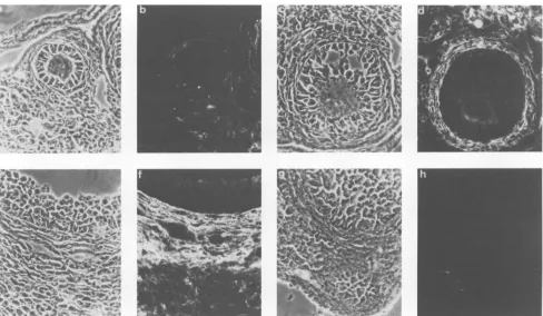
FIG. 1. In situ hybridization experiments with an ecotropic MuLV RNA-specific probe were performed on gonads fromSWR/J mice
inoculatedatbirthwithSWR-RFMuLV(29).(A,C, E,andG)Phase-contrast micrographsand(B, D, F,andH)thesamefieldsseenin dark field.(Athrough D) Ovarian sectionsthroughthree small(Aand B;magnification, x380)andtwomaturefollicles(CandD;magnification,
x152).Thecal cells that surroundfollicles beyond stage type5a (30)as well as some stromal cells arelabeled. Youngerfollicles remain unlabeled.(EthroughH) Transverse sections(toluidinebluestain) throughseminiferous tubules of testis from both inoculated(EandF)and uninoculated(G and H)males showinglabeled clustersofcellsbetween the seminiferoustubules(magnification, x152).
VOL. 63, 1989
on November 10, 2019 by guest
http://jvi.asm.org/
FIG. 2. (a through f) Sections through the ovaries of SWR/J females inoculated at birth with SWR-RF MuLV. (g and h) Section through a mature follicle of an uninoculated SWR/J female (negative control). Shown are phase-contrast micrographs (a, c, e, and g) and thesamefields stained in indirect immunofluorescence with antibodies againstgp7O(b, d,f, and h). (a through f) Sections through ovarian folliclesatdifferent stages ofdevelopment showing thecal cells andsomestromal cells withanti-gp7Ostaining (type 3b [a andb], type6[c
andd], and type 8 [e andfl)(30). The exposure time forpanelh wasadjusted to be thesame as thatforpanels b, d, and f. Magnification, x400.
widely different levels of virus expression. Male and female gonads thus appear as heretofore unsuspected targets for MuLV infection. Although the mechanisms which make thecal and interstitial cells such efficient targets for virus expressionareunknown, itis striking thatbothcelltypes are alsosteroidhormone producers. Whether such a correlation is merelyfortuitous is unknown, however.
FIG. 3. Sectionthroughthetestis ofaSWR/Jmaleinoculatedat
birth withSWR-RFMuLV. Phase-contrast micrograph (a)and the
same field stained in indirect immunofluorescence with antibodies against gp7O (b). The section shows a cluster ofinterstitial cells
locatedbetween theseminiferoustubules positivefor gp7Oantigen (magnification, x400).
Itremainstobe determinedwhether thepatternof expres-sion disclosed here is exclusively
specific
for the SWR-RF MuLV isolate used throughout our study. However, webelievethat this isnot likely for variousreasons. First, this virus is derivedfrom the endogenous ecotropic proviruses carriedbyRF/Jmice(designatedEmv-16andEmv-17) which are structurally related to AKR/J endogenous ecotropic proviruses (20). Second, ecotropic MuLV RNAs are also detected in the ovaries ofAKR/J mice (28). This suggests that the ecotropic MuLV produced in AKR/J mice might display apatternofinfectivityinSWR/Jgonadsverysimilar if notidenticaltoSWR-RFMuLV.Thenucleotidesequence determination of a molecular clone encoding SWR-RF MuLV (in progress in our laboratory) should clarify the structuralrelationships between theseviruses.
One may ask whether the virus reservoir in the
gonads
leads to oncogenesis in infected mice. The
possibility
that the virusinoculated into SWR/J micewilleventuallytrigger
leukemogenesis remainstobe documented. Inthiscontext, however, it is worth
pointing
out that tumors ofthe ovary havebeenoccasionallydetected in our stockofSWR/J-RF/J
hybrid females carryingEmv-16-Emv-17. Thesefemalesalso expresshighlevelsof
ecotropic
MuLV RNAin theirovarian thecal cells (28). However, these tumors havebeen charac-terized asgranulosacell tumors (J. Gaillard, personalcom-munication), i.e., they derive from follicular cellsthatdonot
appear to be
productively
infectedby
ecotropicviruses,
and itshouldbenotedthat spontaneousmalignant
granulosa cell tumors are found in SWR/Jand SWR-derived mice (3). As far as AKR/J mice areconcerned,
it isintriguing
that McEndy et al. (24) have shown that ovariectomy of AKRon November 10, 2019 by guest
http://jvi.asm.org/
[image:5.573.53.541.62.346.2] [image:5.573.47.282.501.665.2]ECOTROPIC MuLV INFECTION OF MURINE GONADS 2139
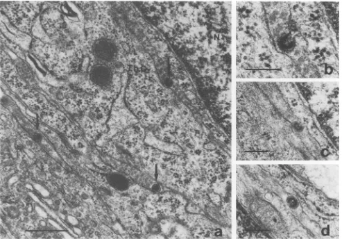
FIG. 4. (a)Productionof virusby thecal cells from the ovary of inoculated SWR/J females. Budding particles (arrows) are clearly visible
intheintracellular space occupied by collagenous fibrils between the thecal cells (magnification, X42,500;bar, 0.5 ,um). N, Nucleus; Th.C., thecal cell. (b) Same cell as in panel a, seen at a higher magnification(x87,500:bar, 0.2pLm).(c and d) Other viruses observed around thecal cells. The average diameter of these particlesis 100 nm(bar. 0.2
Rm).
mice reduces the incidence of leukemia from 74%amongthe controls to 45% among the ovariectomized females, thus pointing out the influence of the ovary on the incidence of
spontaneous leukemia. In contrast, orchidectomy has been shown to increase the incidence of leukemia in AKR mice from 65 to84%(37). Obviously, the endocrine functions and the capacity to produce ecotropic MuLV inherent in the gonads were notseparatedinthese experiments, preventing
us from assessing the relative importance that these ele-ments may have individually in leukemogenesis.
Neverthe-less, the resultspresented indicate that both male and female gonads might be a particular site where continuously
pro-duced ecotropic MuLVs would be available to recombine with endogenous xenotropic or mink cell focus-forming (MCF)-like
sequences.
resulting in recombinant MCFvi-ruses (8, 9.
15.
16, 33, 40). The production of suchrecom-binant viruses is thought to be a prerequisite forthe
devel-opmentof T-celllymphomas (8, 26), whichoccurwith ahigh incidence in AKR/J mice. The existence of such reservoirs
appears to.beespeciallyimportant ifone recallsthatthymic lymphocytes have beenreported to beresistant toinfection by ecotropic virus
(4.
13. 17). and one of the models for T-cell lymphomagenesis assumes that the generation of MCF virusesis needed in order toinfectaspecifictarget cellwhich is refractory to infection by ecotropic MuLV (11).
Oncogenesis would result from the activation ofsome dis-tinct cellulargenes(forareview, see reference 27) by either
anMCF virus orbyapseudotyped MuLV (MCFenvelope, ecotropicgenome) (12).
Finally, it is not clear whether the MuLV pattern of expression in the ovaries of inoculated SWR/J mice bears
any relevance to the integration ofMuLV sequences in the progeny. We demonstrated that ecotropic MuLV particles
are produced in the vicinity of the oocytes; however, this doesnotimplythat thetargetcellsforthevirusinfectionare
the oocytes themselves. More studies involving
manipula-tionof theoocyte or eggtransferexperimentsorboth should allowto understand the infectivity process which results in
transgenic pupsfrom infected SWR/J mothers.
It hasbeen recently shownthat sperm-associated
retrovi-ruses are common in mouse epididymal semen, indicating
that retroviruses are expressed in the male reproductive
tract (21). Our results show that Leydig cells express viral
messenger and proteinproduct;moreover,infectious ecotro-pic MuLV particles can be recovered from the semen of infected SWR/J males (J.-J. Panthier, unpublished data). In apparent contradiction. male virus carriers do not transmit endogenous ecotropic proviruses to their progeny after
mating with nonviremic females. This implies either that spermatozoa are not infectedorthatthe infectionefficiency VOL. 63,1989
on November 10, 2019 by guest
http://jvi.asm.org/
[image:6.612.56.549.73.417.2]g"W.
[image:7.612.79.570.74.683.2]0'''0
'"
Ws'';'~~~~~~~~~~~~~~~~~;,tW;''
.. ...
,I,,*i,;S;,:w4,;w,sL;s;
FIG. 5. Immunoperoxidase localization ofgp7Oviralantigen in Leydig cells ofinoculatedSWR/Jmales. Sections areobserved unstained. Patches of black deposits were often seen near the Golgiapparatus.Thegranular aspect of the intense black deposit is typical of the enhancing method used (39) (see Materials and Methods). Note the extensive smooth endoplasmic reticulum characteristic of Leydig cells in panels b and d.Magnifications: panela, x25,000 (bar,1,um);panelb, x55,000(bar, 0.5 pm);panel c. x25,000 (bar,1 pm); panel d, x42,000 (bar, 0.5
[.m).
on November 10, 2019 by guest
http://jvi.asm.org/
ECOTROPIC MuLV INFECTION OF MURINE GONADS 2141
'tW''%j f.'7F A_ VAM~JW:WYkM M
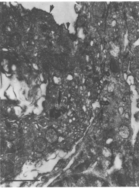
FIG. 6. Immunoperoxidase localization ofgp7O viral antigen in
testis of inoculated males. A black deposit of DAB was often
observedatthe surface ofinterstitial cells (arrowheads) and in the
cell itself(arrow). Magnification, x20,000.
of free virus particlespresent inspermistoo low inutero to
be easily detected by progeny testingorboth.
ACKNOWLEDGMENTS
We thank FernandoPlata for his generousgiftof antibodies and forhelpfulcommentsonthemanuscript;Jean Gaillard for perform-ingthehistologicalexamination of ovarian tumors; Patricia Baldacci for critical reading of the text; and Martine Maury and Monique Agauguefor excellent technical assistance.
This workwassupported bygrants from the Centre Nationalde la
Recherche Scientifique (Unite Associee 1148),the Institut National de la Santd et de la Recherche Medicale (851004), the Fondation
pourla Recherche Medicale,the LigueNationaleFrancaise contre le Cancer, and the Fondation AndreMeyer.
LITERATURE CITED
1. Basgall, E. J., M. M. Soong, and W. A. F. Tompkins. 1986.
Visualization ofcytoskeletalelements and associated retroviral
antigens by immunogold transmission electron microscopy of
detergent-extracted cells. Scanning Electron Microsc. 4:1419-1425.
2. Bautch, V. 1986. Genetic background affects integration
fre-quency ofecotropic proviral sequences into the mouse germ
line. J. Virol. 60:693-701.
3. Beamer, W. G., P. C. Hoppe, and W. K. Whitten. 1985.
Spontaneous malignant granulosa cell tumors in ovaries of
youngSWR mice. Cancer Res. 45:5575-5581.
4. Blank,K. J., and T. Pincus. 1980. Evidence that endogenous ecotropicvirus isnotexpressedin AKRthymic lymphoidcells of chimeric hosts. J. Exp.Med. 152:458-462.
5. Bloom, W.,and D. W.Fawcett.1975.Atextbook ofhistology,p. 805-857. The W. B. SaundersCo., Philadelphia.
6. Buckler,C.E., S. P.Staal,W. P.Rowe,and M. A. Martin.1982. Variation inthenumberofcopies and inthegenomic organiza-tion ofecotropic murine leukemia virus proviral sequences in sublines ofAKRmice.J. Virol.43:629-640.
7. Buetti, E., and H. Diggelmann. 1980. Murine leukemia virus proteins expressedonthe surface ofinfected cells in culture. J. Virol.33:936-944.
8. Chattopadhyay, S. K., M. W. Cloyd, D. L.Linemeyer, M. R. Landers,E. Rands, and D. R. Lowy. 1982. Cellularoriginand roleof minkcellfocus-forming viruses in murinethymic lym-phomas.Nature(London)295:25-31.
9. Chattopadhyay,S. K.,M. R. Lander, S.Gupta,E.Rands,and D. R. Lowy. 1981. Origin of mink cytopathic focus forming (MCF)viruses: comparisonwithecotropicandxenotropic
mu-rine leukemia virusgenomes.Virology 113:465-483.
10. Chattopadhyay,S.K.,M. R.Lander,E.Rands,and D. R.Lowy. 1980. Structure ofendogenousmurine leukemia virus DNAin
mousegenomes. Proc. Natl. Acad.Sci. USA77:5774-5778. 11. Cloyd, M. W. 1983. Characterization oftarget cells for MCF
virusesinAKRmice. Cell 32:217-225.
12. Cuypers,H. T.,G. Selten,W.Quint, M. Zilstra,E. R. Maan-dag,W. Boelens, P. van Wezenbeek, C.Melief, and A. Berns. 1984. Murine leukemia virus-induced T-celllymphomagenesis: integrationofprovirusesinadistinct chromosomalregion.Cell 37:141-150.
13. Gisselbrecht, S., C. Blaineau,M. A.Hurot, F. Pozo,andJ. P. Levy. 1978. Prevalence of non-T-cells in thereplicationof the
N-tropic, type C virus of young AKR mice. Cancer Res. 38:939-941.
14. Herr, W.,and W. Gilbert.1982.Germ-lineMuLVreintegrations
inAKR/J mice. Nature(London)296:865-868.
15. Herr,W., and W.Gilbert. 1983.Somatically acquired
recombi-nant murine leukemiaprovirusesinthymicleukemia of AKR/J mice.J. Virol. 46:70-82.
16. Holland,C. A., J. W. Hartley, W. P. Rowe, and N. Hopkins.
1985. At leastfour viral genes contributing tothe leukemoge-nicity of murine retrovirus MCF 247 in AKR mice. J. Virol. 53:158-165.
17. Horak, I., L. Enjuanes, J. C. Lee, and J. N. Ihle. 1981. Resistance of culturesof normalT-cellstoinfection withmurine type C viruses. J. Virol. 37:483-487.
18. Jenkins, N. A., and N. G. Copeland. 1985. High frequency germline acquisition of ecotropic MuLV proviruses in SWR/ J-RF/J hybrid mice. Cell43:811-819.
19. Jenkins, N. A., N. G.Copeland, B. A. Taylor,and B. K. Lee. 1981.Dilute(d) coatcolour mutationof DBA/2J miceis associ-ated withthesiteofintegrationofanecotropicMuLVgenome. Nature(London)293:370-374.
20. Jenkins, N. A., N. G. Copeland, B. A. Taylor, and B. K. Lee. 1982. Organization, distribution, and stability ofendogenous ecotropic murine leukemiavirus DNA sequences in
chromo-somesofMusmusculus.J.Virol. 43:26-36.
21. Kiessling, A. A., R. C. Crowell, and R. S. Connell. 1987.
Sperm-associated retroviruses in the mouse epididymis. Proc. Natl. Acad. Sci. USA84:8667-8671.
22. Langdon,W.Y.,T.S.Theodore,C. E.Buckler,J.H.Stimpfling, M. A.Martin,and H. C. MorseIII.1984.Relationshipbetween
a retroviralgerm line reintegration and a new mutation at the Ashen locus inB10.F mice.Virology 133:183-190.
23. Lerner,R.A.,C. B.Wilson,B.C. DelVillano,P.J.McConahey,
and F. J. Dixon. 1976. Endogenous oncornaviralgene expres-sion inadult and fetal mice: quantitative, histologic, and phys-iologic studies of the major viral glycoprotein, gp7O. J. Exp.
Med. 143:151-166.
24. McEndy,D.P.,M. C.Boon,andJ. Furth. 1944.On theroleof
thymus,spleenandgonadsinthedevelopmentofleukemiaina
high-leukemiastock of mice. Cancer Res. 4:377-383.
25. Mowat,M.,andA.Bernstein.1983.Linkageof the Fv-2 geneto anewlyreinsertedecotropic retrovirus in Fv-2
congenic
mice. J. Virol. 47:471-477.26. Mucenski,M.L.,H.G.Bedigian,M. M.Shull,N.G.Copeland,
VOL. 63, 1989
on November 10, 2019 by guest
http://jvi.asm.org/
[image:8.612.57.286.71.380.2]and N.Jenkins. 1988. Comparative molecular analysis of lym-phomafrom six inbred mouse strains. J. Virol. 62:839-846. 27. Nusse, R. 1986. The activation of cellular oncogenes by
retro-viralinsertion. Trends Genet. 2:244-247.
28. Panthier, J. J., and H. Condamine. 1987. Expression of ecotro-pic MuLV in ovaries ofSWR/J-RF/J hybrid mice. Ann. Inst. Pasteur Virol. 138:409-422.
29. Panthier, J. J., H. Condamine, and F. Jacob. 1988. Inoculation ofnewbornSWR/J femaleswith anecotropic murineleukemia virus can produce transgenic mice. Proc. Natl. Acad. Sci. USA 85:1156-1160.
30. Peters, H. 1969. Thedevelopment ofthe mouse ovaryfrom birth tomaturity. Acta Endocrinol. 62:98-116.
31. Pinter, A., W. J. Honnen, and J. S. Li. 1984. Studies with inhibitors of oligosaccharide processing indicate a functional role for complex sugars in the transport and proteolysis of Friend minkcellfocus-inducingmurineleukemia virusenvelope proteins. Virology 136:196-210.
32. Plata, F., J. Kalil, M. T. Zilber, M. Fellous, and D.Levy.1983. Identification of a viral antigen recognized by H-2-restricted cytolytic T lymphocytes on a murine leukemia virus-induced tumour. J. Immunol. 131:2551-2556.
33. Quint, W., W. Boelens, P. van Wezenbeek, T. Cuypers, E. R. Maandag, G. Selten, and A. Berns. 1984. Generation ofAKR mink-cell focus-formingviruses: a conserved single copy xeno-tropic-like provirusprovidesrecombinant long terminal repeat sequences. J. Virol. 50:432-438.
34. Quint, W., W. Quax, H. Van Der Putten, and A. Berns. 1981.
Characterization ofAKR murine leukemia virus sequences in AKRmousesubstrains andstructureofintegratedrecombinant genomesintumourtissues. J. Virol. 39:1-10.
35. Rowe, W. P., and C. Kozak. 1980. Germ-line reinsertions of AKRmurine leukemia virus genomes in Akv-1congenicmice. Proc. Natl. Acad. Sci. USA 77:4871-4874.
36. Rowe, W. P., and T. Pincus. 1972. Quantitative studies of naturally occurring murine leukemia virus infection of AKR mice. J. Exp. Med. 135:429-436.
37. Rudali,G.,B.Desormeaux,and L.Juliard.1956. Action
d'hor-monesmalesetfemellessurlaleucemogenesedessouris AkR. Bull. Cancer43:445-449.
38. Steffen, D. L., B. A. Taylor, and R. A. Weinberg. 1982. Con-tinuing germ line integration of AKV proviruses during the breeding of AKR mice and derivative recombinant inbred strains. J. Virol. 42:165-175.
39. Strauss, W. 1982. Imidazole increases the sensitivity of the cytochemicalreactionforperoxydasewithdiaminobenzidineat aneutralpH.J. Histochem. Cytochem. 30:491-493.
40. Thomas, C. Y., and J. M. Coffins. 1982. Genetic alterations of RNA leukemia viruses associated with the development of spontaneous thymic leukemia in AKR/J mice. J. Virol. 43: 416-426.
41. Weiss, R. 1984. Experimentalbiology and assay of retroviruses, p. 209-260. In R. Weiss, N. Teich, H. Varmus, andJ. Coffin (ed.),RNAtumourviruses,2nded. ColdSpringHarbor Labo-ratory,Cold Spring Harbor, N.Y.