JOURNALOF VIROLOGY, June1990, p.3012-3018 0022-538X/90/063012-07$02.00/0
Copyright C)1990, AmericanSocietyforMicrobiology
The Adeno-Associated
Virus
rep
Gene
Suppresses
Herpes Simplex
Virus-Induced
DNA
Amplification
REGINE HEILBRONN,* ALEXANDER BURKLE, SONJASTEPHAN,
AND HARALDZURHAUSEN
Deutsches Krebsforschungszentrum, ImNeuenheimerFeld506, D-6900 Heidelberg, FederalRepublic of Germany
Received 22 January 1990/Accepted22 March1990
Herpessimplex virus(HSV) induceswithinthe host cellgenomeDNAamplificationwhichcanbesuppressed by coinfection with adeno-associated virus(AAV). To characterize the AAV functions mediatingthiseffect,
cloned AAV type 2 wild-type or mutantgenomes weretransfected into simian virus40 (SV40)-transformed
hamster cells together with the six HSVreplicationgenes(encoding UL5, UL8, major DNA-binding protein,
DNApolymerase, UL42,andUL52)whichtogetherarenecessaryand sufficient for the inductionofSV40DNA
amplification (R. Heilbronn and H. zur Hausen, J. Virol. 63:3683-3692, 1989). The AAV rep gene was
identifiedasbeingresponsibleforthecomplete inhibition ofHSV-inducedSV40DNAamplification. Likewise, repinhibited origin-dependentHSV replication.repneither killed the transfected host cellsnorinterfered with
gene expression from the cotransfected amplification genes. This points to a specific interference with
HSV-induced DNA amplification.
Adeno-associated viruses (AAVs) are members of the parvovirus family, a group of small single-stranded DNA
viruses with unique replication properties. Incontrast tothe autonomousparvoviruses whichcanreplicate independently
inproliferating cells, the AAVs relyonhelper viruses,either
adenovirusesorherpesviruses, for efficient replication (fora review, seereference 4). However, cells treated with chem-ical or physical carcinogens can support low-level AAV replication in the absence ofa helper virus (13, 34, 45, 46). Thus, thereplication defectiveness ofAAVisnotabsolute.
The autonomous and helper-dependent parvoviruses have unique biological properties in common. Members of both
groups efficiently suppress tumor growth in animals,
irre-spective of the mode of tumor induction. Parvoviruses
inhibit spontaneous tumor formation (37) and tumors
in-duced by various oncogenic viruses aswell asby chemical carcinogens (9, 10, 19, 28, 30, 39). In addition, there is evidence from seroepidemiological studies that high anti-body titers against the human AAV types 2, 3, and 5 (AAV-2, AAV-3, and AAV-5) areassociated withareduced
cancerincidence(11, 27, 35). The mechanisms, however, by
which parvoviruses exert their oncosuppressive effect are
not yet understood. Inanattempt tounravel the underlying molecular mechanisms, the effect of parvoviruses was
stud-ied in various invitro transformation systems. Minute virus
ofmice,anautonomousparvovirus, suppressed transforma-tion of mouse fibroblasts by simian virus 40 (SV40) (29). Likewise, AAV suppressed the transformation ofhamster fibroblasts by different adenovirus strains(5) and the trans-formation of the mouse fibroblast cell line C127 by bovine papillomavirus. AAV-2p78reP appearedtobe responsible for thiseffect(16).
Three viral functions have been mapped on the
4.65-kilobase(kb) AAV-2genome(Fig. 1). The 145-base-pair (bp)
terminalrepeat structuresserve asoriginsof replication. The
right-hand open reading frames (ORFs) encode the three capsid proteins (cap), whereas the ORFs on the left-hand
sideofthegenomecode forrep,afamily of multifunctional
*Corresponding author.
nonstructuralAAVproteins. The mRNAscodingforp78reP
and the spliced p68reP start at the p5 promotor, and those coding for p52reP and the spliced p40reP start at the p1l promotor. The rep proteins are required for AAV DNA
replication (17, 40). p78/68reP is responsible for the
accumu-lation of replicative intermediates, whereas p52I40reP is required for the generation of single-stranded monomer
AAV and for the packaging of the virus (6). The role of p78168reP in the accumulation ofreplicative intermediates is also reflected by theirbinding to the AAV origins of
repli-cation(18).TherepproteinsarerequirednotonlyforAAV
DNAreplicationbutalso for AAVgeneregulation. Depend-ing on the presence or absence ofhelper adenovirus
func-tions, theyeither activateor repressthe AAVpromotors in
trans (3, 41). Although most of these experiments were performed with human cells, the different AAV mutants exhibited thesamereplicationphenotypeinrodentcells(16;
R. Heilbronn, unpublished data).
Wehave shownpreviouslythat AAVcanseverelyinhibit herpesvirus- or carcinogen-induced DNA amplification (2, 13, 34). DNA amplification plays a central role not only in thedevelopmentofdrug resistance (20, 26, 36)butalsoin the
course oftumor development, where amplified oncogene
sequencesare afrequent finding (forareview, seereference
1). We therefore decided to analyze the AAV-mediated
inhibition ofDNAamplificationatthemolecularlevel. This
was possible by cotransfecting cloned AAV mutants in combination with the recentlyidentified set ofherpes
sim-plex virus (HSV) amplificationgenes (15), thusintroducing
theamplification-inducing and -inhibitingfunctions into the
samefraction of cells.Bytheuse of AAVwild-type (wt)or
mutantgenomes, weshow herethatrepalone is sufficientto completely suppress DNA amplification induced by theset of six HSVamplification genes.
MATERIALS ANDMETHODS
Recombinant plasmid DNAs. The cloned AAV-2 wt ge-nome (pAV2) was obtained from C. Laughlin (24). AAV-2
was subcloned into the BamHI site of Bluescript (Strata-gene), resulting in plasmid pTAV2 (Fig. 1). ori mutants 3012
on November 10, 2019 by guest
http://jvi.asm.org/
p5 p19 p40
rep cap
pTAV-2
pTAV2-4
pTAV2-3
pTAV2-6
pTAV2-7
pTAV2-2
pTAV2-8
FIG. 1. Structure of AAVwtandmutantgenomes. The AAV-2
genome is represented schematically with the inverted repeats servingasorigins of replicationattheends of thegenome(_).The
ORFs encoding the nonstructural proteins (rep) and three capsid proteins (cap) are shown(O). Construction of the AAV mutant
genomes is explained in the text. Frameshift deletions (A) and insertions (0)areindicated. Mutantphenotypesareindicatedtothe right of the plasmid designations.
(pTAV2-2, pTAV2-4, and pTAV2-7) were generated from
pTAV2 by using the BalI sites at nucleotide positions 121 and 4554. repmutantswere generated either by introducing a 4-bp frameshift insertion at position 1045 by using the
unique BamHI site (pTAV2-3 andpTAV2-8)orby introduc-inga 150-bpdeletion encompassingthep1l promotorregion
(positions 814to964,pTAV2-2). Both mutationsinactivated
the rep genes starting from the promoters P5 and p19- cap
mutants(pTAV2-6, pTAV2-7, andpTAV2-8)weregenerated by introducinga164-bp deletion between positions3326and
3490.
The series of plasmids carrying the HSV amplification
genes pH6 (pol and DBP), pH7 (IE175 and IE110), pH8
(UL42andUL52), pH9 (UL5andUL8), andtherespective
ORFsexpressed under thecontrol of thehuman cytomega-lovirus (HCMV) immediate early (IE) promoter (-598 to
+52) (pCM-UL5, -UL8, -UL9, -pol, -DBP, -UL42, and
-UL52) have been described before (15). pHlO carries the HSV
oris
(15). pCMcat (kindly provided by Hubert Stop-pler)expressesthechloramphenicol acetyltransferase (CAT)gene under the control of the same HCMV IE promoter fragment (-598 to +52) as the pCM-UL series of HSV expressionconstructs.
Cells and viruses. The Elonall cell line andpropagationof HSV-1 strain 17 have been described elsewhere (15, 33).
AAV-2waspropagated in HeLa cells with adenovirus 2 as
thehelperasdescribed previously (46).
Transfection procedure. Calcium phosphate
coprecipita-tionfollowed byadimethyl sulfoxide shockwasperformed exactly as described before (15). Each transfection experi-ment was repeated at least three times with two different
plasmidpreparations, leadingto similar results.
AAV replicationassay. HeLa cells (3 x
105)
were plated onto 6-cm-diameterdishes. The next day, cells were trans-fected with AAV-2mutantplasmidswhichhad beenexcisedfrom the vector with PvuII. After the dimethyl
sulfoxide-shock, cells were infected with HSV-1 diluted in 1 ml of
medium(multiplicityofinfection, 10)andincubated at37°C
for 40 h. Thecultureswerelysedinsitubythreefreeze-thaw
cycles. The disrupted cells were pelleted and used for the analysis of AAV DNA replication. Genomic DNA was
extracted by digestion withproteinase K, repeated extrac-tions with phenolandchloroform, digestion with RNaseA,
and dialysis against lx TE (15). The supernatants were
treated at 56°C for 30 min to inactivate the helper virus
(HSV) and then used forthe titration of infectious AAV in
1:10 dilution steps on HeLa cells grown on 96-well plates.
Cells were infected with HSV-1as the helper virus 16 h later.
At 40 hafter HSV infection, cells were dotted onto Gene-Screen (Dupont, NEN Research Products, Boston, Mass.)
andhybridized to 32P-labeled AAV-2 DNA.
Analysis of genomic DNA. For the analysis of AAV DNA
replication, total genomic DNA was doubly digested with XbaI and DpnI. A 5-,ug sample of each digest was run on
0.7%agarosegels, blotted onto GeneScreen Plus (Dupont, NEN Research Products), and hybridized to a32P-labeled,
full-length AAV-2 probe. SV40 DNA amplification was assayed on Southern blots of SacI-digested genomic DNA hybridized to32P-labeled SV40 DNA, and HSV
oris
replica-tion was assayed with the DpnI assay as described before(15).
CAT assays.Protein contents of freeze-thaw extracts were determined by using the Bradford assay (Bio-Rad
Laborato-ries, Munich, Federal Republic of Germany). CAT enzy-matic activity was assayed by using defined amounts of heat-treated (10 min at 60°C) protein extract (8). Final concentrations in the assay were 1.25 ,uCi of
"4C-chloram-phenicol per ml and 1 mmol of acetyl coenzymeAperliter. Ethyl acetate-extracted reaction products wereanalyzed by thin-layer chromatography. Spots representing unreacted chloramphenicol and acetylated chloramphenicol were
ex-cised, and radioactivity was quantitated by liquid scintilla-tion counting.
RESULTS
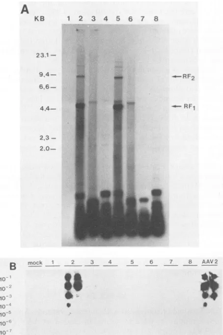
Replication phenotypeofAAV mutantplasmids. Single and double mutationsweregeneratedin clonedwtAAV-2 DNA (pTAV2) according to published genetic data (Fig. 1) (17, 40). The phenotypes of this series of AAV plasmids were
verified by transfecting the individual mutants into HeLa cells with HSV-1 as the helper virus. At 40 h after HSV infection, cultures were processed for the determination of
AAVDNAreplicationand, in parallel, productionof infec-tious viral particles (Fig. 2). With pTAV2 (wt), the typical
AAV replication intermediates appeared (Fig. 2A, lane 2).
Titrationof freeze-thaw supernatants of transfected cellson
fresh HeLa cells in the presenceof HSV-1asthehelpervirus
clearly demonstrated therescue of infectious viralparticles
from pTAV2 (Fig. 2B, lane 2). HSV-1 served as a helper
virus with an efficiency equal to that of adenovirus type 2
(data not shown). The cap mutant pTAV2-6 gave rise to
replicative intermediates (Fig. 2A, lane 5) whose slightly
shorterlengths(comparedwith thatofwtpTAV2)aredueto the 150-bp deletion within the cap ORF. The
cap-negative
phenotype ofpTAV2-6 wasdocumented by the inability of the mutant to generate infectious viral
particles (Fig. 2B,
lane5). Asexpected, repmutants didnotgiveriseto either
replicative intermediates orinfectious AAV particles (Fig.
2AandB, lanes4 [pTAV2-3],7 [pTAV2-2], and 8 [pTAV2-8]).Transfection oforimutantswithdeletions of the terminal repeats but intact rep genes (pTAV2-4 and pTAV2-7) gave rise to a high AAV-2-specific
background
hybridization
signal on the Southern blot without the
typical replication
intermediates(Fig. 2A,lanes 3 and6).Theabsenceof similar
hybridization signals in all the lanes withrep mutants
(Fig.
2A, lanes 4, 7, and 8) raises the possibility that rep geneexpression per se mediates
unspecific
initiation of DNAsynthesis on the AAV
template
in the absence of originsequences. However, no infectious
particles
were formed.a
1%
A
-A
on November 10, 2019 by guest
http://jvi.asm.org/
[image:2.612.68.297.69.192.2]A
KB 1 2 3 4 5 6 7 8
23.1
994-
666-
4.4-223
2.0-B
10
10'
10
10,
lo
10
-RF1
nmock 2_ 3 _4_ 7 8 AAV2
FIG. 2. PhenotypesofAAV-2mutantswithHSVashelpervirus.
HeLacellsweretransfected withAAV-2 wtormutant genomesand
infectedwithHSV-1. At40 hp.i.,cultureswerefreeze-thawed three
timesandcellswerepelleted.GenomicDNAwasextracted from the
pelletfor Southern blot analysis (A). Supernatants were usedfor
titration ofinfectious AAVparticles (B). (A)Southernblotanalysis
of XbaI-DpnI-digested genomic DNA hybridized to 32P-labeled
AAV DNA. XbaI was a noncut enzyme for AAV DNA. DpnI digested the transfected procaryotic plasmid DNAs visible in the
lower part of the figure, whereas AAV molecules replicated in
eucaryotic cells were resistant to DpnI and appearas thetypical replicativeintermediatesinlanes 2and5. RF1(4.65 kb)represents
adouble-strandedmonomer, andRF2(9.3 kb)isadouble-stranded
dimer. In addition, higher oligomeric forms are visible faintly.
TransfectedDNAs(20 p.g)are asfollows:lane1,Bluescript;lane2,
pTAV2 (wt);lane3, pTAV2-4(orinegative);lane4,pTAV2-3 (rep negative);lane5,pTAV2-6 (cap negative);lane6,pTAV2-7 (oriand cap negative); lane 7, pTAV2-2 (ori and rep negative); lane 8, pTAV2-8 (rep and cap negative). Because of the high level of background hybridizationwhichwasmostlikelyduetosingle-and partiallydouble-strandedreplicativeintermediatesof allsizeclasses,
itisdifficulttodetectsingle-stranded,full-sizeAAVmolecules.The
bandsaround 5kbvisible inlanes2to6 mostlikelyrepresentasmall
amountofinputplasmidDNAresistant toDpnI.The varioussizesof
these bands correspond to the different sizes of the mutants. (B)
TitrationofinfectiousAAV-2particles generatedaftertransfection of
AAV mutants andsubsequentHSV-1 infection. Supernatantswere
treatedat56°Cfor30mintoinactivate thehelpervirus(HSV-1) and
titrated in duplicate in 1:10 dilution steps on HeLa cellsgrown in
96-wellplates.After16h,cellswereinfectedwithHSV-1asthehelper
virus.After40h,cellsweredotted ontoGeneScreenandhybridizedto
32P-labeledAAV-2DNA.Lanes:mock,nosupernatant;1to8,sameas
inpanel A; AAV2, titrationofheat-inactivated(30min,56°C)
predi-lutedAAV-2stockvirususedas apositivecontrol.
Thetransfections described abovewere
repeated
inElonallcells with similar results.
Effect of AAV mutants onDNAamplification inducedbythe
HSV amplification genes. HSV infection leads to DNA am-plification of chromosomally integrated SV40 DNA
se-quences. This effect can be suppressed by coinfection with
AAV (2, 34). Recently, we have identified the six HSV-1 genes which together are necessary and sufficient for the induction of SV40 DNA amplification. The locations ofthe amplification genes (UL5,UL8, DBP,pol, UL42, and
UL52)
on the HSV genome are depicted in Fig. 3. Transfection of
the set of plasmids carrying the HSV
amplification
genes (pH6, pH8, pH9, and pH7) induced a strongamplification
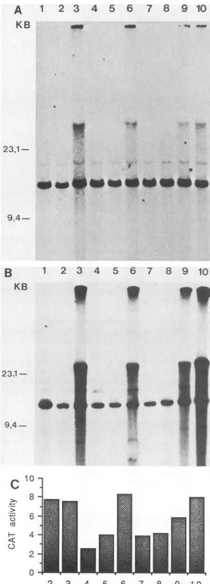
signal (Fig. 4A, lane 3)asdescribed before (15).
Cotransfec-tion of pTAV2 (wt) with pH6, pH7, pH8, and pH9 sup-pressed the amplification effect (Fig. 4A, lane 4).
Bluescript
DNA or cloned AAV-2 wt DNA (pTAV2) was used as a
negative control (Fig. 4A, lanes 1 and 2).
To evaluate which of the AAV-2 functions mediated the suppression of DNAamplification, individual AAV-2mutant
genomes (Fig. 1) were cotransfected. pTAV2-4 (ori nega-taive) (Fig. 4A, lane 5) as well as pTAV2-6 (cap negative)
(Fig. 4A, lane 7) suppressed the induction of DNA
amplifi-cation as well as pTAV2 (wt),whereas pTAV2-3 (rep nega-tive) did not affect HSV-induced DNA amplification
(Fig.
4A, lane 6). The conclusion that the rep geneis responsible
for this effect was confirmed by theuse of double mutants. pTAV2-7(ori and cap negative) suppressed DNA amplifica-tion (Fig. 4A, lane 8), which is in line with thefactthatthis
mutant carried an intact rep gene. However, double mutants with adisrupted rep gene, pTAV2-2 (ori and rep negative) and pTAV2-8 (rep and cap negative), were unable to sup-pressHSV-induced DNA amplification (Fig. 4A, lanes 9and 10). In conclusion, the rep gene is responsible for the suppression ofHSV-induced DNA amplification.
TheAAV rep gene suppresses DNA amplification induced by HSV amplification genes under control of a heterologous promoter. Several targets can be envisaged for the rep gene-mediated inhibition of HSV-induced DNA amplifica-tion. Toexclude the possibility that rep interferes with HSV IE gene-mediated transactivation of the early promoters which drive the HSVamplification genes, we asked whether rep is able to inhibitDNA amplification induced by the setof amplification genes driven by a heterologous promoter (pCM-UL5,-UL8, -DBP, -pol,-UL42,and-UL52;Fig. 3).A
transfection experiment similar to the one described above was performed, with replacement of the authentic HSV amplification genes by the HCMV IE-driven constructs. Again, the controls, i.e., transfection of either Bluescript DNA orpTAV2, did not show any DNA amplification (Fig. 4B,lanes 1 and 2), whereas the set of HCMVIE-driven HSV amplification genes induced a strong amplification effect (Fig. 4B, lane 3) as described before (15). This effect was suppressed upon cotransfection of pTAV2 (wt), pTAV2-4 (ori negative), or pTAV2-6 (cap negative) (Fig. 4B, lanes 4, 5, and 7).pTAV2-3 (repnegative), however, did not have an effect onHSV-induced DNA amplification (Fig. 4B, lane 6). Again, double mutants confirmed the conclusion that rep mediates this inhibition. The mutant pTAV2-7 (oriand cap negative) was able to suppress DNAamplification (Fig. 4B, lane 8),whereas the two double mutants with disrupted rep genes, pTAV2-2 (ori and rep negative) and pTAV2-8 (rep andcap negative), did not influence DNA amplification (Fig. 4B, lanes 9 and 10). Transfection ofequimolar amounts of pTAV2-7 (ori and cap negative) compared with the HCMV IE-driven HSV amplification genes (1 ,ug of each) still led to J. VIROL.
on November 10, 2019 by guest
http://jvi.asm.org/
[image:3.612.69.296.71.412.2]HSV1
UL
us
_0 -_ _ 4 O
ULSUL8 UL9 dbp pol
pH9 pH6
Ip U I MII p 1 11 pCM-UL5 pCM-ULS pCM-DBP pCM-POL
_ol
_0
UL42 UL52
i
pHs
IE110 IE175
I,
pH7
pCM-UL42 pCM-ULS2
FIG. 3. Structure of HSV-1 genome and cloned amplification genes. Schematic representation of the 150-kb HSV-1 genome with the unique long (UL) and unique short(Us)regions and the flanking inverted repeats (_ and =).The replication- and amplification-inducing genes areindicated: UL5, -8, -9, -42, -52, dbp (single-strandedDNA-binding protein), andpOI(DNA polymerase). These genes are early genes which have to be transactivated by the HSV IE genes,IE110 (ICPO),andIE175(ICP4), for efficient transcription. Thearrowsindicate the direction of transcription: black arrows are genes necessary for both HSV DNA replication andSV40DNAamplification(UL5,UL8, dbp, pol, UL42, UL52,and IE175);speckled arrows indicate genes required only for HSV DNA replication (UL9 andIE110).The set of plasmids covering the individual genes are shown. Below, the pCM series of expression constructs is represented. These constructs are named accordingto theORF they express under the control of the HCMV IE promotor.
complete suppression of DNA amplification (data not shown). These experiments confirm that the rep gene func-tion alone mediates complete suppression of HSV-induced DNAamplification. Inaddition, the HSV IE gene-mediated
transactivation of early genes does not represent the target
for this interference.
rep mediates suppression of DNA amplification by
mecha-nismsother than nonspecific down regulation of expression of HSV amplification genes. To monitor the transfection
effi-ciency of the above described experiment, an HCMV IE promoter-driven CAT construct (pCMcat) was cotransfected (atamolarratio of1:20compared with pTAV2-7) and CAT enzyme activity was assayed in parallel to the amplification assayinFig.4B. CAT enzymeactivitywasin the same range in alltransfections (Fig.4C), reflecting comparable transfec-tion efficiencies. Inaddition, this result excludes the
possi-bilitythatthesuppression of DNAamplificationin the range
of 100-fold isdueto arep-mediatedrepressionof the HCMV IE promoter, which drives the HSV amplification genes. However, someminor, two- to threefold reduction of
pCM-catexpressionwasobserved whenever AAVwtor mutants
withintact rep genes werecotransfected (Fig.4C). To study the influence ofrep on pCMcat expression in moredetail, pCMcat was transfected into Elonall cells together with
increasingamountsof pTAV2-7 underconditionsidenticalto
theonesusedfor theamplificationstudies described above. A 2-fold inhibition of CAT expression was observed with
equimolar concentrations of pTAV2-7, and up to 3.5-fold
inhibition was observed with a 25-fold molar excess of pTAV2-7 versuspCMcat(Fig. 5). These results confirm the aboveconclusion that this mild reduction ofpCMcat
expres-sioncannotexplainthedrasticinhibition ofDNA amplifica-tion. Our results are in line with recent reports which describeamoderate(abouttwofold) rep-mediatedinhibition of CATexpressionfroman SV40 orbovinepapillomavirus
promoter(3, 16). Weconclude that rep mediates inhibition of HSV-induced SV40 DNA amplification by mechanisms other than down regulation of the expression of the HSV
amplification genes.
repinhibitsorigin-dependent HSVreplicationparalleltothe
inhibition ofDNAamplification. The six HSV amplification
genesencode proteinsnecessary butnot sufficient for HSV
DNA replication. An additional gene coding for a HSV
origin-binding protein(UL9) isrequired for origin-dependent
HSV replication (15, 44). We therefore asked whether rep could also interfere withorigin-dependentHSV replication.
The sevenHSVreplication genes under HCMV IE promoter control were transfected into Elonall cells together with
increasingamounts of pTAV2-7 andaplasmid carrying the HSV
oris
(pH10) totest fororigin-dependent HSVreplica-tion.
oris
replicationwasassayedwith theDpnI assay(Fig.2A).
oris
replication inducedbythe seven HSV replicationgenes was suppressed by increasing amounts ofpTAV2-7
(ori and capnegative) (Fig. 6), thusparallelingthe suppres-sion of SV40 DNAamplificationwhichwasmeasured in the
sameexperiment (datanotshown).
oris
replicationappearednot tobe repressedasstronglyasSV40 DNAamplification,
butthismightbe dueto ahighercopynumber of transfected
oris
sequencescomparedwith the single integrated copyofSV40. Insummary, repmarkedlyinhibits HSV
oris
replica-tion inducedbythe sevenHSVreplication genesinparallel
totheinhibitionofSV40 DNAamplification.At present, we
donotknow whichofthedifferentrepproteins is
responsi-ble for the above-described effects. We have generated a
HCMV IE promoter-driven construct for the rep78 ORF
which leads to suppression of DNA amplification and
oris
replication similar to those ofpTAV2-7 (ori and cap
nega-tive) (data not shown). However, the
rep52
ORF was em-bedded in the rep78 ORF, so an additional role ofp52reP
cannotbe excluded.
DISCUSSION
AAV inhibits DNA amplification induced by HSV or
carcinogens. Inthis report,weshow that the AAV rep gene isresponsible for the inhibition of HSV-induced DNA
am-plification.
SV40DNAamplificationasamodelsystemfor the
amplifi-cation of authentic cellular genes. Two lines of evidence support the notion that DNA
amplification plays
an impor-tantrole in tumor development. On the onehand,
amplifi-cationof cellularorintegrated
viral DNA sequencescanbe induced in cell linesbychemicalorphysical carcinogens, by
avarietyofchemotherapeutic drugs,
and alsoby
viruses likeon November 10, 2019 by guest
http://jvi.asm.org/
[image:4.612.161.459.59.218.2]A
23
4
5
6
7
8
9
10
KB _ _*_
23.1-
9A4-B
1 2 3 4 5 6 7 8 910
KB
23.1-
9.4
-J
-_E
_ _ w _
-'f4
3
_,
HSV
(13, 15, 20, 25, 26,
33,36).
Onthe otherhand,amplified
oncogene sequences are frequently detected in tumor cell linesaswell asin primary tumors (1). The amplificationof cellular genes after carcinogen treatment has been well
documented,asin thecaseofinducibledihydrofolate reduc-tase(DHFR)gene amplification, which leadsto methotrex-ate resistance (20, 26, 36). Since inducible amplification of cellular genes is a rare event whose detection requires
selection for the amplified phenotype, short-term assay
systems were developed. A variety ofcarcinogenic agents
and also viruses like HSV induce a high degree
(over
100-fold)ofDNAamplificationofchromosomally
integrated
SV40 DNA sequences in SV40-transformed hamster cells within 2to5days (2, 13, 15, 20, 25, 33).This madethese cell linesaconvenient model system foranalysis of the
amplifi-cation-inducing or -inhibiting potential of many different
agentsin a short-termassay system. Parallelanalysis dem-onstrated thatalthough SV40 DNA amplification occurredat amuchhigherrate than DHFRgene amplification, the two eventsexhibited the samedoseresponses and time courses and, in addition, occurred in thesamesubpopulation of cells (20, 21). This parallelism suggested that similar intracellular
events lead to SV40 as well as DHFR gene amplification.
Thus, SV40 DNA amplification can be considered to be a suitable model system for the molecular analysis of the mechanisms leading to DNA amplification. This is further supportedby the observation that AAVappears tointerfere with both SV40 DNA amplification and DHFRgene ampli-fication: AAV severely inhibitsnotonlycarcinogen-induced
SV40 DNA amplification (34) but also carcinogen-induced resistanceagainst methotrexate, which has been associated with amplification of the DHFR gene (A. 0. Yalkinoglu, J. R. Schlehofer, and H. zur Hausen, submitted for publica-tion). It will be interestingtoseewhether the AAVrep gene
is alsoresponsible forthese carcinogen-induced effects. Targets forrepinterferencewithDNAamplification. Three different mechanismscanbeenvisaged for therep-mediated
interference with DNA amplification: aninterference ofrep
with the expression ofthecotransfected HSVamplification
genes, aconcentration-dependent toxicity ofrepforthehost cell, and a specific interference ofrepwithDNA amplifica-tion.
Interference ofrep with the expression of the
cotrans-fected HSV amplificationgenescould be excluded. Aminor
10-lo- "
>~8
4
2
0
2 3 4 5 6 7 8 9 10
FIG. 4. Effects ofwtandmutantAAVgenomesonHSV-induced
SV40 DNA amplification. (A) Southern blot of SacI-restricted genomic DNA ofElonall cells transfected with the set of HSV amplification-inducing genes (pH9, pH6, pH8, and pH7) and the different AAV-2 mutant plasmids. The blot was probed with a
32P-labeled BstXI-KpnI fragment of SV40 DNA. The 15-kb (KB)
band which is present in everylane corresponds to the genomic restrictionfragment carrying the integrationlocus ofSV40,whereas
the bands around 40 kb represent amplified copies ofthis locus. Bandswith intensities far below the level of1copy per cellalways becamevisible in all thelanesuponlongexposureoftheblots and mostprobably represent cellularsequences cross-hybridizing with SV40 DNA. The following combinations ofDNAs weretransfected (seeFig. 1and 3 forreference): lane 1, Bluescript DNA,20 ,ug;lane 2, pTAV2 (wt),20jig;lane3,pH9, pH6, pH8,andpH7, 4 ,ugeach; lane4,sameaslane 3pluspTAV2 (wt), 10,Ig;lane 5, same as lane 3plus pTAV2-4(orinegative), 10 jug;lane 6, same as lane 3 plus pTAV2-3 (repnegative),10 ,ug; lane 7, same as lane 3plus pTAV2-6 (capnegative), 10 ,ug;lane 8,same aslane 3pluspTAV2-7 (oriand capnegative),10jig;lane9,sameaslane3pluspTAV2-2(oriand repnegative),10jig;lane10, same as lane 3 plus pTAV2-8 (rep and cap negative), 10jig. (B) Asdescribed forpanel A, but with the HSVamplification genes under the control of aheterologous con-stitutive promotor, HCMV IE (pCM-UL5, -UL8, -DBP, -pol, -UL42, and -UL52; 1 jig each). For theCATassay performedin parallel,0.5jigofpCMcatwascotransfected. Lanescorrespondto those inpanelA.(C)CAT assayswere performed in parallel to the amplificationassay in panel B with 30jigof protein extract for 1 h. CATenzymatic activity is representedaspercent CATconversion. Numbers on the x axis correspond to lanes in panel A.
4m MO ow
14" !..
.i ,;ul.,
0-n
on November 10, 2019 by guest
http://jvi.asm.org/
[image:5.612.80.286.66.638.2]pCMcat
16000~~~~~~,
46
-^- C C.5 0.0050.5 5
,.g pTAV2-7
FIG. 5. Influence of AAV rep gene on HCMV IE promoter-driven CAT gene expression. CAT assays were performed with
extractsfromElonallcellstransfected with 0.2 jigof pCMcat inthe presenceof increasingamounts ofpTAV2-7 (oriandcapnegative).
CATenzymatic activity wasassayed with 10,ug ofprotein extract for1hasoutlined in Materialsand Methods. Autoradiograms ofthe thin-layer chromatographiesare showntothe right. Quantitation of theenzymeactivity is given totheleft aspicomoles of
acetylchlo-ramphenicolgeneratedperminute permilligram of total protein.
(2-to3-fold) effect ofrepon gene expression doesnotaffect thisconclusion, sincethe repressionof DNA amplificationis in the range of100-fold. Moreover, to exclude competition between the HCMV IE promoter driving the HSV amplifi-cation genes and the AAV promoters drivingtherep genes, we cotransfected an HCMV IE promoter-driven construct
for the rep78 ORF which suppressed HSV-induced DNA amplification in a manner similar to that of pTAV2-7 (R. Heilbronn, unpublished data). Thus, the amplification-in-ducing HSVgenesand the amplification-inhibiting AAVrep
genes behave similarly, irrespective of whether they are
expressed by their cognate or by a heterologous promoter
(HCMV). This further argues againstan interference at the level of gene expression under natural coinfection
condi-tions.
An alternative mechanism ofrep interference with DNA
amplification could beaconcentration-dependent toxicityof
rep for the host cell. Low-level rep expression is certainly
not toxic, since AAV is known to establish latency in cell cultures with high frequency (7, 12) in the absence of helper viruses. The AAV promoters drivingrep are active without
ahelpervirus, though atalowlevel(3, 41, 42). However,it
- 10 0.1 g pTAV2-7
DpnI + pCM-seriesM
-+ + + + + oriS
_0
__ * _-ori5
marker transfection
FIG. 6. repinhibitsoris replication induced by HSVreplication genes. Elonall cells were transfected withthe set of seven HSV
replication genes(UL5, UL8, UL9, DBP, pol, UL42, and UL52; 1
,ugof each) together withpHl0(oris, 1 jig) andincreasingamounts
of the rep-expressing plasmid pTAV2-7 (0.1, 1, and 10 jg). A
Southern blot ofEcoRI-HindIII-DpnI-digested genomic DNA was
probedwitha32P-labeledorisfragment. -,Notpresent; +, present.
iswell established that AAV infection alone already leads to
a reduced plating efficiency of the infected cells (43; R.
Heilbronn and A. Burkle, unpublished observation).
Treat-mentofAAV-infected cells with carcinogens leads to AAV
DNA
replication
and a concomitant drastic reduction ofplating efficiency and killing of the cells, irrespective of
whether infectious progeny is produced (14, 43, 45, 46).
Furthermore, overexpressionofNS1, the rep gene homolog
ofautonomous parvoviruses, leads to cell toxicity (31, 32).
Toxicity of overexpressed rep has also been assumed
be-cause it proved difficult if not impossible to generate cell lineswhich constitutively express rep(23). Inspiteof all the
aforementioned observations, toxicityof rep does not seem toplayarole within the 48 h of the DNA amplification assay,
because there is no major cell killing by rep. This can be concluded from the amplification experiments described in thisreport inwhichCAT expressionfromtransfected
pCM-cat was not
significantly
lowered by the expression ofrep,whereas DNA amplification induced by the cotransfected HSV amplification genes was completely suppressed. We
cantherefore exclude the possibility that repleads to loss of the successfully transfected cell.
Fromourdata, weconclude that repinterferes with DNA amplification by a specific mechanism. Many intracellular targetscanbeenvisaged. However,since repinterferes with bothSV40 DNAamplificationand HSV
oris
replication, oneshould consider that rep might directly interact with the HSV replication and amplification complex. Recently, La-bow and Berns reported rep-mediated inhibition ofhybrid
virus genomes carrying AAV terminal repeats attached toan
SV40replicon (22). Itis difficulttocomparethetwosystems,
but itappears that repinhibits replication of the AAV/SV40
hybrid virus through the AAV termini. This assumption is further supportedby the recent demonstration thatp78/68reP
binds to the AAV terminal repeats (18). Whatever intracel-lulartarget repmight use for the inhibition of inducible DNA amplification, further detailed analysis of rep gene
interfer-ence with DNA amplification will hopefully lead to an understanding of AAV-mediated oncosuppression as well.
ACKNOWLEDGMENTS
We are grateful to M. Boshart, C. Laughlin, and H. Stopplerfor plasmids. We thank M. MarquardandG.Steinhauserfor secretarial assistance during preparation of the manuscript. The stimulating discussion and critical reading ofthemanuscript byM.Boshartand J. Kleinschmidt is acknowledged.
Part of this work was supported by grant HE1598/1-1 from the Deutsche Forschungsgemeinschaft.
LITERATURE CITED
1. Alitalo, K., and M. Schwab. 1986. Oncogene amplification in tumorcells. Adv. CancerRes. 47:235-281.
2. Bantel-Schaal, U., andH. zur Hausen. 1988. Adeno-associated viruses inhibit SV40 DNA amplification and replication of herpes simplex virus inSV40-transformed hamster cells. Virol-ogy 164:64-74.
3. Beaton, A., P.Palumbo, and K. I. Berns. 1989. Expression from theadeno-associated virus p5 and p19 promoters is negatively regulatedin trans by therep protein. J. Virol. 63:4450-4454. 4. Berns, K. I., and M. A. Labow. 1987. Parvovirus gene
regula-tion.J. Gen. Virol. 68:601-614.
5. Casto,B.C.,andC. R. Goodheart. 1972. Inhibition of adenovi-rus transformation in vitro by AAV-1. Proc. Soc. Exp. Med. Biol. 140:72-78.
6. Chejanovsky, N., and B. J. Carter. 1989. Mutagenesis of an AUG codon in theadeno-associated virus rep gene: effects on viral DNAreplication. Virology 173:120-128.
7. Cheung, A. K. M., M. D.Hoggan, W. W. Hauswirth, and K.I.
on November 10, 2019 by guest
http://jvi.asm.org/
[image:6.612.76.278.73.224.2] [image:6.612.100.258.568.654.2]3018 HEILBRONN ET AL.
Berns. 1980. Integration of theadeno-associated virus genome into cellular DNA in latently infected human Detroit6cells. J. Virol. 33:739-748.
8. Crabb,D. W., and J. E. Dixon. 1987. A method forincreasing the sensitivity of chloramphenicol acetyltransferase assays in extracts of transfected culture cells. Anal. Biochem. 163:88-92. 9. Cukor, G., N. R. Blacklow, S. Kilbrick, andI. C. Swan. 1975. Effect of adeno-associated virus on cancer expression by her-pesvirus-transformed hamster cells. J. Natl. Cancer Inst. 55: 957-959.
10. De la Maza, L. M., and B. J. Carter. 1981. Inhibition of adenovirusoncogenicity in hamsters by adeno-associated virus DNA.JNCI 67:1323-1326.
11. Georg-Fries, B., S. Biederlack, J. Wolf, and H. zur Hausen. 1984. Analysis of proteins, helper dependence, and seroepide-miology of a new human parvovirus. Virology 134:64-71. 12. Handa,H., K. Shiroki, and H. Shimojo. 1977. Establishment and
characterization of KB cell lines latently infected with adeno-associated virus type 1. Virology 82:84-92.
13. Heilbronn, R., J. R. Schlehofer, A. 0. Yalkinoglu, and H. zur Hausen. 1985. Selective DNA amplification induced by carcin-ogens (initiators): evidence for a role of proteases and DNA polymerasealpha. Int. J. Cancer 36:85-91.
14. Heilbronn, R., J. R. Schlehofer, and H. zur Hausen. 1984. Selective killing of carcinogen-treatedSV40-transformed Chi-nese hamster cells by a defective parvovirus. Virology 136: 439-441.
15. Heilbronn, R., and H. zur Hausen. 1989. A subset of herpes simplex virus replication genes induces DNA amplification withinthe host cell genome. J. Virol. 63:3683-3692.
16. Hermonat, P. L. 1989. The adeno-associated virus rep78 gene inhibits cellulartransformation induced by bovine papillomavi-rus. Virology 172:253-261.
17. Hermonat,P. L., M. A. Labow, R. Wright, K.I. Berns, and N. Muzyczka. 1984. Genetics of adeno-associated virus: isolation andpreliminarycharacterization of adeno-associated virus type 2mutants. J. Virol. 51:329-339.
18. Im, D.-S.,and N. Muzyczka. 1989. Factors that bind to adeno-associatedvirus terminal repeats. J. Virol. 63:3095-3104. 19. Kirschstein, R. L., K. 0. Smith, and E. A. Peters. 1968.
Inhibition ofadenovirus 12 oncogenicity by adeno-associated virus. Proc.Soc. Exp. Biol. Med. 128:670-673.
20. Kleinberger, T., S. Etkin, and S. Lavi. 1986. Carcinogen-mediated methotrexate resistance and dihydrofolate reductase amplification in Chinese hamster cells. Mol. Cell. Biol. 6: 1958-1964.
21. Kleinberger, T., E. Sahar, and S. Lavi. 1988. Carcinogen-mediated co-activation of two independent genes in Chinese hamster cells. Carcinogenesis9:979-985.
22. Labow, M. A., and K. I. Berns. 1988. The adeno-associated virusrep geneinhibitsreplication of an adeno-associatedvirus/ simian virus 40 hybrid genome in cos-7 cells. J. Virol. 62: 1705-1712.
23. Labow, M. A., L. H. Graf, and K. I. Berns. 1987. Adeno-associatedvirus gene expression inhibits cellular transformation byheterologous genes. Mol. Cell. Biol. 7:1320-1325.
24. Laughlin, C. A., J. D. Tratschin, H. Coon, and J. B. Carter. 1983. Cloningof infectious adeno-associated virus genomes in bacterial plasmids. Gene 23:65-73.
25. Lavi,S. 1981. Carcinogen-mediated amplification of viral DNA sequences in simian virus 40-transformed Chinese hamster embryocells. Proc. Natl. Acad. Sci. USA78:6144-6148. 26. Lee, T.-C., N. Tanaka, P. W. Lamb, T. M. Gilmer, and J. C.
Barrett. 1988. Induction of gene amplification by arsenic. Sci-ence241:79-81.
27. Mayor, H. D., S. Drake, J. Stahmann, and D. M. Mumford. 1976. Antibodiestoadeno-associated satellite virus and herpes simplexinsera fromcancerpatients and normal adults. Am. J.
Obstet. Gynecol. 126:100-104.
28. Mayor, H. D., G. S. Houlditch, and D. M. Mumford. 1973. Influence of adenoassociated satellite virus on adenovirus-induced tumors in hamsters. Nature (London)241:44-46. 29. Mousset, S., and J. Rommelaere. 1982. Minute virus ofmice
inhibits cell transformation bysimianvirus 40. Nature(London) 300:537-539.
30. Ostrove, J. M., D. H. Duckworth, and K.
I.
Berns. 1981. Inhibition ofadenovirus-transformed cell oncogenicity by ad-eno-associated virus. Virology 113:521-533.31. Ozawa, K., J. Ayub, S. Kajigya, T. Shimada, and N. Young. 1988. The gene encoding the nonstructural protein of B19 (human) parvovirus may belethal in transfected cells.J. Virol. 62:2884-2889.
32. Rhode, S. L., III. 1987. Construction of a genetic switch for inducibletransactivationof geneexpression ineucaryotic cells. J. Virol. 61:1448-1456.
33. Schlehofer, J. R., L. Gissmann, B. Matz, and H. zur Hausen. 1983. Herpes simplex virus-induced amplification of
SV40
se-quences in transformed Chinese hamster embryo cells. Int. J. Cancer 32:99-103.34. Schlehofer, J. R., R. Heilbronn, B. Georg-Fries, and H. zur Hausen. 1983. Inhibition of initiator-induced
SV40
gene ampli-fication inSV40-transformedChinese hamstercells byinfection with a defective parvovirus. Int. J. Cancer 32:591-595. 35. Sprecher-Goldberger, S., L. Thiry, N.Lefebvre,D. Dekegel, andF. de Halleux. 1971. Complement-fixation antibodies to adeno-virus-associated viruses, adenoviruses, cytomegaloviruses and herpes simplex viruses in patients with tumors and in control individuals. Am. J. Epidemiol. 94:351-359.
36. Tlsty, T. D., P. C. Brown, and R. T. Schimke. 1984. UV radiation facilitates methotrexate resistance and amplification of the dihydrofolate reductase gene in cultured 3T6 mouse cells. Mol. Cell. Biol.4:1050-1056.
37. Toolan, H. W. 1967. Lack of oncogenic effect of the H-viruses for hamsters. Nature (London) 214:1036.
38. Toolan, H. W., S. L. Rhode III, and J. F. Gierthy. 1982. Inhibition of7,12-dimethylbenz(a)anthracene-induced tumors in Syrian hamsters by prior infection withH-1 parvovirus. Cancer Res. 42:2552-2555.
39. Toolan, H. W., and N. Ledinko. 1968. Inhibition byH-1 virus of the incidence of tumors produced by adenovirus 12 in hamsters. Virology 35:475-478.
40. Tratschin, J. D.,I. L. Miller, and B. J. Carter. 1984. Genetic analysis of adeno-associated virus: properties of deletion mu-tants constructed in vitro and evidence for an adeno-associated virus replication function. J. Virol. 51:611-619.
41. Tratschin, J. D., J. Tal, and B. J. Carter. 1986. Negative and positive regulation in trans of gene expression from adeno-associated virus vectors in mammalian cells by a viral rep gene product. Mol. Cell. Biol. 6:2884-2894.
42. Trempe, J. P., and B. J. Carter. 1988. Regulation of adeno-associated virus gene expression in 293 cells: control of mRNA abundance and translation. J. Virol. 62:68-74.
43. Winocour, E., M. F. Callaham, and E. Huberman. 1988. Pertur-bation of the cell cycle by adeno-associated virus. Virology 167:393-399.
44. Wu, C. A., N. J. Nelson, D. J. McGeoch, and M. D.Challberg.
1988. Identification of herpes simplex virus type 1 genes re-quired for origin-dependent DNA synthesis. J. Virol. 62:435-443.
45. Yakobson, B., T. Koch, and E. Winocour. 1987. Replication of adeno-associated virus in synchronized cells without the addi-tion of a helper virus. J. Virol. 61:972-981.
46. Yalkinoglu, A. O., R. Heilbronn, A. Burkle, J. R. Schlehofer, and H. zur Hausen. 1988. DNA amplification of adeno-associ-ated virus as a response to cellular genotoxic stress. Cancer Res. 48:3123-3129.