0022-538X/87/061938-11$02.00/0
Copyright ©1987, American SocietyforMicrobiology
Creation of
a
Chimeric
Oncogene: Analysis
of the Biochemical and
Biological
Properties of
a
v-erbBlsrc
Fusion
Polypeptide
MARTIN L. PRIVALSKY
Department ofBacteriology, University ofCalifornia atDavis, Davis, California 95616 Received 19 November 1986/Accepted 16 March 1987
A novel gene wascreated that linkedcomplementary portionsoftwodifferent
tyrosine
kinase oncogenes:v-erbBand v-src. Thev-erbB/srcchimera encodedaglycoproteinexhibitingthe subcellular distribution of the v-erbB protein but containing the kinase catalytic domain of the v-src parent. Fibroblasts expressing the
v-erbB/srcgeneproduct becametransformedto anoncogenicstateandcloselyresembledcellsexpressingthe v-erbB parent oncogene. Our results indicated that v-erbB sequencescanbefunctionally replaced bysequences derived from a different oncogene, v-src, and that important determinants of the transformed phenotype
appear to beencoded in oncogene sequences distinct from thosedefiningthe kinasecatalytic domain itself.
Over 80distinctoncogenicretroviruseshave beenisolated in the yearssince 1908 (49). Thegenetic lociresponsible for oncogenesis by many of these viruses have been identified and characterized (6). Many of these retroviral oncogenes,
althoughdistinct fromoneanother,appear tobe classifiable into interrelated families. The largest of these oncogene
families is thatrepresentedby the loci that encode tyrosine-specific protein kinases (6, 25). Members of this group
include the v-srcgene of the Rous sarcomaviruses(RSV), the v-erbBgene of avianerythroblastosis virus (AEV), the
v-ablgeneof Abelson leukemia virus, thev-yes geneof Y-73 virus, the v-fpsgenes ofthe Fujinami and PRC-II viruses, the v-fms and v-fes genes oftwo strains of feline sarcoma
virus, and the v-ros gene ofUR-2 virus(reviewed in
refer-ence25).
The tyrosine-specific protein kinasefamily ofoncogenes
encodes enzymes that phosphorylate tyrosine residues in
specific substrate (target) polypeptides, an activity
appar-ently involved in their mechanism of action (25). Many of these oncogene kinases recognize overlapping sets ofthe samein vitro and in vivotargetpolypeptides (25). Members of the
tyrosine
kinasefamily
share a number of additional properties. All share a segment of conserved coding se-quence,termed thekinase domain, whichappears todefineaportion of theenzymeactive site (3,6,25,30). With certain
exceptions,
many of thetyrosine kinase oncogene proteinsare membraneassociated, although the exact nature ofthe
membrane association varies from oncogene to oncogene
(6). Virtually allofthe tyrosine-specific protein kinase on-cogenes are capable of oncogenic transformation of fibro-blasts in vitro, and many also induce fibrosarcomas in animals (6).
Despite these
similarities,
different members of thetyro-sine kinasefamily, such as the AEVv-erbB and RSV v-src oncogenes, also demonstrate many divergent
characteris-tics. Although bothpossess an archetypic kinase domain, mostofthe AEV v-erbB protein is unrelated in amino acid sequence to the RSV v-src polypeptide. Even within the
relatively conserved kinase domain itself, there is a 64%
divergence of amino acid sequence between v-erbB and v-src. Reflecting these structural differences, v-erbB and v-srcalso exhibit divergentbiochemical and oncogenic
prop-erties. The v-src oncogene protein is synthesized on free
polysomes andassociateswith the innersurface of the host
cellplasma membrane because of posttranslational addition
ofmyristic acid to its N terminus (8, 17, 33, 42,45;reviewed inreference 32). The v-srcprotein is notglycosylated,is not exposed on the surface of a transformed cell, and induces
primarily fibrosarcomas in susceptible host animals (6). In contrast, the v-erbB oncogene polypeptide is a transmem-braneglycoprotein that is synthesized on rough endoplasmic reticulum and subsequently transported to the host cell
plasma membrane (4, 23, 24, 40, 41). AEV principally induces arapidlylethalerythroleukemia, although fibrosar-comas canalso be detected(14, 22).
Our ultimategoalis a betterunderstandingof the mecha-nismof action of the v-erbB oncogene. We reasoned that,
since the v-erbB and v-srconcogenes display both related and unrelated structural andfunctionalmotifs,ananalysisof chimeric oncogene constructs would permit us to better understand therelationshipof structure to function in both oncogenes. We report here the construction ofa chimeric
v-erbB/srconcogenethat links the transmembrane
glycosyl-ated domain of the v-erbB genetothe kinasedomain of the v-srcgene. Thischimeric gene, whentransfectedinto avian cells, gave rise to aglycoprotein fullycapable of oncogenic transformation of fibroblasts, demonstrating that the transmembrane N terminus of the v-erbBprotein can
func-tionally replacethe myristylated membrane-association do-main of the v-src polypeptide. Thebiological properties of the chimeric oncogene most
closely
resemble those ofthe v-erbB parent and are distinct from those of the v-src oncogene. Our results indicate that certain of the differences intransformationphenotype manifestedby RSV- and AEV-infectedfibroblasts are due to (i) differencesin N-terminal sequences, perhaps affecting target protein specificity oraccessibility, rather than (ii) divergences within the kinase active site itself.
MATERIALS ANDMETHODS
Virus, cells, and molecular clones. Chicken embryo
sec-ondarycells consisting largely of fibroblasts were obtained
fromSPAFASflockC/OorC/Eembryos. Allfibroblast cell
cultures were maintained and propagated in DME 8+1
(Dulbeccomodified Eagle medium supplemented with 10% tryptosephosphate broth, 8%fetal bovine serum, 1%
heat-inactivatedchicken serum, 1mg of streptomycin per ml, 100 U ofpenicillin perml, and 2.5 ,ugof amphotericin perml; components obtainedfromGIBCO Laboratories).
1938
on November 10, 2019 by guest
http://jvi.asm.org/
A CHIMERIC v-erbBlsrc ONCOGENE 1939 Stocks of the Schmidt-Ruppin A strain of RSV were
generouslyprovidedbyKathryn Radke. Molecular clones of
the ES-4strainofAEVwere provided byBjorn Vennstrom (50). A molecular clone of the v-src sequences of the Schmidt-Ruppin A strain of RSV, representing the EcoRI B DNA fragment from the RSV genome subcloned into pBR322, was obtained from Nancy Quintrell and J. Michael
Bishop(13).
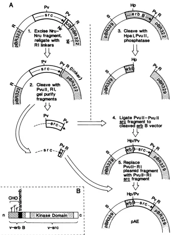
Construction ofa chimericv-erbB/srcgene. The N-terminal extracellular and transmembrane domains of the v-erbB
coding region were joined to the kinase and C-terminal
domains of the v-src coding region by anHpaIlPvuII blunt-end ligation, as follows (see Fig. 1).
(i) Steps 1 and 2:preparation ofthe v-srcmolecular clone. ThepBR322 subclone of the v-src EcoRI B fragment
con-tainsU3sequences from the RSV long terminal repeat which
would interfere with subsequent generation of infectious
chimeric virus genomes (13). We therefore reversed the
orientation of the EcoRI B v-src fragment in the pBR322 vectorby EcoRI cleavage and religation; this reversed clone was subsequently cleaved by NruI and religated in the presence of an excess of EcoRI oligonucleotide linkers
(Pharmacia, Inc.) to delete theundesired U3 sequences (see
Fig. 1). The plasmid DNA was then cleaved with PvuII restriction endonuclease, and the 870-base-pair fragment
(representingthe kinase domainand all but the C-terminal-most 11 aminoacidcodons of the v-srccoding region; 12, 46) waspurified by preparative agarose gel electrophoresis.
Step 3: preparation of the v-erbB molecular clone. The
creation andproperties ofthe Alu321 mutantofthe v-erbB
gene, representing a fully transformation-competent,
in-frame insertion ofanHpaI linker at anAluI site withinthe
v-erbBcoding region, havebeen previously described(36). The v-erbB gene carrying this HpaI oligonucleotide linker
insertionwassubclonedasaSalI-PvuII DNAfragment into the plasmid vector pBR329, cleavedwith HpaIand PvuII,
and treated with bacterial alkaline phosphatase to prevent
self-ligation.
Step 4: creation of a chimeric
v-erbB/src
oncogene. Thegel-purified PvuII-to-PvuII fragment ofv-srcisolatedin step 1 was ligated to the phosphatase-treated v-erbB pBR329 vector prepared in step 3, and the ligation products were
transformed intoHB101 hostcells. A molecularclone
rep-resenting thedesired constructionwasidentifiedby restric-tion endonuclease mapping; thecorrect nature ofthe clone
and preservation of the reading frame through the
HpaII
PvuII junction were confirmed by subsequent DNA se-quence analysis. The resulting construct(pErbB/src7)
rep-resents anin-framelinkage ofthefirst119N-terminal amino acid codons of the v-erbB coding region to amino acid codons 226 to 515ofv-src (12, 46,51).Step5:completionof the construction. Useofthe
870-base-pair PvuII-to-PvuIIv-srcfragmentintheligationin step 3of
ourconstruction resulted inamolecularclone,
pErbB/src7,
whichlacked 11 amino acid codonsthat arepresent on the veryC terminus of thewild-typev-srccodingsequence. ThepErbB/src7
plasmid DNA was thereforesubsequently
cleaved with Pvull and EcoRI, and the missing v-src se-quences were introduced in the form of a135-base-pair
PvuII-EcoRI DNA fragment obtained from the v-src plas-mid. The final plasmid clone,pErbB/src7-36,
contains anintact coding domain
beginning
at the N terminus of theparental v-erbB
coding
sequence andterminating
atthevery Cterminusof thev-src sequence.Constructionofaninfectious molecular clone and transfec-tionof avian fibroblasts. The v-erbB/src chimericoncogene
was subsequently reconstructed into aninfectious form by
replacing
the SalI-to-EcoRI sequences in pAEV-11-3R, aninfectious molecular clone of the wild-type AEV genome, with the corresponding SalI-to-EcoRI sequences from the
pErbB/src7-36
plasmid (47).Theresultingvector,referredto here aspChimera,
therefore encodes a variantofthe AEV DNA genome in which theoriginal
v-erbB sequences havebeen
replaced
by
the chimeric v-erbBlsrc gene. ThepChimera
vector possesses an intact v-erbA locus and iscapable of
generating
infectiousvirus when transfected intoavian fibroblasts in the presence ofa suitable
helper
virus(pAEVchimera,
like theoriginalAEV genome,isreplication
defective)
(36, 47).Chicken
embryo secondary
cells weretransfected witha molecular clone(pRAV-1OR)
of the Rous-associated virus type 1 (RAV-1) genomealone,
the RAV-1 helperplus
theerbBlsrc chimera
plasmid,
or the RAV-1helper plus
wild-typeAEV(AEV-11-3R) byacalcium
phosphate
coprecipita-tiontechnique
(47).Approximately
100 ,ug ofpRAV-1OR
DNA plus 500 ,ug each ofeither
wild-type
or chimeraAEVplasmid
DNA was used for each 60-mm(diameter) plate.
Thetransfectedavian cellswerepassaged
1:5 every 2days
in DME 8+1 at390C. Culture supernatants from the transfect-ed fibroblasts weresubsequently
used toinfect fresh fibro-blasts orbone marrow cells.Assaysforoncogenic transformation of cells.The
ability
of infected fibroblasts to grow in soft agar(demonstrate
sub-strate-independent
growth)
was tested aspreviously
de-scribed,
with fibroblast-conditioned medium inplace
of afeeder cell
monolayer (36).
Hexoseuptake
was determinedby
measurement of[1-3H]deoxyglucose
transport(39,
44).
Actin cables were visualizedby
fluorescentstaining
ofpermeabilized
fibroblasts withrhodamine-conjugated
phal-loidin (2).Plasminogen
activator protease secretion was determinedby
useofacasein-agarose overlay plaque
assay(20). The
ability
ofthe mutant to transformerythroid
cellprogenitors
wasassayed by
amethylcellulose colony
methodwith bone marrow cells derivedfrom 1- to 2-week-old SPAFAS chickens(21).
Immunoprecipitation analysis and in vitro kinase assay. Infected fibroblasts were radiolabeled for 2 h at
39°C
in RPMImediumcontaining
250,Ci
of[35S]methionine
perml(800
Ci/mmol;
500puCi/106
cells).
The cellswerethenlysed,
and the
lysates
wereimmunoprecipitated
aspreviously
described,
witheithertumor-bearing-rabbit (TBR)
serum or serumdirectedagainst purified
RSVvirions(35, 37,
41).
Theimmunoprecipitated
proteins
wereanalyzed
by
sodiumdo-decyl
sulfate(SDS)-10% polyacrylamide
gel
electrophoresis
and visualized
by
fluorography.
The in vitro kinase assay was
performed
aspreviously
described for the viral src
protein (7, 35),
with the TBR serum used in theprotein
analysis
describedabove.Subcellular fractionation and
tunicamycin
treatment.Ap-proximately 107
fibroblasts infectedby
theerbB/srcchimera were radiolabeled with[35S]methionine
for 2 h at39°C
asdescribed above. The cells were then swelled on ice in
hypotonic
buffer(5
mMKCl,
1 mMMgCl2,
20 mMHEPES[N-2-hydroxyethylpiperazine-N'-2-ethanesulfonic
acid; pH
7.1],
10mMN-ethylmaleimide,
0.5%aprotinin),
scrapped
offthe culturedish with arubber
policeman,
and broken openby
30strokes ofaloose-fitting plunger
inaDouncehomog-enizer.Thedifferentsubcellularfractionswere
subsequently
isolated
by
differentiationcentrifugation
andisopycnic
band-ing
aspreviously
described for theparental
v-erbB andv-srcproteins (10, 40).
Samples
of the different subcellular frac-tions wereadjusted
to 0.5 MNaCl-1
mg of bovine serumVOL.61, 1987
on November 10, 2019 by guest
http://jvi.asm.org/
albumin per ml-1% (wt/vol) Nonidet P-40 and subjected to
immunoprecipitation with src-directed serum, and the
immu-noprecipitates were analyzed by SDS-polyacrylamide gel
electrophoresis.
Oneplate oftwoduplicate cultures of infected fibroblasts
(approximately 106 cells per plate) was treated with 1 Fig of
tunicamycinfor 4 h at 39°C; the other plate was not exposed to the inhibitor. Both cultures were then washed and incu-bated for 2 h more in RPMI medium containing 500 ,uCi of
[35S]methionine perplate, retaining 1 ,ugof tunicamycin per ml in the labeling medium in the treated culture. The cells were subsequently washed with phosphate-buffered saline
and lysed, and the lysates were analyzedby immunoprecip-itation and SDS-polyacrylamide gelelectrophoresis.
RESULTS
Achimeric
v-erbB/src
oncogene was created by recombinant DNA methodology. The N-terminal extracellular,glycosyl-ated, and transmembrane domains of the v-erbB coding
region were fused to the C-terminal kinase domain of the v-srccoding region by an HpaI-PvuII ligation, using molec-ular clones of each of these two oncogenes and standard
recombinant DNA techniques (Fig. 1A). This construction resulted in in-frame linkage of the first 119 codons of the
v-erbB sequence to the C-terminal 301 codons of v-src. A
single extra codon was introduced at the site of fusion
because ofthe method of construction; avirtually identical
in-frame insertion at the same site in the v-erbB protein sequence has beenpreviously shown to have no detectable
effect on oncogenic or biochemical properties (36). The resulting construction places the v-src kinase domain in a
similar position, relative to the v-erbB transmembrane
do-main,asthe v-erbB kinasedomainitreplaces(Fig. 1B). The veryC-terminal region of the v-erbB polypeptide, previously
implicated in erythroid transformation, possesses no obvi-ous cognate in v-src and is absent from the chimeric con-struct (51).
The
v-erbB/src
chimeric oncogene encodesastable polypep-tideof theexpected molecular weight. An infectious form ofthechimeric oncogene was created by completely replacing the v-erbB sequences of pAEV-11-3R, amolecular clone of the AEV genome, with the
v-erbB/src
chimeric gene. Thefinal infectious construct (referred to as pChimera) was
subsequently transfected into avian fibroblasts in the pres-enceofanRAV-1genome. A RAV-1helper virus is required
by AEV and AEV-based vectors for replication (22, 26). Parallelcell cultures were transfected by the RAV-1 genome
alone or the RAV-1 genome and an unmodified AEV
genomic clone to serve as negative and positive controls,
respectively. Virus stocks recovered from the transfected cells were used to infect fresh fibroblast cells, and the infected cells were propagated for at least five passages before being assayed for their biological and biochemical
properties.
The polypeptides encoded by the chimeric virus were
analyzed by immunoprecipitation of extracts of infected
fibroblasts metabolically labeled with
[35S]methionine
(Fig.2). A polypeptide doublet of heterogeneous molecular
weight (49,000 and 53,000 apparent molecular weight [49K and 53K polypeptides, respectively]) could be detected by
v-src-directed antiserum, (TBR serum) in fibroblasts
in-fected by the chimera(lane 2), which was not present in cells
infected by the RAV-1 helper alone or by the AEV parent
(lanes 1and 3). The TBR serum was obtained from a rabbit
bearing
an RSV-induced tumor and therefore cross-reactswith a number of helper viral structural proteins (7, 35, 37, 48) (Fig. 2). The pattern of reactivity of the TBRserum can be compared to the pattern of structural and
p74gager-bA
proteins detected in these same cells by a serum directed against purified virion proteins (Fig. 2, lanes 4 to 6). The 49K-53K protein was immunoprecipitated by neither the virion-specific antiserum (Fig. 2, lanes 4 to 6) nor normal rabbit serum (data not shown). Partial proteolysis mapping (notshown) confirmed theidentity of the 49K-53Kproteinas av-erbBlsrc chimeric polypeptide.The chimeric v-erbBlsrc polypeptide is glycosylated. The heterogeneoussize of the chimeric oncogene protein synthe-sizedin chimera-infected cells closely resembled the pattern displayed by the parental AEV erbB protein (24, 41), al-though the chimera pattern migrated at a position some 15,000 daltons smaller than that of the wild-type v-erbB protein. This is the pattern predicted if the v-erbB-derived N terminus is glycosylated in the chimera as it is in the parental v-erbB protein (thev-erbBlsrc chimera protein should be 132 amino acid codons smaller than the v-erbB parent).
Thishypothesis was confirmed by the use of tunicamycin, aspecific inhibitor of N-linked protein glycosylation (Fig. 3). Fibroblasts infected by the chimera and radiolabeled with [35S]methionine in the absence of tunicamycin synthesize the 49K-53Kprotein doublet as described above (lane 4; labeled gp49 and gp53). A duplicate culture of chimera-infected fibroblasts, treated with tunicamycin, synthesized a single, smaller polypeptide of homogenous size (about 46,000 in apparent molecular weight, labeled p46 in lane 3). The
polypeptide synthesized by the chimera in the presence of tunicamycin was virtually identical in size to the primary translation (unglycosylated) product predicted from the nu-cleicacidsequenceof the v-erbBlsrc chimeric oncogene. No such protein was detected in fibroblasts infected by RAV-1 helper virus alone (lanes 1 and 2). Serving as an internal control, the effectoftunicamycin on theproteinsencoded by the helper virus can also be seen in Fig. 3. Tunicamycin treatment resulted in synthesis of a smaller form of the normally glycosylated RAV-1 envelope precursor protein (gPr92) but had no visible effect on the unglycosylated gag-relatedpolypeptides ofthehelpervirus (p27, for exam-ple).
The v-erbB/src chimeric protein follows the biosynthetic pathway of thev-erbB parent.The subcellularlocations of the v-erbBand v-srcproteins are distinct from one another. The v-src protein is synthesized on free polysomes and associ-ates with the plasma membrane posttranslationally (33, 42; reviewed in reference 32). The v-erbB protein is synthesized on rough endoplasmic reticulum and subsequently translo-cated to lighter membrane fractions (4, 23, 40). The glycosylation observed for the v-erbBlsrc chimera protein
strongly suggested that the N-terminal v-erbB sequences were capable of directing the chimeric polypeptide to the rough endoplasmic reticulum system despite the present of the v-src catalytic domain. This was confirmed by subcel-lular fractionation of chimera-infected fibroblasts (Fig. 4).
Fibroblasts infected by the chimera were radiolabeled for 2 h with [35S]methionine and lysed, and the various subcellularfractions were separated by differential-velocity and isopycnic centrifugations. Most of the v-erbBlsrc chi-meric protein was found in the P-154 membrane and nuclear wash fractions by these methods (Fig. 4A, lanes 3 and 5). When the membranes in the P-154 pellet were further fractionated by density, the majority of the v-erbBlsrc pro-tein was found at the 40%/50% sucrose interface (lane 7). Both the nuclear wash and the40%/50% sucrose interface
on November 10, 2019 by guest
http://jvi.asm.org/
A CHIMERIC v-erbBIsrc ONCOGENE 1941
A
Hp1. Excise Nru-" N Nru fragment,
religatewith Z RI linkers
Pv
Egt~~&
I ..
ID c
f
o3
I ..
I
I I'
Hp
er
(0111
4. Ligate Pvu 11- PvuII
srcfragmentto
cIeaved
erb B vectorHp/Pv
Al.,V
4
D
EB
CHOQ
,n/E/ E
Kina,s,e .D,o,m.ar:-
Ic,
v-erb B v-src
L_-_--_-_--________-___-_-_-__-_-__
FIG. 1. Construction ofachimeric v-erbBIsrconcogene. (A) Flow chart of the construction scheme used. The details, of and rationale behind thestepsusedin theconstruction are explained in Materials and Methods. Briefly, aPvuII-PvulI fragmentfrom the RSV DNA genome wasexcised byrestriction endonuclease cleavage and used to replace theHpaI-PvuIIDNA fragment originally in the v-erbB pAE-SAL-RI-Alu321 molecular clone(steps 1 to 4). APvuII-EcoRIfragment fromtheRSVclone(containing theC-terminal11amino acid codonsmissing from the PvuII-PvuII fragment described above)was nextexcisedand used toreplace thePvuII-EcoRIfragment contributedbythepBR329 vector(step5). The resulting chimeric oncogene contains the N-terminal coding sequences of v-erbB linked at an HpaI-PvuII site to the C-terminalcodingsequencesofv-src.Abbreviations:R=EcoRI,S=Sall,Pv =PvuII,Hp=HpaI,and Nr=NruI.Sequences contributed
by v-erbB are represented as hatched boxes, and sequences contributed by v-src are represented by open boxes. Arrows indicate the orientation ofthereading frames of bothgenes.(B) Schematic ofthechimericoncogenepolypeptide. Theexpectedstructureofthechimeric polypeptide synthesized by the v-erbB/src fusion oncogene is shown schematically. v-erbB-related sequences are hatched; v-src-related sequencesareshownasopenboxesorstippled. CHO, Possible sites ofN-linkedproteinglycosylationinv-erbB;transmemb.,transmembrane domain; kinase domain, region ofconservedaminoacidsequencethoughttodefinethe active site of thetyrosine-directedproteinkinases. VOL.61, 1987
on November 10, 2019 by guest
http://jvi.asm.org/
[image:4.612.139.488.106.583.2]TBR-se rum
antI-virus
seruiS...
..::~.:.I....Z
_ .f.P4.%.
_53K
'49K
12
3
<
:
I 0
w
3-4 5 6
< c Er W
0
lm
env
_gPr92
-\p74erb
Ap27
gag\p
19gag
preparations in vitro, but none ofthe known erbB-directed antisera appeartoberecognizedassubstratesby the v-erbB kinase (19, 31). We were therefore interested in character-izing the in vitro kinase activity ofourv-erbBlsrcchimeric protein.
The chimeric polypeptide was immunoprecipitated with TBR serum, theimmunoprecipitateswerewashed and incu-bated with [-y-32P]ATP, and theproducts of the reactionwere analyzed by SDS-polyacrylamide gel electrophoresis and
autoradiography (Fig. 5). Intense kinase activity directed against immunoglobulin G (IgG) heavy chain could beseen in immunoprecipitates of the v-erbBlsrc chimeric protein (lane 4). No kinase activity wasdetected in control immu-noprecipitates with normal rabbitserum(lanes 1, 3, and 5), in TBR immunoprecipitates of cells infected with RAV-1 only (lane 2), or in TBR immunoprecipitates of wild-type
AEV-infected cells (lane 6). The TBR serum used in this analysis had no detectable activity againstc-srcpolypeptide (lane 2; also, reference 37). Similar immunoprecipitates of theparental v-erbBprotein with v-erbB-directedserum were essentially negative in these assays (datanotshown). These results demonstrate that the v-src kinase domain in the chimeric polypeptide retains the structural and enzymatic
env
IN--gPr92
..of...
Ll
FIG. 2. Proteins synthesized by the chimeric oncogene in in-fected fibroblasts. Avianfibroblasts infected by theRAV-1 helper virus alone (lanes 1and 4), by the RAV-1helper and thev-erbBlsrc chimera (lanes2and5), orbythe RAV-1helperand wild-typeAEV (lanes3and6) weremetabolically radiolabeled with[35S]methionine andlysed, and v-src-related proteinswereimmunoprecipitated with TBR serum(lanes1 to3) oranti-gagserum(lanes4 to6). 49K-53K indicates the 49,000-53,000-molecular-weight polypeptide doublet synthesized by the chimera. Helper virus-encoded polypeptides include thep27,andp19gagproteins,aswellasgPr92,an envgene product. Also visible in these immunoprecipitates isp74gagerbA, the product ofthe AEVv-erbAoncogene. Molecularweight standards, runinadjacent lanes, are not shown.
represent fractions highly enriched for endoplasmic reticulum (40). This pattern observed for the gp49/53 chi-meric protein is virtually identicaltothepatternseenforthe
parental v-erbB polypeptide (Fig. 4B), indicating that the N-terminal domain of the v-erbB parent directed the subcellular location of the chimeric protein (40; data not shown). Incontrast, the subcellular distribution ofthev-src
parental proteinwas very different, withmost ofp60vsrc in solubleandlight-density (plasma membrane) fractionsunder thesameconditions (10;data notshown).
The v-erbB/src chimera protein possesses in vitro kinase
activity similar to that of the v-src parent polypeptide. The
v-src protein possesses strong tyrosine kinase activity in vitro, phosphorylating bothitself (autophosphorylation) and the heavy chain ofmany src-directed immunoglobulins (9,
35). Incontrast, the kinase activity ofthe v-erbB protein is much moredifficultto demonstrate in vitro. Relatively low levels of autophosphorylation and phosphorylation of
cer-tain target polypeptides have been reported for v-erbB
27gag
-* -p27~
1 2 3 4
+
_
+ - [image:5.612.59.296.74.390.2]tunica-RAV
CHIMERA
mycinFIG. 3. Tunicamycintreatmentofchimera-infected cells.
Fibro-blastsinfectedby the chimera plus the RAV-1 helper virus (lane 3
and4)orby the helper virus alone (lanes 1 and 2)wereradiolabeled
with [35S]methionine in the presence (lanes 1 and 3) or absence
(lanes2 and4) of tunicamycin. Thecellsweresubsequently lysed,
the lysates were immunoprecipitated with TBR serum, and the
immunoprecipitateswereanalyzed by SDS-polyacrylamide gel
elec-trophoresis and autoradiography.Glycosylated (gp49 and gp53) and unglycosylated (p46) forms of the chimeric oncogene protein are
indicated. Helper virus-encoded proteins also detected by TBR
serum includethe normally glycosylated gPr92env protein and the
unglycosylated p27Rag protein. Molecular weight standards, run in adjacent lanes,arenot shown.
a._...
...p
J9P53
-gp49
[image:5.612.344.527.318.605.2]zP46
...
4il
on November 10, 2019 by guest
http://jvi.asm.org/
A CHIMERIC v-erbBlsrc ONCOGENE 1943
1
2
3 4
5
67
r
_
. _
_ _
~~gPr92
A
U
~~~gp53
cgp681
L
_^
_ ,
_
_
,gp65
.t ._ 0
a) LO O XCO?
ii ~~~~~~~
Wx
a)
mL
3
o
It°
sucrose
FIG. 4. Subcellular localization of thev-erbB/src chimeric pro-tein.Fibroblasts infected by either thev-erbBIsrc chimera (panel A) orby wild-type AEV (panel B) were metabolically radiolabeled with [35S]methionine for2h and thenlysed, and the different subcellular fractions were isolated as described in Materials and Methods. Equal amounts of each fraction were immunoprecipitated with src-directed TBR serum (panel A) or anti-v-erbB serum (panel B), andthe immunoprecipitates were analyzed by SDS-polyacrylamide gel electrophoresis and autoradiography. The different fractions were loaded on theelectrophoretogram as follows (lanes): 1, total celllysate sample before fractionation; 2, 154,000 x gsupernatant; 3,154,000x g pellet; 4,purified nuclear fraction; 5, detergent wash of crude nuclei. Lanes 6 and 7 represent the 154,000 x g pellet further fractionated by density. Material at the 20%/35% (wt/vol) sucroseinterface, lane 6; material at the40%/50%sucrose interface, lane7.
propertiesnecessaryforTBRimmunoglobulin
phosphoryla-tion, properties that are not shared by the v-erbB parent
kinase domain (virtually no activity against TBR serum by thewild-typeAEVv-erbB protein was detected; Fig. 5, lane 6).
To better compare the kinase activity of our chimeric oncogene protein with thatoftheRSV parental v-src
poly-peptide,
we assayed in parallel the abilities of these twopolypeptides to function in the in vitro immunoglobulin kinaseassay(Table 1). Immunoprecipitatesfrom fibroblasts
infected by the
v-erbBlsrc
chimera demonstrated slightlyhigher kinase activity percell in thisassay than did
immu-noprecipitates derived from cells expressing the v-src par-ent. However, there was also slightly moregp49/53v-erbB/src protein in chimera-infected cells (detected as
[35S]methio-nine radiolabel) thanpp6O-src protein in RSV-infected cells(Table 1). Partial-proteolysis mapping (data not shown)
indicatedthatboth v-src and thechimericprotein
phosphory-lated thesamesite(s) withintheIgG molecule. Weconclude
that there is little or no significant difference between the in vitrokinase activities ofthe
v-erbBlsrc
and v-srconcogeneproteins.
The chimeric v-erbBlsrc polypeptide is fully capable of
transforming fibroblasts to an oncogenic state. Fibroblasts infected by the chimera quickly developed the distinctive
spindle-shaped, fusiform, criss-crossed morphology exhib-itedbycells transformedby theAEV parent
(Fig.
6band c; reference 44). This transformed morphology was differentfrom the round, loosely adherent morphology exhibited by
fibroblasts transformed by the RSV parent (panel d) and from theflat,nonrefractile, organized monolayers of untrans-formedfibroblasts infected by the RAV-1 helper alone (panel a).
Infectedfibroblasts were also tested for four other
pheno-typic manifestations ofoncogenic transformation (Table 2; reference 44). Chimera-infected fibroblasts were capable of
anchorage-independentgrowth, a relatively stringent test of oncogenic transformation, yielding soft-agar colonies indis-tinguishable in number and morphology from those gener-ated by AEV-infected cells. Chimera-infected fibroblasts also demonstrated high levels of plasminogen activator pro-tease secretion, anothercharacteristic of oncogenic transfor-mation (44), comparable to those demonstrated by AEV-infected cells although much lower than the protease levels seen in RSV-infected cells. Chimera-infected fibroblasts
contained few intact actin cable bundles, similar to the
disaggregation of actin cables seen in AEV- and RSV-induced transformation, whereas most fibroblasts infected by the helper alone retained actin cables. Whentested for
hexoseuptake,afifthcriterion of fibroblastoncogenic
trans-formation,fibroblasts infected by the chimera demonstrated
1
2
3
4
5 6N
T
N
T
N
T
a* .. _ *I
CHIMERA
RAV
AEV
IgG-heavy
chain
-
sera
FIG. 5. In vitro kinase assay of v-erbBIsrc chimera protein. Fibroblasts infectedbythe RAV-1helpervirus alone(lanes1and2), by the helper andthechimera(lanes 3and 4),orbythehelperand theparentalAEV (lanes5and6)were lysed,and thelysateswere
immunoprecipitatedwithnormal rabbit(N)serum(lanes 1, 3,and5) orRSV TBR(T)serum(lanes 2, 4,and6).Theimmunoprecipitates
werewashed, incubatedwith[-y-32P]ATPfor15minat23°C,washed again,andanalyzedbySDS-polyacrylamide gelelectrophoresisand autoradiography.Molecularweightstandards,runinadjacentlanes, areindicatedontheleft.
VOL.61, 1987
on November 10, 2019 by guest
http://jvi.asm.org/
[image:6.612.59.300.75.266.2] [image:6.612.330.554.323.635.2]TABLE 1. Kinaseactivityofthev-erbBlsrcchimeric oncogene
protein comparedwith that of thev-srcparent
[35S]methionine-Kinaseactivity labeled
ACM/0
ocgn Kinase/labeledProtein
.cpm)/lSa
oncogene protein ratioinfectedcellsa protein (102)
(CpM)/106(12
infectedcellsb
v-erbBIsrc 592,200 2,595 2.28
chimera
v-src 225,138 1,474 1.53
parent
aKinaseactivity wasmeasured as describedpreviously (9, 35). Briefly,
cells infectedbyeither the v-erbB/src chimera virusorwild-type
Schmidt-Ruppinwerelysed,and the extractsweleimmunoprecipitatedwith RSV TBR
serum as described in the legendto Fig. 5. Theimmunoprecipitates were
washed and incubated with 2,Cieach of[y-32P]ATPfor 15minat23°C,and theradioisotope incorporatedinto IgG heavychainwasmeasuredby
SDS-polyacrylamide gel electrophoresis and aliquid scintillationcounting
tech-nique. Kinaseactivityisrepresentedas32pcountsperminuteincorporated
intoIgG heavychainduringthe 15-miri incubation(1,000cpm=0.151 fmol of
phosphate incorporated).
b Infected cultures offibroblasts, preparedand maintained in parallel to
those used inthekinaseassaydescribed abovewereradiolabeled for 2 h with
[35S]methionine as previously described (41). The cells were lysed, the
extractswereimmunoprecipitatedwith TBRserum,and the
immunoprecipi-tateswereanalyzed by SDS-polyacrylamide gel electrophoresis. The
radio-labeledv-srcandv-erbBIsrc proteinbandswerevisualizedby
autoradiogra-phyandquantitated byexcision andaliquidscintillationcounting technique.
slightly but consistently elevated levels of deoxyglucose uptakerelative to thatof untransformed cells. This elevation
was statistically significant and reproducible iti over five inidependentassays.
[image:7.612.313.556.90.155.2]A trivial explanation of the oncogenic properties of the v-erbB/src chimeric construct would be accidental contami-nation of our stocks by wild-type virus or a revertant or TABLE 2. Fibroblasttransformationparametersexhibitedby
chimera-infected fibroblasts
% of cells
No. of Hexose with No.of
Virus used forinfectiona
cinesoft
uptake intact caseinolyticagarb (cpm)c actin plaquese
RAV-1only 0 700 85 0
RAV-1 + AEV 1,022 13,000 31 489
RAV-1 + chimera 1,058 1,839 23 345
Schmidt-RuppinRSV NT 3,066 16 3,740f
aInfected chicken embryo fibroblasts were cultured for at least five
passages before the transformation phenotype was assayed. All assays
representtheaverageof at leasttwo determinations.
bInfected fibroblasts were trypsinized and counted,and 105cells were
platedinto softagarmedium. Thenumber ofmacroscopicfibroblast colonies
visible perplate after a 10-day incubation at 39°Cis presented. NT, Not
tested.
IApproximately 105infected fibroblastswereincubated for 5minat39°C
with 4,uCiof[3H]deoxyglucose.Thecellswerethenextensively washed, and
theradiolabelremainingcellassociated, expressedascountsperminute,was
determinedby liquidscintiliationcounting.
dInfected fibroblasts were plated on cover slips, washed, fixed, and permeabilized, and the actin cables were visualized with rhodanline-conjugated phalloidin. The number ofcellsexhibitingintact actincables is presentedas apercentageof thetotalnumber of cellscounted(about 300 cells perassay).
eInfected(RAV,AEV,orChimera)fibroblastsweretrypsinized,and5 x
105cellswereplatedinto 60-mm(diameter) petri plates.Thecellswerethen
washedandoverlaidwith caseinagaroverlay mnediumaspreviously described
(20).Thenumber ofzonesofcaseinolysis (plaques)werecounted aftera16-h
incubation at37°C.
fRSV-infectedcellswereplatedat 5 x 10⁴cellsperplate,and the number
ofplaquesobservedwasmultiplied by10.
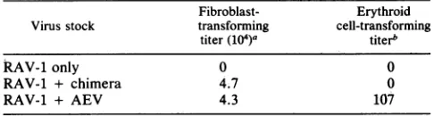
TABLE 3. Erythroid cell transformationbythe chimera
Fibroblast- Erythroid Virus stock transforming cell-transforming
titer(104)a titerb
kAV-1only 0 0
RAV-1 + chimera 4.7 0
RAV-1 + AEV 4.3 107
aDeterminedby exposing 5 x 10⁵ uninfected fibroblasts to a series of
dilutions of the virusstock, incubating the cells for 12 hat39°C, followedby
trypsinization and plating of the cells in soft agar medium. Fibroblast-transforming titerisexpressedasthe number ofsoftagarcoloniesinducedper
milliliter of theoriginal virus stock.
bDetermined byuseofamethylcellulose-bonemarrowcolonyassay(21)
andexpressedasthe number ofmacroscopic erythroid cell colonies induced per4ml of theoriginalvirus stock.
recombinant form of the chimeric virus. We therefore
ex-tracted genomic DNA from infected fibroblasts and
sub-jected the DNA to restriction endonuclease-Southem blot-ting analysis by using restriction enzyrhes and hybridization probes that would distinguish the AEV genome from that of the chimera. The restrictiondigestion pattern obtained from the DNA from chimera-infected cells was identical to thatof theoriginal constructionandruledoutlarge-scale rearrange-mentswithinthe chimeric oncogene or possible contamina-tion withwild-type AEV or RSV (data not shown).
The chimeric v-erbB/srcgene does not transform erythroid cells in an unsupplemented bone marrow colony assay. The
wild-type v-erbB protein is capable of oncogenic transfor-mation of immature erythroid cells as well as fibroblasts (5, 14, 18, 22). This erythroid cell-directed activity has been localized, in part, to the very C-terminal domain of the
v-erbB protein (51; unpublished data). However, actual
determinationof target cellspecificity inthetyrosine kinase family of oncogenes appearstobe acomplex phenomenon; a number of other tyrosine kinase oncogenes, including
v-src, appears to becapable ofatleastlimited erythroidcell
transformation activity (28, 29, 38). It was therefore of
interestto testtheability ofourchimerato transformavian
erythroid cells inan in vitrobonemarrow colonyassay. Stocks of the v-erbBlsrc chimera showed no erythroid
cell-transforming activity in our assay, in contrast to the hundreds of erythroid cell colonies induced by the AEV
parent(Table 3).Both the AEVparentalvirus stock and the
chimera stock possessed approximately equal
fibroblast-transforming titers (Table3). Weconcludethat theerythroid cell-transforming potential ofthechimerais at least 2 orders ofmagnitudelower than thatof thev-erbB parent oncogene
despite the presence of an intact, expressed copy of the v-erbAgene inthechimeric construct.
DISCUSSION
Nature of the
v-erbB/src
chimericoncogene. The chimericconstruction we have generated should encode a
46,000-molecular-weight primary translation product bearing the extracellular, glycosylated, andtransmembrane domains of the AEV v-erbB protein linked to the kinase catalytic domain of the RSV v-src polypeptide. The amino acid sequence Val-Ala-Ile-Lys (VAIK), a highly conserved do-main in allof the knowntyrosinekinases (25), is thought to represent a portion of the ATP-binding site (30) and is
situated 74 aminoacid codons C terminal to the end of the transmembrane domain in theparental v-erbB protein. The
on November 10, 2019 by guest
http://jvi.asm.org/
[image:7.612.57.297.100.197.2]A CHIMERIC v-erbBlsrc ONCOGENE 1945
a.
RAV-only
b.
CHIMERA
c.
AEV
d.
RSV
.VIq?" ,. .
i0?!
IhFIG. 6. Morphology of fibroblasts infected by the chimera. Representative microscope fields offibroblastmonolayers infected by the RAV-1helper virus alone (a), the helper plus the chimera (b), the helper plus the AEV parent (c), or the RSV parent (d) are shown. Bar, about 20,um.
chimeric construction places the same VAIK sequence in
the v-srckinasedomain 86aminoacidsfrom the end of the transmembrane domain contributed by the v-erbB gene.
The abilityofour chimeric construct to transform fibro-blasts suggeststhatthe exactspacing betweenthecatalytic and membrane-association domains ofthe tyrosine kinases is not crucial for biological activity. This result supports
evidence
previously
obtainedfromsite-directedmutagenesis experiments on v-erbB (36). Furthermore, the ability toexcise the kinase domain ofv-src and introduce it into an
unrelated protein sequence background (v-erbB-encoded sequences) in an active form strongly suggests that the
kinase region is itself a functionally and conformationally discrete domain. This is in agreement with data obtained
from site-directed mutagenesis and
partial-proteolysis
map-ping (11, 17, 34).
Biochemicalpropertiesofthechimera-encodedpolypeptide.
The chimeric
v-erbBlsrc
oncogene encoded a46,000-molecular-weightprimary translation product thatwas con-sistent with the peptide predicted from the construction. This chimeric protein wasglycosylated in infected cells to multiple species of higher apparent molecularweight, indi-cating that the N-terminal glycosylation sites contributedby
the v-erbB sequences could be fullyrecognized by the host cell glycosyl transferases despite the linkage of C-terminal sequencesfrom the normallyunglycosylated v-srcprotein.
Glycosylation of the v-erbB/src protein also implied the presence in the chimera of an appropriate signal sequence capable ofdirecting the association of the chimericprotein with microsomal fractions. The subcellular distribution of thev-erbBlsrc protein bore out thisprediction; the chimeric protein is found in the same subcellular fractions as the v-erbB protein, principally fractions enriched for rough endoplasmic reticulum (40). This distribution of the chimera is distinct from that of thev-srcpolypeptide(10), confirming that the N-terminal sequences of these two oncogene pro-VOL.61,1987
on November 10, 2019 by guest
http://jvi.asm.org/
[image:8.612.60.560.70.496.2]teins are crucial in determining their biosynthetic pathways. The actual location of this signal sequence on theparental
v-erbB protein remains unclear. We feel that the strongest hypothesis is that thetransmembrane sequence of the v-erbB protein is itself the signal sequence for membrane associa-tion. We base this hypothesis on theabsence of an obvious consensus signal sequence at the extreme Nterminusof the AEV-ES-4 v-erbB coding region (unpublished data) and on the properties of a mutant ofv-erbB we have isolated that lacks the transmembrane domain (A-transmemb.; manu-script inpreparation). If the extreme Nterminus of v-erbB, which is retained in our A-transmemb. mutant, encoded a signal sequence, the mutantpolypeptidewouldbeexpected
to be sequestered into microsomal fractions and perhaps secreted. Instead, the A-transmemb. v-erbB protein is syn-thesized as asolublecytoplasmic protein(unpublisheddata).
The chimeric polypeptide is fully capable ofacting as a protein kinase in vitro, phosphorylating src-directed IgG heavy chain at levelscomparable to thoseexhibited by v-src protein itself. This result for the chimera contrasts to the properties of thev-erbB protein, which is a relatively poor kinase in vitro and does notrecognize as asubstrate any of the erbB- orv-src-directed immunoglobulins yet tested (19, 31). The kinase domain in ourchimeratherefore appears to retain the in vitro enzymaticproperties demonstratedby the RSV parent.
Biological properties of the
v-erbB/src
chimera. Thev-erbBlsrc chimera was fully capable of transforming
fibro-blasts to an oncogenic state, as judged by morphology,
growth in soft agar, plasminogen activator protease secre-tion, and loss of actin cables, although hexose uptake was onlyslightly elevatedcomparedwith levels inuntransformed
cells.
The morphologyofv-erbB/src-transformedcells was iden-tical to that offibroblaststransformed by the v-erbBparent (fusiform cells forming criss-crossed monolayers) and was readily distinguished from that of the round, refractile,
poorlyadherent cellstransformed by the RSV parent(44).It therefore appears that replacement of N-terminal v-src se-quenceswith v-erbB sequences can alter at least one aspect of the transformed phenotype. Intriguingly, a number of
different mutations within the v-src gene are also known to yield a fusiform morphology (1, 15, 43). These fusiform
mutations, as a group, tend to map totheN-terminalportion
ofthe v-src sequence (15, 16, 27). The morphological prop-erties of cells infected by our chimera maytherefore be due to the absence of src sequences that are necessary forfull manifestation of the transformed state, perhaps resulting in alteration of the substrate specificity of thechimera tyrosine kinase. An alternative hypothesis is that the distinct subcellular localizations of the v-src and
v-erbBlsrc
chimera proteins results in different accessibility of the kinases to host cell protein substrates that are important in determina-tion of morphology. This latter hypothesis is supported by a study which revealed that several fusiform mutants of RSV synthesized a v-src polypeptide with altered subcellular distribution (43). A shared theme in both of these hypotheses is that the v-src kinase domain itself is not the sole determi-nant of the transformed phenotype and that the differences seenin the phenotype of v-src- and v-erbB-transformed cells cannot be solely attributed to the divergence in amino acid sequence within the kinase domains of these two oncogene polypeptides.Ourchimeric oncogene failed to transform erythroid pro-genitor cells detectably in our in vitro bone marrow assay
despite theability of the construct to synthesize a
(presum-ably)functionalv-erbApolypeptide. This result is consistent with evidenceobtained from AEV mutantsthat suggests that the C-terminal domain of the v-erbB protein is intimately
involvedinerythroidtargetcell specificity(51; unpublished
data). This C-terminal domain is missing from our chimeric construct. On the other hand, our results appear to be
inconsistent with those of Kahn et al., who demonstrated that the parental v-src gene, in association with v-erbA, could induce erythroidcelltransformation similar to that of wild-type AEV (29). It has been reported that v-src-transformed erythroid cells propagate only under a very narrow rangeof pH and temperature (29). It is possible that our relatively simple bone marrow culture conditions, al-though capable of supporting growth of wild-type AEV-transformed erythroid cells, are not capable of permitting
propagation of perhaps more fastidious v-erbBlsrc-trans-formed erythroid precursors or that a very low level of erythroid cell-transforming ability exists for our chimeric oncogene but could not be detected within the statistical limitations ofour assay.
Inconclusion, at least twomembers of the tyrosine kinase family of viral oncogenes, v-erbB and v-src, share close functional and structural interrelatedness, to the point that domains of one polypeptide can be interchanged with do-mains of the other and yield a fully functional polypeptide
product. Thepropertiesof theresultingchimera suggest that the N-terminal v-erbB sequences play an important role in determining fibroblast morphology, perhaps byaffectingthe accessibility or affinity of the chimeric protein for certain target polypeptides. It has been previously suggested that the ratio of pp36 to pp42 phosphorylation or differences in fibronectin attachment in v-src- versus v-erbB-transformed
cells may account for the differences in morphology of fibroblasts infectedbyAEV versus RSV(19, 27, 43).Weare presently analyzing the phosphorylation pattern of known targetsubstrate polypeptides in our chimera-infected cells to testthis hypothesis.
ACKNOWLEDGMENTS
The expert and enthusiastic technical assistance of Cathy Judelson isgratefullyacknowledged.Ialso thank Blake Millikanfor helpfuldiscussions.
This workwassupported by Public Health ServicegrantCA38823 fromthe NationalCancer Institute.
LITERATURE CITED
1. Anderson, D. D., R. P. Beckmann, E. H. Harms, K. Nakamura, and M. J. Weber. 1981. Biological properties of "partial" transformation mutants ofRous sarcoma virus and character-izationoftheirpp60sc kinase.J. Virol. 37:445-458.
2. Barak, L. S., R. R. Yocum, E. A. Nothnagel, and W. Webb. 1980. Fluorescence staining ofthe actin cytoskeleton in living cells with7-nitrobenz-2-oxa-1,3 diazole phallacidin. Proc. Natl. Acad. Sci. USA77:980-984.
3. Barker, W. C., and M.0.Dayhoff. 1982. Viral src geneproducts are related to the catalytic chain of mammalian cAMP-dependent protein kinases. Proc. Natl. Acad. Sci. USA 79: 2836-2840.
4. Beug, H., and M. J. Hayman. 1984. Temperature-sensitive mutants ofavian erythroblastosis virus: surface expression of the erb B product correlates with transformation. Cell 36: %3-972.
5. Beug, H., A. von Kirchbach, G. Doderlein, J. F. Conscience, and T. Graf. 1979. Chicken hematopoietic cells transformed by seven strains ofdefective avianleukemiavirusesdisplay three distinct phenotypes ofdifferentiation. Cell 18:375-390.
on November 10, 2019 by guest
http://jvi.asm.org/
A CHIMERIC v-erbBIsrc ONCOGENE 1947 6. Bishop, J. M., and H. E. Varmus. 1984. Functions and originsof
retroviral transforming genes, p. 999-1108. In R. Weiss, N. Teich, H. Varmus, and J. M. Coffin (ed.), RNA tumor viruses, 2nded. Cold SpringHarbor Laboratory, Cold Spring Harbor, N.Y.
7. Brugge, J., and R. L. Erikson. 1977. Identification of a transfor-mation-specific antigen induced by an avian sarcoma virus. Nature(London) 269:346-348.
8. Buss, J. E., M. P. Kamps, and B. M. Sefton. 1984. Myristic acid is attached to thetransforming protein of Rous sarcoma virus during or immediately after synthesis and is present in both soluble andmembrane-bound forms of the protein. Mol. Cell. Biol.4:2697-2704.
9. Coliett,M. S.,and R. L.Erikson.1978. Protein kinase activity associated with the avian sarcoma virus src geneproduct. Proc. Natl. Acad. Sci. USA 75:2021-2024.
10. Courtneidge, S. A., A. D. Levinson, and J. M. Bishop. 1980. The protein encoded by the transforming gene of avian sarcoma virus (pp6Osrc) and a homologous protein in normal cells (pp6OPrOtOSrC) are associated with theplasma membrane. Proc. Natl.Acad. Sci. USA 77:3783-3787.
11. Cross, F. R., E. A. Garber, and H.Hanafusa. 1985. N-terminal deletions in Rous sarcoma virus p60src: effects on tyrosine kinase andbiological activities and on recombination in tissue culturewith thecellular src gene. Mol. Cell. Biol. 5:2789-2795. 12. Czernilofsky, A. P., A. D. Levinson, H. E. Varmus, J. M. Bishop, E.Tischer, and H. M. Goodman. 1983.Nucleotide sequence of anavian sarcoma virus oncogene (src) andproposed amino acid sequence for the gene product. Nature (London) 301:736-738.
13. DeLorbe, W. J., P. A. Luciw, H. M. Goodman, H.E.Varmus, andJ. M. Bishop.1980. Molecular cloning andcharacterization of avian sarcoma virus circular DNA molecules. J. Virol. 36:50-61.
14. Engelbreth-Holme,J., and A. Rothe Meyer. 1935. On the con-nection between erythroblastosis (haemocytoblastosis), mye-losis and sarcoma in chicken. Acta Pathol. Microbiol. Scand. 12:352-377.
15. Feuerman, M. H., B. R.Davis, P. K. Pattengale, and H. Fan. 1985. Generation ofarecombinant Moloney murine leukemia virus carrying the v-src gene of avian sarcoma virus: transfor-mation in vitro and pathogenesis in vivo. J. Virol. 54:804-816.
16. Fujita, D. J., J. Bechberger, and I. Nedic. 1981. Four Rous sarcomavirus mutantswhich affect transformedcell morphol-ogyexhibit altered src geneproducts. Virology 114:256-260. 17. Garber, E. A., F. R.Cross, and H. Hanafusa. 1985. Processing
ofp60v-srctoitsmyristylatedmembrane-bound form. Mol. Cell. Biol. 5:2781-2788.
18. Gazzolo, L., J. Samarut, M. Bouabdelli, and J. P. Blanchet. 1980. Earlyprecursors in theerythroidlineagearethe specific target cells of avian erythroblastosis virus in vitro. Cell 22:683-691.
19. Gilmore, T., J. E. DeClue, and G. S. Martin. 1985. Protein phosphorylation at tyrosine is induced by the v-erb B gene product in vivo and in vitro. Cell 40:609-618.
20. Goldberg, A.R.1974.Increasedprotease levels intransformed cells:acasein overlay assay for the detection ofplasminogen activatorproduction. Cell 2:95-102.
21. Graf,T.1975. In vitrotransformation of chicken bonemarrow cells with avianerythroblastosisvirus. Z. Naturforsch. Sect. C Biosci. 30:847-849.
22. Graf, T.,B.Royer-Pokora,G. E.Schubert,andH. Beug.1976. Evidence for themultipleoncogenic potentialof cloned leuke-mia virus: in vitro and in vivo studies with avian erythro-blastosis virus.Virology71:423-433.
23. Hayman,M.J.,and H. Beug. 1984.Identification ofaformof the avianerythroblastosisvirus erbBgeneproductatthecell surface. Nature(London) 309:460-462.
24. Hayman,M.J.,G.Ramsey, K. Savin, G. Kitchener, T. Graf, and H. Beug. 1983. Identification and characterization ofthe avianerythroblastosisviruserbBgeneproductas amembrane glycoprotein.Cell32:579-588.
25. Hunter, T., andJ. A. Cooper. 1985. Proteintyrosinekinases. Annu. Rev.Biochem. 54:897-930.
26. Ishizaki, R., andT. Schimizu. 1979. Heterogeneityof strain R avian(erythroblastosis)virus. Cancer Res. 30:2827-2831. 27. Iwashita, S., N. Kitamura, and M. Yoshida. 1983. Molecular
events leading to fusiform morphological transformation by partial src deletion mutant of Rous sarcoma virus. Virology 125:419-431.
28. Kahn, P., B. Adkins, H. Beug,and T. Graf. 1984. srcandfps
containingavian sarcomaviruses transform chickenerythroid cells. Proc. Natl. Acad. Sci. USA 81:7122-7126.
29. Kahn, P., L. Frykberg, C. Brady, I. Stanley, H. Beug, B. Vennstrom, and T. Graf. 1986. V-erb A cooperates with
sar-coma oncogenes in leukemic cell transformation. Cell 45:349-356.
30. Kamps, M. P., S. S. Taylor, and B. M. Sefton. 1984. Direct evidence that oncogenic tyrosine kinases and cyclic AMP-dependentproteinkinase havehomologous ATP-bindingsites. Nature(London)310:589-592.
31. Kris,R.M.,I.Lax,W.Gullick,M. D.Waterfield,A.Ullrich,M. Fridkin, andJ. Schiessinger. 1985. Antibodies against a syn-theticpeptideas aprobefor the kinaseactivityof the avian EGF receptor and v-erb Bprotein. Cell 40:619-625.
32. Krueger, J. G., E. A. Garber, and A. R. Goldberg. 1983. Subcellular localization of
pp6Asrc
in RSV-transformed cells. Curr.Top. Microbiol. Immunol. 107:51-124.33. Lee,J.S.,H. E.Varmus,andJ.M.Bishop. 1979.Virus-specific messengerRNAsin permissivecells infectedbyaviansarcoma virus. J. Biol. Chem. 254:8015-8022.
34. Levinson, A. D., S. A. Courtneidge, and J. M. Bishop. 1981. Structural and functional domains of the Roussarcoma virus transforming protein (pp60s1). Proc. Natl. Acad. Sci. USA 78:1624-1628.
35. Levinson,A. D.,H. Oppermann, L. Levintow,H. E. Varmus, andJ.M.Bishop. 1978. Evidence that thetransforminggeneof aviansarcomavirus encodesaproteinkinaseassociated witha
phosphoprotein. Cell 15:561-572.
36. Ng, M., and M. L. Privalsky. 1986. Structural domains of the avianerythroblastosisviruserbBprotein requiredfor fibroblast transformation: dissectionby in-frameinsertional mutagenesis.
J. Virol. 58:542-553.
37. Oppermann, H., A. D.Levinson, H. E. Varmus, L. Levintow, andJ. M. Bishop. 1979. Uninfected vertebrate cells containa protein that is closely related to the product of the avian sarcomavirustransforminggene (src). Proc. Natl. Acad. Sci. USA 76:1804-1808.
38. Palmieri, S. 1985. Transformation oferythroid cells by Rous
sarcomavirus (RSV). Virology140:269-280.
39.
Paulieri,
S.,H.Beug, and T.Graf. 1982. Isolation andcharac-terization of four new temperature-sensitivemutants of avian
erythroblastosisvirus(AEV). Virology 123:296-311.
40. Privalsky,M.L., andJ.M. Bishop. 1984. Subcellular localiza-tionof the v-erb Bprotein,theproductofatransforminggeneof avianerythroblastosisvirus. Virology135:356-368.
41. Privalsky, M. L., L. Sealy, J. M.Bishop, J. P. McGrath, and A. D.Levinson.1983. Theproductof the avian
erythroblastosis
virus erb B locus isaglycoprotein.Cell32:1257-1267. 42. Purchio, A.F.,S.Jovanovich,and R. L. Erikson.1980.Sites of
synthesis of viral proteins in avian sarcoma virus-infected chickencells. J.Virol. 35:629-636.
43. Rohrschneider, L.,andS.Reynolds.1985.
Regulation
ofcellular morphology by the Rous sarcoma virus src gene:analysis
of fusiformmutants.Mol. Cell. Biol.5:3097-3107.44. Royer-Pokora, B.,H.Beug,M.Claviez,H.J.Winkhardt,R. R. Friis,and T.Graf.1978.Transformationparametersinchicken fibroblasts transformed by AEV and MC29 avian leukemia viruses. Cell 13:751-760.
45. Schultz,A.M.,L. E.Henderson,S.Oroszlan,E. A.Garber,and H.
Hanafusa.
1985.Amino terminalmyristylation
oftheprotein
kinase
p605rc,
a retroviraltransforming
protein.
Science 227:427-429.46. Schwartz,D.C.,R.Tizzard,and W.Gilbert.1983. Nucleotide sequence ofRoussarcomavirus. Cell 32:853-861.
VOL. 61,1987
on November 10, 2019 by guest
http://jvi.asm.org/
47. Sealy, L., M. L.Privalsky, G. Moscovici, C. Moscovici, and J. M. Bishop. 1983.Site-specific mutagenesis of avian erythroblastosis virus: v-erb B is required for oncogenicity. Virology 130:
155-177.
48. Sefton, B. M., K. Beemon,and T.Hunter.1978.Comparison of theexpression of thesrcgeneof Roussarcomavirus in vitro and invivo. J. Virol. 28:957-971.
49. Teich, N. 1984. Taxonomy of retroviruses, p. 25-208. In R. Weiss,N.Teich,H.Varmus, and J. M.Coffin (ed.),RNAtumor
viruses, 2nded. Cold Spring Harbor Laboratory, Cold Spring Harbor,N.Y.
50. Vennstrom, B., L. Fanshier, C. Moscovici, and J. M. Bishop. 1980. Molecular cloning of the avian erythroblastosis virus
genome and recovery ofoncogenic viruses by transfection of
chicken cells. J. Virol. 36:575-585.
51. Yamamoto, T.,Y.Nishida, N.Miyajimi, S.Kawai, T. Ooi,and
K. Toyoshima. 1983. The erbBgene of avianerythroblastosis virus isamemberofthesrcgenefamily. Cell 35:71-78.

![FIG. 4.fractionsgeltein.orandsrc-directedfurtheroflanecellwere[35S]methionine3,Equalsucrose 154,000 by crude Subcellular localization of the v-erbB/src chimeric pro- Fibroblasts infected by either the v-erbBIsrc chimera (panel A) wild-type AEV (panel B) we](https://thumb-us.123doks.com/thumbv2/123dok_us/1358960.89384/6.612.330.554.323.635/fractionsgeltein-directedfurtheroflanecellwere-methionine-equalsucrose-subcellular-localization-fibroblasts-infected.webp)