City, University of London Institutional Repository
Citation
:
Powell, T., de Jong, S., Breen, G., Lewis, C. M. and Dima, D. ORCID:
0000-0002-2598-0952 (2018). Telomere length as a predictor of emotional processing in the brain.
Human Brain Mapping, doi: 10.1002/hbm.24487
This is the published version of the paper.
This version of the publication may differ from the final published
version.
Permanent repository link:
http://openaccess.city.ac.uk/21228/
Link to published version
:
http://dx.doi.org/10.1002/hbm.24487
Copyright and reuse:
City Research Online aims to make research
outputs of City, University of London available to a wider audience.
Copyright and Moral Rights remain with the author(s) and/or copyright
holders. URLs from City Research Online may be freely distributed and
linked to.
City Research Online:
http://openaccess.city.ac.uk/
[email protected]
R E S E A R C H A R T I C L E
Telomere length as a predictor of emotional processing in the
brain
Timothy R. Powell
1| Simone De Jong
1,2| Gerome Breen
1,2| Cathryn M. Lewis
1,2,3|
Danai Dima
4,51
Social, Genetic and Developmental Psychiatry, Institute of Psychiatry, Psychology and Neuroscience, King's College London, London, United Kingdom
2
National Institute for Health Research Biomedical Research Centre for Mental Health, Institute of Psychiatry, Psychology and Neuroscience at the Maudsley Hospital and King's College London, London, United Kingdom
3
Department of Medical and Molecular Genetics, Guy's Hospital, King's College London, London, United Kingdom
4
Department of Psychology, School of Arts and Social Sciences, City, University of London, London, United Kingdom
5
Department of Neuroimaging, Institute of Psychiatry, Psychology and Neuroscience, King's College London, London, United Kingdom
Correspondence
Dr Danai Dima, Department of Psychology, School of Arts and Social Sciences, University of London, 10 Northampton Square, London EC1V 0HB, UK.
Email: [email protected]
Funding information
European Union’s Horizon 2020 Research and Innovation Programme , Grant/Award Number: Marie Sklodowska-Curie grant agreement 658195; Medical Research Council (MRC), Grant/Award Number: MRN014863/1; NARSAD 2014 Young Investigator Award, Grant/Award Number: 22471; NARSAD Young Investigator Grant , Grant/Award Numbers: 60373, YI 60373; Psychiatry Research Trust Grant , Grant/Award Number: 92; European Union's Horizon 2020 Research and Innovation Programme, Grant/Award Number: 658195; Medical Research Council, Grant/Award Number: MRN014863/1; Department of Health; National Institute for Health Research; South London and Maudsley NHS Foundation Trust; King's College London
Abstract
Shorter telomere length (TL) has been associated with the development of mood disorders as
well as abnormalities in brain morphology. However, so far, no studies have considered the role
TL may have on brain function during tasks relevant to mood disorders. In this study, we
exam-ine the relationship between TL and functional brain activation and connectivity, while
partici-pants (n = 112) perform a functional magnetic resonance imaging (fMRI) facial affect recognition
task. Additionally, because variation in TL has a substantial genetic component we calculated
polygenic risk scores for TL to test if they predict face-related functional brain activation. First,
our results showed that TL was positively associated with increased activation in the amygdala
and cuneus, as well as increased connectivity from posterior regions of the face network to the
ventral prefrontal cortex. Second, polygenic risk scores for TL show a positive association with
medial prefrontal cortex activation. The data support the view that TL and genetic loading for
shorter telomeres, influence the function of brain regions known to be involved in emotional
processing.
K E Y W O R D S
bipolar disorder, emotional faces, fMRI, polygenic risk score, telomere length
1
|
I N T R O D U C T I O N
Biological aging, as opposed to chronological aging, is the concept that
two individuals born on the same date, may be aging at different rates
at any one particular time, at the cellular level (Jin, 2010). Telomeres
are repeat DNA sequences at the end of chromosomes that shorten
upon each cell division, and relative telomere length represents a
com-monly used measure of cellular age (Stewart, Chaiken, Wang, & Price,
2012). Characteristically, older individuals who have experienced a
greater number of cell divisions have shorter telomere lengths (TLs),
relative to younger individuals (Saretzki & von Zglinicki, 2002).
How-ever, shorter TL relative to one's chronological age, is associated with
increased risk for age-related diseases, and both environmental and
genetic factors have been found to moderate the rate of TL
shorten-ing (Shammas, 2011).
In the realm of psychiatry, TL is becoming of increasing interest
after the realization that TL shortening appears to be accelerated in
those who have experienced maltreatment and those with psychiatric
disorders (Drury et al., 2012). Faster shortening of telomeres has been
found after life stress (Epel et al., 2004), exposure to violence during
Received: 30 August 2018 Revised: 20 November 2018 Accepted: 24 November 2018 DOI: 10.1002/hbm.24487
This is an open access article under the terms of the Creative Commons Attribution License, which permits use, distribution and reproduction in any medium, provided the original work is properly cited.
© 2018 The Authors.Human Brain Mappingpublished by Wiley Periodicals, Inc.
childhood (Shalev et al., 2013), experiencing highly disadvantaged
environments (Mitchell et al., 2014) and early life adversity (Ridout
et al., 2018). Short TL in psychiatry has been linked primarily to
patients suffering from major depressive disorder (Hartmann,
Boeh-ner, Groenen, & Kalb, 2010; Lung, Chen, & Shu, 2007;
Monroy-Jara-millo, Dyukova, & Walss-Bass, 2017; Ridout, Ridout, Price, Sen, &
Tyrka, 2016; Schutte & Malouff, 2015), post-traumatic disorder
(Lindqvist et al., 2015; Shalev et al., 2013), anxiety disorders
(Monroy-Jaramillo et al., 2017; Shalev et al., 2013) and bipolar disorder
(Barbé-Tuana et al., 2016; Lima et al., 2015). However, not all patients have
shortened TL, and evidence suggests that TL may actually be marking
the effects of specific environmental risk factors for psychiatric
disor-ders, or the severity of specific cognitive biases or symptoms, rather
than representing a definitive case/control predictor (Elvsåshagen
et al., 2011; Powell, Dima, Frangou, & Breen, 2018; Vincent et al.,
2017). One of the key cognitive biases associated with mood
disor-ders is in the way patients process emotion, while demonstrating
altered patterns of brain activation compared with healthy controls
(Mourão-Miranda et al., 2012; Stuhrmann, Suslow, & Dannlowski,
2011). Interestingly, functional magnetic resonance imaging (fMRI)
studies have revealed regional differences in brain activation during
reading of facial emotions between young and old participants,
(Ebner, Johnson, & Fischer, 2012), suggesting the way we process
emotion may be subject to the effects of age. Premature biological
aging may, therefore, affect or predict, the way an individual
pro-cesses emotion, and consequently their susceptibility to developing a
mood disorder.
Although the reasons behind functional changes to the normal
brain as we age are likely complex, there is relevant research linking
TL with the health and function of neurons. Literature to-date
sug-gests that factors associated with shorter TL such as heightened
corti-sol, decreased mitochondrial number, immuno-inflammatory
activation, and oxidative stress can dynamically influence neuron
sur-vival, synapse formation, the generation of action potentials, and
region-specific differences in brain volume (Epel & Prather, 2018; King
et al., 2014; Mamdani et al., 2015; Mottahedin et al., 2017; Nilsonne,
Tamm, Månsson, Åkerstedt, & Lekander, 2015; Powell et al., 2018;
Wang & Michaelis, 2010; Warren et al., 2018). Consequently,
environ-mental stressors may cumulatively impact upon TL, the health of
neu-rons, and the functional reorganization of the brain as we age.
As well as telomeres representing a potentialstatebiomarker
cap-turing environmentally mediated stress on the brain at a given period
of time, studies suggest that genetic regulators of TL may represent
traitbiomarkers. Our recent work revealed that the strongest genetic
risk variant for shorter TL directly increases risk for childhood-onset
major depression in a UK sample, suggesting that for some mood
dis-order subtypes TL may play a causal role (Vincent et al., 2017).
Although the reason behind this association needs to be verified, both
in vitro and nonhuman animal studies have shown that shorter TL
dur-ing development inhibits the proliferation and differentiation of neural
stem cells, and consequently could evoke enduring differences to
neu-ral morphology, organization and connectivity, relevant to mood
disor-ders (Ferron et al., 2009; Liu, Nemes, & Zhou, 2018; Zhou et al.,
2016). Indeed, animal studies have shown that a genetic deficit in the
telomere lengthening enzyme telomerase has drastic effects on the
brain and evokes depression-like behaviors (Zhou, Ning, Lee,
Ham-bly, & McLachlan, 2016).
Consequently, TL itself may possess state biomarker properties,
encapsulating the influences of the environment as we age, on brain
functionality, whereas genetic risk for telomere length alone, may
rep-resent a trait biomarker whereby it captures the enduring
neurodeve-lopmental effects of genetic risk on brain organization and function. In
this study, we attempt to better understand the relationship between
TL and functional brain activation and connectivity, by studying
partic-ipants (controls, relatives of BD subjects, and BD subjects) performing
an fMRI facial affect recognition task. In addition, we generated
poly-genic risk scores for TL (PRS-TL), which encapsulates single nucleotide
polymorphisms (SNPs) that predict TL, into an individualized score.
We used PRS-TL to better understand whether genetic risk for
shorter telomeres could represent a trait biomarker for altered
face-related activation and connectivity.
2
|
M A T E R I A L S A N D M E T H O D S
2.1
|
Participants
Buccal DNA was available from 217 individuals of White British
ancestry who had participated in the Vulnerability to Bipolar
Disor-ders Study (VIBES), described previously (Frangou, 2009; Powell et al.,
2018). The sample comprised 63 patients with BD, 74 first-degree
rel-atives (siblings = 35; offspring = 39) and 80 unrelated healthy
volun-teers (Supporting Information Table S1). The telomere length was
significantly negatively associated with age in the entire sample
(beta =−0.18; t=−2.72; p= .007) but not with sex (beta = 0.07;
t= 1.09; p= .29). From those, 112 underwent a neuroimaging
ses-sion; the final study sample comprised euthymic patients with BD
(n = 41), their healthy first-degree relatives (n = 25), and
demographi-cally matched unrelated healthy individuals (n = 46) (Table 1). All
par-ticipants were of White British ancestry. They were assessed using
the structured clinical interview for the Diagnostic and Statistical
Manual of Mental Disorders, 4th edition, revised (DSM-IV) for Axis I
diagnoses (First, Spitzer, Gibbon, & Williams, 2002a, 2002b). Patients
that fulfilled the criteria for BD, type I according to the DSM-IV
(American Psychiatric Association, 1994) were included. The relatives
were carefully selected from the VIBES sample based on the absence
of any lifetime history of psychopathology. The sample included
17 BD patients-sibling pairs from 17 different families. Unrelated
healthy individuals were selected based on the absence of family
his-tory and personal lifetime hishis-tory of psychiatric disorders. In all
partici-pants, current IQ was assessed using the Wechsler Adult Intelligence
Scale 3rd Edition (Wechsler, 1997) and psychopathology was rated
using the Hamilton Depression Rating Scale (Hamilton, 1960) (HDRS),
Young Mania Rating Scale (Young, Biggs, Ziegler, & Meyer, 1978)
(YMRS) and Brief Psychiatric Rating Scale (Lukoff, Nuechterlein, &
Ventura, 1986) (BPRS). Psychopathology was assessed weekly in
patients over a period of 1 month prior to testing and at each
assess-ment they scored below 7 in the HDRS and YMRS. Patients were also
required to have remained on the same type and dose of medication
highly correlated (all r> 0.73, p< .0001). To avoid collinearity, we
used the total BPRS score as a covariate in subsequent neuroimaging
analyses because, unlike the two other scales, it is applicable to
noncli-nical populations.
The study was approved by the Joint South London and
Mauds-ley and Institute of Psychiatry research ethics committee. All
partici-pants provided written informed consent before study participation.
2.2
|
Telomere length assessment
Buccal DNA was extracted using a standardized procedure described
previously (Freeman et al., 2003). DNA samples had good purity ratios
(260/280 ratios of between 1.7 and 1.9), as measured using the
Nano-drop, ND1000 (Thermoscientific, Wilmington, DE). Telomere length
was quantified using the output from two separate quantitative
real-time polymerase chain reactions (qPCRs). The first qPCR assays the
telomere repeat region (TTAGGG), and the second qPCR assays a
sin-gle copy gene (albumin). The ratio between the telomere repeat region
and the single copy gene was calculated to determine relative TL. Five
calibrator DNA samples were included in every plate to account for
inter-plate variability, see Powell et al. (2018) for further details. Both
reactions were performed on the ABI Prism 7900HT Sequence
Detec-tion System, with the output generated using SDS Software
ver-sion 2.2.
2.3
|
Polygenic risk score telomere length (PRS-TL)
construction
DNA extracted from buccal swabs was genotyped on the Psych Chip
(Illumina Infinium PsychArray-24). Data quality was controlled in
PLINK v1.07 (Purcell et al., 2007) using the same parameters as
described in Coleman et al. (2016) and Dima, de Jong, Breen, and
Frangou (2016). To generate polygenic risk scores for telomere length
(PRS-TL), we obtained the genome-wide association study (GWAS)
summary statistics from the largest TL GWAS to-date (Codd et al.,
2013). SNP positions were lifted over from hg18 to hg19 build using
UCSC LiftOver tool (Kuhn, Haussler, & Kent, 2013).
To obtain the optimalp-value threshold with which to generate
our PRS-TLs, we utilized corresponding genetic and TL data (adjusted
for age and sex) from all unrelated individuals in the full VIBES sample
(n = 136). Polygenic risk scores were generated within PRSice
(Euesden, Lewis, & O'Reilly, 2015), whereby, we identified the optimal
p-value threshold in our base dataset (TL GWAS summary data),
which explained the most variance in adjusted TL in VIBES, our target
dataset. Subsequently, we output individualized PRS-TL for the
112 individuals with fMRI data at thisp-value threshold.
2.4
|
Facial affect recognition paradigm
Three negative facial emotions (fear, anger, and sadness) were
exam-ined in three separate event-related experiments presented in a
ran-dom order during a single acquisition session. Each experiment lasted
for 5 min. In each experiment, 10 different facial identities (six female,
four male; www.paulekman.com) depicting 150% intensity of an
affec-tive or a neutral facial expression were used. Faces were presented in
alternation with a fixation cross in a pseudorandom order. The fixation
cross, neutral faces and affective faces were each displayed for 2 s
and repeated 20 times (each facial identity was shown twice as a
neu-tral expression and twice with an effective expression), giving a total
[image:4.595.47.553.62.312.2]of 60 images in each experiment. The inter-stimulus interval followed TABLE 1 Demographic, clinical, and behavioral data
BD patients (n = 41) HI (n = 46) Healthy relatives (n = 25)
Age 44.3 (11.9) 40.3 (13.2) 39.7 (13.7)
Sex (male/female) 20/21 25/21 13/12
IQ 117.9 (17.9) 112.6 (14.5) 115.8 (18.5)
HDRS total scorea 4.8 (5.3) 0.1 (0.5) 0.14 (0.4)
YMRS total scorea 1.4 (3.0) 0.2 (0.6) 0.0 (0.0)
BPRS total scorea 27.5 (4.0) 24.3 (0.7) 24.1 (0.4)
Telomere length 1.63 (0.58) 1.69 (0.53) 1.49 (0.58)
Age of onset (years) 24.7 (8.0) – –
Duration of illness (years) 20.2 (10.5) – –
Depressive episodes (n) 5.7 (7.5) – –
Manic episodes (n) 5.6 (7.7) – –
Lithium (n) 12 – –
Any medication (n) 38 – –
Any antidepressant (n) 9 – –
Any antipsychotic (n) 8 – –
Any anticonvulsant 13 – –
Correctly identified emotional faces, % 90.3 (4.1) 93.1 (4.8) 90.1 (5.2)
Response time to emotional faces, sb 1.4 (0.20) 1.10 (0.24) 1.09 (0.14)
Unless otherwise indicated, data are expressed as mean (standard deviation). Bipolar disorder (BD); Healthy individuals (HI); number (n); seconds (s); Hamil-ton Depression Rating Scale (HDRS); Young Mania Rating Scale (YMRS); Brief Psychiatric Rating Scale (BPRS); Global Assessment of Functioning (GAF). aScores for BD patients are significantly greater than those for HI and unaffected first-degree relatives (p< .019).
bBD patients had longer mean response times compared with HI and to unaffected first-degree relatives (p< .009).
a Poisson distribution and was varied between 3 and 9 s (mean
inter-val, 5 s). Participants were instructed to press the right or left button
with their dominant hand on an MRI-compatible response box to
indi-cate whether the face was affective or neutral in each trial.
Partici-pants were familiarized with the task off-line 1 hr before the scan.
Response time and accuracy data were collected.
2.5
|
Image acquisition
Anatomical and functional imaging data were acquired during the
same session using a General Electric Sigma 1.5 Tesla. A
high-resolution T1-weighted structural image was acquired for each
partici-pant in the same session in the axial plane for co-registration
(inver-sion recovery prepared, spoiled gradient-echo sequence; repetition
time = 18 ms, echo time = 5.1 ms, flip angle = 20, slice thickness =
1.5 mm, matrix size = 256 ×192, field of view = 240 ×180 mm,
voxel dimensions = 0.9375×0.9375×1.5 mm).
For the facial affect recognition paradigm, 450 T2-weighted MR
images reporting blood-oxygen-level dependent (BOLD) contrast
were acquired (repetition time = 2,000 ms, echo time = 40 ms, flip
angle = 70, slice thickness = 7 mm, matrix size = 64 × 64, voxel
dimensions = 3.75×3.75×7.7 mm).
2.6
|
Functional neuroimaging data analysis
Data were analyzed in SPM8 (www.fil.ion.ucl.ac.uk/spm/software/
spm8/). Data from each paradigm were analyzed separately. FMRI
images were realigned, normalized, and smoothed using an 8-mm
full-width-half maximum Gaussian kernel. Each participant's fMRI data
from the three event-related experiments (fear, anger, or sadness)
were concatenated and vectors of onset representing correct
responses were convolved with a canonical hemodynamic response
function. Six movement parameters were also entered as nuisance
covariates. The means of the three sessions as well as the transition at
the end of each session were also modeled and images for the affect
> neutral faces contrast were produced for each participant. Contrast
images from each participant were entered into second-level analyses
using a one-samplet-test to identify clusters of increased task-related
activation with a family wise error (FWE) peak-level whole-brain
cor-rected p< .05 and minimum cluster size (k) > 20. The BPRS total
score and familial relatedness were added as covariates. The effect of
group (patients, relatives, and controls) was tested using a one-way
analysis of variance with the BPRS total score as covariate.
Supra-threshold clusters were identified using a FWE whole-brain corrected
peak-level ofp< .05,k> 20.
2.7
|
Effect of TL and PRS-TL on face-related
activation
Contrast images from each participant were entered into second-level
analysis of regression in SPM8 to identify clusters of correlation with
TL and PRS-TL atp< .05 with a FWE whole-brain corrected
peak-level and cluster size (k) > 20, applying task-specific masks. In our
pre-vious article (Powell et al., 2018), we found a positive effect of lithium
medication on TL, for that reason we include lithium status in all
subsequent analyses. Additionally, a recent meta-analysis study by
Rao et al. (2016) showed that TL is decreased in medicated patients
with antipsychotics. Thus, age and BPRS scores were included as
cov-ariates in all models, alongside sex, subject group, lithium and
antipsy-chotic status, and family relatedness included as fixed factors.
2.8
|
Dynamic causal modeling (effective
connectivity)
For the facial affect recognition paradigm, previous studies implicate
the inferior occipital gyrus (IOG), fusiform gyrus (FG), AMG, and
ven-tral prefrontal cortex (VPFC), most consistently identified in the right
hemisphere (Dima, Stephan, Roiser, Friston, & Frangou, 2011;
Fair-hall & Ishai, 2007; Torrisi, Lieberman, Bookheimer, & Altshuler, 2013).
In our previous studies, the strategy for determining the most
parsi-monious model for facial affect processing employing the same
partic-ipants was detailed (Dima et al., 2011, 2013; Dima, Roberts, &
Frangou, 2016). In summary, we produced a basic 4-node DCM in the
right hemisphere with endogenous connections between volumes of
interest (VOI) specified in the IOG, FG, AMG, and VPFC. The main
effect of “all faces” was modeled as driving input to the IOG
(Supporting Information Figure S1). We then created all possible
models derived through permutation of condition-specific responses
(affective faces) on the forward coupling strength toward the VPFC.
Seven models were produced for the facial affect recognition
par-adigm (Supporting Information Material; Supporting Information
Table S2 and Supporting Information Figure S1). To summarize the
strength of effective connectivity and quantify its modulation, we
used random effects Bayesian Model Averaging (BMA) to obtain
aver-age connectivity estimates across all models for each participant
(Penny et al., 2010; Stephan et al., 2010). BMA DCM connections
(n = 10) and modulations (n = 3) were extracted and tested separately
using multivariate analysis of covariance models.
2.9
|
Effect of TL and PRS-TL on effective
connectivity
To test the effect of TL on effective connectivity (DCM connections and
modulations) they were entered into a multivariate model with age, sex,
group (BD patients, first-degree healthy relatives, and unrelated healthy
controls), BPRS scores, lithium status and relatedness as covariates. Since
we are conducting 10 analyses for the connections and 3 for the
modu-lations, we correct p-values for multiple testing using false discovery
rates (FDR) implemented in Matlab using the Benjamini and Hochberg
(1995) procedure. The same analysis path was repeated for PRS-TL.
3
|
R E S U L T S
3.1
|
Telomere length, behavioral task performance
and medication
We used a univariate generalized linear model to compare patients,
relatives, controls on TL, with age and sex as covariates. There was no
p= .39; Table 1). No other significant correlations were found
between TL and symptom severity (based on the total score of the
HDRS, YMRS, and BPRS), age of onset, duration of illness, number of
depressive, and manic episodes (p> .13).
There was a main effect of group on response time (p= .004),
with patients being slower than the other two groups (p< .007;
Table 1). Patients' medication type and dose did not affect their
per-formance on the task (all p> .40). No significant correlations were
found between TL and task performance (accuracy, reaction time).
We examined the effect of medication (on lithium vs. not on
lith-ium, on antidepressants vs. not on antidepressants; typical
antipsy-chotic, atypical antipsyantipsy-chotic, none; carbamazepine, lamotrigine,
sodium valproate, none) on telomere length in patients with BD. We
first tested the effect of each individual class and then considered all
classes together. We found no effect of lithium (t39= 1.19,p= .22), of
antidepressants (t39= 0.49, p= .68), antipsychotics (F3, 38= 0.10,
p= .89), or anticonvulsants (F4, 37= 1.15,p= .34). When all
medica-tions and their interacmedica-tions were considered, the main effects and
inter-actions between medication classes were not significant (p> .53).
3.2
|
Facial-affect brain activation
In all participants, activation in the affect > neutral faces contrast was
found in the visual association and prefrontal cortical areas
(Supporting Information Table S3). A group effect in the contrast
affect > neutral faces was noted in the right ventral ACC and right
superior frontal gyrus, where patients showed, respectively, increased
and decreased activation compared with their relatives and unrelated
controls (Supporting Information Table S4). For more details please
see Dima, Roberts, & Frangou, 2016.
3.3
|
Effect of telomere length on face-related
activation
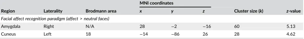
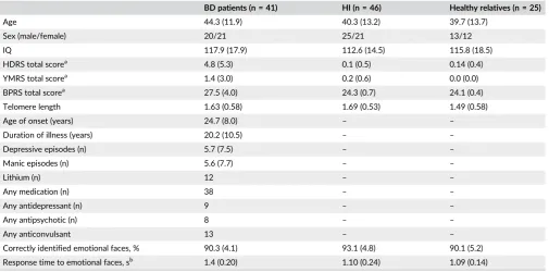
Only significant positive correlations were found between TL and
face-related activation in the amygdala and cuneus (Figure 1; Table 2).
There was no effect of group on these brain areas or an interaction
between TL and group.
Since, both age and TL are included in the regression models, and
[image:6.595.127.471.327.730.2]there is a potential for collinearity we performed additional analysis
FIGURE 1 Telomere length positively correlated with task-related activation in the (a) Amygdala, (b) Cuneus.“Fitted”response shows cross-subject average and“plus error”shows individual subject's contrast values [Color figure can be viewed at wileyonlinelibrary.com]
excluding age as a covariate from the regression model (the results
were almost identical; Supporting Information Table S5) and tested the
effect of age on face-related brain activation (not significant,p> .05).
3.4
|
Effect of telomere length on effective
connectivity
There was no effect of TL on effective connectivity (DCM
connec-tions;F= 0.98;p= .482). However, there was a main effect of TL on
facial affectmodulation on the three forward connections toward the
VPFC (F3,109= 2.25, p= .035). TL positively influenced the facial
affect modulation from the IOG to the VPFC (F1,112= 3.14;pFDR=
.048). Groups' differences in effective connectivity are described in
Supporting Information Material.
3.5
|
Polygenic risk score telomere length (PRS-TL)
High resolution polygenic risk scoring revealed that ap-value
thresh-old of p= .011 within our base dataset (TL GWAS), consisting of
1,793 SNPs, predicted 3.25% of the variance in TL within VIBES
(p= .036). We subsequently utilized this as our instrumental variable,
and outputted individualized PRS-TL within VIBES at this threshold.
Testing PRS-TL on facial affect brain activation, we find a positive
correlation between PRS-TL and the medial prefrontal gyrus (mPFC)
(x= 4,y= 40,z= 24;z-score = 4.84;k= 42 voxels; Figure 2). There
was no interaction between PRS-TL and TL on face-related activation
while performing the facial affect recognition task that survives FWE;
even when we lower the threshold top< .001 (uncorrected) there is
no interaction. There was no effect of PRS-TL on effective
connectiv-ity, intrinsic connections or facial affect modulation and no interaction
of PRS-TL with TL could be detected on connectivity (p> .31).
4
|
D I S C U S S I O N
This is the first study to-date to demonstrate a link between TL and
emotional brain activity and connectivity, as well as a link between
genetic risk for TL and emotional brain activation. First, TL was
posi-tively associated with activation in the amygdala and cuneus, and with
connectivity between the posterior regions of the face network and
the VPFC, during a task where participants were asked to label facial
emotions. Second, the cumulative impact of risk-alleles for TL,
expressed as a polygenic risk score (PRS-TL), showed a positive
asso-ciation with mPFC activation during facial affect.
4.1
|
The effect of TL on the facial affect-processing
network
Our results demonstrated that longer TLs are significantly associated
with activity in the amygdala and cuneus when participants are asked
to label facial emotions. The amygdala is central to emotion, involved in
detecting the valence and intensity of expressed emotions (Whalen
et al., 2009), especially negative emotion and its volume has also been
positively associated with TL (King et al., 2014). It is thought to enhance
the mental representation of other states such as those we perform
here in labeling the emotion in strangers' faces (Ashworth et al., 2001).
When considering TL as a state biomarker that captures the detrimental
effects of age, cortisol, and oxidative stress, the result does
comple-ment previous findings, whereby longer TL marks healthier brain cells
that retain the ability to generate action potentials (Uttara, Singh,
Zam-boni, & Mahajan, 2009). Furthermore, we know from rodent studies
that the amygdala is a brain region particularly sensitive to the effects
of oxidative stress, and consequently long buccal TL could represent an
individual's reduced exposure to reactive oxygen species, and thus
more functional cells in this brain region (Cano-Europa et al., 2008).
In terms of a link with mood disorder pathology, longer TL being
associated with increased amygdala activation is perhaps a little surprising,
as mood disorders are generally associated with shorter telomeres and
have previously been associated with amygdala hyper-reactivity
(Hamilton et al., 2012; Wegbreit et al., 2014). Consequently, it could be
that mood disorders associated with amygdala hyper-reactivity represent
a subtype which is not usually marked by shorter TL, and so further work
will be needed to test whether TL may be useful in differentiating patients
with high and low amygdala activation in response to emotional stimuli.
Alternatively, higher amygdala activity may represent a compensatory
response, rather than pathological feature of mood disorders. In which
case, higher amygdala activity (and longer TL) would be marking a healthy
response to brain pathology. Future studies should investigate if amygdala
hyperactivity in patients reflects amygdala dysfunction, by testing
whether it is associated with behavioral deficits and poor clinical outcome,
or whether it reflects a compensatory mechanism (Kim et al., 2012).
Perhaps more in keeping with the literature on mood disorders was
the finding that longer TLs were positively associated with higher
activa-tion in the cuneus during emoactiva-tion labeling. Our results add support to the
notion of increased involvement of the visual cortices when attention is
directed to the emotional valence of the stimuli (Dima et al., 2011;
Gre-goriou, Gotts, Zhou, & Desimone, 2009). Research until now has focused
primarily on amygdala activation and its connectivity with bottom-up and
top-down brain areas during the categorization of affective faces.
[image:7.595.48.551.75.141.2]How-ever, recently equal attention has been drawn to the occipital lobe and its TABLE 2 Peak coordinates of significant positive correlations between telomere length and task related activation (n = 112; allp< .05
FWE,k> 20)
Region Laterality Brodmann area
MNI coordinates
Cluster size (k) z-value
x y z
Facial affect recognition paradigm (affect > neutral faces)
Amygdala Right N/A 28 −2 −16 60 5.13
Cuneus Left 18 −14 −86 26 28 4.62
coupling to the VPFC as it seems that this coupling is a critical component
of the brain network processing faces (Dima et al., 2011; Pessoa &
Adolphs, 2010; Piech et al., 2010; Tsuchiya, Moradi, Felsen, Yamazaki, &
Adolphs, 2009). The finding that increased cuneus activation is associated
with longer TL is one which is complemented by our connectivity results
that showed that the facial affect information forwarded from the IOG to
the VPFC is significantly increased with longer TLs. Similarly, in brain
tis-sue, no decrease in TL has been observed in studies quantifying telomere
length in the occipital cortex (Teyssier, Ragot, Donzel, & Chauvet-Gelinier,
2010) of subjects with psychiatric disorders and Holstege et al. (2014)
showed that there is a differential effect of aging on brain tissues taken
from the same individual, where the occipital cortex has the longest
telo-meres compared with all other brain areas. Our results contribute to this
discussion by highlighting a potential role of TL in the occipital lobe, and
by supplementing the current belief that TL in the occipital lobe is less
susceptible to the effects of age and disease, while playing a role in
emo-tional processing.
4.2
|
The effect of PRS-TL on the facial
affect-processing network
When we perform the genetic analysis, we find a positive association
between PRS-TL and mPFC activation during facial affect labeling.
The mPFC has been found to play a critical role in emotion facial
dis-crimination (Winston, O'Doherty, & Dolan, 2003), emotional appraisal,
expression and regulation (Etkin, Egner, & Kalisch, 2011) and memory
for emotional facial expressions (Mattavelli, Cattaneo, & Papagno,
2011). Neuropsychological studies support this by demonstrating that
patients with mPFC damage are impaired in recognizing emotional
expressions and this deficit is associated with abnormal social
behav-ior (Mah, Arnold, & Grafman, 2005) and reduced emotional
respon-siveness (Heberlein, Padon, Gillihan, Farah, & Fellows, 2008).
In animal work, studies working on TL in rats have found that
female rats exposed to nurturing foster care outside of the homecage
exhibited longer TL in the mPFC than females exposed to
maltreat-ment or normal care (Asok, Bernard, Rosen, Dozier, & Roth, 2014).
Similarly, prenatally-stressed animals had shorter telomeres than
con-trols in the mPFC (Blaze et al., 2017). Pivotally, a study using mice
deficient for Tert, a key component of the enzyme telomerase
respon-sible for the maintenance of telomere length, demonstrated the
requirement of this telomere-regulating enzyme for mood stability
(Zhou, Wu, et al., 2016). Specifically, they showed that the
re-expression of Tert in the mPFC rescued the depressive phenotype of
Tert−/−mice, thus revealing a novel role of Tert in emotional control
in the mPFC (Zhou, Ning, et al., 2016). Our results provide empirical
[image:8.595.137.459.47.400.2]support for the role of the PRS-TL in the mPFC and further suggest FIGURE 2 Association between polygenic risk score for telomere length (PRS-TL) and activation in the right medial frontal gyrus.“Fitted” response shows cross-subject average and“plus error”shows individual subject's contrast values [Color figure can be viewed at
wileyonlinelibrary.com]
that TL could be a mediator of emotional processing in the mPFC.
Fur-ther studies will now need to understand why the mPFC is selectively
affected by PRS-TL, as opposed to other brain regions.
4.3
|
Considerations and conclusions
There are three main limitations of the current study which should be
acknowledged. First, DNA used to measure TL was ascertained from
buccal swabs rather than from brain tissue, meaning it may have
use-ful predictive biomarker properties, but may lack construct validity
(Powell, Fernandes, & Schalkwyk, 2012). Subsequently, it remains
unclear to what extent peripheral TL correlates with brain-region
spe-cific TL, and what precise neural mechanisms contribute to the
associ-ation between TL and functional activity. However, previous studies
have found that TL assessment using peripheral DNA is an acceptable
surrogate since genetic influences on the regulation of TL appear
tis-sue independent (Dlouha, Maluskova, Kralova Lesna, Lanska, &
Hubacek, 2014; Friedrich et al., 2000). Furthermore, there is some
ani-mal work (Zhou, Wu, et al., 2016) which provides clues about
connec-tions between TL and the brain, but far more research is needed.
Second, as TL changes over time, future studies incorporating
longitu-dinal designs are needed to test whether the engagement of
telomere-eroding behaviors (e.g., smoking) or protective behaviors
(e.g., exercise) impact on the functional processing of emotions. Third,
TL likely represents a marker sensitive to the effects of a wide variety
of environmental stress, and so further work will be needed to
under-stand what factors affecting TL are particularly important in
modulat-ing brain activity (e.g., cortisol levels, diet, levels of reactive oxygen
species, inflammatory cytokine levels) (Kordinas, Ioannidis, &
Chatzi-panagiotou, 2016; Lin, Epel, & Blackburn, 2012; Révész, Milaneschi,
Verhoeven, & Penninx, 2014).
To the best of our knowledge, this study provides the first
evi-dence linking TL and genetic risk for TL to brain activation and
con-nectivity while categorizing emotional faces. We demonstrated that
TL is associated with brain activity in regions known to be involved in
emotional processing and are part of the facial-affect processing
net-work. Increased activation in amygdala and cuneus during emotional
processing is predicted by longer TL. The current results also indicate
the effect of genetic load for TL on brain function affects a key region
for emotion, the mPFC. It is worth noting that shorter TL do not
equate to developing a mood disorder, BD (Martinsson et al., 2013;
Simon et al., 2006) or major depression disorder (Schaakxs,
Verhoe-ven, Voshaar, Comijs, & Penninx, 2015; Vincent et al., 2017), rather TL
may represent one of several pathways that increase vulnerability to
mood disorders, through differential emotional processing. Future
imaging telomere studies with larger and longitudinal samples would
be uniquely informative in mapping the spatial distribution of TL on
brain processes during emotional processing as patients and healthy
individuals age, and may lead to the discovery of a biological
mecha-nism linking TL maintenance with emotional brain function and the
regulation of neural networks. TL could prove to be a faster and
cheaper way of subtyping mood disorders based on their predicted
emotional responses.
A C K N O W L E D G M E N T S
We thank Dr Sophia Frangou for giving us access to the study dataset.
Drs Powell, Breen, and Dima and the laboratory work were supported
in part by the NIHR Biomedical Research Centre (“BRC”) and NIHR
Dementia Biomedical Research Unit (“BRU”) hosted at King's College
London and South London and Maudsley NHS Foundation Trust and
funded by the National Institute for Health Research under its
Bio-medical Research Centres funding initiative. The views expressed are
those of the authors and not necessarily those of the BRC or the
BRU, the NHS, the NIHR or the Department of Health. Dr Powell is
funded by a Medical Research Council Skills Development Fellowship
(MRN014863/1). The current project was part-funded by a Psychiatry
Research Trust Grant awarded to TRP and DD (Grant Reference:
92 Branthwaite). Additional support for Dr Dima was provided by the
NARSAD 2014 Young Investigator Award (Leichtung Family
Investiga-tor, Grant ID: 22471; Dr. Dima). Dr de Jong is partially supported by
NARSAD Young Investigator Grant (YI 60373) and European Union's
Horizon 2020 Research and Innovation Programme(Marie
Sklodowska-Curie grant agreement 658195).
O R C I D
Danai Dima https://orcid.org/0000-0002-2598-0952
R E F E R E N C E S
American Psychiatric Association. (1994).Diagnostic and statistical manual of mental disorders (4th ed.). Washington, DC: American Psychiatric Press.
Ashworth, F1., Pringle, A., Norbury, R., Harmer, C. J., Cowen, P. J., & Cooper, M. J. (2001). Neural response to angry and disgusted facial expressions in bulimia nervosa.Psychological Medicine,41, 2375–2384. Asok, A., Bernard, K., Rosen, J. B., Dozier, M., & Roth, T. L. (2014). Infant-caregiver experiences Alter telomere length in the brain.PLoS One,9, e101437.
Barbé-Tuana, F. M., Parisi, M. M., Panizzutti, B. S., Fries, G. R., Grun, L. K., Guma, F. T.,…Rosa, A. R. (2016). Shortened telomere length in bipolar disorder: A comparison of the early and late stages of disease.Revista Brasileira de Psiquiatria,38, 281–286.
Benjamini, Y., & Hochberg, Y. (1995). Controlling the false discovery rate: A practical and powerful approach to multiple testing.Journal of the Royal Statistical Society,57, 289–300.
Blaze, J., Asok, A., Borrelli, K., Tulbert, C., Bollinger, J., Ronca, A. E., & Roth, T. L. (2017). Intrauterine exposure to maternal stress alters Bdnf IV DNA methylation and telomere length in the brain of adult rat off-spring.International Journal of Developmental Neuroscience,62, 56–62. Cano-Europa, E., Pérez-Severiano, F., Vergara, P., Ortiz-Butrón, R., Ríos, C.,
Segovia, J., & Pacheco-Rosado, J. (2008). Hypothyroidism induces selective oxidative stress in amygdala and hippocampus of rat. Meta-bolic Brain Disease,23, 275–287.
Codd, V., Nelson, C. P., Albrecht, E., Mangino, M., Deelen, J., Buxton, J. L., …Samani, N. J. (2013). Identification of seven loci affecting mean telo-mere length and their association with disease.Nature Genetics,45, 422–427.
Coleman, J. R., Euesden, J., Patel, H., Folarin, A. A., Newhouse, S., & Breen, G. (2016). Quality control, imputation and analysis of genome-wide genotyping data from the Illumina HumanCoreExome microarray.
Briefings in Functional Genomics,15, 298–304.
Dima, D., de Jong, S., Breen, G., & Frangou, S. (2016). The polygenic risk for bipolar disorder influences brain regional function relating to visual and default state processing of emotional information. Neuroimage Clinical,12, 838–844.
the facial network during affect processing by CACNA1C and ANK3 risk genes for bipolar disorder.JAMA Psychiatry,70, 1303–1311. Dima, D., Roberts, R. E., & Frangou, S. (2016). Connectomic markers of
dis-ease expression, genetic risk and resilience in bipolar disorder. Transla-tional Psychiatry,6, e706.
Dima, D., Stephan, K. E., Roiser, J. P., Friston, K. J., & Frangou, S. (2011). Effective connectivity during processing of facial affect: Evidence for multiple parallel pathways. The Journal of Neuroscience, 31, 14378–14385.
Dlouha, D., Maluskova, J., Kralova Lesna, I., Lanska, V., & Hubacek, J. A. (2014). Comparison of the relative telomere length measured in leuko-cytes and eleven different human tissues. Physiological Research,63, S343–S350.
Drury, S. S., Theall, K., Gleason, M. M., Smyke, A. T., De Vivo, I., Wong, J. Y.,…Nelson, C. A. (2012). Telomere length and early severe social deprivation: Linking early adversity and cellular aging.Molecular Psychiatry,17, 719–727.
Ebner, N. C., Johnson, M. K., & Fischer, H. (2012). Neural mechanisms of reading facial emotions in young and older adults.Frontiers in Psychol-ogy,3, 223.
Elvsåshagen, T., Vera, E., Bøen, E., Bratlie, J., Andreassen, O. A., Josefsen, D., … Boye, B. (2011). The load of short telomeres is increased and associated with lifetime number of depressive episodes in bipolar II disorder.The Journal of Affective Disorders,135, 43–50. Epel, E., Blackburn, E., Lin, J., Dhabhar, F., Adler, N., Morrow, J., …
Cawthon, R. M. (2004). Accelerated telomere shortening in response to life stress.Proceedings of the National Academy of Sciences of the United States of America,101, 17312–17315.
Epel, E. S., & Prather, A. A. (2018). Stress, telomeres, and psychopathology: Toward a deeper understanding of a triad of early aging.Annual Review of Clinical Psychology,14, 371–397.
Etkin, A., Egner, T., & Kalisch, R. (2011). Emotional processing in anterior cingulate and medial prefrontal cortex.Trends in Cognitive Sciences,15, 85–93.
Euesden, J., Lewis, C. M., & O'Reilly, P. F. (2015). PRSice: Polygenic risk score software.Bioinformatics,31, 1466–1468.
Fairhall, S. L., & Ishai, A. (2007). Effective connectivity within the distrib-uted cortical network for face perception. Cerebral Cortex, 17, 2400–2406.
Ferron, S. R., Marqués-Torrejón, M. A., Mira, H., Flores, I., Taylor, K., Blasco, M. A.,…Farinas, I. (2009). Telomere shortening in neural stem cells disrupts neuronal differentiation and neuritogenesis.The Journal of Neuroscience,29, 14394–14407.
First, M. B., Spitzer, R. L., Gibbon, M., & Williams, J. B. W. (2002a). Struc-tured clinical interview for DSM-IV-TR axis I disorders, research version, non-patient edition. New York, NY: Biometrics Research, New York State Psychiatric Institute.
First, M. B., Spitzer, R. L., Gibbon, M., & Williams, J. B. W. (2002b). Struc-tured clinical interview for DSM-IV-TR axis I disorders, research version
(Patient ed.). New York, NY: Biometrics Research, New York State Psy-chiatric Institute.
Frangou, S. (2009). Risk and resilience in bipolar disorder: Rationale and design of the vulnerability to bipolar disorders study (VIBES). Biochemi-cal Society Transactions,37, 1085–1089.
Freeman, B., Smith, N., Curtis, C., Huckett, L., Mill, J., & Craig, I. W. (2003). DNA from buccal swabs recruited by mail: Evaluation of storage effects on long-term stability and suitability for multiplex polymerase chain reaction genotyping.Behavior Genetics,33, 67–72.
Friedrich, U., Griese, E., Schwab, M., Fritz, P., Thon, K., & Klotz, U. (2000). Telomere length in different tissues of elderly patients.Mechanisms of Ageing and Development,119, 89–99.
Gregoriou, G. G., Gotts, S. J., Zhou, H., & Desimone, R. (2009). High-fre-quency, long-range coupling between prefrontal and visual cortex. Sci-ence,324, 1207–1210.
Hamilton, J. P., Etkin, A., Furman, D. J., Lemus, M. G., Johnson, R. F., & Gotlib, I. H. (2012). Functional neuroimaging of major depressive disor-der: A meta-analysis and new integration of base line activation and neural response data. The American Journal of Psychiatry, 169, 693–703.
Hamilton, M. (1960). A rating scale for depression.Journal of Neurology, Neurosurgery, and Psychiatry,23, 56–62.
Hartmann, N., Boehner, M., Groenen, F., & Kalb, R. (2010). Telomere length of patients with major depression is shortened but independent from therapy and severity of the disease.Depression and Anxiety,27, 1111–1116.
Heberlein, A. S., Padon, A. A., Gillihan, S. J., Farah, M. J., & Fellows, L. K. (2008). Ventromedial frontal lobe plays a critical role in facial emotion recognition.Journal of Cognitive Neuroscience,20, 721–733.
Holstege, H., Pfeiffer, W., Sie, D., Hulsman, M., Nicholas, T. J., Lee, C. C.,… Sistermans, E. A. (2014). Somatic mutations found in the healthy blood compartment of a 115-yr-old woman demonstrate oligoclonal hemato-poiesis.Genome Research,24, 733–742.
Jin, K. (2010). Modern biological theories of aging.Aging and Disease,1, 72–74.
Kim, P., Thomas, L. A., Rosen, B. H., Moscicki, A. M., Brotman, M. A., Zarate, C. A., Jr.,…Leibenluft, E. (2012). Differing amygdala responses to facial expressions in children and adults with bipolar disorder.The American Journal of Psychiatry,169, 642–649.
King, K. S., Kozlitina, J., Rosenberg, R. N., Peshock, R. M., McColl, R. W., & Garcia, C. K. (2014). Effect of leukocyte telomere length on total and regional brain volumes in a large population-based cohort.JAMA Neu-rology,71, 1247–1254.
Kordinas, V., Ioannidis, A., & Chatzipanagiotou, S. (2016). The telomere/te-lomerase system in chronic inflammatory diseases. Cause or effect?
Genes,7, 60.
Kuhn, R. M., Haussler, D., & Kent, W. J. (2013). The UCSC genome browser and associated tools.Briefings in Bioinformatics,14, 144–161. Lima, I. M., Barros, A., Rosa, D. V., Albuquerque, M., Malloy-Diniz, L., &
Neves, F. S. (2015). Analysis of telomere attrition in bipolar disorder.
Journal of Affective Disorders,172, 43–47.
Lin, J., Epel, E., & Blackburn, E. (2012). Telomeres and lifestyle factors: Roles in cellular aging.Mutation Research,730, 85–89.
Lindqvist, D., Epel, E. S., Mellon, S. H., Penninx, B. W., Révész, D., Verhoeven, J. E.,…Wolkowitz, O. M. (2015). Psychiatric disorders and leukocyte telomere length: Underlying mechanisms linking mental ill-ness with cellular aging.Neuroscience and Biobehavioral Reviews, 55, 333–364.
Liu, M.-Y., Nemes, A., & Zhou, Q.-G. (2018). The emerging roles for telo-merase in the central nervous system.Frontiers in Molecular Neurosci-ence,11, 160.
Lukoff, D., Nuechterlein, K. H., & Ventura, J. (1986). Manual for expanded brief psychiatric rating scale (BPRS).Schizophrenia Bulletin,12, 594–602. Lung, F. W., Chen, N. C., & Shu, B. C. (2007). Genetic pathway of major depressive disorder in shortening telomeric length.Psychiatric Genetics,
17, 195–199.
Mah, L. W., Arnold, M. C., & Grafman, J. (2005). Deficits in social knowl-edge following damage to ventromedial prefrontal cortex.The Journal of Neuropsychiatry and Clinical Neurosciences,17, 66–74.
Mamdani, F., Rollins, B., Morgan, L., Myers, R. M., Barchas, J. D., Schatzberg, A. F.,…Sequeira, P. A. (2015). Variable telomere length across post-mortem human brain regions and specific reduction in the hippocampus of major depressive disorder.Translational Psychiatry,5, e636.
Martinsson, L., Wei, Y., Xu, D., Melas, P. A., Mathé, A. A., Schalling, M.,… Backlund, L. (2013). Long-term lithium treatment in bipolar disorder is associated with longer leukocyte telomeres.Translational Psychiatry,3, e261.
Mattavelli, G., Cattaneo, Z., & Papagno, C. (2011). Transcranial magnetic stimulation of medial prefrontal cortex modulates face expressions processing in a priming task.Neuropsychologia,49, 992–998.
Mitchell C, Hobcraft J, McLanahan SS, Siegel SR, Berg A, Brooks-Gunn J, …, Notterman D (2014). Social disadvantage, genetic sensitivity, and children's telomere length.Proceedings of the National Academy of Sci-ences of the United States of America, 111,5944–5949.
Monroy-Jaramillo, N., Dyukova, E., & Walss-Bass, C. (2017). Telomere length in psychiatric disorders: Is it more than an ageing marker?The World Journal of Biological Psychiatry,25, 1–19.
Mottahedin, A., Ardalan, M., Chumak, T., Riebe, I., Ek, J., & Mallard, C. (2017). Effect of Neuroinflammation on synaptic organization and function in the developing brain: Implications for neurodevelopmental and neurodegenerative disorders. Frontiers in Cellular Neuroscience,
11, 190.
Mourão-Miranda, J., Almeida, J. R., Hassel, S., de Oliveira, L., Versace, A.,… Phillips, M. L. (2012). Pattern recognition analyses of brain activation elicited by happy and neutral faces in unipolar and bipolar depression.
Bipolar Disorders,14, 451–460.
Nilsonne, G., Tamm, S., Månsson, K. N., Åkerstedt, T., & Lekander, M. (2015). Leukocyte telomere length and hippocampus volume: A meta-analysis.F1000Res,4, 1073.
Penny, W. D., Stephan, K. E., Daunizeau, J., Rosa, M. J., Friston, K. J., Schofield, T. M., & Leff, A. P. (2010). Comparing families of dynamic causal models.PLoS Computational Biology,6, e1000709.
Pessoa, L., & Adolphs, R. (2010). Emotion processing and the amygdala: From a‘low road’to‘many roads’of evaluating biological significance.
Nature Reviews. Neuroscience,11, 773–783.
Piech, R. M., McHugo, M., Smith, S. D., Dukic, M. S., Van Der Meer, J., Abou-Khalil, B., & Zald, D. H. (2010). Fear-enhanced visual search persists after amygdala lesions.Neuropsychologia,48, 3430–3435.
Powell, T. R., Dima, D., Frangou, S., & Breen, G. (2018). Telomere length and bipolar disorder.Neuropsychopharmacology,43, 445–453. Powell, T. R., Fernandes, C., & Schalkwyk, L. C. (2012). Depression-related
behavioral tests.Current Protocols in Mouse Biology,2, 119–127. Purcell, S., Neale, B., Todd-Brown, K., Thomas, L., Ferreira, M. A., …
Sham, P. C. (2007). PLINK: A tool set for whole-genome association and population-based linkage analyses. American Journal of Human Genetics,81, 559–575.
Rao, S., Kota, L. N., Li, Z., Yao, Y., Tang, J., Mao, C.,…Xu, Q. (2016). Accel-erated leukocyte telomere erosion in schizophrenia: Evidence from the present study and a meta-analysis.Journal of Psychiatric Research,79, 50–56.
Révész, D., Milaneschi, Y., Verhoeven, J. E., & Penninx, B. W. (2014). Telo-mere length as a marker of cellular aging is associated with prevalence and progression of metabolic syndrome.The Journal of Clinical Endocri-nology and Metabolism,99, 4607–4615.
Ridout, K. K., Levandowski, M., Ridout, S. J., Gantz, L., Goonan, K., Palermo, D.,…Tyrka, A. R. (2018). Early life adversity and telomere length: A meta-analysis.Molecular Psychiatry,23, 858–871.
Ridout, K. K., Ridout, S. J., Price, L. H., Sen, S., & Tyrka, A. R. (2016). Depression and telomere length: A meta-analysis.Journal of Affective Disorders,191, 237–247.
Saretzki, G., & von Zglinicki, T. (2002). Replicative aging, telomeres, and oxidative stress. Annals of the New York Academy of Sciences, 959, 24–29.
Schaakxs R, Verhoeven JE, Voshaar RCO, , Comijs, H. C., Penninx, B. W. J. H. (2015). Leukocyte telomere length and late-life depression.The American Journal of Geriatric Psychiatry, 23,423–432.
Schutte, N. S., & Malouff, J. M. (2015). The association between depres-sion and leukocyte telomere length: A meta-analysis.Depression and Anxiety,32, 229–238.
Shalev, I., Moffit, T. E., Sugden, K., Williams, B., Houts, R. M., Danese, A.,… Caspi, A. (2013). Exposure to violence during childhood is associated with telomere erosion from 5 to 10 years of age: A longitudinal study.
Molecular Psychiatry,18, 576–581.
Shammas, M. A. (2011). Telomeres, lifestyle, cancer, and aging.Current Opinion in Clinical Nutrition and Metabolic Care,14, 28–34.
Simon, N. M., Smoller, J. W., McNamara, K. L., Maser, R. S., Zalta, A. K., Pollack, M. H.,…Wong, K. K. (2006). Telomere shortening and mood disorders: Preliminary support for a chronic stress model of accelerated aging.Biological Psychiatry,60, 432–435.
Stephan, K. E., Penny, W. D., Moran, R. J., den Ouden, H. E., Daunizeau, J., & Friston, K. J. (2010). Ten simple rules for dynamic causal modelling.NeuroImage,49, 3099–3109.
Stewart, J. A., Chaiken, M. F., Wang, F., & Price, C. M. (2012). Maintaining the end: Roles of telomere proteins in end-protection, telomere repli-cation and length regulation.Mutation Research,730, 12–19.
Stuhrmann, A., Suslow, T., & Dannlowski, U. (2011). Facial emotion proces-sing in major depression: A systematic review of neuroimaging find-ings.Biology of Mood & Anxiety Disorders,1, 10.
Teyssier, J. R., Ragot, S., Donzel, A., & Chauvet-Gelinier, J. C. (2010). Telo-meres in the brain cortex of depressive patients.Encephale,36, 491–494. Torrisi, S. J., Lieberman, M. D., Bookheimer, S. Y., & Altshuler, L. L. (2013). Advancing understanding of affect labeling with dynamic causal model-ing.Neuroimage,82, 481–488.
Tsuchiya, N., Moradi, F., Felsen, C., Yamazaki, M., & Adolphs, R. (2009). Intact rapid detection of fearful faces in the absence of the amygdala.
Nature Neuroscience,12, 1224–1225.
Uttara, B., Singh, A. V., Zamboni, P., & Mahajan, R. T. (2009). Oxidative stress and neurodegenerative diseases: A review of upstream and downstream antioxidant therapeutic options.Current Neuropharmacol-ogy,7, 65–74.
Vincent, J., Hovatta, I., Frissa, S., Goodwin, L., Hotopf, M., Hatch, S. L.,… Powell, T. R. (2017). Assessing the contributions of childhood maltreat-ment subtypes and depression case-control status on telomere length reveals a specific role of physical neglect.Journal of Affective Disorders,
213, 16–22.
Wang, X., & Michaelis, E. K. (2010). Selective neuronal vulnerability to oxi-dative stress in the brain.Frontiers in Aging Neuroscience,2, 12. Warren, K. N., Beason-Held, L. L., Carlson, O., Egan, J. M., An, Y., Doshi, J.,
et al. (2018). Elevated markers of inflammation are associated with lon-gitudinal changes in brain function in older adults.Journals of Gerontol-ogy. Series A: Biological Sciences & Medical Sciences,73, 770–778. Wechsler, D. (1997). Wechsler memory scale–third edition manual. San
Antonio, TX: The Psychological Corporation.
Wegbreit, E., Cushman, G. K., Puzia, M. E., Weissman, A. B., Kim, K. L., Laird, A. R., & Dickstein, D. P. (2014). Developmental meta-analyses of the functional neural correlates of bipolar disorder.JAMA Psychiatry,
71, 926–935.
Whalen, P. J., Davis, F. C., Oler, J. A., Kim, H., Kim, M., & Neta, M. (2009). Human amygdala response to facial expressions of emotion. In P. J. Whalen & E. A. Phelps (Eds.),The human amygdala(pp. 265–288). New York, NY: The Guilford Press.
Winston, J. S., O'Doherty, J., & Dolan, R. J. (2003). Common and distinct neural responses during direct and incidental processing of multiple facial emotions.NeuroImage,20, 84–97.
Young, R. C., Biggs, J. T., Ziegler, V. E., & Meyer, D. A. (1978). A rating scale for mania: Reliability, validity and sensitivity.The British Journal of Psy-chiatry,133, 429–435.
Zhou, Q.-G., Wu, H.-Y., Zhou, H., Liu, M.-Y., Lee, H.-W., Liu, X.,…Suh, H. (2016). Reactivation of Tert in the medial prefrontal cortex and hippo-campus rescues aggression and depression of Tert−/−mice. Transla-tional Psychiatry,6, e836.
Zhou, Y., Ning, Z., Lee, Y., Hambly, B. D., & McLachlan, C. S. (2016). Short-ened leukocyte telomere length in type 2 diabetes mellitus: Genetic polymorphisms in mitochondrial uncoupling proteins and telomeric pathways.Clinical and Translational Medicine,5, 8.
S U P P O R T I N G I N F O R M A T I O N
Additional supporting information may be found online in the
Sup-porting Information section at the end of the article.
How to cite this article: Powell TR, De Jong S, Breen G, Lewis CM, Dima D. Telomere length as a predictor of
emo-tional processing in the brain. Hum Brain Mapp. 2018;1–10.
![FIGURE 1Telomere length positively correlated with task-related activation in the (a) Amygdala, (b) Cuneus.subject average and “Fitted” response shows cross- “plus error” shows individual subject's contrast values [Color figure can be viewed at wileyonlinelibrary.com]](https://thumb-us.123doks.com/thumbv2/123dok_us/1387478.91912/6.595.127.471.327.730/telomere-positively-correlated-activation-amygdala-individual-contrast-wileyonlinelibrary.webp)