by
Ian Stuart McLennan
Being a thesis submitted to the Australian National University
for admission to the Degree of Doctor of Philosophy
Department of Pharmacology
performed by myself, except where otherwise acknowledged, and that it has not been submitted in any previous application for a degree.
TABLE OF CONTENTS
Page
Acknowledgements 1
Papers Published 2
Abstract 3
Abbreviations 5
CHAPTER 1 - GENERAL INTRODUCTION 6
A. Neuronal differentiation and determination 7
B. Neuronal cell death and maturation 14
C. Nerve growth factor 21
D. Conclusions 34
CHAPTER 2 - MATERIALS AND METHODS 35
Materials 36
A. Nerve growth factor 36
B. Collagen 36
C. Chemicals 36
Animals 37
Methods 39
A. Culture methods 39
I. General 39
a. Terminology 39
b. Culture plates 39
c. Basic salt solutions 39
d. Basic culture medium 40
e. Inactivation of serum 40
f. Preparation of conditioned medium 40
g. Preparation of extract medium 40
a. Culture medium 43
b. Dissociation procedure 43
c. Skeletal muscle cultures 43
d. Cardiac muscle cultures 43
e. Liver and lung cultures 45
f. Salivary gland cultures 45
III. Neuronal cultures 45
a. Culture medium 45
b. Spinal cord cell cultures 45
c. Ciliary, superior cervical and dorsal root
ganglia cultures 48
B. Chromatographic methods 48
I. Processing of fractions 48
II. Elution profiles 51
III. Columns 51
C. Bioassays for factors influencing: 51
I. Ciliary ganglion fibre outgrowth 51
II. Ciliary ganglion choline acetyltransferase 51 III. Spinal cord choline acetyltransferase 51
IV. Superior cervical ganglion enzymes 53
V. Dorsal root ganglion fibre outgrowth 53
D. Enzyme assays 53
I. Preparation of homogenates 53
II. Choline acetyltransferase 53
CHAPTER 3 - BIOASSAYS FOR THE DETECTION OF FACTORS
AFFECTING CILIARY GANGLION DEVELOPMENT 54
Introduction 55
Results 58
A. Preliminary observations 58
B. Fibre outgrowth 59
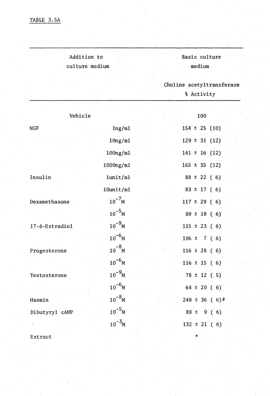
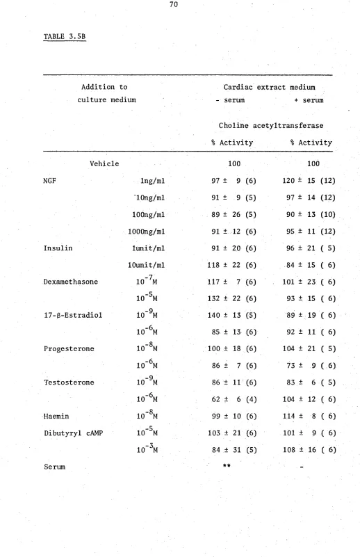
C. Maintenance of choline acetyltransferase activity 61 D. Effect of exogenous compounds on maintenance of
choline acetyltransferase 62
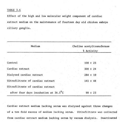
E. Preliminary fractionation of cardiac extract medium 63
Discussion 86
CHAPTER 4 - COMPARISON OF BIOASSAYS AND FACTORS 95
Introduction 96
Results 100
A. Preliminary observations 100
B. Distribution of factors in extract media 100
C. Chromatography of ciliary factors 102
D. Comparison of chromatography fractions in the
different assays 103
Interpretation of results 118
Discussion 120
CHAPTER 5 - SPECIFICATION OF TRANSMITTER TYPE IN THE
SUPERIOR CERVICAL GANGLION 123
Introduction 124
Results 127
C. Determination of SCG in vitro 127 D. Effect of glucocorticosteroids on SCG phenotype in vivo 128
Discussion 139
CHAPTER 6 - CONCLUSION 142
ACKNOWLEDGEMENTS
I am greatly indebted to my supervisor, Dr I.A. Hendry, for the tolerance, excellent guidance and friendship which he has shown throughout this project.
I am also indebted to Dr C.E. Hill and Professor D.R. Curtis for many helpful discussions and criticism of this manuscript. Thanks also to Mrs R.E. Bonyhady for collaboration and friendship during the last two years.
For excellent technical assistance I wish to thank Ms P.J.
Campbell, Mr C. Claudianos, Ms G.S. Jones, Ms W.J. Fiedler and Ms S.L. Tan. Thanks also to Ms J. Dutton, Dr E. Wienawa-Narkiewicz and Mr L.S. Sterling for their help in the preparation of this thesis.
During the tenure of my Australian National University Ph.D. Scholarship the following papers have been published or submitted for publication.
McLennan, I.S. and Hendry, I.A. 1978. Parasympathetic neuronal survival induced by factors from muscle. Neurosci. Lett., 10, 269-273.
McLennan, I.S. and Hendry, I.A. 1980. Influence of cardiac extracts on cultured ciliary ganglia. Devi Neurosci., in press.
McLennan, I.S., Hill, C.E. and Hendry, I.A. 1980. Glucocorticosteroids modulate the choice of transmitter in the developing superior cervical ganglion. Nature, Lond., 283, 206-207.
Hill, C.E., Hendry, I.A. and McLennan, I.S. 1980. Factors influencing the development of cholinergic neurones in the superior cervical ganglion of the rat. Role of calcium and macromolecules. Neurosci., in press.
Hill, C.E., Hendry, I.A. and McLennan, I.S. 1980. Factors influencing transmitter type in sympathetic ganglion cells. In: Histochemistry and Cell Biology of Autonomic Neurones, S.I.F. cells and Paraneurones.
II International Symposium on Nervous Transmission. Raven Press (Helsinki), in press.
McLennan, I.S., Hill, C.E. and Hendry, I.A. Comparison of factors
involved in enzyme induction and nerve fibre outgrowth in sympathetic, parasympathetic and sensory ganglia. Submitted for publication.
ABSTRACT
The objective of this thesis was to study the involvement of exogenous factors in neuronal development, using tissue culture techniques. The involvement was examined of such factors in the induction of fibre outgrowth from chicken ciliary and nerve growth factor-insensitive dorsal root ganglia (DRG), maintenance of choline acetyltransferase (CAT) in chicken ciliary ganglia and induction of a cholinergic phenotype in the normally adrenergic rat superior cervical ganglia (SCG) in vitro.
A variety of conditioned media and media containing tissue extracts (extract media) were found to induce fibre outgrowth and maintain CAT activity in cultured ciliary ganglia. The optimal culture conditions which result in this induction and maintenance by cardiac extract medium have been defined.
The induction of CAT in the SCG could be prevented by inclusion of physiological levels of glucocorticosteroids, but not other steroids, in the cultured medium. The glucocorticosteroids appeared to prevent the induction of cholinergic properties by determining the SCG.
ABBREVIATIONS
-Ca-Mg BSS -calcium and magnesium basic salt solution
CAT choline acetyltransferase
DMEM Dulbecco’s modified essential medium
DRG dorsal root ganglion
Hanks BSS Hanks basic salt solution
HEPES N -2-hydroxyethypiperazine-N -2-ethanesulphonic acid
LMC lateral motor column
NGF nerve growth factor
NGF-As nerve growth factor antiserum
SCG superior cervical ganglion
CHAPTER 1
GENERAL INTRODUCTION
A. Neuronal differentiation and determination
The vertebrate neural crest is a transient embryonic structure originating after the closure of the medullary plate. Neural crest cells are able to differentiate into a diversity of cellular phenotypes
(Weston, 1970) and are the scource of neuroblasts to the sympathetic (Detwiler, 1937; Hammond and Yntema, 1947), parasympathetic
(Campenhout, 1946), enteric (Yntema and Hammond, 1945; Le Douarin and Teillet, 1973) and sensory ganglia (Weston, 1963). Additionally, neural crest cells differentiate into glia (Weston, 1970), neuroendocrine
cells such as the adrenal medullary and parafollicular cells (Le Douarin and Le Lievre, 1970), melanocytes and various elements of skeletal and connective tissue (Weston, 1970).
The neural crest cells contributing to any tissue originate from specific axial levels. For instance, enteric ganglia are derived from two regions of the neural axis, the vagal region (between somites 1 to 7) and the lumbosacral region (behind the 28th somite) (Le Douarin and Teillet, 1974). The cervico-dorsal crest (between somites 8 to 28) gives rise to the orthosympathetic chains and plexus (Le Douarin and Teillet, 1973), while the adrenomedullary paraganglion is derived from cells between somites 18 to 24 (adrenomedullary region) (Teillet
and Le Douarin, 1974).
tube and the myotome and into the somitic mesenchyme where it divides segmentally, migrating further within the somites than between them (Detwiler, 1937; Weston, 1963; Johnston, 1966).
There are a number of inductive influences which are responsible for the differentiation and determination of neurones from neural crest cells and these influences may occur at different stages of development. Firstly, neural crest cells may be determined to express specific
phenotypes prior to the onset of migration, either as a result of primary induction during the formation of the neural crest or as the result of exposure to diffusible or circulating inducers from surrounding tissues. Secondly, the neural crest cells may be induced during their migration as a result of interaction with other cells on or near their
i i i t
migratory pathway or, equally important, as a result of migration away from potential inducers. Lastly, the tissue surrounding the site of termination of migration may contain the principal inducer cells. In the case of neuronal differentiation the position of axon terminals may be as important as the position of the perikarya. Temporal factors may also play an important role in regulating neural crest differentiation as the neural crest is formed in an anterior to posterior sequence with emigration from it, at any given axial level, taking only 12 to 15 hours (Weston and Butler, 1966).
cells, which are characteristic of the host’s axial level rather than the donor's axial level (Weston, 1970; Noden, 1975; Le Douarin et al.3 1975). When "adrenomedullary" level neural crest cells, for instance, are transplanted into a host at the vagal level they migrate into the splanchnic mesoderm and give rise to enteric ganglia; whereas, in normal development the "adrenomedullary" level neural crest cells do not penetrate into the gut (Le Douarin and Teillet, 1974).
Furthermore, neural crest cells can be induced to express
different phenotypes in vitro by different culture conditions (Cowell and Weston, 1970). For instance, chicken embryo extract promotes the induction of pigment cells (Maxwell, 1976), horse serum promotes
neural induction (Greenberg and Schrier, 1977) and surface interaction with pigmented epithelium promotes chondrogenesis (Newsome, 1976).
It should be noted that although these studies indicate the
majority of neural crest cells are undetermined prior to their migration, they do not exclude the possibility that neural crest cells are exposed to important inductive influences during this stage of development. Indeed this seems likely since a small number of cells appear to be already determined prior to their migration. When vagal neural crest cells are transplanted into the trunk region of a host a few cells migrate to the gut and are incorporated in the host's enteric ganglia, their normal developmental fate, while the majority of the transplanted cells give rise to sensory neurones, adrenergic ganglia and adreno medullary cells, the normal developmental fate for trunk region cells
(Le Douarin and Teillet, 1974). This determination of neural crest
cells prior to migration has also been observed for melanocyte migration, parasympathetic ganglion and cephalic mesenchyme formation (Twitty, 1945;
Neural crest cells are subjected to inductive influences during their migration. In particular, the induction of neural crest cells into adrenergic sympathetic neuroblasts is thought to result from the movement of ventrally migrating neural crest cells across the somitic mesoderm, under the influence of the neural tube and notochord (Weston and Butler, 1966).
Adrenergic properties can be first detected in sympathetic neuroblasts as they aggregate to form sympathetic ganglia primordia
(Allan and Newgreen, 1977; Cochard
et at. 3
1978, 1979). At this time catecholamines and tyrosine hydroxylase can be detected in cells around the dorsal aorta, an apparently ectopic position, suggesting that the cue for expression of adrenergic properties occurs during the migration of the neuroblasts rather than during the condensation into a ganglion (Cochardet al.}
1979).Cohen, A.M. (1972) studied the influence of the migratory pathway on sympathetic neuroblast induction by culturing neural tube and parts of the associated tissue on the chorioallantoic membrane of host
embryos. The development of fluorescence in these explants required the presence of somitic mesoderm, the tissue through which presumptive adrenergic neuroblasts migrate, but not the ventral half of the axial trunk, which contains the normal final site of sympathetic ganglia.
In vitro
the influence of competent somitic mesoderm (mesoderm which has been induced by the ventral neural tube) on neural crest cells depends on a direct contact between the two tissues, suggesting the involvement of surface active molecules (Norr, 1973). In these cultures thep l u r i p o t e n t c e l l s r a t h e r t h a n t h e m a t u r a t i o n o f a d e t e r m i n e d s u b -
p o p u l a t i o n o f n e u r a l c r e s t c e l l s . A c a u t i o n a r y n o t e i s r e q u i r e d ,
h owever , as t h e a p p e a r a n c e o f c a t e c h o l a m i n e f l u o r e s c e n t c e l l s may n o t
b e a r e l i a b l e i n d i c a t i o n o f t h e i n d u c t i o n o f n e u r a l c r e s t c e l l s t o
a d r e n e r g i c n e u r o b l a s t s . D u r in g d ev el o pme nt c a t e c h o l a m i n e s a r e t h o u g h t
t o p l a y an i m p o r t a n t r o l e as d e v e l o p m e n t a l i n d u c e r s , which i s
i n d e p e n d e n t o f t h e i r f u n c t i o n as n e u r o t r a n s m i t t e r s (McMahon, 1974),
and a d r e n e r g i c p r o p e r t i e s h a v e b e e n d e t e c t e d i n n o n - n e u r o n a l c e l l s
d u r i n g d e v e l o p m e n t . For i n s t a n c e , n o r a d r e n a l i n e and a d r e n a l i n e h a v e
b e e n d e t e c t e d i n a n e u r a l s e a u r c h i n embryos ( Bunznikov
e t a l , 3
1968)and t h e y o l k o f c h i c k e n eggs ( I g n a r r o and Shideman, 1968a).
C a t e c h o l a m i n e s from t h e l a t t e r s o u r c e a r e a c c u m u l a t e d by s u c h t i s s u e s
as t h e h e a r t p r i m o r d i a by p a s s i v e d i f f u s i o n ( I g n a r r o and Shideman,
1968a, b ) .
A l t h o u g h i t i s l i k e l y t h a t c o m p et en t s o m i t i c mesoderm i s i n v o l v e d
i n the i n d u c t i o n o f a d r e n e r g i c p r o p e r t i e s i n n e u r a l c r e s t c e l l s i t i s
u n l i k e l y t o be t h e s o l e i n d u c t i v e s o u r c e f o r a d r e n e r g i c n e u r o b l a s t s
i n v i v o .
C a t e c h o l a m i n e f l u o r e s c e n t n e u r o n e s can be d e t e c t e d i n c u l t u r e so f n e u r a l c r e s t c e l l s i n t h e a b s e n c e o f c o m pe t en t s o m i t i c mesoderm,
s u g g e s t i n g t h a t some n e u r a l c r e s t c e l l s may h a v e a p r e d i s p o s i t i o n t o
become a d r e n e r g i c p r i o r t o t h e i r m i g r a t i o n (Cohen, A.M., 1977) . The
p o s s i b i l i t y t h a t t h e c o l l a g e n s u r f a c e u s e d i n t h e s e c u l t u r e s mimics t h e
i n d u c e r on c o m p e t e n t s o m i t i c mesoderm n eed s t o be e l i m i n a t e d .
Whi le t h e m i g r a t o r y pat hway i s i n v o l v e d i n a d r e n e r g i c d e ve l op me nt
i t i s n o t e s s e n t i a l f o r c h o l i n e r g i c e n t e r i c g a n g l i a f o r m a t i o n . When
a l l p o s s i b l e m i g r a t o r y i n f l u e n c e s a r e p r e v e n t e d by t r a n s p l a n t i n g n e u r a l
c r e s t c e l l s d i r e c t l y i n t o a n e u r a l h i n d g u t mesoderm, and t h e n c u l t u r i n g
t h i s mesoderm on t h e c h o r i o - a l l a n t o i c membrane, normal i n t r i n s i c p l e x u s e s
thus formed were always non-fluorescent with significant levels of choline acetyltransferase, even when the neural crest cells were
derived from an 'adrenergic' axial level (Le Douarin and Teillet, 1974; Smith
et al.3
1977).Even after the formation of a ganglionic complex many para
sympathetic neuroblasts retain an ability to express different phenotypes in response to different environments. When newly formed ciliary
ganglia, in which some neurones already have a limited capacity to synthesise acetylcholine, are grafted into younger embryos at the trunk neural crest level, the ganglionic cells migrate and differentiate into cells characteristic of the trunk neural crest. These include
adrenergic ganglia, the suprarenal gland, enteric ganglia and various non-neuronal cells (Le Douarin
et al. 3
1978). Similar phenomena also occur in tissue culture. Immature dorsal root ganglion (DRG) cells will differentiate into melanocytesin vitvo
(Cowell and Weston, 1970), and the normally adrenergic neurones of the superior cervical ganglion(SCG) can be induced to differentiate into a cholinergic phenotype under certain culture conditions (Patterson, 1978; Bunge
et al.3
1978) (see Chapter Five for a more extensive discussion of this phenomenon).In summary, the differentiation of a neural crest cell is influenced by its cellular environment. Migration of neural crest cells plays an
important role during this differentiation in that it defines the inductive influences to which the neural crest cells will be exposed. Most neural crest cells maintain a pluripotentiality (in that they are
After a neurone has reached its final position in the embryo and mitosis has ceased, it extends an axon and interacts with the environ ment around the tissue which it innervates. This interaction appears to be important for the subsequent maturation and survival, or death, of the neurone.
An overpopulation and subsequent cell loss is a common feature of developing cell populations (Glucksmann, 1951; Lockshin and Beaulton,
1974). This phenomenon appears to be ubiquitous in developing nervous systems, having been detected in peripheral and central centres and across phylogenetic lines (Prestige, 1970, 1974; Jacobson, 1978). For instance, there is a 50% reduction in the number of chicken ciliary ganglion neurones during stages 35 to 39 of the developing chicken embryo (Landmesser and Pilar, 1974b) and a 75% reduction in the number of ventral horn cells in Xenopus laevis during development (Hughes,
1961). The number of degenerating neurones is maximal during this period suggesting that the reduction in cell numbers results from cell death (Hughes, 1961; Bibb, 1978).
The phase of cell death is temporally correlated with the period of synaptogenesis and, in general, appears to be related to the size of the peripheral field (Prestige, 1970). Decreasing the size of the peripheral field by ablation of the target tissue (Beaudoin, 1955; Jacobson, 1978) increases the extent of the cell death. For instance, in the absence of their target tissue 92% of chicken ciliary ganglion neurones die during stages 35 to 39 whereas only 50% die in the normal
animal (Landmesser and Pilar, 1974b) . Conversely, increasing the size of the peripheral field by decreasing the number of innervating
limbs (Hollyday and Hamburger, 1976) causes a neuronal hyperplasia. In the cervical motor column of the chicken embryo the hyperplasia resulting from an increased peripheral field can be explained solely on the basis of a reduction in normally occurring cell death (Hollyday and Hamburger, 1976). However, an increased proliferation of neurones, as well as a decreased cell death, appears to be involved in the
hyperplasia of spinal ganglia of
Rana pipiens
(Bibb, 1978).During development an excess of axons attempts to innervate any tissue. The trochlear nerve of an immature Peking duck, for instance, contains 30 times as many fibres as that of a mature duck. As there is only a 42% loss of trochlear cells during development this excess must be due to an overproduction of both neurones and axons per neurone
(Sohal and Weidman, 1978). The excess axon production appears to be regulated by the target tissue as in the trochlear nerve of normal Peking duck embryos the axon to neurone ratio, on day 12 of incubation, is 20 to 1 whereas it is 1 to 1 in the absence of the peripheral field
(Sohal
et al.3
1978).An overproduction and subsequent elimination of immature synapses, resulting in a decrease in motor unit size, also occurs during development (Redfem, 1970; Bagust
et al.3
1973; Conradi and Ronnevi, 1975; Zelena, 1976). For instance, 73% of endplates in the soleus muscle of 11 day old rats have three or more axon terminals, whereas in 26 day old rats this poly-innervation is virtually absent, with only 2% of endplates having more than one axon terminal (Riley, 1977). The loss of poly-innervation appears to be unrelated to neuronal cell death. In chicken muscleThe phenomenon of neuronal cell death during development has been postulated to occur as a result of competition for a limited supply of synaptic sites or trophic substances (Hamburger, 1958; Landmesser and Pilar, 1976). This contention is supported by the inverse correlation between the size of the peripheral field, and thus presumably the availability of synaptic sites and trophic substances, and the extent of cell death, and by observations which suggest that many of the neurones which are eliminated during development are biochemically competent and have made appropriate synapses* prior to their death. The best demonstration of this phenomenon comes from studies by
Landmesser and Pilar on the development of the chicken ciliary ganglion. They found that all neurones of this ganglion, including those which die, undergo a peripheral field-dependent maturation to a secretory state, characterised by a large increase in the formation of polyri bosomes and rough endoplasmic reticulum (Pilar and Landmesser, 1976). All neurones, with axons in the postganglionic trunk, receive functional
innervation, and inappropriate synapses by the preganglionic nerve could not be detected (Landmesser and Pilar, 1974a). Similarly, the majority of chicken (Landmesser and Morris, 1975; Oppenheim and Chu-Wang, 1977; Landmesser, 1978) and Xenopus laevis (Lamb, 1976) motoneurones do not appear to form inappropriate synapses prior to cell death. Furthermore, that many of the neurones which die are biochemically competent, prior to their elimination, is implicit in the observation that cell death is decreased when the peripheral field size is increased.
Formation of inappropriate synapses does, however, occur during development. The initial, but not subsequent, projection of motor axons in Xenopus leavis is to the mesenchyme nearest to their point of entry. Thus, the presumptive knee flexor muscles during early limb bud formation receives innervation from the entire lumbar lateral motor column (LMC). All neurones contributing to this projection
subsequently die and the muscle is reinnervated by neurones in the more rostral region of the LMC (Lamb, 1976, 1977). The death of the early projecting neurones is not due to some type of competition for the flexor muscles as removal of the rostral region of the LMC does not prevent the loss of innervation from the caudal region (Lamb, 1979). A similar incorrect innervation of the flexor muscle of Ambystoma mexicana also occurs during initial innervation (McGrath and Bennett,
1979).
The loss of poly-innervation and reduction in motor unit size, in contrast to neuronal cell death, does not appear to be related to a limited supply of synaptic sites or trophic substance since surgical removal of all but a few motor axons to the soleus muscle of a rat pup slows but does not prevent the loss of poly-innervation and reduction in motor unit size, even though some muscle fibres are left uninnervated
(Brown et at., 1976).
As the neurone matures the nature of its dependence on its
peripheral field varies. Initially, neuronal development is independent of the target tissue. Up to stage 34, chicken ciliary neurones will survive, extend axons and maintain immature synapses with the
with the preganglionic trunk (Marwitt
et dl.3
1971; Pilar and Landmesser, 1972). Furthermore,in vitro
studies indicate that the sensitivity of these neurones to acetylcholine is also regulated by the target tissue (Pilaret al.3
1979). The development of neuronal dependency on its target tissue has been characterised into three stages (Prestige, 1970; Fortune and Blacker, 1976). Prior to the stage of normal motoneurone death, in the toad, ablation of the target tissue results in the cessation of neuronal maturation, but cell death does not occur until the stage of normal cell death has been reached(Hughes and Tschumi, 1958). Indeed, if metamorphosis is prevented by hypophysectomy the neurones will survive indefinitely in the absence of their peripheral field (Race, 1961). The morphological changes associated with the death of these peripherally deprived neurones differs from that observed during normal cell death (Decker, 1978)
(for similar observations in the chicken ciliary ganglion see Pilar and Landmesser, (1976)). During the stage of normal cell death, ablation of the target tissue causes immediate cell death which is morphologically indistinguishable from normal cell death (Decker, 1978). After the stage of normal cell death, ablation of the target tissue causes only a small immediate cell loss followed by a slow decline in neuronal numbers (Hughes and Tschumi, 1958).
Neuronal activity, or the associated muscle contractions, appears to be essential for the loss of excess motoneurone innervation to occur. Blockade of cholinergic receptors on muscle with d-tubocurarine
no effect (Laing and Prestige, 1978; Olek and Edwards, 1978).
Furthermore, nerve stimulation increases (O'Brien
et al.3
1978), and decreased neuronal activity slows down (Benoit and Changeux, 1975), the rate at which poly-neuronal innervation of muscle is lost.These results suggest that muscle is directly involved in the destruction of excess motoneurone production. Muscle could achieve this by the release of neurotoxic substances or by withdrawing support, such as synaptic sites or trophic substances, to part of its innervation. An active destruction of the excess innervation by muscle is supported by ablation studies. Removal of the limb bud of
Xenopus laevis3
late in the period of cell death, slows but does not prevent cell loss (Hughes, 1974). Clearly, however, active neuronal destruction by the target tissue cannot be the major cause of cell death, otherwise ablation of the target tissue would decrease rather than increase cell death.The mechanism by which cholinergic stimulation of muscle can alter its ability to maintain synapses has not been determined. Electrical activity of the innervating nerve is essential to suppress the
appearance of extrajunctional receptors in developing (Braithwaite and Harris, 1979) or mature muscle (Edward, 1979). It is possible that depolarisation of the muscle membrane induces the aggregation of some membrane component(s), which are involved in the association of nerve terminals with muscle, and thereby decreases the number of synaptic sites on, or release of trophic substances by, the muscle. That stimulation of muscle decreases its ability to form synapses is
supported by studies using adult animals. When the muscle is paralysed with botulinum toxin (Fex
et al. 3
1966) or local anaesthetics (JansenStimulation of the paralysed muscle, however, will maintain the muscle in a refractory state (Jansen
et al.3
1973). Furthermore, botulinum toxin or a-bungarotoxin poisoning will induce sprouting in maturemotor nerves (Duchen, 1970; Pestronk and Drachman, 1978). Alternatively, cholinergic stimulation of neuromuscular activity has been reported to cause the release, by muscle, of proteolytic enzymes and these enzymes have been postulated to digest and eliminate poorly competing
terminals (O’Brien
et al.3
1978).C. Nerve growth factor
Although it has been known for over fifty years that the peripheral field effects neuronal maturation the mechanisms by which the peripheral field and the developing neurone interact are poorly understood. The little that is known can be attributed to the study of the action of one remarkable protein, nerve growth factor (NGF).
The physiological role of NGF is presumed to be the mediation of neuronal-target tissue interactions. It appears to be involved in
the regulation of neuronal cell death, fibre outgrowth and the biochemical maturation of immature sympathetic and sensory neurones. Despite
extensive studies the nature of the involvement of NGF in these
processes, particularly
in vivo3
is still sketchy and the investigation of the intracellular mechanism by which NGF exerts its influence onreceptive neurones is but in its infancy. The physicochemical properties of NGF, however, are now well characterised.
Mouse salivary gland NGF is a complex of three proteins with a molecular weight of 140,000 dalton (Varon
et at. 3
1967). This complex has been designated 7S NGF on the basis of its sedimentary coefficient, and the subunits have been designated a, ß and y (Varonet at.3
1968). The stoichiometry of the complex is ß (Greeneet at. 3
1971) and1 - 2 Zn^+ (Pattison and Dunn 1975, 1976a).
All the nerve growth promoting activity of NGF resides in the ß
occurs with the loss of eight amino acids from the amino terminal and one from the carboxy terminal (Angeletti, R.H. et at. 3 1973b; Moore
et at.3 1974). This molecular species has been designated 2.5S NGF
(Bocchini and Angeletti, P.U. 1969). The extent of such cleavage varies with the method of purification. No differences have been observed, however, in the biological actions of ß- and 2.5S NGF
(Mobley et a7.jl976). The amino acid sequence of ß-NGF has been determined (Angeletti, R.H. and Bradshaw, 1971; Angeletti, R.H.
et at.3 1973a, b; Mobley et al.3 1974), and has a structural relatedness
to pro-insulin, relaxin and non-suppressible insulin-like activity, suggesting that these proteins have evolved from a common precursor
(Frazier et at.3 1972; Rinderknecht and Humbel, 1976; James, R.
et at. 3 1977).
The y subunit of NGF is a family of five closely related proteins with a molecular weight of 26,000 dalton (Stach et at.3 1976). y-NGF is an arginyl-estero-peptidase (Greene et at. 3 1968), the physiological function of which appears to be the conversion of pro-3-NGF to ß-NGF
(Berger, E.A. and Shooter, 1977). After cleavage of pro-ß-NGF, y-NGF has been postulated to remain bound to the C terminal of ß-NGF and to combine with y-NGF to give the 7S NGF complex (Berger, E.A. and Shooter, 1977). While in this complex the enzyme activity of y-NGF is inhibited (Greene et at. s 1969). This inhibition is mediated by the Zn^+ bound to the 7S complex (Pattison and Dunn, 1975, 1976a, b).
The a subunit of NGF has no known biological function and its properties have not been extensively studied. Its molecular weight is
approximately 30,000 dalton (Varon et at.s 1968).
The physicochemical properties of NGF and the structural
The two classical target tissues for NGF are sensory and
sympathetic neurones. Not all sensory neurones, however, are responsive to NGF. The smaller mediodorsal DRG neurones respond to NGF with a marked hypertrophy, whereas the larger ventrolateral cells are
unresponsive both in vivo and in vitro (Weis 1970, 1971; Ebendal and Hedlund, 1975).
The response of sympathetic and sensory neurones to NGF varies according to their stage of development. In the mouse, sympathetic neurones do not develop an absolute dependence on NGF for survival or fibre outgrowth until between the fifteenth and eighteerith days of gestation, which is several days after the formation of the SCG. Prior to this stage explanted SCG will survive, extend neurites and synthesise tyrosine hydroxylase, in vitro3 in the absence of NGF, or the presence of NGF antiserum (NGF-AS). NGF will, however, stimulate neurite out growth and the synthesis of tyrosine hydroxylase in these cultures
(Coughlin et at. 3 1977, 1978). The situation in chickens is slightly different. SCG from eight day old chicken embryos will not spontaneously extend fibres in vitro and are unresponsive to NGF. Fibre outgrowth in response to NGF develops between the ninth and tenth days of incubation
(Partlow and Larrabee, 1971).
DRG are only sensitive to NGF for a short period of their development. In the chicken, DRG from four day old embryos will not extend fibres in vitro in the presence of NGF, but will extend fibres in appropriate culture conditions (Luduenä, 1973; Letoumeau, 1975).
to the induction of fibre outgrowth, being also observed for the NGF induced increase in substance P content of cultured DRG (Schwartz and Costa, 1979).
Sensory and sympathetic neurones are not the only cells to respond to NGF. A number of studies indicate that NGF can influence the central nervous system, although it is uncertain which cells are affected. NGF stimulates ornithine decarboxylase in rat brain (Roger
et al. 3
1974; Lewiset al. 3
1978), optic nerve regeneration in the newt (Turner and Glaze, 1977; Glaze and Turner, 1978) and behavioural recovery after hypothalamic lesion in adult rats (Berger, B.B., 1973) and is required for the normal developmental changes in surface properties of chicken tectal cells to occurin vitro
(Merrellet al. 3
1975). There isconflicting evidence as to whether NGF influences central monoaminergic neurones. NGF is retrogradely transported by a number of neurones, including some in monoaminergic nuclei(Ebbott and Hendry, 1978). The noradrenergic neurones of the locus coeruleus and the dopaminergic neurones of the substantia nigra, however, either transport NGF poorly or not at all (Ebbott and Hendry, 1978; Schwab
et al. 3
1979).Furthermore, localised injection of NGF and NGF-AS does not alter tyrosine hydroxylase levels in the locus coeruleus or substantia nigra
(Schwab
et al. 3
1979). Bjerre and co-workers, however, have reported that local injections of NGF or NGF-AS into the locus coeruleus affects the growth of lesioned ascending monoaminergic axons into an irisimplanted in the diencephalon (Bjerre
et al. 3
1974; Steneviet al. 3
1974). At variance with these observations is that NGF does not stimulate the regrowth of axons of central neurones which have been destroyed by 6-hydroxydopamine (Konkol
et al.3
1978), and that the innervation of denervated iris by immature central aminergic neurones transplanted into the anterior eye chamber is not influenced by NGFNGF may also influence the development of some neuroblasts. In rat embryo gut mesenchyme there is a transient appearance of adrenergic fluorescence in presumptive neuroblasts (Cochard
et al. 3
1978).Injection of NGF into rat embryos, during a critical stage, increases the number of fluorescent cells and delays the loss of these cells from the gut. This treatment also increases the number of apparently ectopic adrenergic neuroblasts in liver parenchyma and heart primordium
(Kessler
et al.3
1979). Furthermore, NGF will increase the number of fluorescent cells in cultured neural crest, which had been previously induced by exposure to the ventral tube. The inductive influence of the ventral tube could not, however, be substituted by exogenous NGF (Norr, 1973). Whether NGF increases the number of fluorescent cells seen in these studies by increasing neuroblast division, regulating phenotypic expression or by increasing catecholamine synthesis and inhibiting neuroblast death, in a manner analogous to its action on immature sympathetic neurones, is unknown.In addition to its effects on neurones, NGF inhibits the bio synthesis of mucopolysaccharides in chondrocytes (Eisenbarth
et al.3
1975), induces fibre outgrowth from a pheochromocytoma cell line (Tischler and Greene, 1975) and increases adenyl cyclase (Nikodijevic
et al.3
1976), dopamine-ß-hydroxylase and tyrosine hydroxylase activity in adrenal medullary cells (Ottenet al.3
1977); although NGF-AS has also been reported to increase tyrosine hydroxylase in the adrenal medulla(Angeletti, P.U.
et al.3
1972). The functional significance of these effects has not been determined.Pertinent to any hypothesis that NGF may mediate target tissue- neurone interactions is the demonstration that NGF is syWtH6sised, or
tissues (Angeletti, P.U. and Vigneti, 1971; Johnson, D.G.
et al. 3
1971; Hendry, 1972). The localisation of NGF within these tissues, howev.er, is unknown. The change in tissue levels of NGF during development has also only been scantily studied. The content of NGF in the hearts of newborn mice is double that found in hearts of two week old mice(Johnson, D.G.
et al.
3
1971), suggesting that embryonic levels of NGF may be significantly higher than in adult tissues. A moleculeimmunologically similar to NGF is synthesised and released by the
adrenal medulla in organ culture (Harper
et al. 3
1976) and by myoblasts, myotubules and cloned myogenic cells (Murphyet al. 3
1977b). Caution, however, is required in relating these experiments to thein vivo
situation as NGF could not be detected in the explants of adrenal medulla prior to their culturing (Harper
et al. 3
1976), and NGF is produced by primary and clonal fibroblast cultures (Ogeret al.3
1974; Younget al.3
1975) and a neuroblastoma cell line (Murphyet al. 3
1975), which are presumably not physiological sources of NGF. Thus, it is possible that NGF is produced by the various cells in response to the culture environment.Adult male mouse salivary gland contains a thousand fold higher levels of NGF than any other mouse tissues (Johnson
et al.3
1971;Hendry, 1972). This NGF probably has an exocrine role. It is located in the secretory tubular portions of the gland (Schwab
et al. 3
1976) and can be released into the saliva (Wallace and Partlow, 1976; Schwabet al.3
1976; Murphyet al. 3
1977a). An exocrine role for NGF is supported by the existence of high levels of NGF in mouse milk (Murphyet al.3
1977a), various snake venoms (Cohen, S., 1959), guinea pig , prostrate gland (Harperet al. 3
1979) and bull semen (Harper, 1979).salivary glands (sialectomy) of adult mice has been reported to decrease the NGF content of plasma, heart and the SCG (Hendry and Iversen, 1973), and to decrease enzyme levels in sympathetic ganglia
(Hendry and Thoenen,1974). The levels of NGF return to normal after a few months indicating that the salivary gland is not the only primary source of NGF (Hendry and Iversen, 1973). Furthermore, premature
enlargement of the submaxillary gland of neonatal rats by testosterone increases tyrosine hydroxylase levels in sympathetic ganglia. This effect is presumably mediated via the serum as it occurs in ganglia not innervating the submaxillary gland, and unilateral sialectomy effects both ipsilateral and contralateral ganglia (Dibner and Black, 1978). In challenge to the proposal that NGF has a role via serum is the failure of Murphy
et at.
(1977a) to observe a decrease in mouse serum levels after sialectomy and the low levels of NGF found in serum of vertebrates other than mice (Johnson, D.G.et at. 3
1971; Ogeret at. 3
1974).
While salivary gland NGF may influence mature sympathetic neurones it is unlikely to be an important source for the developing nervous system. High levels of NGF do not occur in immature mouse salivary glands (Hendry, 1972), and sialectomy of neonatal rats does not decrease tyrosine hydroxylase levels in the iris, indicating that the development of sympathetic innervation of one target tissue is not influenced by the state of another target organ (Dibner
et at. 3
1977).Neurones may receive NGF from associated glial cells, as well as their target tissues. Glial cells can substitute for exogenous NGF in supporting immature DRG neurones
in vitro
(Burnhamet at. 3
1972; Varon1977c; Longo, 1978). Whether glial cells synthesise or release NGF
in vivo is not known. If, however, glial cells are a source of NGF
in vivo their physiological significance is more likely to be associated with maintenance of mature neurones than with developmental phenomena.
NGF has a profound effect on sympathetic neuronal survival, both
in vivo and in vitro. Immature sympathetic neurones in vitro have an
absolute dependence on NGF for their survival (Levi-Montalcini and Angeletti, P.U., 1963). The number of sympathetic neurones which survive in vitro is proportional to the concentration of NGF in the medium (Chun and Patterson, 1977a). Injection of NGF into immature mammals markedly increases the number of neurones in the pre- and paravertebral sympathetic chain (Levi-Montalcini and Booker, 1960a; Hendry, 1976). This increase in neuronal number can be attributed to
a decrease in the naturally occurring developmental cell death (Hendry and Campbell, 1976; Hendry, 1977a). Conversely, injection of anti serum to NGF (NGF-AS) into newborn mice almost totally destroys the sympathetic nervous system (Levi-Montalcini and Booker, 1960b; Levi- Montalcini and Angeletti, P.U., 1966). Thus altering the availability of NGF has the same effect on neuronal survival as altering the size of the peripheral field. That NGF is the physiological mediator of peripheral field regulated neuronal survival is suggested by the
observation that NGF can prevent the neuronal death which results from the peripheral field isolation of the developing neurone (Hendry and Campbell, 1976; Banks and Walter, 1977). Whether the naturally occurring neuronal death during development results from a limiting production of NGF by the target tissue or is due to some other
The effects of NGF on sensory neuronal survival are only poorly documented. Immature chicken DRG require NGF for survival
in vitro
(Levi-Montalcini and Angeletti, P.U., 1963; Blood, 1972). However, to my knowledge the administration of NGF or NGF-AS to developing animals has not been shown to alter the number of neurones in the DRG.
When newborn mice are injected with NGF there is a massive sympathetic fibre outgrowth (Levi-Montalcini and Booker, 1960a) and an increase in the density of sympathetic innervation (Olson, 1967). NGF will also enhance, and NGF-AS inhibit, the regeneration of
sympathetic fibres after lesion with 6-hydroxydopamine (Bjerre
et al.3
1973, 1974) or axotomy (Hendry, 1975).
A similar phenomenon also occurs
in vitro.
NGF will induce a fibre halo from cultured sympathetic and DRG (Levi-Montalcini and Angeletti, P.U., 1968). The latter response is the basis of the first and most commonly used bioassay for NGF (Levi-Montalciniet ail. 3
1954).The physiological significance of the excess fibre outgrowth has only been briefly examined. As previously discussed there is a massive loss of axons during normal development. Does NGF increase fibre out growth in a manner analogous to the manner it increases cell number, that is by decreasing axonal loss? Analysis of this problem is hampered because fibre production, unlike neurone production, is an ongoing process and NGF may enhance this production nonspecifically by its general pleotypic effects.
action is suggested by an ingenious experiment by Campenot (1977) who devised a three chamber system in which dissociated sympathetic neurones could be cultured with their somas and branches of axons in different environments. When NGF was removed from one chamber the axons in that chamber stopped growing and degenerated, even though their somas and proximal axons were still receiving NGF (Campenot, 1977).
A second possible role for NGF-induced fibre outgrowth is that of directed growth. This question arose with the conception of NGF itself. When Levi-Montalcini implanted sarcomas 180 or 37 onto the chorioallantoic membrane of 4-6 day old chicken embryos she found a massive sympathetic fibre outgrowth which penetrated the veins near the sarcomas. This was the first demonstration of a diffusable growth factor and led to the hypothesis that nerve fibres grow down a gradient of growth factor (Levi- Montalcini, 1952). Competitive innervation of target tissues by
sympathetic neurones in vitro has been used to study this phenomenon. Many investigators have reported that densely innervated tissues such as the atrium or vas deferens will preferentially attract growing
sympathetic fibres whereas sparsely innervated tissue will not (Chamley
et at. 3 1973a, b; Chamley and Dowel, 1975; Ebendal and Jacobson,
1977a).
That NGF has chemotaxic properties has been suggested by both
in vivo and in vitro studies. When NGF is injected into the brain of
the higher concentration of NGF. The growth of fibres was not directed by the concentration of NGF per
se
but rather by the relativeconcentration of NGF (Charlwood
et al. 3
1972; Letoumeau, 1978). A chemotaxic response could be induced if fibre outgrowth is stimulated locally by NGF or if random outgrowth occurs and NGF locally stabilises that outgrowth. The demonstration of a localised regulation of axonal survival by NGF (Campenot, 1977) suggests that the latter mechanism may be operative.While these experiments strongly suggest that NGF can be
chemotaxic, appropriate gradients of NGF will have to be demonstrated in the developing embryo before they constitute evidence for chemotaxic guidance of sympathetic neurones to their appropriate target tissue.
The maturation of sympathetic neurones is probably regulated, in part, by NGF. Immature sympathetic neurones respond to NGF by a marked hypertrophy (Levi-Montalcini and Booker, 1960a; Banks
et al.3
1975), and increases in protein content (Thoenenet al. 3
1971), levels of noradrenaline (Crain and Wiegard, 1961), specific activity of tyrosine hydroxylase and dopamine-3-hydroxylase (Thoenenet al. 3
1971; Hendry and Iversen, 1971), and density of target tissue innervation (Bjerreet al. 3
1975). In all these respects the treated neurones resemble their mature counterparts.A problem with the interpretation of these experiments is that administration of NGF during development is likely to alter the gross biochemical characteristics of sympathetic ganglia by increasing
neuronal survival. That the above effects are not solely due to changes in neuronal survival can be deduced from studies using mature
(Bjerre
et al. 3
1975) and to NGF-AS by a transient decrease in the activity of tyrosine hydroxylase and dopamine-ß-hydroxylase (Goedertet al. 3
1978).If a target tissue is to specifically regulate the maturation and survival only of neurones which innervate it, then a mechanism must exist by which the target tissue can transfer information to the perikaryon of a select population of neurones. The demonstration by Hendry, Stöckel and co-workersthat NGF is retrogradely transported from target tissues to the perikaryon, via the axon, provides such a
mechanism (Hendry, 1980).
When NGF is injected unilaterally into the salivary gland or anterior eye chamber then it is selectively accumulated (Hendry
et al.3
1974a, b), and the activity of tyrosine hydroxylase increased (Stöckel and Thoenen, 1975; Paravicini
et al. 3
1975)^ in the ipsilateralrelative to the contralateral SCG. This induction of tyrosine
hydroxylase depends on an intact postganglionic trunk indicating that retrograde transport of some substance, via the axon is required for such enzyme induction (Paravicini
et al.3
1975).That NGF is only influencing neurones which have transported it has been elegantly demonstrated by Hendry. He implanted a depot
preparation of NGF, which slowly released NGF to the surrounding nerve terminals, into the anterior eye chamber. After one week the neurones projecting to the eye were labelled by injecting a small quantity of
125
The exact nature of the primary and subsequent biochemical events elicited by NGF has only been partially determined. The
biological action of NGF seems to result from a pleiotypic activation of a wide variety of anabolic and catabolic pathways rather than selective gene expression (Bradshaw, 1978). This point is perhaps most vividly demonstrated by studies with cultured immature
sympathetic neurones. These neurones express either a cholinergic or an adrenergic phenotype, in response to different culture conditions
(Patterson, 1978). When adrenergic these neurones respond to NGF by an increased synthesis of noradrenaline, and when cholinergic they respond by an increased synthesis of acetylcholine (Chun and Patterson, 1977b). Thus, NGF is a trophic hormone (Varon and Bunge, 1978)
D. Conclusions
Neuronal differentiation and maturation is influenced by the cellular environment around the developing neurone. The peripheral field appears to be involved in the maturation of transmitter
function, and survival or death of the neurones which innervate it. The mechanisms by which the peripheral field influences neuronal development are substantially unknown. If factors released by the periphery are involved in these mechanisms then these factors would be expected to act in vitro to cause neuronal maturation, fibre out growth, neuronal survival and neuronal death (under conditions which neurones normally survive).
The objective of this thesis was to develop tissue culture systems to enable the detection and subsequent characterisation of factors active in neuronal development. In the long term it is hoped that the purified effector molecules, and antibodies prepared to them,can be used as investigative tools with which to study the phenomena
CHAPTER 2
MATERIALS AND METHODS
MATERIALS
A. Nerve growth factor
3-NGF was purified from adult male mice salivary glands using the method of Mobley
et al.
(1976) and assayed spectrometrically(Greene, 1974).
B. Collagen
Stock solutions of rat tail collagen in acetic acid were prepared using the method of Bornstein (1950).
C. Chemicals
The following chemicals were purchased from: a. Calbiochem (Aust.) Pty Ltd,
Co-enzyme A [2341], 6,7-dimethyl-5,6,7,8-tetrahydropterine [31636], 17-ß-estradiol [3301], physostigmine sulphate [32997] and
testosterone [5811].
b. Commonwealth Serum Laboratories, Melbourne,
benzylpenicillin sodium [017.7700] and foetal calf serum (#149-2 and 181-2) [5023].
c. Hopkin and Williams, England,
N-2-hydroxyethypiperazine-N'-2-ethanesulphonic acid (HEPES).
d. Flow laboratories, USA,
e. Grand Island Biological Company, USA,
noninactivated horse serum (# 069 and 073) [605] . f. Radiochemical centre (Australia), Pty Ltd,
[^H] acetic anhydride [TRA 370] and L-[3,5-^H] tyrosine [TRA 200].
g. Robert Bryce and Co. Ltd, NSW, Triton X-100.
h. Sigma Chemical Company, USA,
aldosterone [A-6628], choline bromide [C-1754], collagenase [C-2139], corticosterone [C-2505], dexamethasone [D-1756], dibutyryl-cAMP [D-0627], 5-fluorodeoxyuridine [F-0503], haemin
[H-2250], insulin [1-5500], progesterone [P-0130], tetraphenylboron (sodium) [T-4125] and uridine [U-3750].
i. Worthington Biochemical Corporation, USA, lyophilised trypsin [3707 TRL3].
All other chemicals used were of standard laboratory grade.
Animals
The following animals were used: a. Chickens
Fertile white leghorn eggs were obtained from Marks and Talbot Ltd (Victoria) and incubated in a forced draft incubator at 37°C and 40° humidity.
b. Rats and mice
Outbred wistar rats and outbred mice were bred within the JCSMR.
c . Cat
d. Cows, pig and sheep
METHODS
A. Culture methods
I General a. Terminology
All terminology used in this thesis is in accordance with the recommendations of the tissue culture association (Federoff, 1967).
b. Culture plates
All cultures were grown in collagen coated plastic multiwell
2
plates (Linbro 76-033-05; 16 mm diam., 2 cm surface area) containing 0.5 ml of culture medium, unless otherwise stated. Two forms of
collagen coating were used. In most experiments a thin air dried layer of collagen was prepared by spreading one drop of collagen-acetic acid stock solution over the surface of the well and allowing it to dry. The well was then washed with 165 mM sodium chloride. In other experiments (Chapter 4, Part B, and 5) the wells were thickly coated with a collagen gel as described by Elsdale and Bard (1972).
c. Basic salt solutions
(i) Calcium and magnesium free basic salt solution (-Ca-Mg BSS) This solution contained 130 mM NaCl, 9.4 mM Na^ H P O ^ , 5.6 mM NaH^PO^, 4.2 mM glucose, 5.5 mM KC1 and 1 mg/1 phenol red. The pH was adjusted to 7.4 with NaOH.
(ii) Hanks basic salt solution (Hanks BSS)
d. Basic culture medium
Two variants of Dulbecco modified essential medium (DMEM) were used. DMEM-HCO^ consisted of DMEM supplemented with 44 mM sodium bicarbonate and 100 units benzylpenicillin/ml and was incubated at 36.5°C in a 10% CC>2/90% air mixture. DMEM-HEPES consisted of DMEM supplemented with either 10 or 50 mM HEPES and 100 units
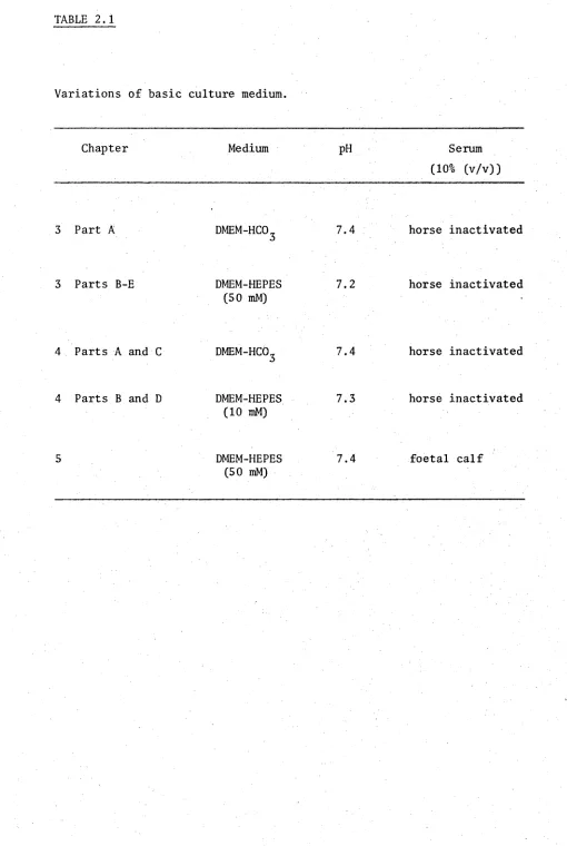
benzylpenicillin/ml and was incubated at 36.5°C in air. These media were supplemented with either horse or foetal calf serum as summarised in Table 2.1
e. Inactivation of serum
Horse serum was inactivated by heating at 57°C for 30 minutes.
f. Preparation of conditioned medium
Conditioned medium was prepared by incubation of basic culture medium with mature non-neuronal cell cultures for two days. For
experiments reported in Chapter 3 the conditioned media were transferred directly to the neuronal cultures whereas for experiments reported in Chapter 4 they were stored at -20°C prior to its use.
g. Preparation of extract medium (i) for chapter 3
Tissue extracts were prepared by homogenising adult female wistar rat tissues in basic culture medium, lacking serum, followed by centrifugation at 50,000xg for lh. The resulting supernatants were sterilised by filtration (0.22 ym) and were supplemented with 10%
inactivated horse serum. For experiments which required the same extract in different media (see Table 3.4) the high molecular weight factors were exchanged into the media by gel filtration. Biogel P4 columns
TABLE 2.1
Variations of basic culture medium.
Chapter Medium PH Serum
(10% (v/v))
3 Part A d m e m-h c o3 7.4 horse inactivated
3 Parts B-E DMEM-HEPES 7.2 horse inactivated
(50 mM)
4 Parts A and C d m e m-h c o3 7.4 horse inactivated
4 Parts B and D DMEM-HEPES 7.3 horse inactivated (10 mM)
5 DMEM-HEPES 7.4 foetal calf
[image:47.550.24.534.47.807.2]of 165 mM sodium chloride. The high molecular weight fraction of the extract was then eluted in the new medium by the addition of 2.0 ml of 165 mM sodium chloride to the column.
(ii) for chapter 4
Extract media of rat organs were prepared using tissue pooled from 15 adult females (Table 4.3). The muscle used was the quadriceps. The ventricle extract media of particular species were prepared from tissue pooled from 2 chickens, 40 mice or 10 rats. Extract media from the other species were prepared by mincing the entire ventricle of one animal and processing a sample of this mince (Table 4.4). In the third experiment (Table 4.5) the ventricle extract media were prepared from 64 newborn, 5 adult male or 5 adult female rats.
The tissue was homogenised in 20 mM HEPES buffer, pH 7.3, to give a 20% (w/v) extract, of which 22 ml was mixed with 18 ml of double
strength DMEM. This mixture was centrifuged at 50,000gforl h, filtered through glass wool and centrifuged at 50,000g for a further 1 h. The pH was readjusted to 7.3, the supernatant sterilized by millipore
(0.22 ym) filtration and 4 ml of heat inactivated horse serum added. The resulting mixture was equivalent to a 10% (w/v) extract in basic culture medium. Rat aortic and atrium extract media were prepared from an
initial 6% (w/v) homogenisation, thus giving a final extract concentration of 3% (w/v).
h. Steroid solutions
Solutions of steroids in basic culture medium were prepared by
II. Non-neuronal cell cultures a. Culture medium
All non-neuronal cultures were grown in DMEM-HCO^, pH 7.4.
During the initial four days of culture the medium was supplemented with 20% (v/v) inactivated horse serum. The serum concentration was then decreased to 10% (v/v). The culture medium was changed every four days, unless otherwise indicated.
b. Dissociation procedure
Rat embryos of 19 day gestation were placed in -Ca-Mg BSS and the required organs were then dissected from the embryos, placed in fresh
3
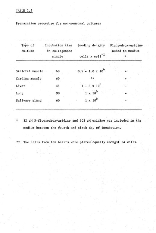
-Ca-Mg BSS, and minced into small pieces (approximately 1 mm ). The tissue was then added to a 1% (w/v) collagenase solution in -Ca-Mg BSS and placed in a gently shaking water bath at 37°C. After a set period, which depended on the organ (see Table 2.2), the tissue was dissociated by trituration with a pasteur pipette and then a 25 gauge hypodermic needle. The dissociated tissue were centrifuged at 200xg for 1 minute, washed in culture medium, resuspended in media, the cells counted in a haemocytometer and plated at the densities indicated in Table 2.2
c. Skeletal muscle cultures
The mononucleated myoblasts began to fuse with each other after the first few days in culture, and by eight days the cultures consisted of spontaneously contracting myotubes on a layer of flat fibroblast-like cells. The myotubes in these cultures resembled those described for rat (Königsberg, 1971), chicken (Shimada
et al.s
1969; Fischbach, 1972) and mouse muscle (Gilleret al.3
1977).d. Cardiac muscle cultures
TABLE 2.2
Preparation procedure for non-neuronal cultures
Type of culture
Incubation time in collagenase
minute
Seeding density
cells x well *
Fluorodeoxyuridine added to medium
*
Skeletal muscle 60 0.5 - 1.0 x 10⁶ +
Cardiac muscle 60 ** +
Liver 45 1 - 5 x 10⁶
-Lung 90 1 x 10⁶
-Salivary gland 60 1 x 10⁶
-82 yM 5-fluorodeoxyuridine and 203 yM uridine was included in the medium between the fourth and sixth day of incubation.
[image:50.550.20.538.26.800.2]cultures the cardiac myocytes stopped contracting and appeared to lose viability. Despite the use of 5-fluorodeoxyuridine these cultures contained a dense layer of fibroblast-like cells which, in cultures older than two weeks, became dominant. Thus, only cultures between one and two weeks old were used as sources of conditioned media. The morphological appearance of these cultures resembled those previously described (Mark et al. 3 1973).
e. Liver and lung cultures
These cultures consisted predominantly of islands of epithelial- like cells and a few isolated fibroblast-like cells. The cultures were maintained for periods of up to five weeks without domination by
fibroblast-like cells. The morphology of the liver and lung cultures was similar to that described for primary cell cultures of liver from neonatal and adult rats (Williams et al., 1971; Williams and Gunn, 1974) and cloned foetal feline lung (Kniazeff et al. 3 1976) respectively.
f. Salivary gland cultures
These cultures consisted of islands of epithelial-like cells interspersed with fibroblast-like cells and a few unidentified cells
(Figure 2.1). In a few cultures isolated groups of fat cells were seen.
Ill Neuronal cultures a. Culture medium
Basic culture and extract media were changed every four days and conditioned media every two days.
b. Spinal cord cell cultures
FIGURE 2.1
Photomicrograph of an unidentified rat embryo salivary gland cell.
[image:52.550.13.546.33.807.2]sharpened edge, was run down dorsal to and on each side of the spinal cord, breaking the attached membranes. The spinal cords were removed intact and placed in culture medium. By using this method spinal cords, essentially free of DRG and meningial membrane, could be dissected
rapidly from the embryo. The spinal cords were minced and dissociated by gentle trituration with a 21 gauge, followed by a 25 gauge,
hypodermic needle. The cells were plated at a density of 0.6 x 10^* cells/well.
The mature spinal cord cultures consisted predominantly of small islands of 10-15 neurones which were interconnected by a dense labyrinth of axons. Non-neuronal cells were also present but never at a density which required the use of 5-fluorodeoxyuridine to control their
proliferation. This was presumably due to the absence of meningial membrane cells. The morphology of the neurones was similar to those described in cultures of chicken (Fischbach, 1972) and mouse (Peacock
et at., 1973) spinal cord.
c. Ciliary, superior cervical and dorsal root ganglia cultures Ciliary and DRG from chicken embryos and SCG from neonatal rats were removed into basic culture medium, trimmed of adherent tissue and one ganglion was cultured per well. Photomicrographs of typical ciliary ganglion cultures are shown in Figures 3.1 and 3.3 and of SCG in Figure
2.2.
B. Chromatographic methods
I Processing of fractions
FIGURE 2.2
Photomicrograph of a cultured neonatal rat superior cervical ganglion.
[image:55.550.22.548.38.809.2]V A f
fäm-
‘'MW?*
H W
:
pps
SSE
% % / v f W
- / 1
'• # '■
-
II Elution profiles
180 Drop fractions were collected and the elution profile
established by measuring the absorbance at 280 and 260 nm, except for column 4.5 where 160 drop fractions were collected and the profile established by protein determination (Lowry et at. 3 1951).
III Columns
The preparation of samples and the running of columns was carried out as indicated in Table 2.3.
C. Bioassays for factors influencing:
I Ciliary ganglion fibre outgrowth
The ability of a source of factor to induce a fibre outgrowth was determined by culturing ganglia for four days and then measuring the magnitude of the outgrowth using a relative scale (0-5). Ganglia which exhibited a 5 response (Figures 3.3 E-F) had a dense fibre halo which extended 0.8 to 1.0 mm from the ganglion. Ganglia which had fewer or shorter fibres were assigned proportionally lower values. Examples of typical responses and their scores are shown in Figures 3. 3 A-F.
II Ciliary ganglion choline acetyltransferase
The level of CAT maintained by different media was assayed by culturing ciliary ganglia for four days and then measuring the activity of CAT as described in Part D of this Chapter.
III Spinal cord choline acetyltransferase