(1) Section 1.2 (bk pages 4-5 Duplicated paragraph removed. (2) Figure 1.1, page 8
Inserted text:
“Molecules involved in the transitions between the subsets are shown at the relevant points, where x'1' means the phenotype observed in the knockout o f molecule x.”
(3) Section 1.2 (ch Negative Selection, page 11 Inserted “to” in paragraph 5.
“In a similar way, the peritoneal cavity microenvironment o f anti-erythrocyte transgenic mice allows auto-reactive B -l cells to accumulate (Murakami,
1992)...”
(4) Section 1.4(b), Memory B cell phenotypes and locations, page 34 Removed “#44” from reference list in paragraph 2.
(5) Section 2.12(a), Cytoskeletal Stabilising Buffer (CSB) Lysis, page 72 Inserted text, paragraph 1:
“CSB buffer components are described in Section 2.13.” (6) Section 2.13, Stock Solutions, page 76
Added CSB lysis buffer recipe. (7) Figure 3.4. Part A, page 90
Deleted text: “IgM and IgD (MD-4)” Now reads:
“ELISPOT data from day 5 after transfer and immunisation demonstrating the equivalent behaviour o f transgenic lines expressing the IgG membrane tail (MG2, MG6 and MG8) compared to lines expressing IgM alone (MM-4) or IgM8E (MSE1 and M8E2).”
(8) Figure 6.2, page 158 Inserted text. Now reads:
“The results are given as the number o f anti-lysozyme IgGi AFCs (closed circles) and the number o f anti-lysozyme IgMa AFCs (open circles) per 107 recipient splenocytes.”
(9) Section 6.3(c), IgG, IgM/G and IgM transgenic lymph node B cells retain differential responses to antigen, page 161, paragraph 2
Deleted text, paragraph 2. Now reads:
“Mature lymph node B cells from Ig G :R A G r‘, IgM /G :R A G l’/" and IgM :R A G l‘/‘ mice were transferred along with TCR transgenic T cells into immunised recipients.”
B cell antigen receptor isotypes:
implications for immune memory
Stephen W. Martin
December, 2001
A thesis submitted for the degree of Doctor of Philosophy of The Australian National University
Medical Sciences Graduate Program, John Curtin School of Medical Research,
This thesis presents research undertaken at the Medical Genome Centre, Division of Molecular Medicine of the John Curtin School of Medical Research, Australian National University, Canberra. This work was performed between July 1998 and December 2001, while I was the recipient of an Australian National University PhD scholarship.
This is to certify that the studies outlined in this thesis are my own work, performed under the supervision of Professor C. Goodnow. Valuable assistance was also provided from other members of the Goodnow lab, the staff of the ACRF Medical Genome Centre and the staff of the John Curtin School of Medical Research, Australian National University, Canberra, as stated in the acknowledgments. This thesis is under 100,000 words.
Acknowledgments
Above all my sincere thanks to my supervisor and mentor Professor Chris Goodnow for his vision, clarity of thought and boundless positive energy which has given direction to this study. Chris is a remarkable scientist and has been an approachable, patient teacher.
Thanks also to all of my fellow PhD students at the ANU for helping make life as a research student both bearable and enjoyable - Jane, Simon, Pete, Adele, Lisa, Jesse, Rui, Nick, Damo, Marie, Francis, Todd, Tony and Yvonne. It has been a pleasure to share the last few years with you.
Thank you to the many members of the Goodnow lab who have made it such an exciting place to work. In particular, Dr. Sarah Townsend provided valuable assistance with adoptive transfer experiments, Dr. Benny Weintraub helped with biochemical experiments and Dr. Carola Garcia de Vinuesa gave expert assistance with immunohistochemical staining. Thank you also to the Medical Genome Centre staff, in particular Katherine Sullivan, who maintained the many mouse strains required for this work. For technical assistance, Geoff Osborne and Sabine Grüninger of the JCSMR FACS facility and the members of the JCSMR Photography and Electron Microscopy Unit have been very helpful.
A human being is part of a whole, called by us the "Universe”, a part limited in time and space. He experiences himself, his thoughts and feelings, as something separated from the rest - a kind of optical delusion of his consciousness. This delusion is a kind of prison for us, restricting us to our personal desires and to affection for a few persons nearest us. Our task must be to free ourselves from this prison by widening our circles of compassion to embrace all living creatures and the whole of nature in its beauty.
ix
Abstract
The B cell antigen receptor (BCR) can be expressed in a number of molecular forms during B cell development, and provides context-dependent signals that lead to a variety of cellular outcomes. Isotype switching during an immune response represents a key, irreversible molecular change in BCR structure, where naive BCR isotypes (IgM and IgD) are replaced by switched isotypes (IgG, IgA, IgE) that become markers of antigen experience. Attempts to study isotype-specific BCR signalling in vivo have been hampered, however, by the changes in BCR affinity, location, cell surface phenotype, activation requirements and lifespan that also accompany antigen priming.
To address the role of isotype-switched BCR expression during an immune response, homogeneous populations of naive B cells derived from anti-lysozyme immunoglobulin transgenic mice were seeded into a T-dependent immune response to lysozyme in vivo. Comparisons were made between transgenic B cells expressing IgM and IgD BCRs and B cells expressing full length IgG or the unique IgG membrane tail domain.
It was found that naive transgenic B cells expressing the IgG membrane tail domain make a more robust response to antigen compared to naive transgenic B cells bearing IgM, both in terms of net clonal expansion and in the production of antibody-forming cells. Transgenic B cells expressing the IgG membrane tail were not activated more efficiently, nor did they have a greater rate of cell division compared to IgM transgenic B cells. Rather, the IgG membrane tail protected B cells from cell death during the process of clonal expansion.
This experimental strategy was verified by showing that B cell developmental differences in the bone marrow, as well as phenotypic variations in the marginal zone and follicular B cell subsets of the various transgenic lines did not account for their responses to antigen in vivo. The only apparent determinant of reactivity to antigen in this system was BCR isotype and the expression of the IgG membrane tail domain.
Publications
Arising from this work:
Martin, S. W. and Goodnow, C. C. (2001) Burst-enhancing role of the IgG membrane tail as a molecular determinant of memory, Nature Immunol., accepted for publication.
Selected conference papers:
Martin, S. W. and Goodnow, C. C. (2001) The IgGi cytoplasmic tail domain is an important regulator of B cell clonal expansion, Keystone Symposium: “B cell Immunobiology and Disease ”, Snowbird Resort, Utah, U.S.A.
Martin, S. W. and Goodnow, C. C. (1999) BCR isotype can influence terminal B cell differentiation: implications for B cell memory, 29th Annual Conference of the Australasian Society for Immunololgy, Dunedin, New Zealand
Other publications:
Contents
Acknow ledgm ents... v
Abstract ... ix
Publications... xi
Contents ... xiii
Figures and Tables... xix
Abbreviations... xxii
Chapter 1. Introduction 1 S e c tio n 1.1. P r e a m b le ... 2
S e c tio n 1.2. B cell d e v e lo p m e n t and d iffe re n tia tio n ...3
1.2 (a) Stem cells and B lineage com m itm ent...3
1.2 (b) B cell development in the adult bone m arrow ... 4
1.2 (c) Selection shapes the mature B cell repertoire...6
Evidence for selection... 6
Negative selection... 7
Antigen-driven selection...12
1.2 (d) B cell subsets in the periphery... 13
The formation of mature B cell subsets...13
Mature, follicular B cells...14
Marginal zone B cells...14
B-1 B cells... 19
1.2 (e) Memory B c e lls ... 20
1 .2 (f) Plasma ce lls...20
1.3 (a) T -in d ep end en t im m une responses... 2 4 1.3 (b) T -d e p e n d e n t im m une re s p o n s e s ...2 4
Activation of antigen-specific B and T lymphocytes... 24
Extrafollicular antibody responses... 25
Germinal centre reactions...27
Isotype switching: mechanism and signals... 28
The remnants of an immune response - memory B cells and long-lived plasma cells...29
S e c tio n 1 .4 . M e m o r y c e lls a n d m e m o r y r e s p o n s e s ...2 9 1.4 (a) Early observations on the nature of im m une m em o ry... 30
1.4 (b) C urrent issues in the study of im m une m e m o ry ... 31
Mechanisms for the persistence of the memory state... 31
Memory B cell phenotypes and locations... ... 33
Mechanisms for the features of the memory response...35
S e c tio n 1 .5 . E v id e n c e fo r is o ty p e -s p e c ific B C R s ig n a llin g in vitro a n d in vivo... 3 7 1.5 (a) B C R isotypes differ in their structure within the m e m b ra n e ...3 7 1.5 (b) Attem pts to d elineate isotype-specific signalling fu n c tio n s ...3 8 In vitro studies...38
In vivo studies...43
In vivo studies of isotype-switched B cell antigen receptors... 45
S e c tio n 1 .6 . T h e c u rre n t s t u d y ... 4 6
Chapter 2. Materials and Methods... 53
S e c tio n 2 .1 . M a t e r ia l s ...5 4
S e c tio n 2 .2 . M o u s e lin e s (d e s c rip tio n a n d h o u s in g ) ... 5 4
S e c tio n 2 .3 . T r a n s g e n ic m o u s e s c r e e n in g ... 5 4
2 .3 (a) P C R s c re e n in g ...5 4 2 .3 (b) Blood s c re e n in g ... 5 6
S e c tio n 2 .4 . In v iv o c e ll tra n s fe rs a n d im m u n is a tio n s ... 5 6
2 .4 (a ) A ntigen/adjuvant preparation... 5 6 2 .4 ( b ) Im munisation re g im e s ...5 7 2 .4 (c) T ran sfer of Ig transgenic and T C R transgenic donor cell suspensions
Section 2.5. Antibody staining reagents for flow cytometry, ELISA, histochemistry and
W estern blotting... ... 61
Section 2.6. Cell isolation from mouse tis s u e ... 63
2.6 (a) Cell preparation...63
2.6 (b) Cell counting... 63
Section 2.7. Flow C ytom etry... 64
2.7 (a) Flow cytometry staining... 64
2.7 (b) Flow cytometry sorting... 65
2.7 (c) Examples of flow cytometry gating strategies... 65
Section 2.8. C F S E Labelling... 68
Section 2.9. E L IS P O T ... 68
Section 2.10. Serum E LIS A ...69
Section 2.11. H istology... 69
Section 2.12. Biochemistry techniques... 71
2.12 (a) Cell stimulation and lysis... 71
Cytoskeletal stabilising buffer (CSB) lysis... 71
TX-100 lysis...72
2.12 (b) Western Blotting... 72
Section 2.13. Buffers, media and stock solutions... 73
Buffers... 73
Media... 74
Stock solutions... 75
Chapter 3. The IgG membrane tail is a regulator of B cell clonal expansion in vivo... 77
Section 3.1. Introduction...78
Section 3.2. IgG-bearing B cells produce more antibody than IgM-bearing B cells during an immune response... 80
Section 3.3. The IgG mem brane tail is the molecular determinant responsible for heightened antibody responses... 80
Section 3.5. increased clonal expansion conferred by the IgG membrane tail is due to
decreased death over successive cell divisions... 87
Section 3.6. Chapter summary...94
Chapter 4. An examination of factors which affect transgenic B
cell responses to antigen in vivo...97
Section 4.1. Introduction... 98
Section 4.2. The location and phenotype of IgG and IgM transgenic B cells during an immune response...98
4.2(a) Histology... 98
4.2(b) FACS... 103
4.2 (c) AFC production...104
4.2(d) Summary...105
Section 4.3. Germinal centre and follicular expansion of IgM/G and IgM transgenic B c e lls... 109
Section 4.4. IgM transgenic B cells do not undergo preferential apoptosis via FasL expressed on transgenic helper T cells... 110
Section 4.5. Do other changes associated with memory play a role in the magnitude of antibody responses?... 118
Section 4.6. Chapter summary...122
Section 4.7. Future W ork... 123
Chapter 5. Marginal zone and follicular B cell subsets vary in
different anti-HEL transgenic lines... 125
Section 5.1. Introduction...126
Section 5.2. Peripheral B cell subsets in IgG, IgM/G and IgM transgenic lines... 126
Section 5.3. Marginal zone and follicular B cell analysis in IgG, IgM/G and IgM transgenic lines... 127
Section 5.4. Marginal zone and follicular subset differences are retained and augmented in RAGT7' mice...135
Section 5.5. Marginal zone and follicular subset differences are present in other HEL transgenic lines... 140
xvii
Section 5.7. Future work... 147
Chapter 6. Line to line variation does not explain differential B cell
responses to antigen in vivo... 149
Section 6.1. Introduction...150
Section 6.2. Line to line variation in construct integration site, transgene copy number
or transgene switch regions does not explain different responses to
antigen... 151
6.2 (a) Variations in the proportion of HEL-binding B cells do not explain
different responses to antigen...156
Section 6.3. Marginal zone and follicular subsets differ between lines but do not
correlate with reactivity to antigen... 157
6.3 (a) The proportions of MZ versus FO subsets in different lines do not
correlate with responses to antigen... 157 6.3 (b) Immature sorted B cells retain differential responses to antigen...160 6.3 (c) IgG, IgM/G and IgM transgenic lymph node B cells retain differential
responses to antigen... 161 6.3 (d) Sorted MZ versus FO subsets retain differential responses to antigen
...166
Section 6.4. The effect of IgD expression on transgenic B cell responses to antigen in vivo...167
Section 6.5. Chapter summary...169
Section 6.6. Future w ork...172
Chapter 7. Biochemical analysis of early signalling by IgG isotype
receptors... 173
Section 7.1. Introduction...174
Section 7.2. IgG transgenic B cells show a different pattern of early activation
compared to IgM transgenic B cells...175
Section 7.3. The IgG BCR may constitutively associate with lipid rafts... 176
Section 7.4. Chapter summary and future work... 185
Section 8.1. Introductory comments... 188
Section 8.2. The role of BCR isotype in B cell responses to antigen...188
8.2 (a) Summary of the major findings:...188
8.2 (b) Validation of the experimental model...190
8.2 (c) Integration of these findings with other studies of switched isotype BCR function... 191
8.2 (d) Candidate signalling pathways for the protective effect of the IgG membrane tail... 194
Section 8.3. A model of the role of BCR isotype during memory responses... 196
8.3 (a) Quantitative and qualitative changes lead to the rapid induction of the memory response... 196
Precursor frequency...197
Cell migration and location...198
Pre-activated state...198
8.3 (b) IgG expression as a component determining the magnitude of the memory response... 199
Section 8.4. Future directions...200
Chapter 9. Appendix... 203
Section 9.1. News and Views Article... 204
xix
Figures and Tables
Figure 1-1. B cell developm ent in the bone m arro w ... 8
Figure 1-2. B cell subsets in the p e rip h e ry ... 22
Figure 1-3. Sequence com parison o f m em brane Ig isotypes from various species at the extracellular, m em brane-spanning and cytoplasm ic re g io n s...40
Figure 1-4. Structure o f the B CR isotypes from the various transgenic strains used in this stu d y ... 50
Figure 2-1. C onstruct inform ation and m em brane tail sequence com parisons across the Ig transgenic lines used in this study ...58
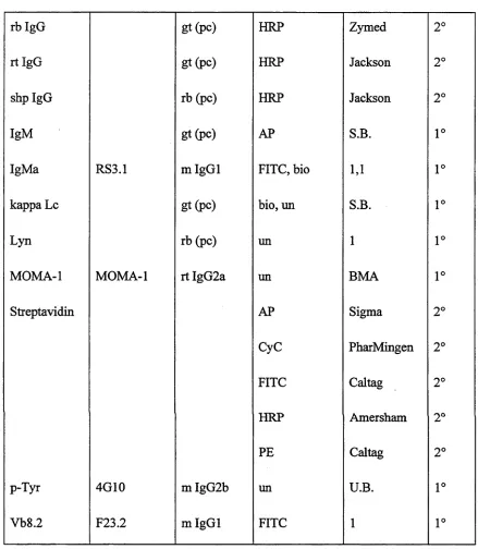
Table 2-1. A ntibody staining reagents used in this study... 62
Figure 2-2. Gating strategies used to visualise H EL-binding B cells in adoptive transfer recipients... 66
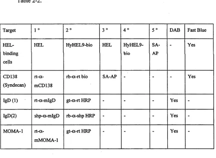
Table 2-2. Schem atic show ing the staining and developing steps for
im m unohistochem ical detection o f various target antigens... 70
Figure 3-1. Seeding a prim ary antibody response w ith naive B confers m em ory- response characteristics when the receptors contain the unique IgG
m em brane ta il... 82
Figure 3-2. The IgG m em brane tail increases the production o f antibody-form ing cells ... 84
Figure 3-3. The IgG tail augm ents clonal e x p an sio n ...88
Figure 3-4. A ll transgenic lines expressing the IgG m em brane tail m ake an equivalent response to antigen in vivo...90
Figure 3-5. IgG, IgM /G and IgM transgenic B cells enter cell division at the sam e tim e yet undergo different net clonal expansion during a T -dependent response to antigen in v iv o... 92
Figure 4-3. F ollicular and germ inal centre expansion o f IgM and IgM /G transgenic B
cells in prim ed recip ien ts... 112
Figure 4-4. C rippling the Fas/FasL pathw ay in transgenic T cell help does not rescue clonal expansion o f IgM transgenic B cells in vivo... 116
Figure 4-5. L ong-term m em ory B cells are poorly generated from nai've transgenic B
cells in short-term prim ed re c ip ien ts...120
Figure 5-1. Peripheral B cells in IgG, IgM /G and IgM transgenic lines vary in CD21 expression profile and in the fraction o f B cells that bind H E L ... 128
Figure 5-2. IgG , IgM /G and IgM transgenic spleens show different recruitm ent o f peripheral B cells into the splenic m arginal zone and follicular subsets by F A C S ... 130
Figure 5-3. Spleen histology show ing the location o f H EL-binding cells in IgG , IgM /G and IgM transgenic m ice... 132
Figure 5-4. Increased entry into the m arginal zone com partm ent by IgG and IgM /G transgenic B cells still occurs on the R A G T 7' b ack g ro u n d ...138
Table 5-1. Cell num bers and the fractions o f H EL -binding B cells in m arginal zone and follicular subsets for each transgenic lin e ... 142
Figure 5-5. C orrelating the effects o f inter-clonal com petition on entry into the m arginal z o n e ...144
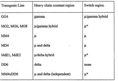
Table 6-1. The type and extent o f sw itch regions used in each transgenic construct .1 5 3
Figure 6-1. A com parison o f im m une responsiveness betw een transgenic B cells from lines G G 4, M G 2, M G 6, M G8, M 6E1, M 6E2, M M 4, D D 6 and M M 4xD D 6
... 154
Figure 6-2. Transgenic B cells on the R A G l'7' background show the sam e pattern o f reactivity to antigen in v iv o...158
Figure 6-3. Im m ature bone m arrow B cells and m ature lym ph node B cells from
xxi
Figure 6-4.
Figure 7-1.
Figure 7-2.
Sorted marginal zone and follicular phenotype spleen cells from IgG or IgM RAG l*7" mice retain differential responses to antigen...164
TX-100 lysis of stimulated IgGiRAGF7' and IgM:RAGF7' transgenic B cells showing differences in tyrosine phosphorylation and Igaß isoforms... 178
The IgG isotype BCR may constitutively associate with a salt-extractable cell compartment corresponding to cytoskeleton-associated membrane rafts
Abbreviations
(v/v) v o lu m e fo r v o lu m e
(w /v ) w e ig h t fo r v o lu m e
°C d eg rees C en tig rad e
Bg m icro g ra m
[i\ m icro litre
p M m ic ro m o la r
aa am in o acid
A F C an tib o d y -fo rm in g cell
A M P 2 -a m in o -2 -m e th y 1-1 -p ropanol
A PC an tig e n -p re se n tin g cell (o r allo p h y co cy an in )
B C IP 5 -b ro m o -4 -ch lo ro -3 -in d o y l phosphate
B C R B cell an tig en re cep to r
bio b io tin y late d
B S A b o v in e seru m alb u m in
C D c lu ste r o f d iffe ren tiatio n
C F A c o m p lete F reu n d 's ad ju v an t
C F S E 5 -(an d 6 -)-carb o x y flu o resc ein diacetate succin im id y l ester
D A B 3 ,3 '-d iam in o b en zid in e tetra h y d ro ch lo rid e
DC d e n d ritic cell
d d H 20 d istilled , d e io n ised w a te r (> 1 8 M Q re sistan c e)
dN T P s d eo x y rib o n u c le o tid e trip h o sp h ates
E D T A e th y len e d iam in e tetra ace ta te
E L I S PO T e n z y m e -lin k e d im m u n o sp o t assay
F A C S flu o re scen c e-a ctiv a ted cell so rter (or g en e ral flo w cytom etry)
F B S fetal b o v in e serum
F IT C flu o re sc e in iso th io cy an ate
FO fo llic u la r B cell subset
g gram
g grav ity
H -2 M H C c o m p le x o f m ice
H E L h en eg g ly so zy m e
xxiii
H R P horse ra d ish p eroxidase
i.p. in traperitoneal
i.v. intrav en o u s
Ig im m u n o g lo b u lin
IgM /G chim eric B C R w ith IgM C H and the IgG
Ig M a Ig M o f th e a allotype
Ig M 8 E ch im eric Ig M B C R w ith 27 aa e x tra celli
L L itre
L PS lip o p o ly sacch arid e
M eO H m ethanol
M H C M ajo r H isto co m p atib ility C om plex
m l m illilitre
m M m illim o lar
M Z m arg in al zone B cell su b set
nM n an o m o lar
P B S p h o sp h ate-b u ffe red saline
P C R p o ly m era se ch ain reaction
P E p h y co ery th rin
pH -lo g 10[H 30 +]
P M S F p h en y lm eth y lsu lfo n y l fluoride
R PM I R o sw ell P ark M em orial Institute
SA strep tav id in
T A C T ris.am m o n iu m ch lo rid e
T A E T ris/ac etate/E D T A b uffer
T B S T ris-b u ffere d saline
T C R T cell an tig en recep to r
T D T (cell)-d ep e n d en t
T E L tu rk ey eg g lysozym e
TI T (cell)-in d ep e n d en t
2 Chapter 1
Section 1.1. Preamble
The immune system is a complex interconnected series of cell types and proteins that has evolved to protect the body from infection. The innate arm consists of physical and antimicrobial chemical barriers to infection as well as series of cells bearing receptors that recognise common conserved patterns amongst pathogens (Hoffmann, 1999). This is found in most forms of multicellular life and represents a complex but evolutionarily primitive form of immune system. By contrast, some form of adaptive immune system is shared by higher vertebrates and consists of cells that use recombined receptors to recognise a vast array of potential pathogen signatures. The hallmarks of adaptive immunity include a pathogen-specific response to clear infection and in many cases a state of immunological “memory” that provides long-term protective immunity against reinfection.
While an appreciation of the ability of the innate immune system to initiate immune responses and to regulate the other arms of the immune system has been relatively recent (Fearon, 1996), the adaptive arm of the immune system has been studied experimentally for over 100 years (Silverstein, 1989). The phenomenon of immune memory, especially with respect to antibody production by B lymphocytes, is well known and yet the underlying cellular mechanisms remain obscure. We know that a first encounter with a pathogen “primes” the immune system so that secondary, or memory immune responses have distinct qualitative and quantitative differences (Ahmed, 1996). Specific antibody is of higher affinity, is produced more rapidly and in much higher quantities compared to a primary response. In addition the antibody isotype profile changes, with switched isotypes such as IgG and IgA dominating. These complex, inter-connected changes make the phenomenon of “memory” very difficult to dissect, and it is still unclear what factors are responsible for the maintenance of the memory state versus the memory response itself.
For example, the role of isotype-switched B cell antigen receptor (BCR) expression on many memory B cells is unknown. At most other stages of B cell development it is well documented that signals through different molecular forms of the BCR influence B cell responses. During B cell production in the bone marrow, pre-BCR mediated signalling is crucial for developmental progression, while immature B cells respond to BCR
antigen-dependent signalling through isotype-switched receptors to the subsequent memory response has yet to be determined.
This introduction will review the role of BCR isotype in shaping B cell development, and outline the features of T-dependent immune responses. The unique qualities of the memory response and memory cells will be discussed, as well as current theories to explain the persistence of the memory state and the features of the memory response. Finally, evidence for isotype-specific signalling, both in vitro and in vivo will be reviewed and placed in context with the current study.
Section 1.2. B cell development and differentiation
B cell development from stem cells in the fetal liver and adult bone through to mature peripheral B cells can be thought of as a series of checkpoints where B cell antigen receptor (BCR) expression and signalling at each stage in B cell development plays an important role in shaping the eventual repertoire (reviewed in Rolink, 1993; Goodnow, 1995; Melchers, 1999; Pillai, 1999; Meffre, 2000; Monroe, 2000). B cell subsets in the bone marrow are shown in Figure 1-1, and peripheral B cell subsets in Figure 1-2.
1.2 (a) Stem cells and B lineage commitment
B lymphopoiesis in the mouse starts in the fetal yolk sac and moves to the fetal liver around day 14 of gestation. B lymphopoiesis is then established in the bone marrow in the first weeks after birth and continues for the life of the animal. In the bone marrow self- renewing hematopoietic stem cells give rise to common lymphoid progenitors that under normal conditions produce T, B and NK cells (Kondo, 1997). The stem cells and
progenitor cells that give rise to lymphoid lineages are thought to express multiple different lineage determinants at low levels. In fact bone marrow-derived stem cells show
remarkable plasticity and under the right conditions can contribute to a wide range of tissues including heart, brain, skeletal muscle and liver (Blau, 2001). Lineage
“commitment” seems to involve the emergence of a more stable pattem of gene expression that reinforces lineage-specific gene expression and actively represses other possible cell fates (Nutt, 1999b; Akashi, 2000; Busslinger, 2000). In the case of B lymphopoiesis in the bone marrow, knockout studies have determined genes important for the development of B cells (Scott, 1994; Zhuang, 1994; Lin, 1995; Schilham, 1996; Wang, 1996). The
4 Chapter 1
developmental progression. It is expressed from the earliest commited B cell stage, and while Pax-5'7' B cells appear to arrest at the early pro-B stage, they remain multipotent and able to form T cells, NK cells, osteoclasts, macrophages, dendritic cells and granulocytes in vitro (Nutt, 1999a) and in some cases in vivo (Rolink, 1999).
1.2 (b) B cell development in the adult bone marrow
The earliest stage of committed B cell precursors express the surface markers AA4.1, B220 and CD43 and are referred to as pre-pro-B cells (Hardy’s Fraction Ao, Aj and A2) (Hardy, 1991; Li, 1993; Allman, 1999). These retain immunoglobulin loci in a germline configuration, express low levels of recombinase activator (RAG) genes and lack
expression of components of the BCR, such as Igocß, suggesting initial B cell commitment is not driven by antigen receptor signals (Allman, 1999).
and BP-1 (Hardy, 1991). During the pro-B cell stage sequential induction of heavy chain immunoglobulin Dh—» Jhand Vh—> DhJhrearrangements occurs. Non-productive
truncated forms of mlgji (Djli) are counter-selected at this stage leading to developmental
arrest at the pro-B stage (Meffre, 2000). Pro-B cells express the Iga, Igß, VpreB and A.5 components of the pre-BCR (Karasuyama, 1993; Meffre, 1996), and recent evidence suggests that a complex exists between calnexin and Igocß on the surface of pro-B cells (Nagata, 1997). Interestingly, crosslinking of Igß in vivo in RAG-deficient mice allowed the pro-B cells to display some features of pre-B cells as if the Igaß complex on pro-B cells delivers a signal for developmental progression (Nagata, 1997). Further evidence that signalling via the calnexin/Igaß complex may be important for pro-B cell progression comes from analysis of Igß'7' (Gong, 1996) and (xMT mice (Ehlich, 1993). In the absence of Igß there are no pre-B cells, but in the pro-B subset Vh —> DhJh rearrangements are
decreased compared to Dh—> Jhjoins. By contrast, in the absence of membrane-bound jll
(and therefore a functional pre-BCR) there are a normal number of VH —> DhJh
rearrangements, suggesting that Igß plays a role in signalling even in the absence of the pre-BCR. This observation fits with a model in which constant checks are imposed on developing B cells to ensure the correct expression of the components of a functional BCR.
After a period of clonal expansion, pre-B cells become small and resting (Hardy’s fraction D). This is the stage of most light chain immunoglobulin gene Vl —» Jl
rearrangement, with a productively rearranged K or X light chain replacing surrogate light chain to form a complete BCR.
When a surface IgM isotype BCR is expressed, the B cell is considered “immature” (Hardy’s fraction E). Immature B cells lose expression of IL-7R and have lower BP-1 expression (Hardy, 1991). The expression of a functional IgM BCR signals a cessation of Ig light chain gene rearrangement, the degradation of RAG proteins, and a loss of mRNA for RAG and pre-BCR components. Immature B cells exit from the bone marrow via the blood and mature further phenotypically in the spleen, eventually contributing to the mature, long-lived recirculating pool.
6 Chapter 1
1.2 (c) Selection shapes the mature B cell repertoire
Evidence for selection
Selection shapes the peripheral B cell repertoire during the transition from immature to mature B cells in the periphery, and also during the formation of specialised mature subsets in the splenic marginal zone and the peritoneal and pleural cavities. The fact that selection occurs at these points seems clear, however the relative importance of “negative” selection to self-antigens, foreign-antigen driven “clonal” selection or “positive” selection to undefined ligands for this process is still in doubt.
Only a small fraction of bone marrow-derived immature B cells are ever incorporated into the long-lived recirculating mature B cell pool (MacLennan, 1986). The exact site of cell loss has been difficult to determine and is probably a combination of apoptosis in the bone marrow and the spleen. Unlike the production of thymus-derived lymphocytes, which occurs in a defined organ and whose rate of emigration can be accurately estimated
(Scollay, 1980), the actual rate of emigration of newly formed B cells from the bone
marrow to the periphery is still uncertain. Estimates originally suggested a turnover rate of 2-5 x 10⁷ IgM+ B cells in the mouse bone marrow compartment daily (Opstelten, 1983; Osmond, 1986). While this may reflect the rate of cell production, it is likely to be a gross overestimate of the number of immature B cells that leave the bone marrow as many tolerance mechanisms operate at that site that lead to clonal deletion (Nemazee, 1989; Hartley, 1993). Also, because of the rapid clearance of apoptotic bodies in the bone marrow the rate of B cell apoptosis is difficult to determine. Recently the emigration rate of B cells from murine BM has been revised to 9xl06 IgM+ cells per day by taking into account the rate of apoptosis (Lu, 1997, 2000).
After entry into the spleen, there is evidence for two B cell populations, one with a very short turnover rate of a few days (Allman, 1993), and one with a half-life of several months (Gray, 1988; Forster, 1990; Allman, 1993; Hao, 2001). The prevailing view is that the short-lived subset represents immature B cells and that a very small fraction (1-10 %) of these enter the long-lived pool (Ron, 1985; Lortan, 1987; Gray, 1988; Forster, 1990; Chan,
1993), although the use of BrdU and other labels that mark proliferating cells rest on the assumption that immature B cells are the only cells derived from proliferating precursors. A subset of mature B cells are also likely to be proliferating by mechanisms such as clonal expansion due to encounter with both T-dependent and T-independent antigens
(MacLennan, 1986; Chan, 1993), making the interpretation of labelling data difficult. The loss of the majority of recent bone marrow emigrant B cells that reach the spleen is
Certainly the “space” available for new B cells in the follicle niches is a critical,
presumably neutral mediator of entry into the long-lived pool, as demonstrated by studies showing the rapid “re-filling” of those areas after depletion (Bazin, 1985b, 1985a). This “clonal competition” (MacLennan, 1986; Lortan, 1987) is also regulated by negative selection forces that affect the ability of self-reactive B cells to compete for follicle entry (Cyster, 1994).
The second piece of evidence for selection shaping peripheral B cell subsets comes from the restricted pattern of J558 Vh gene useage in mature splenic B cells compared to
bone marrow derived immature B cells (Gu, 1991). In addition, the B-l subset is enriched for B cells specific for numerous self and foreign antigens (Wortis, 2001) and recent analyses in rats (Dammers, 1999a) and several transgenic mouse models (Martin, 2000a) show that the splenic marginal zone compartment can be enriched for certain specificities.
Negative selection
Negative selection is a major force in determining the nature of the mature B cell repertoire. The nature and timing of self-antigen exposure leads to a number of negative selection responses in B cells. This process has been most carefully studied in transgenic models that allow the regulated expression of autoantigens (Goodnow, 1992; Goodnow,
1995; Nemazee, 2000).
In the bone marrow, the binding of high avidity self antigen to self-reactive BCRs on immature bone marrow B cells can lead to autoreactive cell deletion (Nemazee, 1989). Self antigen binding to high avidity autoantigens causes developmental arrest at the immature B cell stage, which is separate from subsequent apoptotic loss (Hartley, 1993). It seems that strongly self-reactive immature B cells can alter their antigen receptors by secondary Ig gene rearrangements during developmental arrest, a process termed “receptor editing” (Radic, 1993; Tiegs, 1993). Subsequent expression of a non-autoreactive BCR then allows immature B cells to “escape” the developmental block, although this mechanism still produces an effective “clonal deletion” of the original self-reactive BCR. This process has been modelled in the 3-83 anti-H-2k b transgenic system. On a RAG+/+ background,
“edited” mature B cells carrying endogenous Lc accumulate in the periphery, while on the RAG 7' background B cells fail to populate peripheral lymphoid organs (Spanopoulou,
8 Chapter 1
Figure 1-1. B cell development in the bone marrow
This diagram outlines the major steps in B cell lymphopoiesis in the bone marrow, from hematopoietic stem cell precursors through pre-pro-B cells, pro-B cells, pre-B cells and immature B cells. The expression of relevant cell surface and intracellular markers is indicated by bars underneath each cell subset. Also, the nomenclature for each subset according to Hardy (Hardy, 1991; Li, 1993) is indicated A ^ E . Calnexin is shown in green in a putative receptor complex with Igaß (yellow) on pro-B cells. Rearranged heavy chain
xp
re
ss
ion
Low
+
-f
+
L
General Introduction 11
The importance of receptor editing versus cell death as a mechanism to ensure tolerance against high avidity, abundantly expressed self-antigens is unclear. While one report suggested that evidence of receptor editing is common amongst IgM+^+ peripheral B cells in normal mice (Retter, 1998), most light chain gene rearrangements are to JkI (Nishi,
1985), suggesting receptor editing is not a common occurrence, at least for k+ cells.
For the systemic membrane-bound anti-MHC system, as well as the anti-HEL system where HEL is expressed as a membrane-bound array, the major pathway of tolerance in the bone marrow is developmental arrest, followed by receptor editing or clonal deletion. By contrast, when a lower avidity soluble form of HEL is used, clonal deletion gives way to clonal anergy, in which many self-reactive B cells mature and fill the periphery but are functionally impaired (Goodnow, 1988; Goodnow, 1989; Goodnow, 1995; Goodnow,
1996). Anergic B cells in this system have been shown to have a reduced lifespan due to peripheral follicular exclusion (Cyster, 1994), are unable to up-regulate co-stimulatory molecules (Ho, 1994), and are actively killed via the Fas/FasL pathway by antigen-specific T cells (Rathmell, 1995; Rathmell, 1996). They express lower levels of BCR, flux calcium poorly in response to receptor crosslinking and only generate a subset of the transcriptional responses usually associated with full B cell activation (Glynne, 2000). Sufficiently low avidity systemic autoantigen can actually be ignored by the developing B cell, as in the case where a secreted form of H-2Kb was expressed in mice carrying the 3-83 anti-H-2k,b He and
Lc genes (Nemazee, 1991). Clonal ignorance has also been observed in anti-HEL double transgenic mice expressing sufficiently low concentrations of circulating HEL (Goodnow,
1989; Cyster, 1994).
Tolerance induction is not simply limited to the encounter of systemic autoantigen in the bone marrow. For example, restricted expression of membrane-bound H-2k in the liver led to the deletion of anti-H-2k,b 3-83 transgenic B cells after exit from the bone marrow, probably in the spleen during the transition to the mature cell compartment (Russell, 1991). To model negative selection in fully mature, peripheral B cells the Cre/loxP inducible gene- inversion system was used to switch BCR He expression on mature B cells from an
innocuous specificity to recognise anti-H-2k b (Lam, 1998). This sudden exposure of mature 3-83+ B cells to membrane-bound H-2b molecules led to very rapid elimination of those cells, indicating that sufficiently strong tolerising signals can negatively select even phenotypically mature B cells. In a similar way, the peritoneal cavity microenvironment of anti-erythrocyte transgenic mice allows auto-reactive B-l cells to accumulate (Murakami,
system was used to model the effect of sudden antigen encounter on peripheral B cell subsets (Carsetti, 1993, 1995). It was found that the major B cell subset affected by antigen encounter were “transitional” cells in the bone marrow and spleen, leading the authors to speculate that immature “transitional” B cells in the bone marrow and spleen are sensitive to apoptosis and are primary targets for negative selection in the periphery.
At present it is unclear why immature B cells often respond to antigen binding with a tolerance response while mature B cells often respond to antigen binding with activation (Monroe, 2000). Extrinsic factors that lead to B cell tolerance must include a lack of appropriate T cell help and co-stimulatory signals during self-antigen binding. Also, the extrinsic microenvironmental differences between bone marrow and peripheral lymphoid tissue may play a role in determining whether immature B cells respond with
developmental arrest (Melamed, 1998) or apoptosis (Carsetti, 1995). For example, the culture of immature B cells with unfractionated bone marrow protects immature cells from apoptosis due to BCR crosslinking (Sandel, 1999; Sandel, 2001). Intrinsic factors may also affect B cell responses to antigen. For example, antigen receptor crosslinking in an
immature B cell line and in thymocytes does not induce antigen receptor movement into lipid microdomains (Ebert, 2000; Sproul, 2000), which is an important early event in full antigen receptor signalling in mature B and T cells (Ebert, 2000; Sproul, 2000). In a similar way, pro-apoptotic and anti-apoptotic members of the Bcl-2 family alter their expression levels during the immature to mature B cell transition, with Bcl-2 most highly expressed in pro-B and mature B cells, but poorly expressed in immature B cells (Li, 1993). Bcl-xL is expressed most highly in pre-B cells but is nearly absent from mature B cells (Choi, 1996). A1 expression is low in immature and transitional cells but is highly
expressed in mature B cells (Tomayko, 1998). There is evidence that the relative levels of these anti-apoptotic members may make immature B cells more sensitive to cell death compared to mature B cells. Over-expression of Bcl-2 and Bcl-xL, for example, lead to the impaired deletion of autoreactive immature B cells in the bone marrow (Hartley, 1993; Fang, 1998).
Antigen-driven selection
Superimposed upon negative selection during B cell development is the process of antigen-driven selection by active immune responses that lead to antigen-specific B cell clonal expansion. This process, coupled with the culling of self-reactive clones by negative selection may be sufficient to explain the makeup of the mature B cell repertoire. In
General Introduction 13
Some researchers invoke a third process, termed “positive” or “ligand” selection, to explain observations such as Vh gene useage and clonal enrichment in certain B cell subsets (Gu, 1991; Martin, 1999; Pillai, 1999). In this model they envisage that B cells are selected and maintained on the basis of their specificity in the pre-immune repertoire by contact with some self or foreign antigen. It is certainly clear that peripheral B cell survival requires functional BCR expression, as conditional ablation of BCR expression leads to rapid mature B cell death (Lam, 1997). Also, survival signals can be transmitted through the BCR, for example when B cell survival in CD45'* mice was rescued by binding self antigen (Cyster, 1996). However, in contrast to CD4+ (Takeda, 1996; Kirberg, 1997) and CD8+ (Tanchot, 1997) T cells, which require MHC contact for survival, there is no way at present to distinguish whether a BCR must contact some specific ligand or whether “tonic” BCR signalling is sufficient for survival. For B cells, which lack obvious common ligands and which could potentially interact with a vast array of different molecules with varying avidity, only one case has been reported in which the development of a peripheral B cell clone is dependent on the presence of a specific ligand (Hayakawa, 1999). In that study, the formation of anti-Thy-1 B-la B cells was shown to be dependent on the presence of the Thy-1 antigen. While that study showed that expression of CD5 on a given B cell clone required the presence of the cognate antigen it failed to determine whether that interaction was a true positive selection signal or simply an antigen-driven phenotypic change.
In summary, it is unclear whether true positively selecting ligands act to shape the B cell repertoire. It seems the term “positive” selection is currently used loosely in B cell biology to include not only ligands that are absolutely required for the simple maintenance of mature lymphocytes (as in T cell biology), but also for the antigen-driven process of clonal expansion. Currently a combination of the processes of negative selection and antigen (self or foreign)-driven clonal expansion seems a simpler explanation for the patterns of selection observed in the B cell repertoire. It may be that genuine, pre-immune, positively selecting ligands, such as those operating on peripheral T cells may not operate in B cell biology.
1.2(d) B cell subsets in the periphery
The formation of mature B cell subsets
During these transitional stages IgD expression gradually increases, from IgDlo/' through to IgDhl. In some studies this transition is divided into HSAhl B220io IgM+ IgD' immature and H SA 10 B220hl IgM+ IgD+ mature subsets (Brink, 1992; Allman, 1993). Other studies use more comprehensive markers to define transitional type 1 (IgD10, IgMhl, C D 2 l’°, CD23', CD62L'), transitional type 2 (IgD10, IgMhi, CD21hi, CD23+, CD62L') and mature B cell subsets (follicular: IgDhi, IgM 10, CD21int, CD23+, CD62L+ and marginal zone: IgDlo/', IgMhl, CD21hl, CD23') (Carsetti, 1995; Loder, 1999). T1 and T2 subsets are absent from lymph nodes and the T2 phenotype is seen only in the spleen, consistent with the immature to mature B cell transition occurring almost exclusively in this location (Loder, 1999). T1 cells appear first during ontogeny, followed by T2, mature follicular and finally marginal zone cells. On the basis o f cell transfer experiments, T1 cells give rise to T2 cells that are then capable o f forming mature B cells (Loder, 1999).
The origin o f the marginal zone subset is less clear. Some marginal zone B cells are memory cells, derived presumably from mature follicular B cells during T cell-dependent responses (MacLennan, 1990). As well, many naive marginal zone B cells are derived from mature, recirculating B cells. Marginal zone B cells appear after mature B cells during ontogeny (Loder, 1999), appear after follicular B cells during reconstitution o f hind limb shielded irradiated rats (Lane, 1986) and are rapidly repopulated by transfer o f mature recirculating thoracic duct lymphocytes into marginal zone depleted rats (Kumararatne,
1981b). Despite these findings, both CD45' ' and B t/fld mice show severe defects in mature follicular phenotype B cells (IgM'° CD21med CD23"), and yet have phenotypically normal marginal zone subsets (IgMhl CD21hl CD23‘) (Loder, 1999). This raises the possibility that at some low level T1 or T2 cells can directly form marginal zone cells.
Mature, follicular B cells
Mature follicular (IgD+ IgM 10 CD21med CD23+ CD62L+) B cells constantly recirculate through secondary lymphoid tissues, blood and afferent lymphatic vessels and rely on functional BCR expression for their survival (Lam, 1997). They continue this process until triggered by antigen, a process that will be described further in the next section. As a population they have a half-life o f the order o f several months, based on BrdU labelling (Forster, 1990), disruption o f IL-7 signalling (Grabstein, 1993; Sudo, 1993), and more recently gene-targeting experiments (Hao, 2001).
Marginal zone B cells
General Introduction 15
metallophilic macrophages on the follicular face and a ring of marginal zone B cells, dendritic cells and marginal zone macrophages as an outer boundary (Cyster, 2000; Vos, 2000). Blood constantly percolates through the MZ via the marginal sinus and outward into the red pulp and this close proximity to blood-borne antigens is thought to allow rapid MZ B cell activation during T-independent type 2 and memory B cell responses.
In normal mice, marginal zone B cells consist approximately 5-10 % of splenic B cells and are phenotypically distinct from other peripheral B cell subsets. They have been characterised as CD21hl, CD23', IgMhl, IgDlo/' (Gray, 1984b; Loder, 1999; Martin, 2000b) and CDld+ (Amano, 1998; Roark, 1998). In the mouse, marginal zone B cells may express higher levels of CD38, CD40 (Oliver, 1997a) and the co-stimulatory molecules B7.1 and B7.2 (Oliver, 1999) compared to follicular B cells. In addition, marginal zone B cells are non-dividing (Liu, 1988), enlarged (Oliver, 1997a) and non-recirculating (Kumararatne,
1981a; Kumararatne, 1981b).
The marginal zone B cell population of the spleen has been associated with two functions. Firstly, marginal zone B cells are involved in responses against T-independent type 2 antigens such as bacterial polysaccharides from encapsulated bacteria. Blood-bome versions of these antigens localise very rapidly to the area of the marginal sinus, trapped by resident macrophages (Humphrey, 1981; Gray, 1984a; Matsuno, 1989; Peset Llopis, 1996; Martin, 2000a). The key role of the marginal zone in allowing immune responses against these antigens is underlined by the fact that splenectomised patients and children under the
age of 2 years, are unable to mount effective immune responses to encapsulated bacteria (Mond, 1995).
Because marginal zone B cells are able to form in the absence of T cells (Dammers, 1999a) and exposure to exogenous antigens (Kumararatne, 1981a), at least part of this subset can be considered naive. However, B cells bearing evidence of somatic
The nature of the signals that lead to selection and maintenance of B cells in the follicular and marginal zone compartments is poorly understood. Memory B cells localise to the marginal zone, and the phenotypic changes that allow this have not been elucidated. As well, it has recently been shown that the pre-immune marginal zone repertoire is not random and may be subject to selective pressures. In one study by Dammers and
colleagues, it was estimated that only 0.03 % of mature recirculating follicular B cells were used to replenish the marginal zone B cell pool in the rat per day (Dammers, 1999b). Evidence for the selection of B cell clones in this process came from an analysis of PC7183 family Vh genes in unimmunised rats (Dammers, 2000). Despite 80 % of B cells carrying
germline-encoded sequences, it was found that the PC-1 and PC-4 family members were enriched in the follicular B cell compartment, and gene sequences from marginal zone B cells showed a shorter CDR3 region length compared to the other subsets tested. This correlation of short CDR3 length with marginal zone occupancy fits with the proposal of Martin and Kearney that the marginal zone is enriched by some form of “positive selection” for B cell clones with “polyreactive” antigen receptors (Martin, 2000a). They have shown using transgenic mice that extreme enrichment of certain clones into the marginal zone compartment can be obtained and requires intact antigen receptor signalling. The 8lx heavy chain transgenic mouse carries a Vh81x gene derived from a multireactive mouse
fetal liver B cell clone (Martin, 1997). The transgenic heavy chain preferentially associates with the Vk1C/Jk5 light chain in surviving peripheral B cells expressing the 8lx He (Chen, 1997) and the resulting 35-1 idiotype+ B cells constitute 94 % of the marginal zone subset and just 5 % of the follicular subset (Martin, 2000b). This enrichment process has also been shown in the M l67 anti-phosphorylcholine Hc-only transgenic where M l67 idiotype+ B cells make up 83 % of the marginal zone subset and only 14 % of the follicular subset (Martin, 2000b). By contrast, the MD2 anti-HEL heavy-chain only line (Martin, 2000b) produces HEL-binding clones with a predominantly follicular phenotype.
Despite the striking accumulation of these clones in the marginal zone, the relative importance of negative and positive and their relevance for physiological selection in normal, diverse B cell repertoires is difficult to assess. There is evidence that
Vh8 lx/endogenous Lc combinations other than Vk1C/Jk5 are the target of negative selection in the 8lx transgenic model, including strong competitive exclusion in a diverse repertoire and a lack of transgene-encoded serum antibody or plasma cells (Martin, 1997). This negative selection may be offset in the case of “polyreactive” 81x/Vk1C,Jk5 clones by
General Introduction 17
some sort of positive signal via the BCR is required for the preferential accumulation of the 35-1 Id+ cells in the marginal zone.
Despite the profound levels of selection shown in this transgenic model, clonal competition is also important for entry into the marginal zone pool. In bone marrow chimeras of 1:1 81x-Tg and non-Tg mixtures, only a tiny fraction of marginal zone cells (3 %) were derived from transgenic donor cells compared to nearly 100 % in normal 8lx transgenic mice (Martin, 1997). Therefore it is possible that the extreme enrichment of clones into the marginal zone in the 8lx Tg model is a transgenic artefact. The strong selection of 35-1 Id+ cells in 8 lx transgenic mice may be due to negative selection of many 8lx Hc/endogenous Lc combinations, allowing the 8lx/ Vk1C,Jk5 bearing clones to enter and enrich the MZ subset by default. Under more normal conditions in the polyclonal, diverse B cell repertoire of the competition bone marrow chimeras, the selective forces that allow 35-1 Id+ cell enrichment are swamped. This important competition effect is also seen in the anti-HEL system, where it has been noticed that in the presence of “competing” B cells of other specificities, the MD2 Hc-only anti-HEL line produces few HEL-binding marginal zone cells. By contrast, in the absence of B cells bearing enodogenous specificities, the MD4 Hc+Lc anti-HEL line produces a large number of HEL-binding marginal zone cells (Mason, 1992; Martin, 2000b).
Several signalling pathways have been implicated in the formation, selection and maintenance of the marginal zone B cell subset. The co-receptor CD 19 seems to be essential for the production of marginal zone B cells (Martin, 2000b; Cariappa, 2001). By contrast, while expression of CD21 is important for the localisation of TI-2 antigens to the marginal zone (Guinamard, 2000), it is not required for the production of marginal zone B cells (Cariappa, 2000). The tyrosine kinase Btk (Bruton’s agammaglobulinemia tyrosine kinase) has been implicated in the enrichment process by which certain specificities in the 8lx and M l67 transgenic lines accumulate in the marginal zone. Even though a marginal zone B cells can develop in Btk*ld animals, the enrichment process in 8 lx and M l67 transgenic mice does not occur on a Btk?ld background (Martin, 2000b), suggesting that enrichment requires a certain level of BCR signalling. Cariappa and colleagues have presented a contrasting model in which “weaker” BCR-derived signals lead to more
marginal zone B cells while “stronger” BCR-derived signals lead to an increased number of follicular B cells (Cariappa, 2001). In their view, the fact that Btk*7' mice show an greater ratio of marginal zone relative to follicular B cells suggests that marginal zone B cells develop to a greater extent when BCR signalling has been decreased. They also argue that marginal zone B cells are absent from Aiolos" mice, due to the fact that a negative
importance of signalling through BCR-associated mechanisms for recruitment into the marginal zone is also suggested by the finding that in anergic MD4 x ML5 mice, anti-HEL transgenic B cells constantly exposed to a self antigen are depleted from the marginal zone. By contrast, in the naive MD4 line anti-HEL transgenic B cells form a normal marginal zone subset (Mason, 1992). In this case decreased BCR signalling through the anergic BCR leads to fewer anti-HEL marginal zone B cells.
Other signalling pathways that are important for the formation and maintenance of marginal zone B cells involve the tyrosine kinase Pyk2, the GTP exchange factor (GEF) Lsc, trophic signals via BAFF receptors (BCMA, TACI and BAFF-R) and signals via NFkB.
The tyrosine kinase RAFTK/Pyk2 is related to Focal Adhesion Kinase and has been implicated in signalling by chemokine receptors, integrins, antigen receptors and ion channels (Avraham, 2000). In the absence of Pyk2, peripheral B cells show an intrinsic defect in marginal zone formation and fail to undergo chemotaxis to chemokines,
suggesting Pyk2 may be part of the pathway that directs marginal zone B cells to or retains marginal zone B cells within their correct anatomical location. Connected to this is the finding that mice deficient for the GEF Lsc show reduced marginal zone B cells due to a lymphocyte-intrinsic defect (Girkontaite, 2001). This may be because normal marginal zone localisation requires the arrest of cell movement in response to blood-borne products, and L sc1' mice show a defect in the serum-dependent inhibition of chemotaxis.
Marginal zone B cells are in close proximity with marginal zone macrophages and metalophilic macrophages, and the finding that the B cell survival factor BAFF (also known as BLyS, TALL, THANK, zTNF4) is expressed on monocytes, macrophages and dendritic cells (Moore, 1999; Schneider, 1999; Shu, 1999) raises the possibility that interactions with other cells is required for marginal zone B cell function. Interestingly, transgenic mice that over-express BAFF show B cell hyperplasia, particulary in the transitional type 2 and marginal zone compartments (Mackay, 1999; Batten, 2000). The signalling pathways responsible for the action of BAFF are not fully known, although reports suggest that BAFF is able to activate NF-kB and JNK (Do, 2000; Xia, 2000; Yan, 2000). An absence of marginal zone B cells has been reported in NFicBp50' ' mice
General Introduction 19
B-1 B c e lls
The study of B-l phenotype B cells has been dominated by two competing theories. One, the “lineage” hypothesis, maintains that B-l cells represent a distinct lineage, abundant in fetal liver hematopoietic precursors but absent from adult bone marrow (Herzenberg, 2000). The alternative “instructive” theory maintains that the B-1 pool is derived from the same lineage as B-2 phenotype cells due to differences in antigen contact, selection and compensatory phenotypic changes (Wortis, 2001). Recent studies outlined below show the remarkable plasticity of phenotype choice among peripheral B cell clones in different situations, lending weight to the “instructive” hypothesis.
B-l B cells have a distinctive phenotype, they are self-renewing and are enriched in the pleural and peritoneal cavities of adult mice. They are CD5+ (B-l a only, B-lb cells are CD5'), IgMhi, IgD10, CD23lo/', CD43+, B220'° and CD1 lbhi (in the peritoneal cavity only) (Hayakawa, 1983). In contrast to B-2 follicular B cells, the B-l pool is enriched for B cell specificites directed against self antigens and bacterial antigens (Fagarasan, 2000b; Martin, 2000a). These are directed against foreign antigens such as bacterial phosphorylcholine, or against self antigens such as phosphatidylcholine, immunoglobulin, DNA, and membrane proteins on erythrocytes and thymocytes (Fagarasan, 2000a; Herzenberg, 2000; Martin, 2000a). B-l cells are thought to be important for the production of so-called “natural” IgM antibodies present in germ-free animals and for IgA production against commensal bacteria across intestinal mucosal surfaces (Macpherson, 2000), as well as T-independent responses to blood-borne antigens (Martin, 2001).
The B-l subset represents a more heavily selected subset than normal mature
follicular B cells and selection of specific clones into the B-l subset is exquisitely sensitive to alterations in BCR signal transduction. For example, mutations in negative regulators of BCR signalling such as SHP-1 (Cyster, 1995) and CD72 (Pan, 1999) tend to increase the B- 1 pool, while mutations in positive regulators of BCR signalling such as Btk (Khan, 1995), CD19 (Engel, 1995; Inaoki, 1997), Cr2 (Aheam, 1996), BLNK (Xu, 2000) and PLC-y2 (Hashimoto, 2000) decrease the B-l pool. Among individual clones this effect is clearly seen. For example, the anti-HEL MD4 transgenic contains essentially no B-l antigen- specific cells. When BCR signalling is altered by the removal of the negative regulator SHP-1, many antigen-specific B-l cells appear in the peritoneal cavity (Cyster, 1995). The modulation of BCR signalling in converting B-l and B-2 B cells can work both ways. For example, transgenic mice expressing a rearranged Vh12 heavy chain generate
phenotypes and the finding that in vitro stimulation of B-2 cells can lead to upregulation of CD5 (Cong, 1991), suggests that antigen-driven selection is a major factor directing B cell entry into the B 1 subset. The importance of antigen contact for recruitment of anti
thymocyte B-l cells into this pool was elegantly demonstrated by Hayakawa and colleagues using heavy chain transgenic animals crossed to Thy-1 deficient mice (Hayakawa, 1999). In the absence of the selecting antigen, B-l a Thy-1 reactive cells were absent.
1.2 (e) Memory B cells
Memory B cells represent a poorly defined subset that are traditionally described as antigen-experienced B cells responsible for heightened “memory” or “recall” antibody responses to thymus-dependent (TD) antigens. The generation, maintenance and
phenotypic and functional characteristics of memory B cells will be dealt elsewhere (see section 1.4 (b)). Recently the B-l and marginal zone B cell subsets have been termed “natural memory” cells, on account of these subsets being enriched for both self-reactive and foreign-reactive B cell clones (Martin, 2000a). This is an extension of an earlier idea the evolutionary pressure has maintained certain B cell specificities because of their ability to react to common pathogens (Decker, 1992; Klinman, 1998). As discussed previously in section 1.2 (d), there is good evidence that antigen exposure is responsible for the
enrichment of self and foreign reactive cells in the B-l subset. Marginal zone B cells also have an activated phenotype, make a rapid response to blood-borne antigens and may be selected into that subset by prior antigen contact. Another traditional “memory” feature, that of long lifespan, may also be shared by B-l and marginal zone subsets. When the production of new B cells in the bone marrow is disrupted in adult mice by the conditional targeting of the RAG2 gene, both B-l and MZ subsets persist at stable levels (Hao, 2001). This may be due to continual cell homeostatic cell division rather than intrinsically long lifespans, but the effect is the same - a pool of antigen-reactive cells poised to respond to infection.
1.2(f) Plasma cells
Plasma cells (PC) represent the terminally-differentiated effector stage of the B cell lineage. Phenotypically they are dedicated to the secretion of antibody and are B220*° (Smith, 1996), syndecan-l+ (Sanderson, 1989), CD38hl (human) (Arpin, 1997), CD38' (mouse) (Oliver, 1997b; Ridderstad, 1998), Class II MHC‘, J-chain+, surface-Iglo/‘ (Smith,
General Introduction 21
these PCs are short lived (Ho, 1986), with most undergoing apoptosis in situ (Smith, 1996). In addition, TD antigens are also capable of driving the formation of germinal centres, which also lead to PC production and the subsequent production of high affinity antibody. Most PC derived from germinal centres home to the bone marrow, have a longer lifespan (Ho, 1986; Manz, 1997; Slifka, 1998b), and are thought to be critical for the maintenance of long-term protective immunity (Benner, 1981; Slifka, 1998a). The localisation of PC in extrafollicular sites in secondary lymphoid tissue as well as the bone marrow has recently been attributed to changes in chemokine responsiveness. PCs lose chemotactic responses to CXCL13, CCL19 and CCL21, while becoming more responsive to CXCL12, the
CXCR4 ligand expressed in extrafollicular sites in the spleen and lymph node, as well as in the bone marrow (Hargreaves, 2001).
B cell terminal differentiation involves reciprocal regulation of the transcription factors Pax-5 and Blimp-1. Pax-5 is downregulated during terminal differentiation
Figure 1-2. B cell subsets in the periphery
(B
-1
a
o
n
ly
)
U 0
■o'
ZT 0
O 0
0 C CT 0 0
O O OcQ co
O O O ^ o
o> iv) r\D 3- 5-IV) co —»•
-r ■ °
") O CD Off-» co 3 0 ^ 0 0 2
° “
O o
Section 1.3. B cells in primary immune responses
Naive mature B cells are capable of two main pathways of differentiation. During an immune response they are induced by a variety of signals to become either memory B cells, or to terminally differentiate into antibody-secreting plasma cells.
1.3 (a) T-independent immune responses
B cell responses to some antigens do not require thymus-derived MHC Class II- restricted aß T cells. The antigens that cause TI responses comprise polyclonal B cell activators such as LPS, and antigens such as bacterial polysaccharides composed of repeated antigenic epitopes thought to induce strong BCR cross-linking (Mond, 1995). These antigens do not normally induce classical B cell memory, but may induce isotype switching, predominantly producing IgM and IgG3. This thesis deals with a T-dependent
system, so the rest of this introduction will describe properties of B cells associated with TD antigens.
1.3 (b) T-dependent immune responses
In contrast with TI antigens, T-dependent (TD) antigens are characterised by their ability to associate with MHC molecules on the surface of activated antigen-presenting cells to allow antigen-specific T cells to be recruited into an immune response. Through recruiting antigen-specific B cells, the TD response leads to immunological memory, protective immunity and high affinity antibody production through antigen-driven selection in germinal centres.
Activation of antigen-specific B and T lymphocytes
General Introduction 25
Antigen-specific helper T cells are primed by DCs via a lengthy specific engagement of T cell receptor with peptide bound to MHC Class II molecules on the surface of the DC (Grakoui, 1999; Bromley, 2001). T cell priming leads to increased responsiveness to the chemokine CXCL13 and decreased responsiveness to the chemokines CCL21 and CCL19 (Ansel, 1999). This allows activated T cells to accumulate near the boundary of the B cell follicle and PALS at the site of initial contact with antigen-stimulated naive B cells (van den Eertwegh, 1993; Toellner, 1996; Garside, 1998). This change in chemokine
responsiveness also allows the eventual accumulation and expansion of helper T cells in the follicle required to sustain germinal centres (Kearney, 1994; Gulbranson-Judge, 1996; Ansel, 1999; Garcia de Vinuesa, 2000). Naive antigen-specific B cells, by contrast, are activated by exposure to soluble antigen picked up from the blood or afferent lymph, and also change their distribution pattern to home to the border of the B cell follicle and PALS, probably in response to chemokine gradients (Cyster, 1999). Antigen-specific B cells are able to trap and process antigen to helper T cells in a highly specific way (Lanzavecchia, 1985), but in the naive state are poor APCs, lacking co-stimulatory function and requiring primed T cell help (Parker, 1993).
As well as antigen-specific contacts, both T cell and B cell activation requires a variety of positive and negative “co-stimulatory” signals. These reinforce the
collaborations made by DC, T and B cells and are essential for the normal spectrum of TD antibody responses by ensuring the full activation of antigen-specific cells. Positive “co stimulatory” pairs important for the priming phase of a TD antibody response include CD28/B7.1, B7.2 (Freeman, 1993a; Freeman, 1993b; Shahinian, 1993; Borriello, 1997), CD40/CD40L (Foy, 1993; Kawabe, 1994; Renshaw, 1994; Xu, 1994; Foy, 1996),
OX40/OX40L (Murata, 2000) and ICOS/B7h (B7RP-1) (Swallow, 1999; Yoshinaga, 1999; Dong, 2001; McAdam, 2001; Tafuri, 2001). Co-stimulatory signalling also provides for negative regulation of immune responses via related molecules such as CTLA-4 (Tivol,
1995; Waterhouse, 1995) and PD-1 (Nishimura, 1999). In addition, censoring mechanisms are active to stop the activation of overtly self-reactive cells. For example, anergic B cells are deficient in providing co-stimulatory signals to T helper cells (Ho, 1994), and so are unable to prevent apoptotic cell death via Fas ligation (Rathmell, 1995; Rathmell, 1996).
Extrafollicular antibody responses