Int. J. Electrochem. Sci., 7 (2012) 8560 - 8577
International Journal of
ELECTROCHEMICAL
SCIENCE
www.electrochemsci.org
Influences of Co-Polymerization 1-Vinylimidazole onto
-Irradiated Poly(Vinylidene Flouride) Membranes
A. Lepit1, N. A. Aini1, N.K. Jaafar1, N. Hashim1,A.M.M. Ali1,2, K.Z.M. Dahlan3, M.Z.A. Yahya4*
1
Ionic Materials & Devices (iMADE) Research Laboratory,
Faculty of Applied Sciences, Universiti Teknologi MARA, 40450 Shah Alam, Malaysia.
2
Institute of Science, Universiti Teknologi MARA, 40450 Shah Alam, Malaysia.
3
Polymer Radiation Processing Division, Malaysian Nuclear Agency, 43000 Kajang, Malaysia.
4
Faculty of Science & Defence Technology, National Defence University of Malaysia, Kem Sungai Besi, 57000 Kuala Lumpur, Malaysia.
*
E-mail: mzay@upnm.edu.my (MZA Yahya) / ajis@sabah.uitm.edu.my (A Lepit)
Received: 23 July 2012 / Accepted: 17 August 2012 / Published: 1 September 2012
Proton exchange membrane (PEM) was prepared by radiation-induced grafting of 1-vinylimidazole (VIm) onto poly(vinylidene fluoride), (PVDF) film. The PVDF film was soaked with VIm mixture solution undergo mutual irradiated by -ray. The different concentrations of VIm from 0.5 to 3.0 M in 1,4-dioxane as solvent and ferrous sulfate as a initiator were prepared. The conductivity of the irradiated PEMs in humidity dependent was determined at different temperatures between 273 and 373 K. The results showed the proton conductivity of the membrane increased up to 10-3 S cm-1 at absorbed dose 60 kGy. Grafting yield, conductivity, spectroscopic, thermal analysis and surface morphology study manifested that VIm was successfully grafted onto the PVDF backbone. The finding showed that grafting yield increased in the PVDF membrane associated with absorbed dose. In fact, the surface morphology of the membrane observed homogenous when grafting with the VIm after irradiation that in compliment with XRD study. The PVDF-co-VIm membrane is expected to be a promising candidate for the PEM fuel cell.
Keywords: proton exchange membrane, -rays, PVDF, 1-Vinylimidazole
1. INTRODUCTION
polymer without altering their inherent properties together with excellent thermal, chemical and mechanical properties [1, 2]. A monomer such as styrene is the state-of-the-art monomer grafting onto a polymer backbone such as PVDF, ETFE, and FEP films where usually used radiation-induced technique [3, 4]. However, a monomer such as 1-Vinylimidazole is not much reported in many literatures using radiation grafting technique onto a backbone polymer as alternative membrane materials. In addition, the state-of-the-art membrane such as Nafion® is considered expensive due to the complex fluorine chemistry involved in the fabrication [5, 6]. It needs to reduce cost, simple and easy preparation of an alternative membrane.
The choice for an alternative membrane can be the PVDF polymer and the 1-Vinylimidazole co-monomer. PVDF is one of the semicrystalline polymers that have been chosen because of its toughness, good thermal stability, chemical, and radiation resistance [7-9]. Meanwhile, polyvinylimidazole (PVIm) is more hydrophilic and water miscible [10] and the grafted VIm as a copolymer is chemically bonded to the surface of polymer films since the grafted species were found to be unextractable with hot water [11]. Therefore, the preparation and synthesis of proton exchange membranes through modification of a PVDF film by means of radiation-induced co-grafting of 1-Vinylimidazole monomer is an attractive option.
In the present work, the preparation and properties of membranes obtained by mutual radiation induced co-grafting of VIm onto PVDF membranes has been investigated. The VIm was co-grafted onto PVDF backbone by -ray irradiation. The grafted films were protonated using a sulfuric acid in order to yield protonated PVDF-co-VIm membranes. The effect of reaction conditions, such as monomer composition and absorbed dose were evaluated. The properties of the PVDF membranes were characterized by conductivity, D.C. polarization, spectroscopic, surface morphology, thermal, and XRD studies. The co-grafting of VIm onto PVDF base film is expected to be a potential candidate for the proton exchange membrane fuel cell application.
2. EXPERIMENTAL 2.1. Materials
PVDF with an average molecular weight of 400,000, 1-Vinylimidazole (purity ≥ 99%), acetone (purity ≥ 99.50%) and Iron (II) sulfate heptahydrate (purity ≥ 99.0%) were purchased from Sigma-Aldrich. 1,4-Dioxane (purity ≥ 99%), and sulfuric acid fuming (purity ≥ 65.0%) purchased from Merck was used to prepare the samples.
2.2. Sample Preparation of PVDF films
2.3. Graft copolymerization
The simultaneous radiation-induced grafting of 1-vinylimidazole (VIm) monomers onto PVDF membranes were irradiated using with Co-60 -rays at 20, 40, 60, 80 and 100 kGy of absorbed dose in nitrogen atmosphere. The PVDF films were cut into circular shape and were then weighed and immersed in the 1-vinylimidazole monomer solution in glass bottles at different concentrations (0.5 – 3.0 M) for 12 hours. The irradiation was carried out at MINTec-Sinagama facilities, Malaysian Nuclear Agency. The dose rate of 60Co -rays used was 0.56 Gy/s.
2.4. Grafting Yield
The grafting of the samples was determined by mass method. The ungrafted monomer and homopolymer were removed and firstly washed thoroughly in deionized water (12 hrs), followed by in methanol (12 hrs) and lastly in deionized water (12 hrs) with the total clean up of 36 hours. The grafting yields were determined using equation (1).
100
i i g
w w w yield(%)
Grafting (1)
where wg is the weight of PVDF-co-VIm and wi is the weight of PVDF only.
2.5. Protonation of the PVDF-co-VIm membranes
The grafted PVDF-co-VIm films were protonated using 0.5 M sulfuric acid at a temperature of 60oC for 2 hours and later cooled at room temperature 12 hours. After completion of the reaction, the membranes were cleaned with distilled water several times until the pH closed to 7 and dried in the oven at 60-70oC.
2.6. Ionic Conductivity Measurement
Ionic conductivity of the membrane was determined from the complex impedance plot obtained using WEIS510 Multichannel EIS System in the frequency range 10 Hz to 1 MHz. The measurements were carried out in temperature range between 298 K and 373 K inside a Humidity & Temperature Chamber (ESPEC-SH 221) at relative humidity (RH) 80%. The membranes were cut into suitable size and sandwiched between two silver electrodes. The conductivity was evaluated using the equation:
A R
t ) σ(S.cm
b
1
Where t is the thickness of the membrane; A is the area of surface between electrode and membrane; Rb is the bulk resistance of the membrane obtained from impedance plot.
2.7. FTIR Analysis
Fourier transforms infrared (FT-IR) spectroscopy analyses of the pristine PVDF, and PVDF-co-PVIm and protonated PVDF-PVDF-co-PVIm membrane was recorded using Varian 3100, Excalibur Series. The spectra of the samples were measured in the transmittance mode at a wave number of 4000 to 400 cm-1 with resolution of 4.0 cm-1.
2.8. Surface Morphology
In-lens field emission scanning electron microscope (FESEM) investigations were carried out on a Supra 40VP, Zeiss, Gemini. The samples were vacuum dried, and coated with gold on the surface thin film to surface morphology characterization.
2.9. X-ray Diffraction Studies
X-ray diffraction (XRD) studies was performed by PANalytical, X-ray diffractometer with a wavelength λ = 1.5418 Å for 2 angles between 5o and 80o. The coherent length (Å) was calculated from Scherrer equation,
cos
94 . 0
D (3)
Where λ is the X-ray wavelength; b is the glancing angle; and is the full width at half
maximum (FWHM). The FWHM was calculated with the Gauss function using OriginPro 8 software.
2.10. Thermal Analysis
The melting and glass transition temperatures of PVDF-co-VIm membrane were determined by DSC200 F3 Netzsh, Differential Scanning Calorimetry (DSC). The samples were heated from room temperature to 350 oC at heating rate of 10oC/min. The experiments were performed with 5-10 mg sample, sealed in alumina hermetic pans under nitrogen atmosphere.
2.11. Transference Number of PVDF-co-VIm
The total ionic transference (tion) number for protonated PVDF-co-VIm systems was measured
time until reaches a constant value. The tion obtained from the plot of normalized polarization
current-time using the relation
T r ion
I I t 1
(4)
where Ir is the current on steady state condition and IT is the total current.
3. RESULTS AND DISCUSSION
3.1. Irradiation of PVDF-co-VIm membrane
Radiation-induced grafting is easy to occur between monomers and polymer substrates. Higher ionizing radiation such as -ray can initiate –CH2 free radical through scissions from the polymer
backbone then induce the grafting, polymerization or crosslinking of vinyl monomer [13]. The VIm could not be grafted onto PVDF film directly, however assisted by a small amount of ferrous salt as a reducing agent. Therefore, when PVDF film immersed in different concentration of VIm solution, the homopolymer (homo-PVIm) was formed during the mutual irradiation by -ray and simultaneously the VIm was co-grafted onto PVDF base film.
3.2. Effect of Absorbed Dose
Figure 1. Grafting yield VIm in 1,4 Dioxane (1:1, v/v) at variation of absorbed dosed onto PVDF film.
3.3. Effect of monomer composition
As VIm was co-grafted onto PVDF base film, composition of monomers will influence the grafting yield of PVDF-co-VIm. The effect of VIm concentration on the grafting yield onto PVDF base film by -ray induced grafting has been studied by mass analysis. Fig. 2 shows the effect of VIm concentration (0.5, 1.0, 2.0, and 3.0 M) onto PVDF backbone after irradiation with -ray. The grafting yield of the monomer reached its maximum grafting yield up to 150% at 2.0 M at 60 kGy of absorbed dose. The increasing trend of percent graft yield up to its maximum at 2.0 M of VIm concentration might be a result of the formation of the highest number of grafting sites (PVDF macroradicals in the presence of VIm monomers) upon irradiation with -ray at 60 kGy of absorbed dose.
[image:6.596.198.397.524.705.2]
After which, the grafting yield starts to decrease due to the free radical scavenging or rapid formation of homopolymer at high concentration of VIm [9]. Furthermore, the leveling off of grafting yield may be attributed to the saturation of active PVDF backbone by homopoly(VIm), which forms a diffusion barrier on the PVDF surface, as is reflected in the grafting curve in Fig. 2. A Similar finding was observed in the graft copolymerization of N-Vinylimidazole onto poly(ethylene terephtalate) fibers [14]. Another result, it can be suggested that VIm is a retarding agent by dissipation of the radiation energy forming a stable state that prevents the free radical formation responsible for the initiation of grafting sites. Therefore, the monomer solution containing excess VIm hinders the grafting of co-monomer onto polymer substrate [15].
3.4. Ionic Conductivity Measurement
In order to investigate the conduction mechanism, the temperature dependence of conductivity was investigated. It was widely accepted that the bulk resistance, Rb could be obtained from the
[image:7.596.90.509.387.581.2]intercept on the real axis at the high frequency end of the Cole-Cole plot of complex impedance [16]. The temperature dependence ionic conductivity for variation of VIm concentration that grafting onto PVDF membrane is shown in Fig. 3.
Figure 3. The conductivity study at different concentration of VIm grafting onto PVDF backbone at 60 kGy of absorbed dosed.
kT Ea oexp (5)
It can be observed that each samples has two activation energies. However, two different activation energy, Ea that clearly observed for the concentration 2.0 M and 3.0 M of VIm. The ionic conductivity suddenly increases at the temperature between 323 and 333 K. For all concentrations, the abrupts change in conductivity which can be suggested that there are phase transition in the grafted polymer membrane. The phase transition at around this temperature could be contributed by the crystallinity transition of the PVDF due to the VIm copolymer. The crystallinity transition that occurs in this temperature triggered a sudden increases in the ionic conductivity. A similar of the phase transition was reported in the PZT/PVDF-HFP composite study [17]. Table 1 shows the activation energy and average conductivity at low and intermediate temperature for all concentrations of 1-Vinylimidazole that grafting onto PVDF membrane. It is obvious that the conductivity –temperature plots have two regions, e.g. low temperature, and intermediate temperature.
Table 1. Activation energy and average conductivity at low and intermediate temperature for different concentrations of VIm
Low temperature (303 – 323 K)
Intermediate temperature (333 – 373 K)
Protonated PVDF-co-PVIm
1-Vinylimidazole concentration (M)
Activation energy, Ea (eV)
[Average conductivity, (S/cm)]
0.5 0.59 0.14
5.17 x 10⁻⁸ 1.89 x 10⁻⁷
1.0 0.53 0.39
1.9 x 10⁻⁴ 1.19 x 10⁻³
2.0 0.43 0.19
3.08 x 10⁻⁴ 4.47 x 10⁻³
3.0 0.25 0.25
1.87 x 10⁻⁴ 2.34 x 10⁻³
Fig. 4 shows log conductivity versus VIm for two activation energy at low temperature (LT) and intermediate temperature (IT) region. The conductivity have been reached up to 10-4 S cm-1 at LT and 10-3 S cm-1 at HT which is assisted by the temperature respectively.
migration easily and consequently increases the ionic conductivity. However, the conductivity of the membrane is not reachable up to 10-2 S/cm due to the formation of crystalline phase increased in hydrophilic part of VIm. Hence, the crystalline-like particles on the membrane influence proton conductivity at high concentration of VIm. The crystall-like particles establishment is produced during grafting VIm onto PVDF films and it might be crystalline phase of polyVIm (PVIm). As a result, the crystalline phase reduces ionic mobility.
Figure 4. The conductivity study at different concentration of VIm grafting onto PVDF backbone at 60 kGy of absorbed for two activation energy (a) low temperature (LT) and (b) intermediate temperature (IT)
The amorphous phase helps a high ionic conductivity in the protonated PVDF-co-VIm membranes. The degree of crystallinity of the semicrystaline PVDF membrane also decreases due to the grafting formation of 1-Vinylimidazole onto PVDF backbone (see Table 3). For all samples (except 0.5 M) the conductivity achieved up to 10-3 S cm-1. The 2.0 M of VIm concentration shows the highest conductivity up to 6.37 x 10⁻³ S cm-1 at 373 K. The protonated PVDF-co-VIm (with 2.0 M VIm), which has the highest ionic conductivity shows the lowest Ea. Higher Ea means that more energy is required to provide a conducive conduction for the migration of ions [19].
3.5. Spectroscopic Analysis
Spectroscopic analysis granted us to examine the effectiveness of copolymerization through radiation-induced grafting technique. Fig. 5 shows the FTIR spectra of pristine PVDF, non-protonated and protonated VIm grafted PVDF membranes with different concentrations of monomer. It shows the appearance of bands at 1500 cm-1 that are assigned as the characteristic C=N stretching band, 1560 cm
-1
[image:9.596.128.471.196.397.2]
of VIm [22]. The ring vibration between 1000 and 1900 cm-1 are assigned as the C-H and C=N as the copolymer are complexed with acidic condition due to protonation of the membrane. Spectroscopic study of PVDF-co-VIm with increasing the grafting yield revealed that intensities of C=C, C=N, and C-H vibration bands of imidazole ring increase as grafting degree of PVIm onto PVDF base membrane increases. Therefore, it can be deduced that VIm has been grafted onto PVDF backbone.
Figure 5. (i) FTIR spectra at differerent range of wavenumber 600 – 1800 cm-1 of (a) pure PVDF, (b) co-VIm (0.5 M), (c) co-VIm (1.0 M), (d) co-VIm (2.0 M), and (e) PVDF-co-VIm (3.0 M) membranes and (ii) FTIR spectra at differerent range of wavenumber 600 – 1800 cm-1 of (a) pure PVDF, (b) protonated VIm (0.5 M), (c) protonated PVDF-co-VIm (1.0 M), (d) protonated PVDF-co-PVDF-co-VIm (2.0 M), and (e) protonated PVDF-co-PVDF-co-VIm (3.0 M) membranes at difference concentration of monomer respectively.
3.6. Surface Morphology
[image:10.596.71.530.161.518.2][image:11.596.98.502.324.638.2]
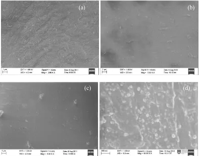
appears more homogenous blend, smooth and fine morphology than that of pristine PVDF. This grafted membrane become more homogenous blend, smooth and fine surface morphology when the PVDF-co-VIm membrane were protonated in sulfuric acid. According to Yahya et al. [23], samples with soft surface and having a low degree of roughness will be useful as an electrolyte since it has sufficient criteria as a good proton conductor as well as could provide a good contact with electrodes. This finding is in a good deal with XRD results that the peaks at 2 = 18o and 20o (see Fig. 7) become a single peak which shifted to 20.2o. The shifting of the peak is due to the surface change become smooth and fine morphology because of grafting and protonation of the membrane. However, it appears little formation of like particles arise from the grafted of VIm onto PVDF base film (Fig. 6 (b) and (c)). The radiation grafting mechanism of VIm may have caused formation of crystal-like particles on surface of PVDF film. In Fig. 6 (d), it can be clearly observed that the appearance of a crystal-like particles onto cross-section of the protonated PVDF-co-VIm at magnification 50.00 K through surface morpholgy study. It can therefore be inferred that the VIm has succesfully grafted deeply onto PVDF film.
Figure 6. Surface morphology of (a) pure PVDF (before grafting), (b) PVDF-co-VIm (after grafting) (c) protonated PVDF-co-VIm (after grafting) and (d) cross-section protonated PVDF-co-VIm
3.7. XRD Studies
The XRD pattern of pristine PVDF, PVDF-co-VIm, and protonated PVDF-co-VIm membranes are shown in Fig. 7 (a), (b) and (c). Wide peaks around 2 = 18o, and 20o reflect semicrystalline nature
(a) (b)
of the PVDF [24]. The peaks are in correlated with and phase due to crystal polymorphism in PVDF [25]. As shown in Fig. 7 (a), there are two peaks (2 = 18o and 20o) in the XRD spectrum of pritine PVDF is observed. These peaks denoted as a 1 peak and peak respectively. However, in
PVDF-co-VIm system (see Fig. 7 (b)) all peaks decrease in intensity and the grafting of these two polymers is observed to reduce the crystallinity of pristine PVDF. As VIm is grafted, the 1 peak
diminished, new 2 peak existed and the peak remained for PVDF-co-VIm. This indicates that a
significant crystalline transformation occurred during radiation-induced grafting of the VIm monomer onto PVDF backbone. Surprisingly, the 2 peak diminished, 1 peak try to develop but not very
obvious peak and peak remained existed when subsequently protonated of PVDF-co-VIM. It seems that the phase and phase crystal try to reorganize at low pH when introduced sulfuric acid group which it interacts with imidazole group to form proton conductor in grafting membrane.
Figure 7. XRD studies of (a) pristine PVDF, (b) PVDF-co-VIm, and (c) Protonated PVDF-co-VIm
[image:12.596.211.385.281.517.2]
because the strong interaction between proton (acid sulfuric) and imidazole ligand group. Therefore, this finding can be said that in compliment with the conductivity and SEM study.
Table 2. The coherent length of pure PVDF, PVDF-co-VIm and protonated PVDF-co-VIM (PVDF-co-VIm(H+)) membranes at 2 = 20o
Membranes Coherent length, L (Å), at 2 = 20o
Pristine PVDF 27.0
PVDF-co-VIm 24.8
PVDF-co-VIm(H+) 24.6
3.8. Thermal Analysis
The glass transition (Tg) for neat PVDF is -30 to – 20oC and melting temperatures (Tm) is
around 160-170oC [27], whereas the Tg neat PVIm is observed at 147.7oC [28] or 163oC [29] or 175.1oC [30]. The crystallization temperature (Tc) and Tm of the grafting PVDF with 1-Vinylimidazole are shown in Fig. 8 and summarized in Table 3. The degree of crystallinity of PVDF-co-VIm membrane is also evaluated. Crystallinity Xc is defined as the ratio of enthalpies,
ref c
H H X
(6)
Quantity H was determined after thermal procedure and quantity Href is the melting enthalpy
of 100% crystalline PVDF. It was determined to Href = 104.7 Jg-1 and Href = 349 Jg-1 for the original crystalline PVDF and PVIm respectively [29, 30].
[image:13.596.138.454.524.707.2]
Fig. 8 (a) shows the result obtained using DSC. These results clearly indicate that the pure PVDF reveals a single peak and another second peak after grafting with VIm. The first peak (Peak 1) and the second peak (Peak 2) is due to the melting peak of pure PVDF and PVIm after grafting onto PVDF film respectively.
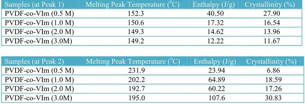
[image:14.596.42.534.538.706.2]The Peak 1 and Peak 2 were evaluated using OriginPro 8 software as shown in Table 3. It shows that the grafted membrane is immiscible. The single peak is observed for pristine PVDF before irradiation at 152oC. The peak remains existing after irradiation however the Tm is shifted to lower temperatures than for pure PVDF. In addition, the degree of crystallinity (Xc) is reduced when the concentration of VIm increased. However, the melting peak of grafted VIm onto PVDF backbone is observed at around 192 to 231oC (Peak 2). The melting reduced from 0.5 to 2.0 M of VIm and bit increasing at 3.0 M of VIm. Simultaneously the Xc increased from up to 1.0 M of VIm, and slightly drop at 2.0 M and a considerable increasing at 3.0 M of VIm. It may reasonable to assume that at 3.0 M of VIm are numerous amounts grafted onto PVDF. Further increase in the monomer concentration leads to a slight decrease in the amount of graft. This may be attributed the increasing in viscosity of the reaction mixture which obstruct the diffusion of monomer towards the polymer matrix and reduce in the grafting yield percentage. Therefore, the reasonably amount required is 2.0 M of VIm in order to graft onto PVDF backbone. This finding suggests that radiation-induced can enhance crystallinity in the side graft chain (hydrophilic part) or otherwise radiation-induced can also reduce the crystallinity in the polymer backbone (hydrophobic part). The increasing crystalline part in at the side chain might be in correlated finding with Masaki et al. [32] due to an unusual behavior during polymerization of poly(N-vinylimidazole) preparation. In this case, Masaki has reported that a large particle results in the precipitation polymerization which is the formation of microgel-like particles as a result polymerization yields by dynamic light scattering (DLS) technique.
Table 3. DSC study for different concentration VIm co-grafting onto PVDF membrane at Peak 1 and Peak 2 respectively
Samples (at Peak 1) Melting Peak Temperature (oC) Enthalpy (J/g) Crystallinity (%)
PVDF-co-VIm (0.5 M) 152.3 40.50 27.90
PVDF-co-VIm (1.0 M) 150.6 17.32 16.54
PVDF-co-VIm (2.0 M) 149.3 14.62 13.96
PVDF-co-VIm (3.0M) 149.2 12.22 11.67
Samples (at Peak 2) Melting Peak Temperature (oC) Enthalpy (J/g) Crystallinity (%)
PVDF-co-VIm (0.5 M) 231.9 23.94 6.86
PVDF-co-VIm (1.0 M) 202.2 64.89 18.59
PVDF-co-VIm (2.0 M) 192.7 60.22 17.26
PVDF-co-VIm (3.0M) 195.0 107.6 30.83
was reported as a phase transition [12, 17], or as an endothermic peak [31] in this temperature region. However, it has not been much dicussed why this phenomena occured in this temperature. In this study we inferred that this phenomena as a phase transition of crystallization in PVDF which crystalline structure try to reorganize and rearrange in this temperature region. There is a strong possibility that the phase crystal transition due to the deformationof -phase result in transformation to -phase in PVDF-co-VIm grafting membrane. The crystallization transition values are summarized in Table 4. The crystallinity transition that occurs in this temperature triggered a sudden increases in the ionic conductivity. It can be clearly observed that the grafting of 1-vinylimidazole onto the PVDF film caused a decrease in crystallization temperature (Tc) as well as showed a slight decrease in the Tm and
Xc onto pristine PVDF. The decrease of Tc caused increasing the flexibility of the polymer chains, thus helps in fast ion conduction. This information can contribute more understanding why in the conductivity study occurs a suddenly increase at Tcregion. The incorporation of 1-vinylimidazole side chain grafts increased the crystallization fraction and restricts the mobility of the molecular chains of the PVDF domain. Thus, the conductivity of the protonated PVDF-co-VIm at 3.0 M of VIm drops due to the crystallization fraction increased. The changes in Tc play major role whether crystallinity
increasing or decreasing in polymer membrane that caused by the flexibility of the polymer chains. Thus, it could be determined in fast or drop in ion conduction. Moreover, it could be speculated that if there is change of crystallization phase, therefore it is related to the onset of a sudden change in conductivity. This information can contribute more understanding that the change in crystallinity is in related with the conductivity studies.
Table 4. DSC study of crystallization phase transition temperature
Samples Crystallization phase temperature (oC)
PVDF-co-VIm (0.5 M) 59.0
PVDF-co-VIm (1.0 M) 42.5
PVDF-co-VIm (2.0 M) 41.6
PVDF-co-VIm (3.0M) 42.2
3.9. Transference Number
found to be 0.96 (1.0 M) and 0.91 (2.0 M) respectively. This suggests that the conductivity of the membranes is predominantly due to the ion.
[image:16.596.212.376.120.317.2]
Figure 9. The polarization current as function of time protonated (a) PVDF-co-VIm (2.0 M) and (b) PVDF-co-VIm (1.0 M) respectively at room temperature
Details of the entire polarization current with different concentrations of VIm that grafted onto PVDF surface membrane is shown in Table 5. The protonated PVDF-co-VIm or PVDF-co-PVIm-H+ are closed unity except for the sample of 0.5 M VIm. The degree of grafting in this concentration normally is less than 5%. Thus, it can be inferred that to provide a good proton conductor membrane, the preparation of VIm concentration before being through -rays irradiation should be more than 0.5 M.
Table 5. Transference number of protonated PVDF-co-VIm membranes with different concentrations of VIm
Concentration Total current (IT), A Residue current (Ir), A Ionic current (Ii), A
0.5 M 6.56 x 10⁻¹⁰ 1.05 x 10⁻¹⁰ 0.60
1.0 M 5.04 x 10⁻⁵ 1.52 x 10⁻⁶ 0.96
2. 0 M 2.68 x 10⁻⁴ 2.32 x 10⁻⁵ 0.91
3.0 M 4.99 x 10⁻⁶ 4.96 x10⁻⁶ 0.99
4. CONCLUSION
[image:16.596.40.555.531.662.2]
homopolymer. Gravimetric analysis and FTIR study confirmed grafting VIm onto the PVDF. The glass transition temperature, melting temperature and crystallinity slightly decrease with the increase of the VIm concentration. Two values of Tm reveal that the PVDF and the VIm copolymers are
immiscible. Reorganization of phase and phase are in correlated with the crystal polymorphism occurred in pristine PVDF, grafted PVDF before and after irradiation and subsequent protonation PVDF-co-VIm. The proton conductivity behaviour is in good agreement with DSC study. The results prove that the phase transition in the PVDF-co-VIm membranes are significantly affected by crystallization of the PVDF and PVIm components. Radiation-induced grafting can enrich the crystallinity in the side graft chain and thus it can also decreases the crystallinity in the polymer backbone (hydrophobic part). In addition, the XRD and SEM morphology studies of the PVDF-co-VIm are complimented. The ionic transference number of the protonated PVDF-co-PVDF-co-VIm membrane reveals that the conductivity of the membranes is preferably due to the ionic conduction. Based on the results presented, it can be said that the PVDF-co-VIm membrane could be a promising candidate as a proton exchange membrane for fuel cells application.
ACKNOWLEDGEMENT
Authors gratefully acknowledge to the Ministry of Higher Education Malaysia and UiTM for the grants (FRGS RMI/ST/FRGS5/3/Fst(33/2009)] and Research Excellence Fund [600-RMI/ST/DANA 5/3/Dst (299/2009)]) respectively. A Lepit thanks to the MOHE and UiTM for the scholarship awarded.
References
1. S. Hasegawa, Y. Suzuki, Y. Maekawa, Radiat. Phys. & Chem. 77 (2008) 617 – 621. 2. R. Rohani, M. M. Nasef, H. Saidi, K. Z. M. Dahlan, Chem. Engine. J. 132 (2007) 27-35. 3. M. M. Nasef, H. Saidi, Mater. Chem. & Phys. 99 (2006) 361-369.
4. L. Gubler, N. Prost, S. A. Gursel, G. G. Scherer, Solid State Ionics 176 (2005) 2849-2860. 5. S. M. Javaid Zaidi, Materials Science Forum 657 (2010) 88-115.
6. T. Itoh, Y. Hamaguchi, T. Uno, M. Kubo, Y. Aihara, A. Sonai, Solid State Ionics 177 (2006) 185-189.
7. A. A. Yousefi, Iranian Polymer J., 20 (2) (2011) 109-121.
8. T. Yuan, J .Q. Meng, G. R. Cai, Y. F. Zhang, Advanced Materials Research, 418 (2012) 639-642. 9. A. Kumar, M. Deka, S. Banerjee, Solid State Ionics 181 (2010) 609-615.
10. M. Anderson, O. Hansson, L. Ohrstrom, A. Isdtrom, M. Nyden, Colloid Polym Sci. 289 (2011) 1361-1372.
11. H. F. Naguib, R.O. Aly, M. W. Sabaa, S. M. Mokhtar, Polymer Testing 22 (2003) 825-830. 12. S. Selvasekarapandian, R. Baskaran, M. Hema, Physica B 357 (2005) 412-419.
13. L. Xu, J. Sun, L. Zhao, Radiat. Phys. Chem. (2011).
14. W. D. Lilac, S. Lee, Korean J. Chem. Eng, 16 (3) (1999) 275-284.
15. Z. Ajji, A. M. Ali, Nucl. Inst. and Meth. in Phys. Res. B, 236 (2005) 580-586. 16. Y. Liu, J. Y. Lee, L. Hong, Solid State Ionics 150 (2002) 317-326.
17. L. F. Malmonge, J. A. Malmonge, W. K. Sakamoto, Materials Research, 6 (4) (2003) 469-473. 18. S .A. Hashimi, A. Kumar, K. K. Maurya and S. Chandra, J. Phys. D: Appl. Phys. 23 (1990)
19. A.S. Kamisan, T.I.T. Kudin, A.M.M. Ali, M.Z.A. Yahya, Electrochimica Acta, 57 (2011) 207 – 211.
20. S. Turmanova, M. Minchev, K. Vassilev, G. Danev, J. Polymer Research, 2008. 15(4): 309-318 21. N. Pekel, N. Sahiner, and O. Güven, Radiat. Phys Chem., 2000. 59(5-6): 485-491.
22. A. Bozkurt, W. H. Meyer, J. Gutman, G. Wagner, Solid State Ionic 164 (2003) 169-176.
23. M. Z. A. Yahya, A.M.M. Ali, M.F. Mohammat, M.A.K.M. Hanafiah, M. Mustaffa, S.C. Ibrahim, Z.M. Darus, M.K. Harun, Journal of Applied Sciences, 6 (2006) 1287 – 1291.
24. G. G. Kumar, P. Kim, A. R. Kim, K. S. Nahm, R. N. Elizabeth, Mate. Chem. Phys. 115 (2009) 40-46.
25. V. Sencadas, S. L. Mendez, J. F. Mano, Therchimica Acta 424 (2004) 201-207.
26. C. Beyler, M. Hirschler, In SFPE Handbook of Fire Protection Engineering, 3rd Ed. P. DiNenno Ed. Quincy, MA: NFPA (2002) 110-131.
27. S. Goswami, A. Dutta, Ionics (2011) in press.
28. S. G. Focil, R. C. Woundenberg, O. Yavuzcetin, M. T. Tuominen, E. B. Coughlin, Macromolecules 40 (2007) 8708 – 2841.
29. N. Pekel, N. Sahiner, O. Guven, Z. M. O. Rzev, Euro. Poly. J., 37 (2001) 2443-2451.
30. S. Rudhziah, N. Muda, S. Ibrahim, A. A. Rahman, N. S. Mohamed, Sains Malaysiana 40(7) (2011) 707-712.
31. A. Gasmi, M. Gouasmia, S. Etienne, Solid State Phenomena, 115 (2006) 151-156. 32. M. Masaki, K. Ogawa, E. Kokufuta, Colloid Polym Sci. 287 (2009) 1405-1415.