A DISSERTATION ON
“A PROSPECTIVE STUDY ON INCIDENCE OF
COMPLICATIONS FOLLOWING
MESH REPAIR VS DARNING
IN COMPLICATED INGUINAL HERNIAS”
Dissertation submitted to
THE TAMIL NADU Dr.M.G.R.MEDICAL UNIVERSITY
CHENNAI
with partial fulfilment of the regulations
for the Award of the degree
M.S.BRANCH–I (GENERAL SURGERY)
DEPARTMENT OF GENERAL SURGERY
THANJAVUR MEDICAL COLLEGE
CERTIFICATE BY THE GUIDE
This is to certify that the dissertation entitled
“A PROSPECTIVE STUDY ON INCIDENCE OF COMPLICATIONS FOLLOWING MESH REPAIR VS DARNING
IN COMPLICATED INGUINAL HERNIAS”
is a bonafide original work of
Dr. M . ARUN
in partial fulfilment of the requirements for
M.S.Branch–I
(General Surgery) Examination of the Tamil Nadu Dr. M.G.R. Medical
University to be held in MAY 2018 under my guidance and
supervision in 2016-2017
Prof Dr.W.EDWINA VASANTHA, M.S
CERTIFICATE
This is to certify that the dissertation entitled
“A PROSPECTIVE STUDY ON INCIDENCE OF COMPLICATIONS FOLLOWING MESH REPAIR VS DARNING
IN COMPLICATED INGUINAL HERNIAS”
is a bonafide original work of
Dr. M . ARUN
in partial fulfilment of the requirements for
M.S.Branch–I
(General Surgery) Examination of the Tamil Nadu Dr. M.G.R. Medical
University to be held in MAY 2018 under my guidance and
supervision in 2016-2017
Prof Dr. M. ELANGOVAN,. MS. FICS
HOD and Professor,
CERTIFICATE
This is to certify that the dissertation entitled
“A PROSPECTIVE STUDY ON INCIDENCE OF COMPLICATIONS FOLLOWING MESH REPAIR VS DARNING
IN COMPLICATED INGUINAL HERNIAS”
is a bonafide original work of
Dr. M . ARUN
in partial fulfilment of the requirements for
M.S.Branch–I
(General Surgery) Examination of the Tamil Nadu Dr. M.G.R. Medical
University to be held in MAY 2018 under my guidance and
supervision in 2016-2017
Dr. JEYAKUMAR ,M.S.,MCh.,
Dean,
DECLARATION
I
Dr. M. ARUN
hereby solemnly declare that the
dissertation titled
“A PROSPECTIVE STUDY ON INCIDENCE OF
COMPLICATIONS FOLLOWING MESH REPAIR VS DARNING
IN COMPLICATED INGUINAL HERNIAS”
is done by me at
Thanjavur medical College, Thanjavur during 2016-2017
under the guidance of Prof.Dr.W.EDWINA VASANTHA,M.S,.
This dissertation is submitted to The Tamil Nadu Dr.M.G.R
Medical University, Chennai towards the partial fulfillment of
requirements for the award of M.S.Degree (Branch-I) in
General Surgery.
Place: Thanjavur
Dr.M.ARUNACKNOWLEDGEMENT
I am grateful to the Dean
Dr.S.JEYAKUMAR,DNB.,M.S.,Mch,
for permitting me to conduct the study and make use of the
resources of the college.
I consider it a privilege to have done this study under the
supervision of my beloved professor and head of the department,
Prof Dr. M. ELANGOVAN, M.S
who has been a source of constant
inspiration and encouragement to accomplish this work.
I am highly indebted to my chief
Prof Dr.W.EDWINA
VASANTHA, M.S,
for her constant help, inspiration and valuable
advice in preparing this dissertation.
I express my deepest sense of thankfulness to my assistant
professors
DR.A.MUTHUVINAYAGAM ,M.S., DR.V.MARIMUTHU,
M.S.,
for their valuable inputs and constant encouragement,
without which this dissertation could not have been completed.
I express my sincere thanks to my fellow post graduates and
junior colleagues for their support and help in completing this
dissertation. It is my earnest duty to thank my family without
whom accomplishing this task would have been impossible.
TABLE OF CONTENTS
S.NO
INDEX
PAGE NUMBER
1
AIMS OF STUDY
7
2
HISTORICAL ASPECTS
9
3
INTRODUCTION
11
4
REVIEW OF LITERATURE
12
5
MATERIALS AND METHODS
54
6
OBSERVATION AND RESULTS
56
7
STATISTICS
66
8
CONCLUSION
80
AIMS OF THE STUDY
To study about various Acute surgical emergencies in groin
hernia
To study duration of hernia before acute presentation
To study the various symptoms of presentation and
complications
To study content of hernial sac
To study the type of surgery done, Litchenstein tension free
mesh hernioplasty VS Two layered darning repair.
HISTORICAL ASPECTS
"Hernia" is derived from Latin term ‘a rupture’. Original Edoardo Bassini (Padua 1889) repair is an anterior approach but he opened posterior wall of inguinal canal and did a sutured three layered repair. In modified Bassini repair transverse arch is sutured to the inguinal ligament below .
De Chauliac -differentiated inguinal from femoral hernias (1363). Kaspar Stromayr- differentiated direct and indirect hernias in 1559. Ambroise Pare (1550) – used trusses in hernia.
“John Hunter, Astley Paston Cooper (1841) Franz Hesselbach, Antonio Scarpa (1832), Pieter Camper, Jules Germain Cloquet (1883), Bogros, Retzius” - understanding anatomy of the groin. “La Roque, GL Cheatle, JP Hoguet, AK Henry, McVay, McEvedy, Lotheissen, McArthur” used strips of external oblique aponeurosis to approximate conjoined tendon to inguinal ligament Kirschner - thigh fascial grafts, Gallie - strips of fascia lata for repair using his specialized needle called Gallie’s needle, Handley - silk with stay lace darning, Meick – first multifilament Nylon, Haxton - monofilament Nylon, Moloney -Nylon darn procedure( 1948) .
EE Shouldice (1945) of Toronto did repair of transversalis fascia
under local anaesthesia as a tension free 4/6 layered tissue
repair.
Usher from Texas in 1958 first used Marlex mesh in posterior
wall for repair as tension eliminating.
Lichenstein - did rolled plug mesh repair of femoral hernia in
1970 and tension free repair of inguinal hernia in 1986.
Newman (New Jersey) did tension free inguinal hernia onlay
mesh repair.
Lichenstein and Amid made this technique popular worldwide
with certain modifications.
Nyhus from Chicago and Condon from Milwaukee popularized
open posterior repair from higher approach. Their technique
probably became basis for TEP/TAPP.
Stoppa and Rives (France) evoluted giant reinforcement of the
visceral sac by blocking Fruchaud’s myopectineal orifice
(MPO)bilaterally.
INTRODUCTION
Inguinal hernia is the most commonly performed surgery in
general surgery. Overall risk of developing hernia is 15% in men
and < 5% in women. Hernia repair has evolved and turned into a
day care procedure nowadays. 75% of all abdominal wall herniae
are groin herniae making it the most common hernia among all
abdominal wall herniae. About 95% of all groin herniae are
inguinal herniae and about 5% are femoral herniae. Inguinal
herniae are about 9 times more common in males than in female.
Femoral herniae are more common in women, but inguinal
herniae are the most common herniae women present with.
It is estimated that 40% cases of femoral herniae present
acutely in emergency department with strangulation /
incarceration. The prevalence of hernia is more in the
extremes of age, which is after an initial peak during infancy
they become more prevalent with advancing age.The
procedures advocated for hernia repair are simple and
decrease morbidity and mortality in a major way.
Incarcerated herniae should be operated in less than 12 hrs
of presentation and strangulated hernia on an emergency
basis to prevent prolonged morbidity and mortality. The
need for study arises because though the treatment of groin
herniae is simple and can be done as a day care procedure
ignorance on the part of the patient might lead to
complications like irreducibility, incarceration, obstruction
and strangulation resulting in prolonged morbidity and
might result in mortality too, which can be prevented if the
presenting patients with hernia undergo treatment on an
elective basis. In spite of various developments in the
treatment modalities of
hernia, the morbidity and mortality
has not reduced significantly.
REVIEW OF LITERATURE
Hernia is derived from the Latin word ‘ rupture’. Hernia is an abnormal protrusion of an organ or tissue through a defect in its
surrounding walls. Hernial defects commonly involves abdominal wall, especially the Inguinal ligament . In areas where aponeurosis and fascia are not covered by muscles , herniation occurs. “The sites most commonly involved being the inguinal, femoral, and umbilical areas, linea alba, lower portion of the semilunar line, and sites of previous surgical scars”.
Groin hernias:
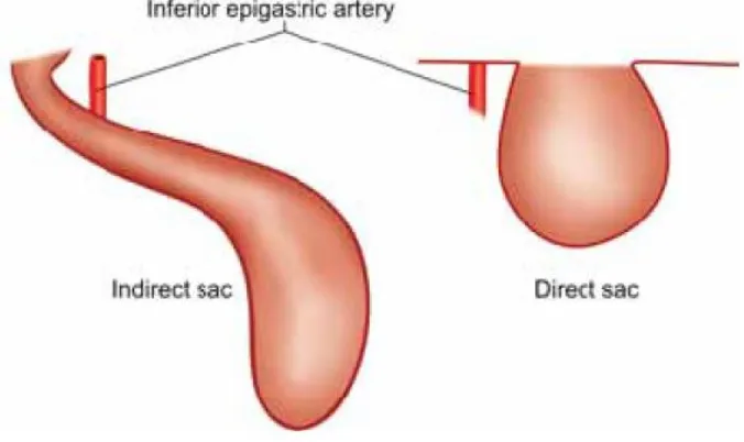
Inguinal hernias are of two types, direct hernia and indirect hernia. Indirect inguinal hernia sac passes from deep inguinal ring obliquely towards superficial inguinal ring and atlast into the scrotum . Direct
inguinal hernia sac bulges outward and forward and deep inguinal ring and inferior epigastric vessels lies lateral to it . Surgical management of indirect and direct inguinal hernia being the same , they need not to be
differentiated from one another. Combined direct and indirect hernia is called pantaloon hernia .
Incidence :
predominate over direct hernias at a ratio of 2 : 1. Direct hernias
are not common in women. Although femoral hernias occur more
frequently in women than in men, inguinal hernias remain the
most common hernia in women. In men femoral hernias are
uncommon. Right sided hernias are more common. This can be
explained by delay in atrophy of the processus vaginalis after the
normal slower descent of the right testis to the scrotum during
fetal development. As the loaded sigmoid colon produces a ‘
tamponading effect on femoral canal’ femoral hernia is also more
common on right side
. Hernia’s incidence, complications like
strangulation hence bowel gangrene increases with age,
Strangulation, the most common serious complication of a
hernia, occurs in only 1% to 3% of groin hernias and is more
common at the extremes of life. Mostly indirect inguinal
hernias goes for strangulation. Also strangulation is most
common in femoral hernia due to narrow neck(15% to 20%),
hence once diagnosed, femoral hernia has to be repaired
immedietly.
Etiology
:
1.
Patent processus vaginalis
2. Failure of the transversalis fascia to retain the visceral sac
in the myopectineal orifice.
The myopectineal orifice of Fruchaud is an area of transversalis fascia that is not protected by the posterior rectus sheath or by muscle. This serves as an area of potential weakness through which groin hernias emerge. Some degree of protection is offered by shutter mechanism, where due to increased intraabdominal pressure the curved fibers of internal oblique and the falx inguinalis flatten and move toward the inguinal ligament. Contraction of transverses abdominis muscle also pulls up and tenses the crura of the internal ring.
3. Mechanical disparity between visceral pressure and strength of abdominal musculature: The increased abdominal pressure may be due to excess cough, constipation, prostatism, and lifting heavy weights. Hernia results when a patent processus vaginalis is present or the endo abdominal fascia is attenuated in these situations . Sudden onset of painful hernia in young individuals after lifting heavy weight is most commonly associated with a patent processus vaginalis . In elderly individuals, as abdominal muscles and fascia weakens with ageing process, inguinal hernias occur equally in sedentary and in physically active men.
Vigorous physical activity as such is not a cause of inguinal hernia formation, abut strenuous strain may aggravate predisposing factors and cause herniation.
The fascia transversalis, derives strength from collagen fibers which gets continuously produced and reabsorbed. Disturbance of this balance leads to attenuation of this fascia.
5. Life-styles factors like smoking : It leads to defective collagen production , which reduces tensile strength. Studies have proven the association between between cigarette smoking and groin hernias. Significantly increased elastolytic activity as well as protease substances in smokers ( thus disturbed protease/
antiprotease balance ) causes destruction of tissue leading to herniation .
6.Abdominal distention and chronic increase in intra abdominal pressure from ascites and peritoneal dialysis can damage the myopectineal orifice and cause a patent processus vaginalis to dilate
7. Genetic factors
A study of congenital indirect inguinal hernias in China on 280 families indicated that the mode of transmission was autosomal dominant, with incomplete penetrance and paternal transmission from their father.
8. Obesity 9 . Pregnancy
10. Chronic constipation
11. Appendicectomy causing injury to ilioinguinal nerve –right sided direct inguinal hernia.
Parts Of Hernia:
Hernia consists of three parts: 1. Sac
Sac is a diverticulum of peritoneum with mouth, neck, body and fundus. strangulation of bowel is a likely complication when neck is narrow. Coverings are derived from layers of the abdominal wall through which the sac passes. The content may be omentum (omentocele)or intestine ( enterocele). Most common content being small bowel, but it can also be large intestine ,appendix or bladder
• When content is a portion of circumference of the intestine it is called Richter’s hernia
• When the bladder or bowel forms part of wall of hernia it is called sliding hernia
• Ovary with or without the corresponding fallopian tube. • Meckel’s diverticulum as content is known as Littre’s hernia;
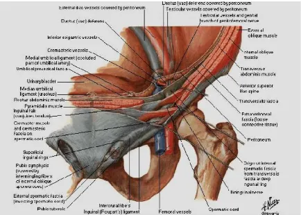
Surgical Anatomy of Inguinal Canal:
Inguinal canal:
In infants ,there is no obliquity of the inguinal canal and the external and deep ring are superimposed. In adults it is 3.75 cm long, directed downwards and medially from the deep to superficial ring. In males spermatic cord passes through the inguinal canal, ilio-inguinal nerve (outside the cord within the inguinal canal) and genital branch of the genito-femoral nerve (within the cord). Cord is covered by cremaster muscle which is a continuation of internal oblique muscle, internal spermatic fascia which is a continuation of transversalis fascia.
Fig 2: Boundaries of Inguinal Canal
Anteriorly it is bounded by External oblique aponeurosis and conjoined muscle laterally.
Posteriorly it is bounded by Inferior epigastric artery, fascia transversalis and conjoined tendon medially.
Superiorly by arched fibres of internal oblique.
Inferiorly by Inguinal ligament and lacunar ligament .
Internal ring is a normal opening in the transversalis fascia and transverses aponeurosis which is U shaped or lambda shaped; it is 1.25 cm above the midinguinal point (between anterior superior iliaciliac spine to pubic symphysis).
[image:23.595.126.460.165.407.2]Fig 3: Showing boundaries of the inguinal canal
An indirect inguinal hernia (hernia though deep ring) travels down the canal on the lateral and anterior aspect of the spermatic cord; whereas the direct hernia pops out directly forward through the posterior wall of the inguinal canal. Neck of the indirect sac is lateral to inferior epigastric
vessels but direct sac emerges medial to the inferior epigastric vessels with a wide neck. Saddle-bag or pantaloon hernia sac has got both medial and lateral component. Inguinal hernia occurs
above and medial to the pubic tubercle. Femoral hernia is below and lateral to pubic tubercle .
Fig 4: Anatomical relations and locations of the inguinal, femoral and obturator hernias.
Inguinal Defense Mechanisms:
Obliquity of inguinal canal creates flap valve mechanism by
approximating anterior and posterior walls of the canal. Cooper described external oblique aponeurosis as outer barrier of the inguinal canal causing step down effect for the spermatic cord. Shutter mechanism of internal oblique ie., roof apposes to floor like a shutter. External ring is guarded by conjoined tendon behind and reflected part of inguinal ligament in front. Internal ring is guarded by fleshy internal oblique. Contraction of external oblique aponeurosis causes pinch cock/slit valve effect by approximation of two crura of external ring. Cremaster muscle on contraction helps cord to plug the external ring—ball valve mechanism.Hormones also play a role to maintain inguinal muscles. Inguinal ligament is formed by thickened posteriorly folded lower border of the external oblique aponeurosis
attached to pubic tubercle; base directing laterally forms medial wall of femoral ring. Cooper’s pectineal ligament is thickened pectineal fascia which is attached to the pectin pubis lateral to the base of the lacunar ligament. Aponeurotic fibers from the inferior crus of the external ring passing superomedially to linea alba is called as Colles’ reflected part of the inguinal ligament.
Cremaster muscle is muscle fascicule embedded in cremaster fascia (Scarpa’s sheath) which are attached to inguinal ligament laterally, internal oblique, transverse abdominis and conjoint tendon, pubic tubercle and pubic crest medially. It is seen properly only in males; sparse in females. It is supplied by genital branch of genitofemoral nerve (L1). It suspends the testis in elevated position. It closes during raise in intraabdominal
pressure; it protects nerves of the inguinal canal. During hernioplasty if cremaster is excised, testis may hang like clapper bell due to loss of suspensory and retractile function of the cremaster. So if cremaster is excised, medial cut end should be sutured to inguinal ligament or pubic tubercle. Stroking medial aspect upper thigh will cause reflex contraction of cremaster with elevation and retraction of testis—
Nerves of inguinal region:
Iliohypogastric nerve arises from the anterior division of L1 nerve runs away from lateral margin of psoas major muscle in frontof quadrates lumborum behind kidney and colon to pierce transversus abdominisjust above the iliac crest; runs between transversus abdominis and internal oblique; pierces the internal oblique 2.5 cm above the anterior superior iliac spine, run between internal oblique and external oblique to reach 2.5 cm above the superficial inguinal ring where it pierces the external oblique aponeurosis to supply skin around the area.
Ilioinguinal nerve also arises from anterior division of L1 nerve runs over the quadratus lumborum, iliac crest pierces the transversus abdominis 3.5 cm medial to anterior superior iliac spine; runs between transverses abdominis and internal oblique for a short distance to pierce internal oblique just below and medial to iliohypogastric nerve lateral to deep inguinal ring; runs in inguinal canal outside the cord in males or round ligament (in females), passes through superficial inguinal ring; runs in front of the scrotum to supply anterior part of the scrotum. The genitofemoral nerve arises from ventral divisions of L1, L2 nerves; passes through the medial part of the psoas major muscle running below in front of themuscle; just above the deep ring it divides into medial genital and lateral femoral branches. Genital branch after piercing the psoas sheath runs in front of the external iliac artery and passes through deep ring to enter the
inguinal canal within the spermatic cord in males or round ligament in females. It supplies cremaster muscle, runs in the cord to reach anterolateral aspect of the scrotum and supplies that part. In females it gives sensory branches to
Fig 5: Nerves in inguinal region
Fascia Transversalis (Cooper’s Fascia
):
fascia and in front of the peritoneum. Inlay preperitoneal mesh repair is done by placing mesh in this space. Thickening of the transversalis fascia at medial side of the internal ring and
in front of the inferior epigastric vessels is called as interfoveolar Hesselbach’s ligament.
Endoabdominal Fascia:
Endoabdominal fascia is connective tissue that lines inner aspect of the entire abdominal musculature. Part in relation to transverse abdominis is called as fascia transversalis. It forms iliac, diaphragmatic and renal fascia.
Inferior epigastric artery (IEA)
Originates from external iliac artery just above the inguinal ligament, runs upwards and medially in the extraperitoneal connective tissue
between peritoneum and posterior lamina of transversalis fascia initially medial to internal ring; pierces the transversalis fascia at the lateral border of the rectus muscle; runs in front of the arcuate line supplying the rectus. Iliopubic tract is confused with inguinal ligament. It is an aponeurotic band extending from iliopectineal arch (thickened medial part of the iliopsoas fascia) and anterior superior iliac spine to superior ramus of the pubis forming deep musculoaponeurotic layer with transverses abdominis
muscle, aponeurosis and transversalis fascia; it runs medially along inferior margin of the deep ring, anterior margin of the external iliac vessels,
.
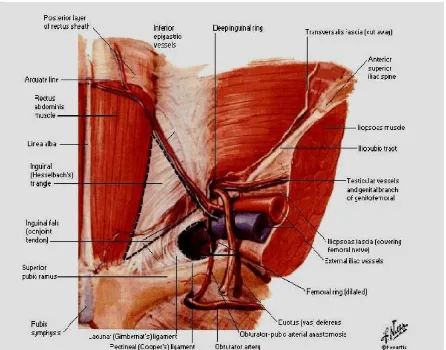
Fig 6: Ligaments related to groin and hernia surgery.
Arteries related to inguinal region :
It is divided into superficial and deep vessels. Superficial
vessels are superficial epigastric, superficial circumflex iliac and superficial external pudendal arteries—are branches of femoral artery. They travel towards umbilicus in subcutaneous plane. Superficial epigastric artery anastomosing with superior epigastric artery. Branches are—muscular, cremasteric, pubic, and cutaneous. Cremasteric branch after travelling laterally towards deep ring supplies cremaster and runs along the spermatic cord. Pubic branch runs medially along
the pubic bone to communicate with pubic branch of anterior division of obturator artery. Often obturator artery may be replaced by this pubic branch of inferior epigastric artery as abnormal/aberrant obturator artery.
Deep circumflex iliac artery arises from lateral side of the external iliac artery behind the inguinal ligament which after piercing the transversalis fascia runs along the iliac crest; at its middle it again pierces the transversus abdominis and internal oblique to reach adjacent to anterior superior iliac spine where it anastomoses with superior gluteal, superficial circumflex iliac, lateral
circumflex femoral arteries. Here it also gives ascending branch which runs in abdominal wall which may be injured in TEP causing large haematoma.
Bendavid circle:
It is the circle where venous network is situated in space of Bogros formed by branches of inferior epigastric, iliopubic, suprapubic, and retropubic veins. They
communicate with aberrant obturator vein which is commonly existing (70%).
During laparoscopy (TEP/TAPP) due to high insufflation pressure veins will collapse and will not bleed during dissection but bleeds later in postoperative
period.
Triangle of doom:
It is observed in laparoscopic hernia procedure. It is a triangle formed medially
by vas deferens, laterally by testicular vessels and peritoneal reflection below with
the apex at internal ring. External iliac vessels lie in this triangle and so extreme care should be taken while dissecting the hernia sac in this triangle.
Preperitoneal Space of Nyhus:
It is the space between transversalis fascia and peritoneum.Medially, it is in front of the urinary bladder and behind pubic bone as retropubic space of Retzius (1858).
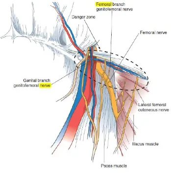
(contains femoral canal, iliac vessels (vein medially and artery laterally) iliopsoas muscle, genitofemoral nerve, lateral femoral cutaneous nerve). External iliac vessels lie in a triangle formed by gonadal vessels laterally, vas deferens medially and peritoneal reflection inferiorly (triangle of doom). Aberrant obturator artery which is an occasional branch of inferior epigastric artery replacing its pubic branch travels across Cooper’s ligament, which during fixation of mesh can cause torrential haemorrhage - circle of death. Triangle of pain is formed by gonadal vessels medially, iliopubic tract laterally and
peritoneal reflection below.
Genitofemoral nerve and lateral cutaneous nerve of thigh traverse this triangle. Injury to these nerves either by dissection causes severepost operative pain. Tacks/staplers should not be placed in this triangle.
Fruchaud’s Myopectineal Orifice (MPO):
It is an osseo-myo-aponeurotic tunnel and through which all groin hernias occur.Medially it is bounded by lateral border of rectus sheath, above by the arched
fibres of internal oblique and transverse abdominis muscle; laterally by the
[image:32.595.87.420.470.679.2]iliopsoas muscle; below by the pectin pubis and fascia covering it.
Fossae :
There are three fossae on the inner surface of the anterior abdominal wall
above the ligament and below the umbilicus. Lateral fossa is lateral to lateral umbilical ligament (which contains inferior epigastric artery) which is the site of
indirect inguinal hernia. Lateral fossa contains deep inguinal ring. Medial fossa is
between lateral and medial umbilical ligament (obliterated umbilical artery). Here direct hernia develops. Supravesical fossa is between medial and median (obliterated
urachus) umbilical ligament which is the site of external supravesical hernia. Internal supravesical hernia develops in relation to urinary bladder directly, as anterior prevesical, posterior retrovesical and lateral paravesical.
CLASSIFICATION OF INGUINAL HERNIA:
Stoppa’s Classification:Type I – Indirect hernia with a normal internal ring measuring <2cm. Inguinal floor is solid.
Type II – Indirect hernia with internal ring >2cm. Inguinal floor solid.
Type III – Indirect hernia + direct hernia + femoral hernia with a weak inguinal floor
Type IV – Recurrent hernia
Nyhus Classification:
It is based on size of the internal ring and posterior wall weakness. Type I: Indirect hernia with normal deep ring.
Type II: Indirect hernia with dilated/widened deep ring. Type III: Posterior wall defect.
a. Direct.
c. Femoral hernia.
Type IV: Recurrent hernia
Gilbert Classification:
Type I: Hernia has got snug internal ring through which a peritoneal sac passes out as indirect sac.
Type II: Hernia has a moderately enlarged internal ring which admits one finger but is lesser than two finger breadth. Once reduced it protrude during coughing or straining.
Type III : Hernia has got large internal ring with defect more than two fingerbreadth. Hernia descends into the scrotum or with sliding hernia. Once reduced it immediately protrudes out without any straining.
Type IV: It is direct hernia with large full blow out of the posterior wall of the inguinal canal. The internal ring is intact.
Type V: It is a direct hernia protruding out through punched out hole/defect in the transversalis fascia. The internal ring is intact.
Type VI: Pantaloon/double hernia. Type VII: Femoral hernia.
TYPE VI and VII are Robbin’s modifications
Bendavid Classification:
Type I: Antero-lateral defect (indirect). Type II: Antero medial. (direct).
Type III: Postero-medial (femoral). Type IV: Posterior-prevascular hernia.
European Classification:
L – lateral ; M – medial; F – femoral P – Primary ; R – Recurrent
Classification Of Femoral Hernia
A) classical type : hernia occurs medial to femoral vein B) special type:
1) prevascular hernia of Narath : sac lies in front of femoral artery 2) External femoral hernia (Hesselbach’s hernia): sac lies lateral to Femoral artery.
3) Laugier’s femoral hernia (lacunar hernia): sac passes through Lacunar ligament
4) Sarafini hernia: sac descends behind femoral vessels
5) Deep femoral hernia (pectineal, cloquet’s hernia): sac passes deep to femoral vessels deep to deep fascia, cannot protrude through saphenous opening.
Types Of Indirect (Oblique) Inguinal Hernia:
This is the most common type of hernia. It is more common in younger age group as compared to direct inguinal hernia which is more common in elderly. It is more common on right side in 1st decade but in 2nd decade the incidence is equal on both sides. Hernia is bilateral in 30% of cases. Sac is thin in indirect type. Neck is narrow and, lies lateral to inferior epigastric vessels
Types:
(1)Bubonocele, where the hernia is limited to inguinal canal;
(2) Funicular where processus vaginalis is closed just above the epididymis. Contents of the sac can be felt separately from testis, which lies below the hernia.
It can occur in any age group. It occurs in a congenital preformed sac (processus vaginalis). More commonly contents descend into the pre-existing sac only when there are precipitating causes which force the content down
Presentation of Inguinal hernias
:
Presentation may be from asymptomatic hernia (which is seen in about 30% of patients) to a painful lump (commonest presentation - 66% of patients), this pain is mild in about 53.9% and severe in less than 1% of patient . focal pain in hernia is unusual ,in such case we should suspect hernia incarceration or strangulation. The most common symptom is a dull feeling of discomfort or heaviness in the groin region, which gets aggravated by activies causing increased intra abdominal pressure. Pain develops as a tight ring of fascia outlining the hernia defect compresses intra-abdominal structures with a
visceral neuronal supply. pain gets worse at the end of the day after prolonged standing
Irreducible hernia:
The contents cannot be reduced back into the abdomen, but
there are no other complications. It occurs because of adhesions between the sac and its contents or from overcrowding within the sac. Irreducibility without other symptoms is almost diagnostic of an omentocele, and any degree of irreducibility can lead to strangulation.
Obstructed hernia:
An irreducible hernia containing intestine which is
strangulation should be anticipated and treated accordingly.incarcerated omental fat can also produce significant pain and tenderness on physical examination.
Incarcerated hernia:
The term “incarceration” can be used to indicate
obstruction or strangulation, but it actually is the lumen of the bowel occupying a hernial sac is loaded and blocked with faeces. here the contents of the bowel can be indented with finger.
Strangulated hernia:
It is an irreversible hernia with obstruction to blood flow. strangulation occurs when the venous outflow is blocked, resulting in
congestion, edema, and tissue ischemia, which leads to reduced arterial flow to hernia contents, gangrene may occur as early as 5-6 hours after the onset of the first symptoms. femoral hernia often undergo strangulation due to the narrow neck .Strangulation causes intense pain in the hernia followed quickly by tenderness, and signs and symptoms of sepsis. Here cough impulse will be absent. Incarceration and strangulation of a groin hernia may present as a bowel obstruction when the tight hernia defect constricts the lumen of the viscus. All patients with symptom of bowel obstruction needs a detailed physical
examination of the groin region for inguinal and femoral hernias. If there is no bowel in the hernia sac, an incarcerated groin hernia may alternatively present as a hard, painful mass that is tender to palpation. The physical exam differs between an incarcerated and a strangulated hernia. The incarcerated hernia may be mildly tender due to venous congestion from the tight defect. The
hernia site indicating the onset of tissue loss. The strangulated hernia clearly requires emergency operation immediately following diagnosis.
History and Clinical Examination:
Hernias develop due to increased intra abdominal pressure ,may be a respiratory, urinary or bowel cause, or while doing a peritoneal dialysis. Usually hernia is noticed while standing or straining, the patient knows that the lump disappears on lying down and he gets it back and becomes uncomfortable after exertion.
Symptoms due to complications of hernia, like abdominal distention and colicky abdominal pain, may be noticed first before the groin lump.
Examination should be done with the patient in both supine and standing position. One should look for “ asymmetry, bulges, or a mass”. Coughing or doing a
Valsalva maneuver can facilitate identification of a hernia
Examination :
1.surgeon is seated with patient standing
2. surgeon visualizes the inguinal canal areas for bulge
3. A provocative cough is necessary to expose the hernia Do the following
1, ask the patient to repeat cough 2. Invaginate scrotum
3.try to feel impulse in inguinal region
4.assess the diameter of internal ring
5.palpate the cord structure by finger rolling perpendicular to long axis of cord structures just medial to internal ring
6.put index cffinger on internal ring , middle finger on external ring and ring finger on femoral ring . Ask the patient to cough . read the impression of the test .
if you feel impulse at index finger then this is indirect inguinal hernia , if it is at
hernia .When attempting to identify a hernia , look for a swelling or mass in the area
of fascial defect
7. place a fingertip into scrotal sac and advance up into the inguinal canal . if the hernia is elsewhere on the abdomen , attempt to define the borders of the defect 8.If the hernia comes from superolateral to inferomedial and strikes the distal tip of the finger ,it most likely is an indirect hernia.
9.If the hernia strikes the pad of the finger from deep to superficial, it is more consistent with a direct hernia.
Investigations:
With ultrasound examination in supine and upright positions supported by a Valsalva maneuver, inguinal hernia is diagnosed with a sensitivity and
specificity of more than 90% . The accuracy of distinguishing indirect from direct hernias, even with the aid of Duplex-ultrasonography, is not higher than 73% . When there is a palpable mass, sonographic examination can differentiate between incarcerated hernia and lymph nodes.
Differential diagnosis:
The differential diagnosis of an inguinal hernia includes the following:
1. Femoral hernia 2. Hydrocele
3. Undesecnded testicle 4. Lymph node
5. Lipoma
6. Femoral artery aneurysm 7. Saphena varix
VARIOUS SURGERIES DONE FOR REPAIR OF HERNIA :
It includes Herniotomy(. excision of hernial sac) Herniorrhaphyor hernioplasty (strengthening of the posterior wall of inguinal canal either by repair or mesh)
Repair may be Pure tissue repair :
Shouldice, Mac Vay (still very useful repair) and modified Bassini
Prosthetic repair: Lichtenstein, Rives, Gilbert, Stoppa, TEP, TAPP
It means repair or strengthening of the posterior wall of the inguinal canal. By principle defective first layer which is transversalis fascia should be used in repair. Strengthening can be done by tissue or prosthetic repair. Strengthening by tissue repair has got various approaches where
fascio-aponeurotic upper part is approximated to iliopubic tract or
Cooper’sligament or shelved edge of the inguinal ligament. Upper leaf taken for repair should be tendinous fascio-aponeurotic layer not fleshy red
muscle. Non absorbable monofilament sutures like polypropylene should be used ideally. In few centers delayed absorbable sutures are used but it is not considered to be ideal. Prosthetic repair is done by placing mesh or prosthesis by onlay /inlay/ sublay /sublay intra peritoneal method. Tissue prosthesis like tensor fascia lata or temporal fascia are not used now. Polypropylene mesh is commonly used with different modifications. Approach for repair may be anterior through inguinal
canal (for tissue repair and mesh repair either onlay or sublay) or
posterior through high supra inguinal pre peritoneal approach.
Shouldice procedure:
This procedure is done under local or regional anaesthesia. Here division of the transversalis fascia from the internal ring to the pubic crest is of the paramount importance. Originally monofilament stainless steel wire 32 or 34 gauge has been used for many years but any non absorbable suture can be used. Continuous suture is considered to be important
because it allows even distribution of tension throughout the floor of the inguinal canal. The repair begins near the pubic crest by approximating the iliopubic tract to the medial flap made of lateral edge of the rectus, internal oblique and transversus abdominis muscles, and the transversalis
two layers are made by a second wire started near the internal ring picking up full thickness of the muscles superiorly to the external oblique
Moloney Darn:
A nylon darn repair using usual elements of the abdominal wall edially to the inguinal ligament laterally in three layers to create a weave that one might considered similar to a mesh.
Bassini’s repair:
Inguinal canal is opened by splitting external oblique. Cord
structure is lateralized after the Cremasteric muscle is dissected, ligated and cut.
Transversalis fascia is opened from medial end of pubic tubercle extending laterally beyond the deep inguinal ring so that the spermatic cord is completely free. The worn out and redundant portion of
transversalis fascia is resected. The inguinal floor reconstruction is done by approximating triple layer consisting of internal oblique muscle, the
transversus abdominis muscle and the transversalis fascia superiorly. The inguinal ligament and the iliopubic tract inferiorly interruptedly
approximated with silk sutures
Modified Bassini’s repair:
1. Transversalis fascia is not opened
2. Conjoint tendon is approximated with inguinal ligament with interrupted nylon
sutures.
3. Deep ring is narrowed (Lytle’s repair)
Fig 9: Modified bassini repair
Cooper’s ligament repair/ McVay repair: It is the only anterior
herniorrhaphy that repairs all of the hernia defects that occurs in the groin. Here interrupted non absorbable sutures are placed in between
transversus abdominis arch and the cooper’s ligament, as far laterally as the medial edge of the femoral vein. Femoral canal is closed by three transitional sutures between the cooper ligament and the
femoral sheath. No sutures are placed lateral to the cord in this layer. Relaxing incision is put on the rectus sheath. If the defect is too large, Marlex mesh patch may be used on the rectus sheath. It is indicated in direct, large indirect, femoral and recurrent hernias.
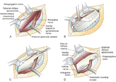
Lichenstein tension free mesh repair:
This is done under local or regional anaesthesia. The femoral ring is routinely evaluated via the space of bogros through a small opening in the canal floor. A sheet of 8 X16 cms mesh is placed over the floor of the
new internal ring is created by overlapping the two tails beyond the cord structures. Medially it is fixed on the rectus sheath and superiorly to the abdominal wall muscles through interrupted sutures
Fig 10: Lichenstein tension- free hernia repair
Rives prosthetic mesh repair:
Placing a mesh between transversalis fascia and the peritoneum. Internal oblique and the transversus abdominis muscle are sutured to the inguinal ligament in front of the mesh.
Stoppa’s giant prosthesis for reinforcement of visceral sac (GPRVS):
external iliac vessels and the iliopsoas muscles are exposed. The
parietalization of the cord structure is achieved and a Dacron mesh with a vertical dimension of 15 cms and a transverse dimension equal to the distance between the right and left anterior superior ileac spine, minus 2 cms is cut into chevrons shape. The mesh is placed in such a way that it widely envelopes the peritoneum and extends well beyond the
myopectineal holes and also protects the midline sub umbilical incisional wound. One stitch fixes the middle of the upper edge of the mesh to the lower margin of Richet’s umbilical fascia.
Laparoscopic Repair:
The two most popular techniques being, 1.Totally extraperitoneal (TEP)
2. Transabdominal preperitoneal (TAPP) approach.
The main difference between these two approaches is the sequence of gaining access to the pre peritoneal space.
TEP TAPP
Dissection begins in the
preperitoneal space using a balloon dissector
the preperitoneal space is accessed after initially entering the peritoneal cavity
preperitoneal dissection is quicker and potential risk for intraperitoneal visceral damage is minimized
Working space is limited and it may not be possible to create a working space if the patient has had a prior preperitoneal operation
More work space is present Can be done in patient who had a
previous preperitoneal operation
Dissection balloons are costly No balloons are used
TECHNIQUE OF DARNING :
Standard inguinal incision was given in the cases and inguinal canal was opened. The nature of hernia was assessed after delivering the sac. The sac was either incised or reduced and the gapping internal ring was
narrowed where required. The posterior wall of inguinal canal was
strengthened with prolene darn. Polypropylene (prolene ) size 1 strength mounted on round bodied atraumatic was used as a suture. The continuous began at the medial end of repair by taking good bite of fascia over pubis, medial end of uinguinal ligament and lowest portion of medial end of rectus sheath. This suturing continued laterally in simple over and over fashion in the form of figure of eight, approximating the lower edge of conjoint tendon to the upper edge of inguinal ligament without tension. Good bites of inguinal ligament where caught on the tip of the needle to safe guard the femoral vessels and at various levels in order to avoid the tearing of inguinal ligament. This is continued slightly beyond the internal inguinal ring by displacing the spermatic cord laterally. The same suture then took the reverse direction as the second layer of darn and was tied medially at the pubic tubercle. Then cord ws placed on darn and anterior wall of inguinal canal was re constituted followed by closure of
Results of Hernia Repair
The true measure of success for the various types of hernia repair is based on the results. The best information on the results of hernia repair is available from large prospective randomized trials, meta-analyses of
clinical trials, and two large national registries, the Danish Hernia Database and the Swedish Hernia Register. The Danish Hernia Database includes more than 98% of inguinal hernia repairs; the capture rate of the Swedish Hernia Register is approximately 80%.10,11 In spite of the randomized nature of some trials, caution must be used when interpreting the results. Many of these patients were highly selected and most trials excluded recurrent hernias, obese individuals, and large inguinal hernias.
Also, some follow-up results were completed by telephone interviews and not by physical examination. The national registries only collect
information on operations, so the incidence of recurrence is lower than if all patients had been interviewed and examined.
There are important differences in the results of primary hernia repair. Hernia recurrence is the primary outcome assessed by most studies. Large series, including multiple types of repairs, have suggested that recurrence ranges from 1.7% to 10%. The results of tissue repairs were often based on reports consisting of personal or single institutional series that were not prospective or randomized and had erratic follow-up periods. Not
surprisingly, recurrence was variable. A recent Cochrane review of 16 studies of more than 4000 tissue repairs has found that the Shouldice repair has a lower recurrence rate than other non mesh repairs.
Follow-up in these series was variable; patients were highly selected and may not have represented the population at large.
Tension-free repairs have a lower rate of recurrence than tissue repairs. Results from the Danish Hernia Database have demonstrated that hernia recurrence resulting in reoperation following the Lichtenstein repair is only 25% that of nonmesh repairs.A recent meta-analysis comparing the Lichtenstein, mesh plug, and bilayered repairs has reported no significant differences in the rate of recurrence, chronic groin pain, other
complications, or time to return to work. Approximately 50% of recurrences are found within 3 years after primary repair.
An extensive systematic review of randomized controlled trials was published in 2002 by the European Union Hernia Trialists Collaboration. The authors reported a meta-analysis of 4165 patients in 25 studies. Based on the available data, the laparoscopic repair resulted in a more rapid return to normal activity and decreased persistent postoperative pain. The recurrence rate for the laparoscopic repair was lower compared with open no nmesh repairs; however, open and laparoscopic mesh repairs had
similar recurrence rates.
A prospective trial sponsored by the Veterans Administration randomized 1983 patients to undergo an open Lichenstein repair or laparoscopic repair, of which 90% were TEP repairs. Most surgeons in this study may have had a suboptimal experience with the laparoscopic approach; only 25 prior repairs were necessary to be eligible to enroll patients, which is consistent with the seemingly high conversion rate of 5%. Despite these factors, the investigators found a twofold higher incidence of recurrence after laparoscopic repair (10%) than open repair (5%). This difference in recurrence remained for primary hernias (10% laparoscopic versus 4% open); however, recurrent hernias repaired by the laparoscopic approach tended to have fewer rerecurrences (10% versus 14%). In a Cochrane review of more than 1000 patients in eight non randomized trials, there was no difference in hernia recurrence between TAPP and TEP repairs.
On inexperience with laparoscopy and surgeon age older than 45 years were bothpredictors of recurrence after laparoscopic repair. What can be concluded from these results? This trial demonstrates that the laparoscopic repair of inguinal hernias may have a definite learning curve to achieve an acceptably low recurrence rate.
STRANGULATED INGUINAL HERNIA
Irreducibility, obstruction and strangulation are the complications of the inguinal hernia. First irreducibility occurs which leads into obstruction and strangulation
Features of strangulated hernia Causes of stangulation
Tense tender irreducible
No impulse on coughing
Shock, toxicity
Features of obstruction when there is no enterocele
Abdomen distension ,vomiting
Rebound tenderness
Narrow neck
Adhesions
Irreducibility
Long time, large hernia with adhesions
Patient with strangulated inguinal hernia presents with
Incision is extended and deepened downwards along the root of the
scrotum. Sac is identified and held. One should also hold the contents in the sac so has to prevent contents getting reduced and also toxic fluid spilling into the peritoneal cavity. Sac is opened carefully; all toxic fluid in the sac is sucked out thoroughly. Content, either bowel or omentum is held with Babcock’s forceps. Viability of the bowel is checked by its colour, texture, peristalsis, mesenteric pulsation or when in doubt by giving 100% oxygen to the patient or after covering the bowel wall using hot mops. If bowel is not viable resection and anastomosis is done. For this, incision may be adequately extended laterally along the lateral abdominal wall.
Closing this wound and separate laparotomy incision is not mandatory. Anastomosis is done using interrupted single layer silk or vicryl sutures. Strangulated omentum is not uncommon which is treated by
omentectomy.Drains are placed into the peritoneal cavity as well as into the inguinal wound.
Fig 18: Garry’s stricture in long standing obstructed hernia
Inguinal canal is strengthened by modified Bassini’s repair using
interrupted monofilament polypropylene sutures. Prosthetic mesh is not used as field is contaminated. In later period if hernia recurs then
prosthetic mesh is used for repair. Biological mesh can be used during the surgery for strangulated hernia. Problems in strangulated hernia are: sepsis, leak and fistula formation, ileus, wound infection, peritonitis, intra
RISK FACTORS FOR GROIN HERNIAS TO PRESENT AS ACUTE EMERGENCIES:
1) advancing age
2) large hernias with small opening 3) delay in hospitalizations
4) coexisting medical complications
In inguinal hernia the probability of strangulation was not more than 2% per year.
But the probability of strangulation for femoral hernia is about 40% per year.
1) Age :
Obstruction and strangulation occurred predominantly in middle age and elderly age groups
Andrew et al 1981 80 yr
Waddington et al 1971 60 yr
MC entee et al 1989 70 yr
Bahadur kulah et al 2001 60yrs
Gallegos et al 1991 65 yrs
Maingot text book 75 yr
2) Sex
In case of inguinal hernia males are twice prone for complication than female sex.
In femoral hernia females are thrice prone than males for complication.
3) Side
Right sided hernia is more prone for complication than left sided hernias
INGUINAL HERNIA RIGHT LEFT Andrew 1981 2.9 1
Anatomical basis for right sided hernias a) right testis descends later than left testis
b) line of attachment of small bowel mesentery is left side of L2 to right iliac fossa
4) Constriction ring
Deep ring is the most common site of obstruction.
5) Types
Femoral hernia is more prone to strangulation than inguinal
hernia.
6) Duration of hernia
Galleos et al 1991: the risk of groin hernia to produce
complication is maximum in first three months due to tight ring.
Later yielding of ring increases so complication decreases.
7) Content
Small bowel is the most common content. Omentum is the next
common. Slider is the next common type with large bowel as
content.
8) Delay in hospitalization
It is an important factor that determines resection and
MATERIALS AND METHODS
This study was a prospective study done in
Thanjavur Medical
College Hospital
from September 2016 to September 2017. The
study group was managed only by department of general surgery.
The subjects of this study were patients who underwent either
Mesh replacement or Darning procedure at our hospital,
Thanjavur Medical College . We evaluated and compared the
efficacy of the above procedures, analgesic requirement in the
first 24 hours, operative time, hospital stay, early postoperative
complication, time until return to work and recurrence between
patients who underwent LMH (Group A) and patients who
underwent DR ( Darn Repair). Tests done for operative
suitability included CBC, Urinanalysis, CXR, ECG. Post operative
patients were followed on weekly basis till return to normal
physical activity achieved. Overall postoperative followup was
done for 10 – 12 months.Patients are of age group of 24 to 86 yrs.
47
cases have been studied. These cases are studied from time of
admission till discharge and followed up in outpatient
department. A detailed history was elicited and clinical
INCLUSION CRITERIA :
1. Both sex, with age >18 years, with inguinal hernias presenting
in emergency.
2. All patients presenting with irreducible / incarcerated /
strangulated inguinal hernias ,
3. Cases which were regular for post treatment follow-up.
4. Patients willing to be part of this study.
EXCLUSION CRITERIA :
1. Patients < 18 years presenting without any features of
obstruction.
2. Ventral, femoral and incisional hernias.
3. patients not fit or willing for surgery
4. Cases which did not come for regular follow-up and who did
not
OBSERVATION AND ANALYSIS
Forty seven cases are studied. Mean age of patients is 60.17 years
DURATION OF HERNIA BEFORE ACUTE EPISODE:
SIDE OF HERNIA MOST COMMONLY INVOLVED
SYMPTOMS :
HERNIA
TYPE
PAIN AND
INCARCERATION
OBSTRUCTION
BOWEL
STRANGULATION
Inguinal
17
30
7
Percentage
36.17%
63.8%
14.8%
In the current study ,36.17% of groin hernia patients presented
with pain and incarceration.
63.8% of patients presented with obstructive symptoms.
Bowel strangulation was present 14.8% of cases.
CONTENT :
According to Andrew et. al the most common content in the hernia sac was small intestine. In the currrent study also the small intestine was the
INCIDENCE OF CO MORBIDITIES
A total of 47 patients were included in this study. They were
divided into two groups, each having 24 and 23 patients in Group
A and group B respectively. Group A underwent LMH repair,
while Group B underwent Darning repair.
S.NO
POST OPERATIVE
COMPLICATIONS
LMH ( N = 24)
DR ( N= 23)
1
SEROMA
4
7
2
HEMATOMA
2
5
3
WOUND SEPSIS
3
5
4
WOUND GAPING
1
3
5
HYDROCOELE
1
6
HOSPITAL STAY
4 DAYS
7 DAYS
7
RETURN TO WORK
21 DAYS
35 DAYS
8
CHRONIC GROIN PAIN
3
6
Seroma was observed in 4 patients in group A and 7
patients in group B.hematoma was observed in 2 patients in
group A and 5 patients in group B. Mean time of hospital stay in
group A was 4 days, while patients in group B remained
hospitalised for more than 7 days due to swelling and
infection. 6 patients developed inguinal swelling in group A,
while 12 patients developed inguinal swelling in group
B..chronic groin pain was seen in 3 patients in group Aand 6
patients in group B.
Wound sepsis was observed in 3 patients in group A while
it was observed in 5 patients in group B. Wound sepsis leading
to removal of skin stitches and therefore wound gaping was
seen in 1 patient in group A and 3 patients in group B.
None of the patients from both groups developed
recurrence during long term followup. Patients in group A,
returned to normal physical activity (walking / stair climbing/
eating, bathing, sitting) in 21 days while group B patients
CHI-SQUARE TEST
STATISTICAL INFERENCE OF AGE
Age DR LR Total Statistical inference
n % n % n %
Below 34yrs 1 4.3% 0 .0% 1 2.2%
X2=4.544 Df=4
.337>0.05 Not Significant
35 to 50 4 17.4% 1 4.3% 5 10.9%
51 to 60 6 26.1% 6 26.1% 12 26.1%
61 to 70 7 30.4% 6 26.1% 13 28.3%
Above 71years 5 21.7% 10 43.5% 15 32.6%
CHI-SQUARE TEST
STATISTICAL INFERENCE OF SIDE
Diagnosis
DR LR Total
Statistical inference
n % n % n %
LIH 7 30.4% 11 47.8% 18 39.1%
X2=1.460 Df=1
.227>0.05 Not Significant
RIH 16 69.6% 12 52.2% 28 60.9%
CHI-SQUARE TEST
STATISTICAL INFERENCE OF PRESENTATION
Presentation
DR LR Total Statistical
inference
n % n % n %
O 9 39.1% 12 52.2% 21 45.7%
X2=0.857 Df=2
.651>0.05 Not Significant
IR 12 52.2% 9 39.1% 21 45.7%
SB 2 8.7% 2 8.7% 4 8.7%
CHI-SQUARE TEST
STATISTICAL INFERENCE OF DURATION
Duration DR LR Total Statistical inference
n % n % n %
1HR 8 34.8% 5 21.7% 13 28.3%
X2=1.561 Df=3
.668>0.05 Not Significant
2HR 7 30.4% 9 39.1% 16 34.8%
3HR 6 26.1% 8 34.8% 14 30.4%
4HR 2 8.7% 1 4.3% 3 6.5%
CHI-SQUARE TEST
STATISTICAL INFERENCE OF CONTENT
Content DR LR Total Statistical inference
n % n % n %
O 13 56.5% 4 17.4% 17 37.0%
X2=9.336 Df=2
.009<0.05 Significant
SB 9 39.1% 19 82.6% 28 60.9%
LB +M 1 4.3% 0 .0% 1 2.2%
CHI SQUARE TEST
STATISTICAL INFERENCE OF COMPLICATION
Seroma
DR LR Total
Statistical inference
n % n % n %
No 16 69.6% 19 82.6% 35 76.1%
X2=1.075 Df=1
.300>0.05 Not Significant
Yes 7 30.4% 4 17.4% 11 23.9%
CHI SQUARE TEST
STATISTICAL INFERENCE OF COMPLICATION
Hematoma
DR LR Total
Statistical inference
n % n % n %
No 18 78.3% 21 91.3% 39 84.8%
X2=1.516 Df=1
.218>0.05 Not Significant
Yes 5 21.7% 2 8.7% 7 15.2%
CHI SQUARE TEST
STATISTICAL INFERENCE OF COMPLICATION
Wound Sepsis
DR LR Total
Statistical inference
n % n % N %
No 18 78.3% 20 87.0% 38 82.6% X2=0.605 Df=1
.437>0.05 Not Significant
Yes 5 21.7% 3 13.0% 8 17.4%
CHI SQUARE TEST
STATISTICAL INFERENCE OF COMPLICATION
Wound Gaping DR LR Total Statistical inference
n % n % n %
No 20 87.0% 22 95.7% 42 91.3% X2=1.095 Df=1
.295>0.05 Not Significant
Yes 3 13.0% 1 4.3% 4 8.7%
CHI SQUARE TEST
STATISTICAL INFERENCE OF COMPLICATION
Chronic Groin
Pain
DR
LR
Total
Statistical
inference
n
%
N
%
n
%
No
18 78.3% 19 82.6% 37 80.4%
X
2=0.138 Df=1
.710>0.05
Not Significant
Yes
5 21.7% 4 17.4% 9 19.6%
‘ T ‘ TEST
STATISTICAL INFERENCE OF AGE
Age
n
Mean
S.D
Statistical inference
DR
23 60.74 13.315
t=-1.548 Df=44
.129>0.05
Not Significant
‘ T ‘ TEST
STATISTICAL INFERENCE OF HOSPITAL STAY
Hosp Stay
N
Mean
S.D
Statistical inference
DR
23 8.30 1.105
t=13.441 Df=44
.000<0.05
Significant
‘ T ‘ TEST
STATISTICAL INFERENCE OF RETURN TO WORK
Return to Work
n
Mean
S.D
Statistical inference
DR
23 32.35 3.973
t=13.498 Df=44
.000<0.05
Significant
CONCLUSION
The following observation has been made from this study:
1) Incidence of acute complication of groin hernia is highest in age
group of 51 to 60 yrs
.
2.The incidence of acute complication of groin hernia is
commoner on the right side than on the left side
3. Majority of them presented with acute symptoms, within 1 year
of development of hernia (34%)
4. The most common presentation being groin swelling with
symptoms of obstruction
5. The most common content found in the sac is small bowel
followed by omentum
6. immediate post operative complications like seroma,
hematoma, wound sepsis was observed more in patients who
underwent Darning repair technique.
7. Hospital stay was minimal in patients who underwent
Litchenstein’s hernioplasty when compared to Darning repair
technique.
8. Patients who underwent Litchenstein’s hernioplasty returned
to work earlier than those patients who underwent Darning
repair.
9. Chronic groin pain was observed in patients who underwent
darnind repair.
BIBLIOGRAPHY
1. Patino.J.F: National University of Colombia: Department of Surgery, Santa Fe Foundation of Bogota, Bogota, Colombia.
2. Lyons.A.S., Petrucelli.R.J II. Medicine; An Illustrated History. New York; Harry N Abrams publishers, 1987.
3. Rutkow.I.M: A Selective History of Hernia Surgery in Late
Eighteenth Century: The treatises of Percivall Pott, Jean Louis Petit, D august Gottieb Richter, Don Antonio de Gimbernat and Pieter
Camper. Surg Clin N Am : 2003:83: 1021-1044.
4. Stoppa R.E: The Midline Preperitoneal Approach and Prosthetic Repair of Groin Hernia, in Fitzgibbon.Jr. R.J, Greenburg.A.G (eds): Nyhus and Codon’s Hernia, 5th ed, Philadelphia: Lippincott Williams and Wilkins, 2002, 199.
5. Ger.R. The management of certain abdominal hernia by intraabdominal closure of the neck of sac. Preliminary communication. Ann. R. Sug engl 1982; 64: 342-344.
6. Arregui M.E. Laparoscopic Preperitoneal Herniorraphy, Paper presented at annual meeting of the society of American Endoscopic Surgeons, 1991, Monterey, C.A.
7. Phillips.E.H., Carroll.B.J, Fallas.M.J., Laparoscopic Preperitoneal Inguinal Hernia repair without Preperitoneal incision; Surg Endosc:
8. Fitzgibbons.R.J. Jr , Camps.T, Cornet.D.A, et al. Laparoscopic Inguinal Herniorraphy. Results of a multicentric trial, Ann Surg; 1995:221:3.
9. Kugel.R.D. The Kugel repair for Groin Hernias. Surg Clin North Am,
2003:83:1119.
10. Skandalakis.J.E, Sandalakis.L.J, Colborn.G.L, Androvlakis.J, Mcclusky.D.A.III, Sandalakis.P.N, Mirilas.P. Surgical Anatomy of the Hernial rings, in Fisher.J.E. Mastery of surgery, 5th ed
Philadelphia, Lippincott Williams and Wilkins, 2007; 168:1859-1887. 11. Quinn.T.H. Anatomy of the Groin: A view from the Anatomist, in Fitzgibbons.R.J. Jr, Greenburg.A.G.(eds), Nyhus and Codon’s Hernia, 5th ed Philadelphia, Lippincott Williams and Wilkins, 2002:55-70. 12. Fitzgibbons.R.J. Jr, Filipi.C.J, Quinn.T.H. Inguinal Hernia in Brunicardi.F.C. et al. Schwartz’s Principles of Surgery, 8th ed. New York. Mc Graw Hill, 2005: 1353-1394.
13. Nyhus.L.M; Iliopubic Tract Repair of Inguinal and Femoral Hernia: The posterior (Preperitoneal) approach, in Baker.R.J, Fisher.J.E.
Mastery of Surgery, 4th ed. Philadelphia, Lippincott Williams and Wilkins, 2001:1943-1951.
14. Halverson.K, Mc Vay.C. Inguinal and Femoral Hernioplasty. Arch Surg: 1970;101:127-135.
15. Rutkow.I.M, Robbins.A.W. Classification System and Groin Hernias. Surg Clin North Am,1998;78:1122-1124.
16.Nyhus.L.M. Individualization of Hernia Repair; A new Era Surgery, 1993;114:1-2.
18.Schumpelick.V, Arit.G. Problems in General surgery. Philadelphia: Lippincott-Raven publishers, 1995: 12: 57-58.
19. Zollinger R.M.Jr. A Unified classification for Inguinal Hernia. Hernia. 1999; 3:195-200.
20. Terranova.O, Santis.LD. et al. The Bassini operation in Baker.R.J, Fisher.J. E. Mastery of Surgery. 4th ed. Philadelphia, Lippincott Williams and Wilkins, 2001: 164: 1904-1912.
21. Rutledge.R.H. Cooper ligament repair of Groin Hernias, in Baker.R.J, Fisher.J. E. Mastery of Surgery. 4th ed. Philadelphia, Lippincott
Williams and Wilkins, 2001: 165:1913-1922.
22. Bendavid.R. The Shouldice Method of Inguinal Herniorrhaphy. in Fisher.J. E. Mastery of Surgery. 5th ed. Philadelphia, Lippincott
Williams and Wilkins, 2007; 171:1888-1898.
23. Stoppa.R.E. Giant Prosthesis for Reinforcement of the Visceral Sac in the Repair of Groin and Incisional Hernia. In Fisher.J. E. Mastery of Surgery. 5th ed. Philadelphia, Lippincott Williams and Wilkins, 2007:175; 1923-1932.
24. Amid.P.K. Linchtenstein Tension Free Hernioplasty. in Fisher.J. E. Mastery of Surgery. 5t