GINGIVITIS USING NEXT GENERATION
SEQUENCING TECHNOLOGY
Dissertation submitted to
THE TAMILNADU Dr. M.G.R. MEDICAL UNIVERSITY
In partial fulfillment for the Degree of
MASTER OF DENTAL SURGERY
BRANCH II
PERIODONTOLOGY
Acknowledgement
I would like to express my gratitude to all the people who supported me in the completion of this thesis.
I take this opportunity to thank Dr. N.S. Azhagarasan, MDS, Principal, Ragas Dental College and Hospital for his support and guidance during my postgraduate course at Ragas Dental College and Hospital.
I express my sincere thanks to Dr. K.V. Arun, MDS, Professor and Head of the Department of Periodontics, Ragas Dental College Chennai, for his valuable advice, guidance and encouragement during my postgraduate course. I am deeply grateful to him for his patience and guidance during the study process.
I would like to extend my thanks to Dr. Swarna Alamelu, MDS,
Reader, who helped me in doing this study.
I also extend my gratitude to Dr. G. Sivaram, MDS, Professor, Dr.B. Shiva Kumar, MDS, Professor, Dr. Ramya Arun, MDS, Reader and Dr.
Archana Meenakshi, MDS, Reader for their continuous guidance and
constant encouragement throughout my study period.
Velkumar, MDS, Senior Lecturer and Dr. M. Divya, MDS, Senior Lecturer for their constant support.
I remain ever grateful to my senior Dr. Guhanathan, Dr. Keerthiha,
Dr. Pavithra and Dr. Kalaivani, for their constant support and
encouragement. I thank my batchmates Dr. Anisha Deborah, Dr. Latha, Dr. Gayathri, Dr. Sakthiganesh and Dr. Manimalla for their support and encouragement. I further extend my thanks to my juniors Dr. Ennet Cynthia , Dr. Asha, Dr. Santosh, Dr. Ali,Dr. Kavi Priya and Dr. Krupa.
I extend my gratitude to Mrs. Parvathi, Mrs. Rosamma, Mr. Chellapan and Mrs. Mala for their timely help during the tenure.
I would like to thank my parents Mr. Ramesh kumar and Mrs.
Gnanam for their love, understanding, support and encouragement
throughout these years.
Above all I’m thankful to The Almighty to have given me the strength
1 INTRODUCTION 1
2 AIMS AND OBJECTIVES 4
3 REVIEW OF LITERATURE 5
4 MATERIALS AND METHODS 36
5 RESULTS 41
6 DISCUSSION 45
7 SUMMARY AND CONCLUSION 53
8 BIBLIOGRAPHY 55
PSD Polymicrobial Synergy and Dysbiosis
DNA Deoxyribonucleic acid
RNA Ribonucleic acid
Rrna RibosomalRibonucleic acid
NGS Next Generation Sequencing
PCR Polymerase Chain Reaction
RT-PCR Real Time Polymerase Chain Reaction
CFU Colony Forming Unit
OUT Operational Taxonomic Unit
SoLiD Supported Oligonucleotide Ligation and Detection
BLAST Basic Local Alignment Search Tool
MSR Mi Seq Reporter Software
HOMD Human Oral Microbiome Database
NO.
1-A Evaluation of abundance of Phyla and their percentage among the health samples
1-B Evaluation of abundance of Phyla and their percentage among gingivitis samples
2-A Evaluation of abundance of top 8 Genera and their percentage among the health samples
2-B Evaluation of abundance of top 8 Genera and their percentage among gingivitis samples
3-A Evaluation of abundance of top 8 Species and their percentage among health samples
3-B Evaluation of abundance of top 8 Speciesand their percentage among gingivitis samples
4-A Comparison of abundance of top 20 species in health vs gingivitis
4-B Comparison of abundance of top 20 species in gingivitis vs health
5-A Species present in health but not present in gingivitis
[image:10.612.101.511.161.611.2]NO.
1 Bar chart describing the comparison of abundance of phyla in health and gingivitis
2-A Bar chart describing the comparison of abundance of top 20 species in health vs gingivitis
2-B Bar chart describing the total comparison of abundance of top 20 species in health vs gingivitis
3-A Bar chart describing the comparison of abundance of top 20 species in disease vs gingivitis
1-A Pie chart describing the comparison of percentage of phyla in health
1-B Pie chart describing the comparison of percentage of phyla in gingivitis
2-A Pie chart describing the comparison of percentage of top 8 genera in health
2-B Pie chart describing the comparison of percentage of top 8 genera in gingivitis
3-A Pie chart describing the comparison of percentage of top 8 species in health
3-B Pie chart describing the comparison of percentage of top 8 species in gingivitis
[image:12.612.103.513.112.467.2]1
INTRODUCTION
Periodontal disease is a chronic inflammatory condition that results in
destruction of the attachment apparatus of the tooth eventually leading to the
loss of teeth. Although the plaque microbiota is said to initiate the disease
process, it is host immune-inflammatory response that does the tissue damage.
It is now widely accepted that the bacterial communities in the biofilm
undergo a shift or what is known as dysbiosis, in which the presence of the
disease associated species may exacerbate the inflammatory reaction to the
commensal bacteria.
A microbiota is an ecological community of commensal, symbiotic and
pathogenic organisms found in all multicellular organisms studied till date
ranging from plants to the highest order of the animal kingdom. They have
been found to be crucial for the immunologic, hormonal and metabolic
homeostasis of the host5.
The salient clinical features of periodontal disease are gingival
inflammation, formation of periodontal pockets and gingival recession.
Gingivitis is a reversible form of periodontal disease that is characterised by
gingival inflammation in response to plaque biofilm. In susceptible individuals
untreated gingivitis may progress to periodontitis.
Ever since the essential role of plaque in gingivitis was first
2
has been a huge leap in our understanding of the microbial profile of the
plaque biofilm and its association to periodontal disease79.
Newer culture independent molecular methods such as those based on
cloning and Sanger sequencing of the 16s rRNA have greatly expanded our
knowledge of the microbial communities in the biofilm. The emergence of
advanced genomic technologies like the high throughput sequencing , also
known as the deep sequencing of the 16s rRNA gene or the next generation
sequencing is a landmark in the development of sequencing techniques and
has immensely contributed to the study of the microbiome.
With all these findings, it is now known that the PSD model might be
involved in the initiation of chronic periodontitis, with the emergence of the
key stone pathogen concept48. There is still a paucity of literature regarding
the knowledge of the microflora in the entire spectrum of periodontal disease,
ranging from health to gingivitis and its transition to periodontitis.
Also, it is now known that microbiome varies from one individual to
another and there is a considerable difference among population and ethnic
group104. Racial and population based difference may be influenced by dietary
and lifestyle pattern. The diversity among the microbiome of individuals is
immense compared to genomic variation: individual humans are about 99.9%
identical to one another in terms of their host genome but can be 80-90%
3
gut21,153. These findings suggest that employing the variation contained within the microbiome will be much more fruitful in personalized medicine, than
approaches that target the relatively constant host genome.
The microbiome of the subgingival environment in various periodontal
conditions has been studied in the past but there is a dearth of information
regarding the microbial profile in gingivitis.
Hence this study was undertaken as a first of its kind to evaluate the
subgingival microbiome in gingivitis and periodontal health in a South Indian
4
AIM AND OBJECTIVES
To evaluate subgingival microbiome from plaque samples of
periodontally healthy individuals and in patients with gingivitis using
NGS technology.
To compare the subgingival microbiome of gingivitis with that of
5
REVIEW OF LITERATURE
[
Oral microbiome:
The human microbiota consists of the 10-100 trillion symbiotic
microbial cells harboured by each person, primarily bacteria in the gut148. The
human body is composed not only of human cells, but is occupied by bacteria,
archaea, fungi and viruses; this ensemble of organisms (microbiota) and their
expressed genes are termed the microbiome. Despite their small size, the
human-associated microbiota have a genetic composition that is at least two
orders of magnitude greater than the human genome and it outnumbers the
cells of human host; the bacterial component alone is estimated to be equal in
number to that of human cells.
Different areas of our body have distinct microbial compositions that
are reflective of that microenvironment. Mucosal surfaces, including the
mouth, intestines, vagina and lung also provide niche environments in which
different microbiomes flourish. In a healthy state, these microbiomes form
symbiotic relationships with the host; the microbes are within a stable and
nourishing environment, whereas the host benefits in terms of metabolism,
immune system priming, and protection from other more pathogenic
6
While there is variability in the microbes inhabiting different
individuals, the microbiomes between individuals have shared core
functionalities that are relevant to the symbiotic relationship that exists
between the microbiome and its host. A disturbance of the levels and function
of the microbiome, termed dysbiosis, can lead to systemic problems with
serious impacts on human health.
The term microbiome was coined by Joshua Lederberg in 2001. Microbiome is defined as the totality of the micro-organisms and their
collective genetic material present in the human body or oral cavity85. This
term has been adopted by the Human Microbiome Project and considered as
the favoured nomenclature to define the complex oral bacterial community,
their genetic elements and environmental interactions, which may be involved
in disease31.
Studies of the diversity of the human microbiome started with Antonie van Leewenhook, who, as early as the 1680s, had compared his oral and fecal microbiota. He noted the striking differences in microbes between these two
habitats and also between samples from individuals in states of health and
disease in both of these sites36.
Thus, studies of the profound differences in microbes at different body
sites, and between health and disease are as old as microbiology itself. What is
new today is not the ability to observe these obvious differences, but rather the
7
differences exist, and to understand how we can affect transformations from
one state to another1,33,95,144.
Turnbaugh et al147 classified human microbiome into 2 types such as core microbiome and a variable microbiome. Core microbiome is one in which
it is common among the individuals in healthy condition in different sites
consisting of a predominant species134,147,157. Variable microbiome is one in
which it is particular to the individual which may be due to lifestyle which
may be unique, phenotypic and genotypic features. Oral microbiome of an
individual is similar to that of their fingerprint as there are difference in the
species and strain level of microbiome30.
Paster et al106 in his study had found that the estimated number of bacterial species in oral cavity was about 700. Periodontal disease condition
becomes nutritionally richer environment for bacteria, thus higher bacterial
diversity can be identified under diseased conditions but there may be
inter-individual differences in the disease group which cannot be ruled out119.
Gingivitis or periodontitis is accompanied by a shift in the oral bacterial
community, structure and composition 2,20,45,61,77.
Gingivitis:
8
bleeding, and increased pocket depth are the characteristics of periodontal
disease9. Moore et al.94 defined gingivitis as a non-destructive periodontal disease of the gingiva surrounding the bacterial biofilm.
Berchier et al.16 stated that the effects of gingivitis are reversible which can be improved by maintaining proper oral hygiene. Dietrich et al35
stated that when left untreated, gingivitis may progress into periodontitis
where as other studies have stated that gingivitis does not always develop into
periodontitis6,145. Page et al.103 added that periodontitis is always preceded by gingivitis.
The American Academy of Periodontology140 defined gingivitis as a non-destructive disease that occurs around the teeth. Bacterial biofilms that are
attached to the tooth contribute to the most common form of gingivitis known
as plaque-induced gingivitis, which acts to initiate the body's host response
thereby leading to the gingival tissues destruction resulting in the destruction
of the periodontal attachment apparatus111.
Gingivitis is a reversible form of periodontal disease characterized by
inflammation of the gingivae in response to a mature dental plaque biofilm. In
susceptible individuals persistent gingivitis may lead to chronic periodontitis,
9
Even though the composition of subgingival plaque is primarily
associated with gingivitis, as quantified by bleeding on probing, rather than the
presence or absence of periodontitis, the presence of periodontitis has
detectable associations with subgingival microbiota that are unrelated to
gingivitis158. In particular, the differences in co-occurrence patterns of taxa
between women with and without periodontitis support a more complex
etoiology of disease than a simple progression from health through gingivitis
to periodontitis120.
Gingivitis and periodontitis were associated with higher microbial
community richness and Shannon indexes, and this association remained after
adjustment for demographic factors, including age, body mass index (BMI),
and socioeconomic status159. This finding is consistent with previous research
by various authors who proposed, with higher diversity meaning that, in
periodontal disease, the oral microbiota is added rather than existing taxa
undergoing replacement22,61. This may correspond to primary ecological
succession in a new environmental niche, as suggested by Abusleme et al2.
Liam Shaw et al120found that many taxa were associated with gingivitis and periodontitis. The abundance of the majority of these taxa
increased with gingivitis severity, and this pattern was not influenced by the
presence of periodontitis. .It would appear that relative bacterial abundances
alone are insufficient to explain the presence of disease, which is consistent
10
to cause disease. He identified distinct signals associated with gingivitis and
periodontitis in subgingival plaque, with a dominant contribution from
gingivitis. Network analysis of observed co-occurrence patterns was consistent
with the role of bridging bacteria like F. nucleatum and F. alocis in the
co-aggregation of periodontal biofilms prior to entrance into subgingival regions.
Although some periodontitis-associated bacteria were also associated with
gingivitis, the major change with periodontitis is in the network of
co-occurrences.
Gingivitis sets the stage for periodontitis to develop by providing an
environment where periodontitis-associated taxa can increase in abundance
and co-aggregate into pathogenic biofilms that may then penetrate to
subgingival regions10,17.
The open contacts between teeth, gingival grooves, bulky and
overhanging restorative margins, claps of removable partial dentures and
calculus which serves for plaque accumulation are known to be plaque
retentive factors152. The various degradative enzymes and toxins such as
lipopolysaccharide (LPS), other endotoxin or lipoteichoic acid (LTA) are
produced by bacteria which may lead to inflammatory response in the
periodontal structures154.
The initial stages of plaque is characterised by gram-positive cocci and
rods while the latter by an increase in gram-negative rods, fusiforms,
11
periodontal disease. The association of plaque to gingivitis was confirmed by
the study done by Loe on the Srilankan tea labourers which popularly came to know as the “Experimental Gingivitis” model80
.
Experimental gingivitis:
Loe et al.79 described the development of gingivitis exclusively in a system of model known as experimental gingivitis.
Gram-positive rods, gram-positive cocci and gram negative cocci were
the initial microbiota of experimental gingivitis. Gram-negative rods and
filaments, spirochaetal and motile microorganisms increase in number
resulting in inflammatory changes leading to gingivitis.
He founded that 56% gram-positive bacteria, 44% gram negative
bacteria are present in plaque induced gingivitis; which also includes 59%
facultative organism and 41% anaerobic organisms. Most predominant
gram-positive organisms includes
S.sanguis, S.mitis, S.intermedius, S.oralis, A.viscosus, A.naeslundii,
and P.micros. The most predominant gram-negative organisms includes
F.nucleatum, P.intermedia, and V.parvula, Haemophilus , Capnocytophaga
12
Experimental gingivitis in man:
Theilade et al141 in an experiment carried out in 11 subjects who had excellent oral hygiene and healthy gingiva wherein they developed
accumulations of plaque and generalized gingivitis after 9 to 21 days without
oral hygiene. It was found that the rate of plaque accumulation was correlated
with the rate of development of gingivitis. Gram-positive cocci and rods were
present initially when the teeth were clean and healthy gingiva.
During the first two days without oral hygiene, there developed the
first phase of plaque. Proliferation of gram-positive cocci and rods were seen
along with an addition of about 20 to 30% gram-negative cocci and rods.
Fusobacteria and filaments began to appear and increased about 7% of the
total flora during the second phase that is about 1-4 days.
Spirilla and spirochetes contributed for about 2% of the total flora
during the third phase; that is after 4-9 days. The composition of the plaque
correlated with the condition of the gingiva in certain areas which helped in
the clinical diagnosis of the gingivitis which in turn correlates with the same
time as this complex flora begin to colonize but the sub-clinical inflammation
started at the first phases of plaque development.
After this experiment the subjects were advised to start the oral
hygiene measures and it was noted that within 1 to 2 days, plaque began to
13
removal of plaque. Plaque index were taken after 7–11 days which returned to
the baseline value.
A was study done by Loe et al 80 from 1970 to 1986 among the tea labourers in Srilanka which included 480 males of age group ranging from 14
– 46 years, who did not perform any conventional oral hygiene measures and
were able to displace large aggregates of plaque and calculus on their teeth.
They were divided into 3 groups: RP group: about 8% of individual had rapid
progression of periodontal disease, MP group: about 81% of individuals had
moderate progression and NP group: about ∼11 % of individuals had no
progression of periodontal disease beyond gingivitis. In RP group the mean
loss of attachment was ∼9 mm, the MP group had ∼4 mm and the NP group
had less than 1 mm loss of attachment at 35 years of age. The mean loss of
attachment in the RP group was ∼13 mm and the MP group ∼7 mm at 45
years of age. The annual rate of destruction in the RP group varied between
0.1 and 1.0 mm, in the MP group between 0.05 and 0.5 mm, and in the NP
group between 0.05 and 0.09 mm. In the RP group, tooth loss already occurred
at 20 years of age and increased throughout the next 25 years. At 35 years of
age, 12 teeth had been lost, at 40 years of age 20 teeth were missing and at 45
all teeth were lost. In the MP groups, tooth mortality started after 30 years of
age and increased throughout the decade. At 45 years of age, the mean loss of
teeth in this group was 7 teeth. The NP group essentially showed no tooth loss.
14
Role of plaque in periodontal disease:
The etiological role of plaque in periodontal disease, the initiation and
the progression of periodontal diseases was clearly explained by Loe‟s
experimental gingivitis but with the time, the ideas about how a shift from oral
health to disease due to dental plaque have been changed79.
Non-specific Plaque hypothesis:
Investigation done by Miller92 opened a window to the non-specific plaque hypothesis. They postulated that it was the quantity of plaque that
determines the pathogenicity without discriminating between the levels of
virulence of bacteria. According to Theilade142, when plaque content with its toxins and breakdown products exceeded the capacity of host response,
disease occurs. He also stated that all bacteria in plaque contribute to the
virulence of the microflora by having a role in either colonisation, evasion of
the defence mechanism, and/or provocation of inflammation and tissue
destruction.
Page RC103 stated that however this hypothesis does not suit for the development of periodontitis, whereas it is applicable only for the
development of gingivitis since periodontitis is a multifactorial disease. The
main lack of this hypothesis is that it failed to prove that why all gingivitis not
15
periodontitis had little plaque whereas in some subjects with mild periodontitis
had increased amount of plaque and also site specificity of the disease is
inconsistent with the concept that all plaque is equally pathogenic.
Specific Plaque Hypothesis:
The specific plaque hypothesis, proposed by Walter J. Loesche83, stated that the quality and not the quantity of plaque mattered as only certain
microorganisms in plaque were thought to be pathogenic. When these specific
bacteria increased in number, virulence factor released by them would lead to
periodontal diseases. For instance in localized aggressive periodontitis,
Aggregatibactoractinomycetacomitans is found to be the specific
pathogen.98,129
Following the development and maturation of dental plaque, with
increase in probing depth, oral microbial flora specifically changes from
gram-positive aerobic species to gram-negative anaerobic species89,131. Socransky
and Haffajee132 identified specific microbial groups with dental plaque. Six inter-related groups were reported. The early colonizer consists of yellow,
green and purple complexes. These complexes help in the colonization of
orange and red complexes. The red complex organisms are associated with
periodontitis which includes Tannerella forsythia, Porphyromonas gingivalis
and Treponema denticola. The main disadvantage of this hypothesis is that it
16
are considered to be the main putative periodontal pathogens are mostly
present in healthy periodontium.
Ecological Plaque Hypothesis:
Philip D. Marsh89 had put forth the Ecological plaque hypothesis. This hypothesis stated that the disease is the result of an imbalance in the total
microflora due to ecological stress, resulting in an enrichment of some oral
pathogens or disease related organisms. More specifically, this hypothesis
proposes that the nonspecific accumulation of plaque leads to inflammation
within the gingival tissues and to the development of gingivitis. This leads to
environmental changes within the gingival sulcus, which in turn favour the
growth of gram-negative and proteolytic species of bacteria. These changes
lead to further inflammatory and immune mediated tissue changes and tissue
destruction, culminating in a predominance of periodontal pathogens and a
greater degree of tissue damage.
Keystone Pathogen Hypothesis:
Hajishengallis et al.48stated that the quantity of normal microbiota and their composition can be changed by some low-abundance microbial
pathogens there by causing inflammatory disease. Socransky et al.131 stated that these keystone pathogens can be detected in higher numbers when the
17
Polymicrobial Synergy and Dysbiosis Model:
Hajishengallis et al48 had described Polymicrobial Synergy and Dysbiosis (PSD) model of pathogenesis. He founded that periodontitis is
initiated by a broadly based dysbiotic, synergistic microbiota as against the
traditional view that it is caused by a single or several pathogens. Thus the
host-microbe homeostasis is altered by this dysbiotic, synergistic microbes and
there by leading to a transition of chronic inflammatory state. Thus the disease
progression is caused by whole microbial community.
The interaction between the micro-organisms present in the plaque and
immune responses results in tissue destruction that includes both the loss of
connective tissue and alveolar bone70.
Microbiological Methods / Assays in Periodontal Diagnosis:
It has been estimated, however, that approximately half of the bacteria
found in the oral cavity have not been, or cannot be, cultivated in the
laboratory151,23,26,139. Therefore, culture studies alone could not provide a
comprehensive description of the microbiota in experimental gingivitis.
The early knowledge of the microbial composition of dental plaque in
gingivitis is based largely on microscopy and cultural methods, which do not
18
Close ended techniques: Culture:
Traditionally identification of the species in any given sample was
achieved by growing it in vitro on suitable media. There are 2 types of media
for culturing, which are the selective media and non-selective media.
Samaranayake115 founded that growth of a broad spectrum of organisms can be enhanced by blood agar which is a common non-selective medium.
Laboratory culturing under special conditions and using a range of media has
allowed isolation of a diverse range of bacteria. However, it is well recognized
that the main drawback of this method is its narrow spectrum. Various authors
founded that about 50% to 60% of distinct bacterial phyla in oral cavity are
still non cultivable62,126,149,136. Moreover, the culture-dependent technique is
expensive, sensitive and needs a highly skilled individual.
Siqueria et al.125 stated that there is a growing need for developing improved methods to cultivate and characterize the as-yet-uncultivated portion
of the oral microbiome so as to unravel its role in health and disease.
Theoretically, all bacteria can grow under proper nutritional and
physicochemical conditions24. However, development of new improved
culture media is still a challenging goal and is mainly due to the highly diverse
microbial community present with each member having different nutritional
requirements. Certain authors has recently suggested a list of
19
the use of culture media with little or no added nutrients and addition of
specific growth factors in the culture media126,143.
A very interesting strategy to ensure the availability of natural growth
factors is to perform incubation in the natural environment using special
devices 43,59,127,137 such as a diffusion chamber or a hollow fibre membrane
chamber which allow diffusion of important growth factors from a natural
environment to the culture via a special membrane8,19,50,74,122.
Though considered gold standard for identification and analysis of
bacteria, bacterial cell culture has its own limitations. Only viable bacteria will
grow, and stringent transport and storage conditions will be needed to identify
those bacteria that are „cultivable‟18,37. Those that are not cultivable would
remain unidentified. This could be due to the fact that many species in the
oral cavity are fastidious and require the presence of other organisms and very
specific growth conditions. If those conditions, that could even be unknown
and unexplored, are not provided, then the bacterial species that are the most
fastidious will simply not grow in culture and will remain unidentified.
Bacterial identification using newer molecular methods will surpass the
limitations associated with culture based methods for identifying
20
Immunologic and enzymatic assays:
Accurate detection of targeted species can be achieved by the
antibody-based detection systems. Raising of antibodies to each species of
interest needs growing the organism in culture before inoculating an animal
and raising antibodies to the bacterial antigens. Wolff, L.F et al.155, Loesche et al.82 used this technique to identify the changes in the pathogens either naturally or in response to treatment in healthy and disease population.
Limitation of immunological assays is that the target organism has to
be cultured to raise antibodies against it. This makes this method useful only
for cultivated species38,93. The antibodies cross reactivity can be tested only on
cultivated species and cannot be done on uncultivated or unknown species.
DNA – DNA hybridization or checkerboard:
DNA-DNA hybridization detects bacteria with the help of a labeled
genomic DNA that is attached to nylon membranes based on hybridization of
target microbes. In periodontal health and disease including the response to the
treatment, the various levels of microbes have been studied with this
technique40,44,47,81,133,156. They used data from population based studies and
grouped 40 species that were found in clusters or „complexes‟ by comparing
the levels of these species in health and disease. Three species,
Porphyromonas gingivalis, Tannerella forsythia (Bacteroides forsythus) and
21
compared to health. These three species were grouped into the „red complex‟
bacteria.
Chairside Diagnostic tools were available based on assays that detect
these species. Red complex bacteria were thought to be the primary etiological
agents for more than 30 years and therapeutic intervention to eradicate these
species were also considered. The main advantage of this technique is that
they are capable detecting multiple species from various samples in a same
time.
Polymerase chain reaction:
Polymerase chain reaction (PCR) was first founded by Mullis et al.97. A part of gene or a specific gene is amplified which can be used to identify the
bacterial species. PCR-based methods were used by researchers in their
studies to detect specific species directly from oral samples. They focused
mainly on the identification of a few species associated with the putative
periodontal pathogens which includes Porphyromonas gingivalis,
Tannerella forsythia, Treponema denticola, and Aggregatibacter
actinomycetemcomitans28,75,101,116,138.
In previous sequence analysis of 16S rRNA genes from oral
microflora, a number of bacterial species were identified as candidates as
putative pathogens for periodontitis, that includes the traditional species, such
22
order to detect the prevalence of causative pathogens, healthy subjects and
diseased plaque samples can be used with the help of species specific PCR
primers.
Real -time polymerase chain reaction:
The technique is similar to PCR but it is capable of detecting the
amplified DNA and helps in simultaneous quantification as the progress of
reaction occurs in real-time46,123.
Open-ended approaches:
This technique is advantageous now since it can detect previously
unknown microbes including those which are non-cultivatable. These
approaches are based on 16 S rRNA sequencing. This approach has been used
to study the microbial population in different ecosystems, enabling the
characterization of hitherto uncultivated microbial communities41,102,121. Using
this approach, the diversity of different colonization niches in the oral cavity
has been explored13,58,108.
16S rRNA sequencing:
This technique is proven to be the most important phylogenetic marker
that amplifies and analyse 16S rRNA genes in a plaque sample and it is a
23
species can be recognized by PCR primers. Various microbes can be
compared at different taxonomic levels with the help of 16S rRNA gene which
consist about 1500 bases72,109,124.
Next Generation Sequencing:
The next-generation sequencing works on the principle that it may
involve oligonucleotide which undergoes cyclical ligation which is of machine
automated or there will be a repeated cycles of polymerase-mediated
nucleotide extensions86, 128,150. In a single machine run, there will be a huge
amount of nucleotide sequence as millions of reactions occur in a massively
parallel process. The types of NGS include:
1. Roche/454 FLX (Life Sciences, Branford, CT, Margulies et al.87) 2. Illumina/ Solexa Genome Analyzer (Illumina, San Diego, CA, Bentley
DR14 , Korbel et al.65)
3. SOLiD (Life Technologies, Carlsbad, CA, Mardis86, Voelkerding et al.150).
4. 4)HiSeq and the Ion Torrent Personal Genome Machine (PGM)
(Rothberg et al.112).
NGS consists of 2 themes which includes a bead to which the fragments
are immobilized to the solid surface and DNA fragments are ligated
oligonucleotide adaptors which serve as primers for sequencing and
24
The pyrosequencing technology is an additional theme in Roche ⁄ 454
which consists of beads which are available on picotiterplate wells which has a
clonal amplification of templates, the laminar fluidics that control the delivery
of deoxyribonucleotide triphosphate and the luminescent which burst upon
deoxyribonucleotide triphosphate incorporation is detected bya high resolution
charge-coupled device camera. Advantage of 454 sequencing is its long read
lengths (400–500 nucleotides) and the amount of sequence generated (0.5
Gb)84. The long reads can handle repetitive regions better than other next
generation sequencing systems100.
SOLiD 35 nucleotide reads which is two-base encoding system and it
provides better sequence fidelity than the one-base next-generation sequencing
systems150 but biased sequence coverage is obtained in AT-rich repetitive
sequences51.This technique requires long running time and only 35% of the
raw reads are useable where as 95% of the raw reads can be used for the 454
system.
Illumina/Solexa Genome Analyzer
This technique is an highly targeted NGS approach which provides
more sequence reads per run, than previous methods thereby allowing for
more in depth coverage14,14,64. In this technique, there will be specific number
of cycles, which detects fluorescent reversible-terminator nucleotides on
25
The target is first amplified in the Solexa system after which only one
of the strands is sequenced with all four deoxyribonucleotide triphosphates in
which each deoxyribonucleotide triphosphate has a unique fluorophore .In 454
system tis sequencing does not occur in one step. In order to prevent insertion
of multiple nucleotide bases, reversible terminator nucleotides are present
91
and adaptor sequences are present at each end of the fragments which is
immobilized with oligonucleotides that are grafted to the surface of a
microfluidic chamber. The DNA templates are hybridized to the immobilized
oligonucleotides by the adaptors after which they are copied using bridge
amplification3.
Fluorescently labelled deoxyribonucleotide triphosphates and the
primer hybridize to one of the adaptors when there is an addition of
polymerase there by initiating DNA sequencing. This is recorded bya
charge-coupled device camera when there is a burst of light incorporation of a
complementary base and the fluorophore is washed away unlike 454
sequencing and is removed from the incorporated base, washed away and the
cycle is repeated63,114.
The Solexa system has an advantage that 1.5 Gb of sequence per run
can be achieved with read lengths which range from 35 to 100 bases out of
which each run requires 3–5 days to complete113.Whereas this system produce
biased sequence coverage that occurs in AT-rich repetitive sequences as they
26
This study used NGS to characterize the composition of the microbial
communities of plaque samples from periodontal health and gingivitis.
Initially, the study of bacterial communities in the oral cavity was
conducted with culture-dependent methods96 . However, because 40% to 60%
of bacteria in the oral cavity cannot be cultivated and so culture-independent
methods such as denaturing gradient gel electrophoresis were used42.
Although several metagenomic studies using 16S ribosomal RNA
(rRNA) gene cloning techniques provided information about bacterial
communities, the studies were limited to insufficient clone numbers and a lack
of statistical significance69. To solve this problem, next-generation sequencing
(NGS) has revolutionized bacterial diversity studies on the oral microbiota of
subjects with a healthy or periodontitis status2,45.
A number of previous high-throughput 16S rRNA sequencing studies
characterized oral bacterial communities to the phylum or genus level
only60,71,76. It is important to distinguish taxa at the species-level, as different
species within the same phylum and/or genus may be health-associated or
pathogenic/disease-associated. The targeting of a highly variable region of the
16S rRNA gene (V1–V3) and the use of a curated human oral 16S rRNA gene
reference set (HOMD), enabled the identification of OTUs (clustered at a
27
Whilst some studies have also recently reported species-level 16S
rRNA gene pyrosequencing analysis of the bacterial communities in
periodontal health, gingivitis and chronic periodontitis, these studies were
cross-sectional in nature and did not examine changes in the same individuals
during the transition from health to disease2,45,55. In the present study a highly
species-rich bacterial community (201–383 OTUs per sample) was revealed in
early health-associated plaque.
The Human Oral Microbiome Database
The goal of creating the Human Oral Microbiome Database (HOMD)
is to provide the scientific community with comprehensive information on the
approximately 700 prokaryote species that are present in the human oral
cavity. Approximately 54% are officially named, 14% unnamed (but
cultivated) and 32% are known o-nly as uncultivated phylotypes. The HOMD
presents a provisional naming scheme for the currently unnamed species so
that strain, clone, and probe data from any laboratory can be directly linked to
a stably named reference scheme128,160.
The HOMD links sequence data with phenotypic, phylogenetic,
clinical, and bibliographic information. Genome sequences for oral bacteria
determined as part of this project, the Human Microbiome Project, and other
sequencing projects are being added to the HOMD as they become available.
28
HOMD. The HOMD site offers easy to use tools for viewing all publically
available oral bacterial genomes.
Microbiome Studies In Periodontal Disease: Chronic periodontitis:
Kroes et al. 66 conducted a study using 16S rRNA gene sequencing and Sanger sequencing in order to evaluate the subgingival microbiome in
chronic periodontitis and founded that there were 77 phylotypes involved in
the disease out of which 48% were novel.
Paster et al.107 did a study using Sanger technique under different periodontal conditions and founded that there was a total of 347 phylotypes
involved in the disease and among them 215 were novel.
Kumar et al.67 founded that 274 phylotypes were associated in the chronic periodontitis while evaluating the subgingival microbiome using
Sanger sequencing in health and periodontitis. Phylotypes associated with
periodontitis were identified as Peptostreptococcus spp., Filifactoralocis,
Megasphaera sp., Desulfobulbus sp., Dialister spp. Campylobacter spp.,
Selenomonas sp., Deferribacteres sp., Catonella sp., Tannerella forsythia,
Streptococcus spp., Atopobium sp., Eubacterium sp. and Treponema sp.
Phylotypes associated with health were Veillonella sp., Campylobacter
29
Eubacteriumsaburreum, Gemella sp., Streptococcus sanguis, Streptococcus
mutans, Capnocytophagagingivalis, Rothiadentocariosa, Eubacterium sp. and
Selenomonas sp.
Griffen et al.45founded 16 phyla, 106 genera and 596 species in chronic periodontitis using high throughput sequencing. He also stated that
health associated species may also be present in the disease but in low
abundance.
Abusleme et al.2 did a study in 22 subjects to understand the ecology of oral subgingival communities in health and periodontitis and elucidated the
relationship between inflammation and the subgingival microbiome using
454-pyrosequencing of 16S rRNA gene libraries and quantitative PCR. He founded
that periodontitis communities had higher proportions of Spirochetes,
Synergistetes, Firmicutes and Chloroflexi, among other taxa, while the
proportions of Actinobacteria, particularly Actinomyces, were higher in
health. Total Actinomyces load, however, remained constant from health to
periodontitis.
Hong et al.52 founded that two variant of microbiome profiles can be seen in periodontitis which includes clusters A and cluster B which was
derived clustering analyses of microbial abundance profiles. In subjects with
cluster A communities there was an increased proportions of different
periodontitis-associated species, health-associated species and core taxa which
30
increased proportions of certain periodontitis-associated organisms, such as
Porphyromonas gingivalis, Tannerella forsythia and Treponema denticola, and
taxa recently linked to periodontitis. The cluster B community showed a
positive correlation with periodontitis extent.
Chi-Ying Tsai146 did a study in Taiwanese individuals with chronic periodontitis with 16S rRNA metagenomic approach. He determined the
subgingival microbiota and demonstrated a microbial shift from health to
disease.
Camelo-Castillo20 evaluated the relationship between the chronic periodontitis-associated subgingival microbiota and clinical inflammation.
Subgingival bacterial samples from periodontal patients were studied by
pyrosequencing PCR products of the 16S rRNA gene and by real-time PCR.
He founded that 16S pyrosequencing revealed that increased inflammation, at
sites with periodontitis, is associated with a more diverse subgingival
microbiota and specific changes in the bacterial composition, involving
“established” periopathogens, symbionts and novel low-abundance
pathobionts.
Gingivitis:
Kistler et al.61 in 40 patients studied using 454pyrosequencing of the 16S rRNA genes and bacterial culture, to characterize the composition of
31
that species-level phylotypes positively and negatively correlated with
gingivitis. Increased community diversity and significant shifts in microbiome
structure after two weeks of oral hygiene abstention was reported. All of the
healthy volunteers developed gingivitis after two weeks. Pyrosequencing
yielded a final total of 344 267 sequences after filtering, with a mean length of
354 bases, that were clustered into an average of 299 species-level Operational
Taxonomic Units (OTUs) per sample. Changes in the relative abundance of
OTUs during the transition from health to gingivitis were correlated to
bleeding on probing (BoP) scores and resulted in the identification of new
health and gingivitis associated taxa. Comparison of the healthy volunteers to
the periodontitis patients also confirmed the association of a number of
putative periodontal pathogens with chronic periodontitis. Taxa associated
with gingivitis included Fusobacterium nucleatum subsp. polymorphum,
Lachnospiraceae sp., Lautropia sp. and Prevotellaoulorum, whilst
Rothiadentocariosa was associated with periodontal health.
Huang et al. 53 founded that 15 genera could distinguish healthy and gingivitis samples with 74 percent accuracy. Temporal shifts in community
structure were observed along the progression from naturally occurring
gingivitis to healthy gingiva to experimental gingivitis.
Park et al104 analyzed bacterial communities in the subgingival paper point samples from 32 Korean individuals with no sign of disease, gingivitis,
32
reads representing 26 phyla, 433 genera, and 1,016 species were detected.
Bacteroidetes, Fusobacteria, Synergistetes, and Spirochaetes were the
abundant phyla in periodontitis subjects, whereas Firmicutes and
Proteobacteria were identified as the dominant phyla in the gingivitis and
healthy subjects, respectively. Although high levels of Porphyromonas,
Fusobacterium, Fretibacterium, Rothia, Filifactor, and Treponema genera were
observed in the periodontitis subjects, Streptococcus, Capnocytophaga,
Leptotrichia, and Haemophilus genera were found at high frequency in the
gingivitis subjects. Species including Porphyromonas gingivalis,
Fusobacterium nucleatum, and Fretibacterium fastidiosum were significantly
increased in periodontitis subjects. On the other hand, Streptococcus
pseudopneumoniae, Haemophilus parainfluenzae, and
Leptotrichiahongkongensis were preferentially observed in the gingivitis
subjects. He founded that showed distinct microbial communities were present
in health, gingivitis and periodontitis.
Huang et al54 compared plaque microbiota changes in gingivitis patients by using tooth brush only in one group and dental scaling in another
group among 91 subjects. He founded that only Actinobaculum, TM7 and
Leptotrichia were consistently reduced by both the treatments, whereas the
different microbial signatures of the two treatments during gingivitis relieve
indicate distinct mechanisms of action. Therefore this study suggests that
33
understanding and potentially comparing the modes of action for clinical
treatments and oral-care products in the future.
Liam Shaw et al120 did a study in 962 Malawian women for periodontal disease and used 16S rRNA gene amplicon sequencing to estimate
the bacterial compositions of supragingival plaque samples.Associations
between bacterial relative abundances and gingivitis /periodontitis were
investigated. He founded that the main differences in supra-gingival plaque
compositions were associated more with gingivitis than periodontitis,
including higher bacterial diversity and a greater abundance of particular
species. However, even after controlling for gingivitis, the presence of
subgingival periodontitis was associated with an altered supra gingival plaque.
A small number of species were associated with periodontitis but not
gingivitis, including members of Prevotella, Treponema and Selenomonas,
supporting a more complex disease model than a linear progression following
gingivitis. This finding confirmed that periodontitis cannot be considered
simply an advanced stage of gingivitis even when only considering supra
gingival plaque.
Diaz et al34 conducted a study using high-throughput 16S rRNA gene sequencing in order to evaluate subgingival microbiome shifts from health to
periodontitis. He founded that the development of gingivitis is also
characterized by distinct shifts. Microbiome shifts resemble microbial
34
adaptation to the changing environment as inflammation ensues.
Gingivitis-associated and core species are proposed as likely mediators of microbiome
transitions.
Andre Paes Batista da Silva et al7 did a study in 175 subjects which measured microbial composition changes during biofilm overgrowth and
subsequent removal among patients with various states of periodontal disease
using 16S rRNA probes (human oral microbe identification microarray) who
were abstained from oral hygiene while using an acrylic stent. At day 21,
participants reinstituted oral hygiene and were followed for 4 weeks.
He founded that Synergistetes is associated with more severe forms of
periodontal disease and plaque accumulation. When oral hygiene was resumed
and another examination performed after 4 weeks, most bacteria in all groups
were reduced to base line levels. However, some bacterial phyla did not
reduce to baseline levels, such as synergistetes. Most importantly, higher
levels of fusobacteria, proteobacteria, firmicutes, synergistetes, and
bacteroidetes are associated with incident increasing PD. He concluded that
these findings further support the potential critical role of synergistetes as well
as the previously characterized fusobacteria, proteobacteria, firmicutes, and
35
Ke Deng et al.29 with gingivitis analysed microbiome in 54 patients with gingivitis from subgingival plaque samples among Chinese population
using high throughput 16s rRNA sequencing extracting 35 genera and 46
species. He proposed that there was a significantly different microbial
community composition of sub gingival plaque between health and gingivitis.
He also showed that the presence of P. gingivalis present among the person
36
MATERIALS AND METHODS
Study population
A sample size of 8 patients was chosen for the study due to the cost
and complexity of the technique involved, which is in accordance with
previous studies by Zheng et al161, Dzink et al.39
Eight individuals, who reported to the outpatient Department of
Periodontics in Ragas Dental College and Hospital Chennai, were enrolled in
the present study, of which 4 were periodontally healthy individuals (control
group) and 4 were gingivitis patients (test group).
Control group consisted of subjects with clinically non-inflamed,
healthy gingiva that had negligible bleeding on probing <10%, probing pocket
depth {PPD} ≤3mm and no clinical attachment loss {CAL}.
Test group consisted of subjects with gingivitis who had bleeding on
probing {BOP}> 20%, with PPD≤ 3mm and no clinical attachment loss.
The study protocol was explained, and written informed consent was
received from each individual before clinical periodontal examination and
37 Inclusion criteria
Subjects must not have any contributing systemic illness
Subjects must meet the criteria of periodontal health and gingivitis as
described above was included in this study.
Exclusion criteria
Patient with systemic disorders, such as diabetes mellitus,
immunological disorders or HIV that may be a contributing factor for
periodontal disease.
Patient who were under medication drugs that alter the microbial
characteristics which includes immunosuppressant drugs or steroids.
Smokers or tobacco users.
Patient with previous history of periodontal treatment in the past 6
months.
Patient who had been prescribed antimicrobial therapy for past 6
months.
Subgingival plaque sampling
All examinations were performed by a single, calibrated examiner. For
the diseased samples, the sites that exhibited bleeding on probing were
selected and the sample was collected in a single Eppendorf tube. The site to
be sampled was first isolated using cotton rolls and air dried gently.
38
plaque was obtained from the sample teeth and with sterilized Gracey curettes.
The plaque from within 2 mm of the sulcus depth was carefully obtained with
a gentle pull motion towards the coronal surface. The tip of the curette was
then inserted in the Eppendorf tube containing ionized molecular water and
shaken until the plaque was removed from the curette. For the healthy plaque
samples, sites that did not exhibit any sign of inflammation and bleeding on
probing were chosen. The same procedures were followed for sampling from
these sites.
The samples obtained were frozen and stored at -20°C until the sample
collection period was completed. All the samples were collected within 2 days
and then sent for processing so as to avoid any degradation.
DNA extraction, 16S rRNA amplification, library construction and sequencing
Genomic DNA was extracted from 8 plaque samples of gingivitis and
periodontally healthy patients with the Fast DNA kit and the FastPrep24-5G
instrument according to manufacturer’s recommendations (MP Biomedicals,
Santa Ana, CA).
Extracted DNA was purified with silica-based spin filters (FastDNA
kit) and DNA was amplified using the 16S V3 (341F) forward and V4 (805R)
reverse primer pairs with added Illumina adapter overhang nucleotide
39
Amplicon synthesis was performed using thermocycling with 8.5 μl of
genomic DNA, 2 μl of amplicon PCR forward primer (2.5 μM), 2 μl of
amplicon PCR reverse primer (2.5 μM), and12.5 μl of 2x KAPA HiFiHotStart
Ready Mix (KapaBiosystems) at 95 °C initial denaturation for 3 min, followed
by 25 cycles of 95 °C for 30 s, 62.3 °C for 30 s, and 72 °C for 30 s, and a final
extension at 72 °C for 5 min.
Reactions were cleaned up with AgencourtAMPure XP beads
(Beckman Coulter Genomics) according to the manufacturer’s protocol.
Attachment of dual indices and Illumina sequencing adapters was performed
using 5 μl of amplicon PCR product DNA, 5 μl of IlluminaNextera XT Index
Primer 1 (N7xx), 5 μl of Nextera XT Index Primer 2 (S5xx), 25 μl of 2x
KAPA HiFiHotStart Ready Mix, and 10 μl of PCR-grade water
(UltraCleanDNA-free PCR water; MO BIO Laboratories, Inc., Carlsbad, CA,
USA), with thermocycling at 95 °C for 3 min, followed by 8 cycles of 95 °C
for 30s, 55 °C for 30s, and 72 °C for 30s, and a final extension at 72 °C for 5
min.
Constructed 16S metagenomic libraries were purified with
AgencourtAMPure XP beads and quantified with Quant-iTPicoGreen and the
KAPA Library Quantification Kit (KAPABIOSYSTEMS). Library quality
control was performed with the Agilent Technologies 2100 Bioanalyzer to
40
Samples were denatured and diluted to a final concentration of 10 pM
with a 20 % PhiX (Illumina) control. Sequencing was performed using the
IlluminaNextseq 500 System. All 8 samples were multiplexed and sequenced
in a single lane on the NextSeq using 2 × 150 bp paired-end sequencing.Data
analysis was done by using 16s metagenomics tool from Base Space Onsite
Operational taxonomic units (OTUs) were assigned to each sequence using
HOMD database.
Statistical analysis
Conventional statistical analysis cannot be done in this study due to
individual variation in the microbial community present in the sub gingival
samples and these data was evaluated according to the previous researchers
45,68,78
. Comparison of abundance among the various species was done by
Mann-Whitney U test and Wilcoxon W test.
At the genus level, circular maximum likelihood phylogenetic tree was
41
RESULTS
The study population consisted of 4 healthy individuals and 4
gingivitis patients, whose subgingival plaque samples were obtained and
subjected to 16s rRNA sequencing using NGS technology.
For each of the 8 plaque samples, bacterial phyla, genera and species
were identified and their relative abundance quantified via taxonomic
assignment against reference database
Healthy group:
There were totally 6 phyla, 21 genera and 37 species found in the
healthy samples.
At the phylum level among the healthy samples Firmicutes were found
more in abundance, followed by Proteobacteria and Candidatus
Saccharibacteria which were about 30%, 23% and 18% respectively as shown
in table 1-A and figure 1-A.
At genus level (top 8) among the healthy samples Veillonella was
found more in abundance followed by Neisseria and Saccharibacteria genera
incertae sedis which were about 22%, 15% 13% respectively as shown in table
2-A and figure 2-A.
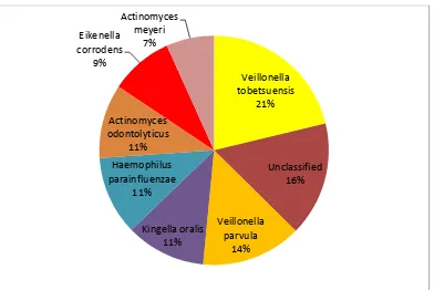
At the species level(top 8) among the health samples Veillonella
42
Kingella oralis which included 21%. 14% and 11% respectively as shown in
table 3-A and figure 3-A. Unclassified species accounted for about 16%
among the top 8 species in health.
Gingivitis group:
There were a total of 7 phyla, 32 genera and 59 species found in the
gingivitis samples.
At the phylum among the gingivitis samples Firmicutes was more in
abundance of about 24% followed by Candidatus Saccharibacteri 20% and
Proteobacteria 17% as shown in table 1-B and figure 1-B.
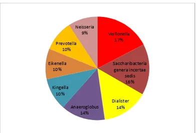
At the genus level(top 8) among the gingivitis samples Veillonella,
Saccharibacteria genera incertae sedis and Dialister had more abundance of
about 17% 16% and 14% respectively as shown in table 2-B and figure 2-B.
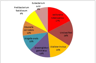
At the species level(top 8) among the gingivitis samples Veillonella
tobetsuensis had more abundance of 17%, followed by Dialister invisus 15%
and Anaeroglobus geminatus 14% as shown in table 3-B and figure 3-B.
Unclassified species contributed for about 16% among the top 8 species in
gingivitis.
Comparison of abundance of phyla among health and gingivitis:
The comparison of abundance of phyla between the health and disease
43
and proteobacteria in the healthy group as compared to the disease group. The
abundance of Candidatus Saccharibacteria, Actinobacteria, Bacteroidetes and
Fusobacteria were found to be higher in the disease group as compared to the
healthy group. Synergistetes was found only in the diseased group.
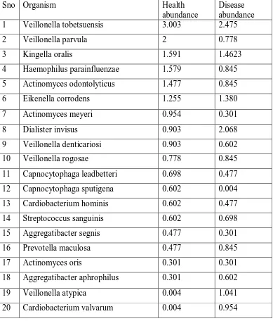
Comparison of abundance of top 20 species in health vs gingivitis:
The comparison of abundance of top 20 species present in health vs
gingivitis has been compared as shown in table 4-A and graph 2-A. The mean
standard deviation value for abundance of species in health is 0.925336 ±
0.7269395 and gingivitis is 0.865053 ± 0.6018219. The comparison of
abundance of top 20 species in health vs gingivitis was not statistically
significant at p=0.276 as shown in graph 2-B.
Comparison of abundance of top 20 species in gingivitis vs health:
Comparison of abundance of top 20 species present in gingivitis vs
healthhas been compared in table 4-B and graph 3-A. The mean standard
deviation value for abundance of species in disease is 0.847391 ± 0.5677049
and health is 0.726773 ± 0.7757812.The comparison of abundance of top 20
species in gingivitis vs health was not statistically significant at p=0.881 as
shown in the graph 3-B
There are about 11 species present in the healthy group but absent in
the gingivitis group that has been listed in the table 5-A. There are about 46
44
group as shown in table 5-B. These organisms may be the causative factor for
the gingivitis.
Comparison of subgingival microbiome in healthversus gingivitis with circular maximum likelihood phylogenetic tree at genus level:
The subgingival microbiome was compared between healthy and
gingivitis sites at genus level and is represented in the form of a circular
phylogenetic tree in figure 3. The tree has been constructed with phyloT
software and is displayed using iTOL as per Letunic and Bork73. The bars in the outer band (green) represent the relative abundance of bacterial genera in
TABLES AND GRAPHS
Table 1-A: Evaluation of abundance of phyla and their percentage among the health samples
Sno Phylum Relative abundance
expressed in logs
Percentage of phyla
1 Firmicutes 3.056 30%
2 Proteobacteria 2.392 23%
3 Candidatus Saccharibacteria
1.799 18%
4 Actinobacteria 1.633 16%
5 Bacteroidetes 1.342 13%
[image:69.595.111.501.439.701.2]6 Fusobacteria 0.004 0%
Figure 1-A: Comparison of percentage of phyla in health
Table 1-B: Evaluation of abundance of Phyla and their percentage among gingivitis samples
Figure 1-B:Comparison of percentage of phyla in gingivitis
Sno Phylum Relative
abundance expressed in logs
Percentage of Phyla
1 Firmicutes 2.775 24%
2 Candidatus Saccharibacteria 2.303 20%
3 Proteobacteria 2.021 17%
4 Actinobacteria 1.653 14%
5 Bacteroidetes 1.447 13%
6 Synergistetes 1.255 11%
7 Fusobacteria 0.069 1%