0022-538X/10/$12.00 doi:10.1128/JVI.01385-10
Copyright © 2010, American Society for Microbiology. All Rights Reserved.
Determinants of Strain-Specific Differences in
Efficiency of Reovirus Entry
䌤
Payel Sarkar and Pranav Danthi*
Department of Biology, Indiana University, Bloomington, Indiana 47405
Received 2 July 2010/Accepted 30 September 2010
Cell entry of reovirus requires a series of ordered steps, which include conformational changes in outer capsid protein1 and its autocleavage. The1N fragment released as a consequence of these events interacts with host cell membranes and mediates their disruption, leading to delivery of the viral core into the cytoplasm. The prototype reovirus strains T1L and T3D exhibit differences in the efficiency of autocleavage, in the propensity to undergo conformational changes required for membrane penetration, and in the capacity for penetrating host cell membranes. To better understand how polymorphic differences in1 influence reovirus entry events, we generated recombinant viruses that express chimeric T1L-T3D1 proteins and characterized them for the capacity to efficiently complete each step required for membrane penetration. Our studies revealed two important functions for the central␦region of1. First, we found that1 autocleavage is regulated by the N-terminal portion of␦, which forms an␣-helical pedestal structure. Second, we observed that the C-terminal portion of␦, which forms a jelly-rollbarrel structure, regulates membrane penetration by influencing the efficiency of ISVP* formation. Thus, our studies highlight the molecular basis for differences in the membrane penetration efficiency displayed by prototype reovirus strains and suggest that distinct portions of the reovirus
␦domain influence different steps during entry.
In comparison to enveloped viruses, the mechanisms by which nonenveloped viruses enter host cells are not well un-derstood. While enveloped viruses use membrane fusion trig-gered by receptor- or pH-mediated activation of viral mem-brane protein for entering host cells (63), nonenveloped viruses lack a membrane and therefore must bypass host mem-branes by mechanisms other than fusion. Nonenveloped vi-ruses from diverse families appear to use a conserved set of cell entry strategies (4, 59). However, a detailed molecular understanding of how their genomic payloads access the interior of host cells has yet to be elucidated. Mammalian reoviruses, which form nonenveloped viruses with two con-centric protein shells, are extensively used as model systems to study different aspects of virus-host interactions, includ-ing virus entry (12, 17).
Successful entry of reovirus into host cells requires a series of events that culminate in delivery of the inner protein shell, or core, into the host cell cytoplasm (12, 17). Reovirus entry is initiated by binding of the viral attachment protein,1, to cell surface carbohydrates (5, 15, 27, 28, 52) and junctional adhe-sion molecule A (6). Following receptor engagement, reovirus is internalized by1 integrin-dependent endocytosis (9, 10, 22, 41, 42, 57). Within endosomes, low pH and cathepsin proteases facilitate stepwise disassembly of reovirus to form a metastable intermediate, the infectious subvirion particle (ISVP), which contains a conformationally altered1 protein, lacks the3 outer capsid protein (which is degraded by proteolysis), and contains a 1 membrane penetration protein that has been cleaved into three particle-associated fragments (3, 10, 14, 20,
21, 55, 57). Generation of the three 1 fragments requires autocleavage of1 at residue 42, which results in the forma-tion of1N and1C (34). In addition,1C is proteolytically cleaved near residue 580 to generate␦andfragments (48). Conformational changes in ISVPs lead to the generation of the next entry intermediate, the ISVP*(11). These rearrangements result in dissociation of 1N,, and1 from the ISVP and lead to exposure of hydrophobic domains within the particle-associated␦fragment (11, 33). The1N fragments released from the particles are necessary and sufficient to mediate mem-brane penetration (33, 65). Memmem-brane penetration by 1N leads to the generation of 4- to 10-nm pores (1, 65), which are thought to facilitate the delivery of the 70-nm cores into the cytoplasm via an as-yet-unknown mechanism.
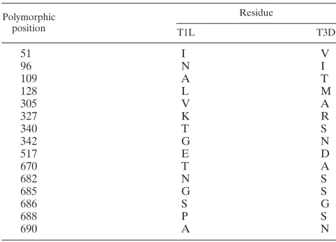
Prototype reovirus strains, type 1 Lang (T1L) and type 3 Dearing (T3D), display numerous differences in the cell entry pathway described above. For example, T1L and T3D differ in efficiency of autocatalytic1 cleavage (49), in the efficiency to form ISVP*s (11) and ultimately in the capacity to permeab-ilize host cell membranes and evoke hemolysis (11, 39). In each case, T3D is more efficient than T1L and reassortant analysis indicates that the difference in efficiency genetically maps to the M2 gene segment (11, 39, 49), which encodes the mem-brane penetration protein, 1 (46). Comparison of the de-duced amino acid sequences of our laboratory stocks of T1L and T3D revealed differences at fifteen amino acid residues in the 708-amino-acid 1 protein, which likely account for the phenotypic differences described above. Although the amino acid sequence of the1N fragment was found to be identical between T1L and T3D, nine and six residues were different within the␦ and domains, respectively (Table 1). The1 protein folds into three␣-helical domains formed by␦and near the base of the 1 trimer and one jelly-roll  barrel domain formed by␦near its apex (38). How these structural * Corresponding author. Mailing address: Department of Biology,
Indiana University, Bloomington, IN 47405. Phone: (812) 856-2449. Fax: (812) 856-5710. E-mail: [email protected].
䌤Published ahead of print on 13 October 2010.
12723
on November 8, 2019 by guest
http://jvi.asm.org/
domains contribute to the various functions ascribed to 1 during cell entry is not known.
In the present study, we used reverse genetics to identify1 domains that account for strain-specific differences between T1L and T3D. Using recombinant reoviruses that encode chi-meric1 proteins derived from portions of T1L and T3D M2 gene segments, we examined the efficiency of 1-regulated steps during reovirus cell entry. We found that presence of the T3D␦domain increases the efficiency of1 autocleavage and ISVP* formation, thereby facilitating membrane penetration. To better define the contribution of the ␦ domain to these steps, we generated additional viruses that express1 proteins with swaps between portions of the T1L and T3D␦domains. Characterization of these viruses indicated a function for the pedestal-forming region in regulating efficiency of autocleav-age, whereas a role for the jelly-rollbarrel domain in regu-lating conformational changes required for membrane pene-tration. These findings suggest independent regulation of1 autocleavage, ISVP* formation, and membrane penetration by distinct portions of the1␦domain during entry of reovirus into host cells.
MATERIALS AND METHODS
Cells. Murine L929 (L) cells were maintained in Joklik minimal essential medium (MEM; Lonza) supplemented to contain 5% fetal bovine serum (FBS) (Sigma-Aldrich), 2 mML-glutamine (Invitrogen), 100 U of penicillin (Invitro-gen)/ml, 100g of streptomycin (Invitrogen)/ml, and 25 ng of amphotericin B (Sigma-Aldrich)/ml. BHK-T7 cells were maintained in Dulbecco modified Eagle medium (Invitrogen) supplemented to contain 5% FBS, 2 mML-glutamine, 2% MEM amino acid solution (Invitrogen), and 1 mg of Geneticin (Invivogen)/ml in alternate passages.
Plasmids.pT7-M2T1L and pT7-M2T3D (35, 36) were used to construct plas-mids, pT7-M2DL, pT7-M2LD, pT7-M2(LD)L, pT7-M2(LD)D, pT7-M2(DL)L, and pT7-M2(DL)D, which express chimeric1 proteins (see Fig. 1 and Fig. 5A). pT7-M2DL and pT7-M2LD were constructed by reciprocally exchanging restric-tion fragments obtained from digesrestric-tion of pT7-M2T1L and pT7-M2T3D with NcoI and SpeI. For the construction of M2(LD)L, M2(LD)D, pT7-M2(DL)L, and pT7-M2(DL)D, a SnaBI site was introduced in pT7-M2T1L and pT7-M2T3D plasmids by QuikChange (Stratagene) site-directed mutagenesis at nucleotides 963 and 739, respectively, in each case replacing a G for a T. These changes do not lead to a change in the amino acid sequence. pT7-M2(LD)D and pT7-M2(DL)L plasmids were constructed by reciprocally exchanging restriction fragments obtained from digestion of pT7-M2T1L(SnaBI) and
pT7-M2T3D(SnaBI) with NcoI and SnaBI. pT7-M2(LD)L and pT7-M2(DL)D plas-mids were constructed by reciprocally exchanging restriction fragments obtained from digestion of pT7-M2T1L(SnaBI) and pT7-M2T3D(SnaBI) with SpeI and SnaBI. Chimeric M2 plasmids were confirmed by sequencing.
Viruses. Recombinant strain T1L (rsT1L), rsT1L/T3D M2, rsT1L/DL M2, rsT1L/LD M2, rsT1L/(LD)L M2, rsT1L/(LD)D M2, rsT1L/(DL)L M2, and rsT1L/(DL)D M2, which contain a wild-type or chimeric M2 gene in an other-wise T1L background were generated by using a four- or ten-plasmid-based reverse-genetics strategy (36). To confirm sequences of mutant viruses, viral RNA was extracted from infected cells and subjected to reverse transcription-PCR using three sets of M2-specific primers. transcription-PCR products were resolved on Tris-acetate-EDTA agarose gels, purified and subjected to sequence analysis. Purified reovirus virions were generated by using second- or third-passage L-cell lysate stocks of reovirus as described previously (25). Viral particles were ex-tracted from infected cell lysates using Vertrel-XF (Dupont) (43), layered onto 1.2- to 1.4-g/cm3
CsCl gradients, and centrifuged at 187,813⫻gfor 4 h. Bands corresponding to virions (1.36 g/cm3) (56) were collected and dialyzed in
virion-storage buffer (150 mM NaCl, 15 mM MgCl2, 10 mM Tris-HCl [pH 7.4]). The
concentration of reovirus virions in purified preparations was determined from an equivalence of one optical density (OD) unit at 260 nm equals 2.1⫻1012
virions (56). Virus titer was determined by plaque assay using L cells (60). ISVPs were generated by incubation of 2⫻1011virions with 200g ofN-p-tosyl-L-lysine
chloromethyl ketone-treated chymotrypsin/ml in a total volume of 0.1 ml at 37°C for 1 h (47). Proteolysis was terminated by addition of 2 mM phenylmethylsul-fonyl fluoride and incubation of reactions on ice. Generation of ISVPs was confirmed by sodium dodecyl sulfate-polyacrylamide gel electrophoresis (SDS-PAGE) and Coomassie brilliant blue staining.
Growth assay.L cells (2⫻105) in 24-well plates were adsorbed in triplicate
with 2 PFU/cell of reovirus at room temperature for 1 h, washed once with phosphate-buffered saline (PBS), and incubated at 37°C for 0 and 24 h. Cells were frozen and thawed twice prior to determination of the virus titer by plaque assay using L cells (60). Viral yield was calculated according to the following formula: viral yield (log10PFU/ml)⫽log10(PFU/ml)24 h⫺log10(PFU/ml)0 h.
Analysis of1N-1C cleavage efficiency.Reducing sample buffers containing 125 mM Tris (adjusted to a final pH of 6.8, 8.3, or 9.8), 10% (vol/vol) glycerol, 2% (wt/vol) SDS, 0.01% (wt/vol) bromophenol blue, and 5%-mercaptoethanol were mixed with an equal volume of dialysis buffer containing 1011virions. The
samples were disrupted by heating at 60°C for 5 min, resolved on SDS–10% PAGE gels and stained using Coomassie brilliant blue. Gels were scanned by using an Odyssey infrared imager (LI-COR), and the intensity of1 and1C bands was determined by densitometry using Odyssey software. The percent uncleaved1 was calculated as described previously (49), according to the following formula: % uncleaved1⫽[1 intensity/(1⫹ 1C intensity)]⫻100.
Analysis of ISVP* generation.ISVPs (8⫻1010) were incubated with 300 mM
CsCl or NaCl at 30°C for 20 min, transferred to ice for 20 min, and incubated with 100g of trypsin/ml at 4°C for 30 min. Trypsin digestion was terminated by addition of SDS-PAGE loading buffer and removal of the samples to dry ice. Generation of ISVP*s was confirmed by SDS-PAGE and Coomassie brilliant blue staining.
Measurement of thermal stability.ISVPs in virion storage buffer were incu-bated at various temperatures for 15 min and transferred to ice for 1 h. Residual infectivity in triplicate samples was assessed by plaque assay (60).
Hemolysis assay.Citrated calf red blood cells (RBCs; Colorado Serum Com-pany) were washed extensively with chilled PBS supplemented to contain 2 mM MgCl2(Mg) and resuspended at a concentration of 30% (vol/vol) in
PBS-Mg. Hemolysis efficiency was analyzed by mixing a 3-l aliquot of resuspended RBCs with virion-storage buffer containing 0 or 300 mM CsCl–1% Triton X-100 (TX-100) in virion storage buffer or ISVPs in a total volume of 30l, followed by incubation at 37°C for 1 h. Samples were placed on ice for 30 min to prevent further hemolysis and centrifuged at 500⫻gat 4°C for 3 min. The extent of hemoglobin release was quantified by measuring theA405of a 1:5 dilution of the
supernatant in a microplate reader (Molecular Devices). The percent hemolysis was calculated by using the following formula: % hemolysis⫽100⫻(Asample⫺ Abuffer)/(ATX-100⫺Abuffer).
Assessment of ISVP* formation after exposure to erythrocytes.Hemolysis reactions were set up as described above. After incubation at 37°C for 40 min, samples were transferred to ice for 20 min and incubated with 100g of tryp-sin/ml at 4°C for 30 min. Trypsin digestion was terminated by the addition of SDS-PAGE loading buffer and removal of the samples to dry ice. Samples were resolved by SDS-PAGE and transferred to nitrocellulose membranes. Mem-branes were blocked for 1 h in blocking buffer (Tris-buffered saline [TBS] containing 0.05% Tween 20 and 5% milk), followed by incubation with a 1-specific MAb 4A3 diluted 1:1,000 in blocking buffer at 4°C overnight.
Mem-TABLE 1. Location and nature of polymorphic residues in T1L and T3D1 proteins
Polymorphic position
Residue
T1L T3D
51 I V
96 N I
109 A T
128 L M
305 V A
327 K R
340 T S
342 G N
517 E D
670 T A
682 N S
685 G S
686 S G
688 P S
690 A N
on November 8, 2019 by guest
http://jvi.asm.org/
[image:2.585.42.283.88.262.2]branes were washed three times for 10 min each time with TBS containing 0.05% Tween 20 and incubated with 1:25,000 dilution of Alexa Fluor-conjugated goat anti-mouse immunoglobulin in blocking buffer. After three washes, membranes were scanned using an Odyssey infrared imager (LI-COR).
RESULTS
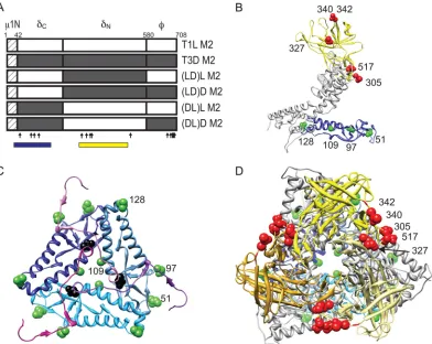
Viruses with chimeric1 are viable.Entry of reovirus into host cells consists of a series of events which include1 auto-cleavage, ISVP formation, ISVP-to-ISVP* conversion, and membrane penetration. Prototype reovirus strains T1L and T3D display differences in efficiency at various steps in this pathway and these differences segregate with the1-encoding M2 gene segment (11, 39, 49). To define 1 domains that contribute to the strain-specific differences in efficiency of cell entry between T1L and T3D, we constructed M2 chimeric gene segments by reciprocally exchanging␦anddomains of T1L and T3D M2 gene segments (Fig. 1). Using plasmid-based reverse genetics, we generated recombinant reoviruses that express either T1L, T3D, or chimeric1 proteins in an other-wise T1L background (36). These viruses were designated rsT1L, rsT1L/T3D M2, rsT1L/LD M2 (containing␦from T1L,
from T3D), and rsT1L/DL M2 (containing␦from T3D and
from T1L). To evaluate whether the differences in1 se-quences affect viral growth over a single cycle of reovirus rep-lication, we infected L cells with each of the viruses at a mul-tiplicity of infection (MOI) of 2 PFU/cell and quantified virus titers at 0 and 24 h after infection. At 24 h after infection, each virus strain showed an equivalent (⬃100-fold) increase in virus titer (data not shown), indicating that the1 chimeric viruses replicate as efficiently as the wild-type virus.
The ␦domain is a determinant of 1N-1C cleavage effi-ciency.The 1 protein undergoes autocleavage during virus assembly or cell entry to form1N and1C (40, 49, 58). This cleavage event affects the release of1N and serves an essen-tial role in controlling membrane penetration and virion infec-tivity (1, 33, 50). When reovirus particles are resolved on de-naturing acrylamide gels under standard conditions, the 1 protein is predominantly found in its autocleaved form,1C. However, increasing the pH of the denaturing sample buffer used for resolving viruses for electrophoresis leads to a greater recovery of nonautocleaved1 (49). Using this strategy, it has been determined that T3D1 is more susceptible to
autocleav-age in comparison to T1L 1 (49). Moreover, these strain-specific differences in cleavage efficiency map to the M2 gene segment (49). Although experimental data for this is as yet lacking, we think it possible that differences in autocleavage efficiency observed for T1L and T3D1 proteinsin vitroalso correlate with the extent of autocleavage during cell entry and that similar regions of 1 may regulate autocleavage under both conditions.
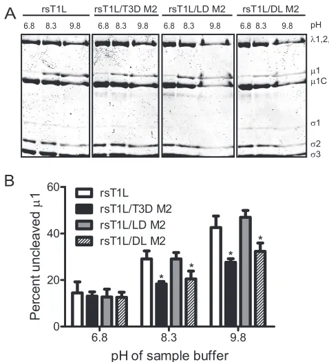
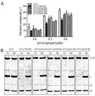
[image:3.585.298.537.66.329.2]To define regions in 1 that influence the efficiency of1 autocleavage, we compared the capacity of1 proteins from rsT1L/LD M2 and rsT1L/DL M2 to undergo autocleavage to rsT1L and rsT1L/T3D M2 (Fig. 2A). For these experiments, virus particles from each virus strain were electrophoresed using sample buffers at pH 6.8, 8.3, or 9.8, and the percentage of1 that remained uncleaved was determined by densitomet-ric analysis (Fig. 2B). For samples resolved under standard conditions of pH 6.8, the percentage of uncleaved 1 was found to be approximately equal for all virus strains tested. As expected, we found that as the pH was increased, a progres-sively greater proportion of1 protein from rsT1L was found in the uncleaved form in comparison to1 from rsT1L/T3D M2. When chimeric1 viruses were analyzed, we found that while1 from rsT1L/LD M2 showed autocleavage efficiency FIG. 1. Viruses with chimeric1 membrane penetration protein.
[image:3.585.69.255.68.149.2]Schematic diagrams of the1-encoding T1L and T3D M2 gene seg-ments are shown, along with cleavage products generated during virus entry. Chimeric M2 gene segments (LD and DL) were constructed by reciprocally exchanging the␦- and-encoding portions of T1L and T3D M2 gene segments. The amino acid sequence of1N is identical for T1L and T3D and is shown as a hatched box. The locations of polymorphic residues in T1L and T3D1 are shown using vertical arrows.
FIG. 2. The ␦ domain affects 1N-1C cleavage efficiency. (A) CsCl-purified preparations of the indicated virus strains were mixed with reducing sample buffers at pH 6.8, 8.3, or 9.8. After incu-bation at 60°C for 5 min, the samples were resolved on SDS–10% PAGE gels and stained with Coomassie brilliant blue. (B) The inten-sities of the1 and1C bands were determined by densitometric analysis using a LI-COR Odyssey scanner. The fraction of uncleaved 1 was determined by dividing the intensity of the1 band by the total intensity of the1 and1C bands. The results are expressed as the mean percent uncleaved1 quantified from three independent exper-iments. Error bars indicate the standard deviations (SD).*,P⬍0.05, as determined by using the Studentttest in comparison to rsT1L at the respective sample buffer pH.
on November 8, 2019 by guest
http://jvi.asm.org/
similar to rsT1L,1 from rsT1L/DL M2 displayed autocleav-age characteristic similar to rsT1L/T3D M2. These data indi-cate that differences between T1L and T3D in efficiency of
1N-1C cleavage are influenced by the ␦, but not the domain.
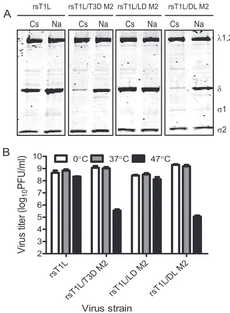
The␦domain affects efficiency of ISVP* formation. Mem-brane penetration by ISVPs requires conversion to ISVP*s, which are characterized by the release of1N andfragments and conformational changes in the particle-associated␦ frag-ment (11, 13, 33). Although the physiologic trigger for ISVP-to-ISVP* conversion is not known,in vitrostudies using ISVPs exposed to high concentrations of Cs or K ions are thought to mimic conditions required for ISVP* formation. Under these conditions, T3D forms ISVP*s more readily than T1L with the difference in efficiency mapping to the M2 gene segment (11). To identify the1 domain that affects ISVP* formation, we incubatedin vitro-generated ISVPs of each virus strain with 300 mM CsCl or with 300 mM NaCl as a control (11) (Fig. 3A). Conformational changes in particle-associated␦ were exam-ined by assessing its protease sensitivity to trypsin digestion. Consistent with previous observations (11), we observed
min-imal protease-mediated loss of T1L␦after treatment of rsT1L with CsCl (Fig. 3A). In contrast, the T3D-derived␦fragment of rsT1L/T3D M2 was lost upon protease treatment in the presence of CsCl. Analysis of chimeric1 viruses indicated that␦from rsT1L/LD M2 displayed protease resistance anal-ogous to rsT1L, whereas␦ from rsT1L/DL M2 showed pro-tease sensitivity similar to rsT1L/T3D M2 (Fig. 3A). These findings indicate that the capacity to undergo ISVP-to-ISVP* conversion is also controlled by the ␦ domain and that the presence of T1L residues within this domain renders this pro-cess inefficient.
Previous studies indicate that analogous to CsCl treatment, heat treatment of ISVPs also promotes a conformational change that increases the protease sensitivity of the␦fragment (45). Heat-induced conformational changes also result in de-creased viral infectivity (16, 44). As an independent measure of the capacity to generate ISVP*s, we assessed the thermal sen-sitivity of our viruses. For these studies, ISVPs of each virus strain were incubated at 0, 37, and 47°C for 15 min, and the residual infectivity of the viruses was determined by plaque assay (Fig. 3B). While the infectivity of rsT1L and rsT1L/LD M2 was unaffected after high-temperature incubation, the in-fectivity of rsT1L/T3D M2 and rsT1L/DL M2 was diminished by ⬃4 orders of magnitude after incubation at 47°C. These findings further support the idea that viruses containing the␦ domain from T1L are less capable of undergoing conforma-tional changes that resemble ISVP-to-ISVP* conversion.
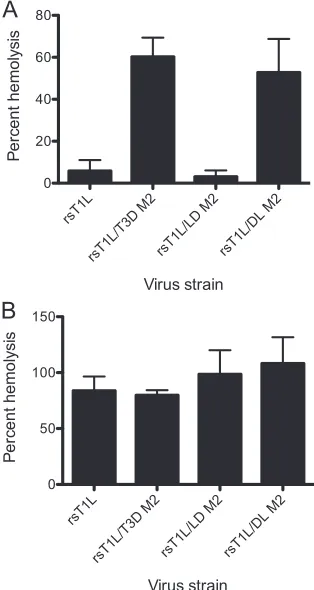
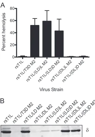
Strain-specific differences in membrane penetration are af-fected by␦.The capacity of reovirus ISVPs to disrupt eryth-rocyte membrane integrity and produce hemolysis correlates with endosomal membrane penetration (13, 16, 18, 44). Reo-virus strains T1L and T3D differ in their membrane-penetra-tion capacities, with T3D being more efficient than T1L, and this efficiency difference has been mapped to the M2 gene segment (11). To identify1 domains that affect membrane penetration capacity of reovirus, we tested ISVPs generated from wild-type and chimeric viruses for the capacity to lyse erythrocytes (Fig. 4A). As expected, while rsT1L failed to produce hemolysis, rsT1L/T3D M2 efficiently lysed erythro-cytes. Analysis of chimeric1 viruses under these conditions showed that rsT1L/LD M2 is incapable of erythrocyte lysis, whereas rsT1L/DL M2 is capable of eliciting erythrocyte lysis. These findings suggest that residues within the␦ domain of T3D M2 are required for efficient membrane penetration by reovirus.
[image:4.585.40.274.66.384.2]Two possible mechanisms may explain why rsT1L and rsT1L/LD M2 fail to elicit hemolysis. First, the decreased ef-ficiency may be a consequence of a diminished capacity to generate ISVP* as described in Fig. 3 above. Second, reduced hemolytic capacity of rsT1L and rsT1L/LD M2 may be related to a decrease in efficiency of pore formation. To distinguish between these possibilities, we performed hemolysis reactions in the presence of CsCl. Addition of CsCl to the reaction allows hemolysis by T1L (11), likely by promoting conforma-tional changes that are required for the release and exposure of the1N fragment (33, 65). Consistent with previous studies, rsT1L produced efficient hemolysis in the presence of 300 mM CsCl to levels that were equivalent to those produced by rsT1L/T3D M2 (Fig. 4B). Similarly, both rsT1L/LD M2 and rsT1L/DL M2 also were capable of erythrocyte lysis in the FIG. 3. The␦domain influences the capacity to undergo
ISVP-to-ISVP* conversion. (A) ISVPs of the indicated viruses were treated with CsCl or NaCl at 30°C for 20 min, chilled on ice for 20 min, and treated with trypsin at 4°C for 30 min. Samples were resolved by SDS-PAGE and stained using Coomassie brilliant blue. The positions of reovirus capsid proteins are shown. (B) ISVPs of the indicated viruses were incubated at 37, or 47°C for 15 min. Residual infectivity was assessed by plaque assay. The results are shown as virus titers for triplicate samples. Error bars indicate the SD.
on November 8, 2019 by guest
http://jvi.asm.org/
presence of CsCl. These findings indicate that the diminished capacity of rsT1L/LD M2 to undergo ISVP-to-ISVP* conver-sion likely contributes to the diminished capacity of this virus to elicit hemolysis.
Viruses expressing chimeric␦domains are viable.The find-ings presented thus far indicate a function for␦domain in1 autocleavage and in ISVP-to-ISVP* conversion. The nine polymorphic differences in the␦domain form two clusters, one each toward the N- and C-terminal ends of ␦, henceforth designated␦Nand ␦C, respectively (Fig. 5A). Four polymor-phic residues (residues 51, 96, 109, and 128) that lie within␦N
are all found within domain I, as described in the crystal struc-ture of1 (encompassing amino acids 30 to 186), which forms an␣-helical pedestal at the base of the 1 trimer (38) (Fig. 5B). The remaining five residues (residues 305, 327, 340, 342, and 517) that are found in␦Clie within or proximal to domain IV, as described in the crystal structure of1 (encompassing amino acids 306 to 514), which forms a jelly-roll  barrel structure at the apex of the trimer (38) (Fig. 5B). To define which portion of ␦ determines strain-specific differences in
1N-1C cleavage and ISVP* formation, we constructed chi-meric gene segments by reciprocally exchanging cDNA frag-ments corresponding to the␦Nand ␦C portions of T1L and
T3D1 (Fig. 5A). These constructs were used to generate four additional recombinant viruses in a T1L background, rsT1L/ (LD)L M2 (containing␦Nfrom T1L,␦Cfrom T3D, andfrom T1L), rsT1L/(LD)D M2 (containing␦Nfrom T1L,␦Cfrom T3D,
andfrom T3D), rsT1L/(DL)L M2 (containing␦Nfrom T3D,
␦C from T1L, andfrom T1L), and rsT1L/(DL)D M2
(con-taining␦Nfrom T3D, ␦C from T1L, and from T3D) (Fig. 5A). To determine whether the chimeric␦viruses were capa-ble of efficient replication, we infected L cells with rsT1L and chimeric viruses at an MOI of 2 PFU/cell, and we quantified the virus titers at 0 and 24 h after infection. At 24 h after infection, each virus strain showed an equivalent (⬃100-fold) increase in virus titer, indicating that the chimeric␦viruses are not compromised for growth and replicate as efficiently as the wild-type virus (data not shown).
Distinct portions of the␦domain regulate autocleavage and ISVP* formation.To determine the contribution of the␦Nand
␦C domains to regulation of 1 autocleavage, we incubated
purified preparations of each virus with sample buffers of in-creasing pHs and compared the fraction of uncleaved 1 present in each preparation using densitometric analysis (Fig. 6A). Analogous to parental viruses rsT1L and rsT1L/T3D M2, we found that the fraction of uncleaved1 for each virus in sample buffer of pH 6.8 was equivalent. As the pH of the sample buffer was increased to 8.3, a substantially greater frac-tion of T1L1 was found in the uncleaved form in comparison to T3D1. Analysis of␦chimeric viruses indicated that at pH 8.3, rsT1L/(LD)L M2 and rsT1L/(LD)D M2 also showed a statistically higher percentage of uncleaved1 in comparison to rsT1L/T3D M2. In contrast, a minimal increase in uncleaved
1 was observed for rsT1L/(DL)L M2 and rsT1L/(DL)D M2. Similar results were obtained when ISVPs of each of these viruses were incubated in sample buffer at pH 9.8, with rsT1L/ (LD)L M2 and rsT1L/(LD)D M2 displaying a higher fraction of uncleaved 1 than rsT1L/T3D M2. In addition to these viruses, the fraction of uncleaved 1 in rsT1L/(DL)L and rsT1L/(DL)D M2 also was found to be greater than that present in rsT1L/T3D M2 at pH 9.8. These findings suggest that the presence of the ␦N portion of T3D increases the efficiency of1 autocleavage and hints at a possible contribu-tion from the␦Cdomain in controlling autocleavage.
To define which portion of␦affects the capacity for ISVP-to-ISVP* transition, ISVPs from each virus strain were incu-bated with either CsCl or NaCl and assessed for protease susceptibility of the particle-associated ␦ fragment, which is characteristic of ISVP* formation (Fig. 6B). As expected, the␦ domain from the parental viruses rsT1L and rsT1L/T3D M2, respectively, displayed protease resistance and protease sus-ceptibility in the presence of CsCl. When chimeric␦viruses were similarly examined, we found that the␦fragment of all viruses was converted to a protease-sensitive form in the pres-ence of CsCl, suggesting that these viruses were capable of forming ISVP*s under conditions in which rsT1L was not. These results suggest that the presence of either the␦Nor the
␦Cfrom T3D is sufficient to confer to T1L1 the capacity for
efficient ISVP-to-ISVP* conversion.
Sequences within ␦C regulate the efficiency of membrane penetration.To determine how␦Nand␦Cinfluence membrane
[image:5.585.84.241.70.365.2]penetration by reovirus, we compared the hemolytic capacities of the chimeric␦viruses to those of the parental viruses (Fig. FIG. 4. The ␦domain modulates membrane penetration. A 3%
(vol/vol) solution of bovine erythrocytes was incubated with 4.8⫻1010
ISVPs of the indicated viruses in virion-storage buffer containing no CsCl (A) or 300 mM CsCl (B) at 37°C for 1 h. Hemolysis was quan-tified by determining absorbance of the supernatant at 405 nm. He-molysis after treatment of an equal number of cells with virion-storage buffer or virion-storage buffer containing 1% TX-100 was considered to be 0 or 100%, respectively. The results are expressed as the mean percent hemolysis for triplicate samples. Error bars indicate the SD.
on November 8, 2019 by guest
http://jvi.asm.org/
7A). We found that rsT1L/(LD)L M2 and rsT1L(LD)D M2 were capable of hemolysis analogous to rsT1L/T3D M2. In contrast, rsT1L/(DL)L M2 and rsT1L/(DL)D M2 were unable to lyse bovine erythrocytes similar to rsT1L. Because rsT1L/ (DL)L M2 and rsT1L/(DL)D M21 proteins are efficiently autocleaved (Fig. 6A) and because these viruses are capable of ISVP* formation when treated with Cs (Fig. 6B), their reduced capacity for hemolysis suggest a function for the ␦ domain subsequent to ISVP* formation. An alternate, equally likely possibility is that despite their capacity to undergo conforma-tional changes that resemble ISVP* formation after incubation with CsCl in vitro, these viruses fail to undergo functional conformational changes that result in ISVP* formation and
1N release during interaction with membranes, as would oc-cur during hemolysis. To test this idea, trypsin sensitivity of␦ in hemolysis reactions was assessed by using immunoblots. Consistent with previous results (11), the ␦ fragments from rsT1L and rsT1L/T3D, respectively, displayed trypsin resis-tance and trypsin susceptibility. Similar examination of our chimeric viruses indicated that ␦ fragments of nonhemolytic
viruses rsT1L/LD M2, rsT1L/(DL)L M2, and rsT1L/(DL)D M2 remained trypsin resistant, whereas those from hemolysis-competent viruses rsT1L/DL M2, rsT1L/(LD)L M2, and rsT1L/(LD)D M2, were protease sensitive (Fig. 7B). Based on these findings we conclude that one or more residues within T3D-derived␦Cpromote hemolysis by increasing the efficiency of ISVP* formation.
DISCUSSION
[image:6.585.97.489.68.380.2]Membrane penetration by reovirus is an exquisitely regu-lated, multistep process controlled by the1 outer capsid pro-tein (12, 17). Successful membrane penetration requires prim-ing of the membrane-penetration machinery through 1 autocleavage (1, 33, 49, 50). In addition, conformational changes in the 1 protein are required to expose domains capable of membrane interaction and pore formation (11, 13, 16). Although the primary sequence of the 708-amino-acid1 proteins of prototype reovirus strains T1L and T3D differ by only fifteen amino acids, these viruses exhibit differences in the FIG. 5. Viruses with chimeric␦fragments. (A) Schematic diagram of the1-encoding T1L and T3D M2 gene segments are shown, along with cleavage products generated during virus entry. Chimeric M2 gene segments (LD)L, (LD)D, (DL)L, and (DL)D were constructed by reciprocally exchanging the␦N- and␦C-encoding portions of T1L and T3D M2 gene segments. The locations of polymorphic residues in T1L and T3D1 are
indicated by vertical arrows. Regions spanning domain I and domain IV are indicated by blue and yellow boxes, respectively. (B) A side view of a1C monomer rendered using UCSF chimera from the crystal structure of1 (PDB:1JMU) is shown with domain I in blue and domain IV in yellow (38, 53). The positions of the polymorphic residues within domains I and IV are shown in green and red, respectively. Residue 97 is labeled since residue 96 was not resolved in the crystal structure and is not in the PDB file. (C) A close-up top view of the region near the autocleavage site is shown, with the domain I comprising␦in shades of blue and the1N fragment in shades of magenta. Polymorphic residues on one of the three chains are labeled as described for panel B. The autocleavage site is shown in black. (D) A top view of a1C trimer is shown, with domain I in shades of blue and domain IV in shades of yellow. Polymorphic residues on one of the three chains are labeled as described for panel B.
on November 8, 2019 by guest
http://jvi.asm.org/
efficiency of autocleavage (49), the propensity to undergo con-formational changes required for membrane penetration (11), and the capacity of these strains to permeabilize host cell membranes (11, 39). To better understand how each of these cell entry steps is regulated by1, we generated and charac-terized recombinant viruses that express chimeric T1L-T3D1 proteins. We found that the efficiency of1 autocleavage is regulated by the␣-helical pedestal domain formed by the N-terminal region of␦(␦N). In addition, we found that the C-terminal portion of␦, which forms the jelly-rollbarrel (␦C), regulates the membrane penetration efficiency of reovirus by controlling the efficiency of ISVP* formation. Collectively, our studies highlight how amino acid polymorphisms within reovi-rus1 affect differences in efficiency of cell entry displayed by prototype reovirus strains and indicate that distinct portions of the reovirus␦domain influence different steps during entry.
Autocleavage of1 resulting in the formation of1N and
1C is required for membrane penetration (1, 33, 49, 50). Recent biochemical evidence along with structural information about 1 from high-resolution cryo-electron microscopic (cryoEM) structures indicate that 1 exists in its unauto-cleaved form in assembled virions and suggest that autocleav-age occurs at an early stautocleav-age during entry (49, 66).
[image:7.585.133.445.64.381.2]Conforma-tional changes in1␦occurring as a consequence of ISVP-to-ISVP* transition have been proposed as a possible trigger for autocleavage (49). However, it is also possible that autocleav-age occurs prior to this step. In support of this idea, a recent 3.3-Å reconstruction of related aquareovirus indicates that VP5, the 1 homolog, exists in its autocleaved form in the ISVP (67). The autocleavage site between Asn42 and Pro43 lies within a pocket on the upper surface of domain I (Fig. 5C), and it is hypothesized that residues on the same or adjacent1 molecules that are in close proximity to the cleavage site may facilitate autocleavage (38). The data presented here offer experimental evidence to support this idea. We found that viruses containing the pedestal forming domain from T3D display higher 1 autocleavage efficiency in comparison to those that contain the similar region from T1L. Based on structural information, residues Thr104, Glu105, and His106 on the helix formed by the same1 subunit and residue Lys113 on the helix formed by the neighboring1 subunit have been implicated in facilitating this process (38). The pedestal region sequences of T1L and T3D1 differ at residues 51, 96, 109, and 128, suggesting that one or more of these residues influ-ences the efficiency of autocleavage. Whether these residues directly participate in the nucleophilic reaction required for FIG. 6. Different portions of␦modulate autocleavage efficiency and ISVP* formation. (A) CsCl-purified preparations of the indicated virus strains were mixed with reducing sample buffers at pH 6.8, 8.3, or 9.8. After incubation at 60°C for 5 min, the samples were resolved on SDS–10% PAGE gels and stained using Coomassie brilliant blue. The intensities of the1 and1C bands were determined by densitometric analysis using the LI-COR Odyssey scanner. The fraction of uncleaved1 was determined by dividing the intensity of the1 band by the total intensity of the 1 and1C bands. The results are expressed as mean percent uncleaved1 for four independent experiments. Error bars indicate the SD.*,P⬍ 0.05, as determined by using the Studentttest in comparison to rsT1L/T3D M2 at the respective sample buffer pH. (B) ISVPs of the indicated viruses were treated with CsCl or NaCl at 30°C for 20 min, chilled on ice for 20 min, and treated with trypsin at 4°C for 30 min. Samples were resolved by SDS-PAGE and stained using Coomassie brilliant blue. The positions of reovirus capsid proteins are shown.
on November 8, 2019 by guest
http://jvi.asm.org/
1N-1C cleavage or whether they affect the efficiency of autocleavage by altering the local structure of1 in that region remains unknown. Comparison of the subatomic-resolution cryoEM structures of ISVPs expressing T1L, T3D, or chimeric
1 may be helpful in better understanding how the␦Ndomain
affects autocleavage.
Our results indicate that virus strains expressing a T1L-derived pedestal [rsT1L/(LD)L M2 and rsT1L/(LD)D M2] re-tain the capacity for hemolysis despite having a lower auto-cleavage efficiency than those containing a T3D-derived pedestal (rsT1L/T3D M2 or rsT1L/DL M2). These findings suggest that differences in autocleavage efficiency observed in parental strains T1L and T3D (49) do not contribute to the differences in membrane penetration efficiency observed by hemolysis or chromium release assays (11, 39). Additional studies with recombinant viruses that encode1 mutants with significantly lower autocleavage efficiency are needed to quan-tify cleavage efficiency required for the release of a sufficient number of1N molecules to mediate membrane penetration. The 1N fragment liberated from 1 by autocleavage re-mains buried in the pedestal domain formed by␦and is not functional for membrane penetration in ISVPs (38). The ex-posure and release of 1N requires conversion of ISVPs to ISVP*s, which leads to massive conformation changes in the particle-associated ␦ fragment, as evidenced by exposure of
new ␦ epitopes and enhanced protease sensitivity (11). Al-though the precise nature of the conformational change is not understood, amino acid substitutions in the jelly-rollbarrel domain of␦that enhance thermal stability of reovirus particles (44) or render them resistant to 33% ethanol (32, 62) prevent conformational changes in␦, implicating the␦jelly-roll bar-rel domain in regulating ISVP* formation. The findings pre-sented here provide additional evidence for the function of the jelly-roll-barrel domain in regulating the efficiency of mem-brane penetration. There are five polymorphic differences be-tween T1L and T3D in this region of the1 protein (Table 1). Two of these residues lie in positions where they can affect interaction between two1 monomers within a trimer (resi-dues 305 and 327) (Fig. 5D). The remaining three resi(resi-dues (residues 340, 342, and 517) are found exposed to the solvent on the outer surface of the1 trimer (Fig. 5D), where they may influence interactions between adjacent1 trimers. Interest-ingly, mutant residues in ethanol- and heat-resistant reoviruses also are found within the same regions described above that can potentially affect interaction between 1 monomers or between1 trimers (38, 44, 62). Thus, we think that residues in the inter- and intratrimer interface both regulate the efficiency of ISVP* formation.
In addition to domain IV, structural studies also implicate domain I in regulating ISVP-to-ISVP* conversion (38, 66). Residues 72 to 96, which are disordered in the crystal structure of1 (38), form a loop that projects sideways and interacts with two other different subunits on an adjacent1 trimer (66). The 72-96 loop contacts one subunit between residues 126 and 129 and one subunit between residues 57 and 65 (66). In addition to participating in1-1 interactions, domain I also is thought to interact with the core particle via a loop formed by amino acids 51 to 62 (66). Three of the four amino acid resi-dues that differ between T1L and T3D (resiresi-dues 51, 96, and 128) lie within these predicted regions of interaction. Thus, it is possible that differences in the efficiency of these viruses to form ISVP* is related to amino acid differences in domain I. Consistent with this idea, characterization of rsT1L/(DL)D M2 and rsT1L/(DL)L M2 indicated that the presence of the T3D-derived pedestal region was sufficient to promote ISVP* in the presence of CsCl. However, this region did not promote ISVP* formation and hemolysis when ISVPs of rsT1L/(DL)D M2 and rsT1L/(DL)L M2 were exposed to erythrocytes, suggesting that polymorphisms within the pedestal domain do not account for differences in the efficiency of T1L and T3D 1 to regulate functional conformational changes that are required for ISVP-to-ISVP* conversion and pore formation upon interaction with the membranes.
[image:8.585.84.238.66.288.2]The cell entry mechanism of reovirus parallels other related and unrelated nonenveloped viruses in many aspects. For ex-ample, the capsid protein needs to be primed for entry by cleavage for closely related rotavirus and also more distantly related viruses, such as adenovirus, Flock House virus, and poliovirus. In some cases, these cleavage events are protease mediated (e.g., cleavage of adenovirus proprotein VI and ro-tavirus VP4) (23, 29), whereas in others capsid protein cleav-age is autocatalytic, similar to reovirus (e.g., cleavcleav-age of Flock house virus␣, poliovirus, and rhinovirus VP0) (7, 26, 31, 37, 54). Although these cleavage events are critical regulators of viral infectivity, where and when this cleavage occurs during FIG. 7. The jelly-roll barrel domain affects efficiency of
mem-brane penetration. (A) A 3% (vol/vol) solution of bovine erythrocytes was incubated with 4.8⫻1010ISVPs of the indicated viruses in
virion-storage buffer at 37°C for 1 h. Hemolysis was quantified by determining absorbance of the supernatant at 405 nm. Hemolysis after treatment of an equal number of cells with virion-storage buffer or virion-storage buffer containing 1% TX-100 was considered to be 0 or 100%, respec-tively. The results are expressed as mean percent hemolysis for six samples. Error bars indicate the SD. (B) A 3% (vol/vol) solution of bovine erythrocytes was incubated with 4.8⫻1010ISVPs of the
indi-cated viruses in virion-storage buffer at 37°C for 40 min, chilled on ice for 20 min, and treated with trypsin at 4°C for 30 min. Samples were resolved by SDS-PAGE and transferred to nitrocellulose membranes. The samples were probed with a1-specific MAb and visualized by using a LI-COR Odyssey scanner.
on November 8, 2019 by guest
http://jvi.asm.org/
virus infection remains unclear. In addition to priming cleav-age, conformational changes that expose membrane-interact-ing hydrophobic domains also are required for cell entry for these viruses. In some cases, interaction with a host cell recep-tor is the trigger for conformational change (e.g., Flock house virus and poliovirus) (2, 24, 61); in others, the low pH envi-ronment of the cellular endosomes modulates conformational changes (e.g., adenovirus) (30, 64). Since the known receptors and endosomal pH appear not to be required to induce con-formational changes in reovirus, the cellular trigger that pro-motes conformational changes needed for cell entry remain unknown. Analogous to the release of reovirus1N (1, 33, 65), adenovirus protein VI (64), Flock house virus␥peptide (8, 51), and poliovirus VP4 (19) also need to be released to mediate the disruption of host cell membranes. Much remains to be elucidated about the mechanisms that govern the function of these released peptides. Through mutational analysis of reovi-rus 1 coupled with biochemical and structural studies, we hope to enhance our understanding of how nonenveloped vi-ruses enter host cells.
ACKNOWLEDGMENTS
We thank members of our laboratory, Karl Boehme and Tuli Muk-hopadhyay, for helpful suggestions and review of the manuscript. We thank Terry Dermody for sharing plasmids used for generating reovi-rus mutants and Ulla Buchholz for BHK-T7 cells.
This research was supported by American Heart Association Mid-west Affiliate Award 09SDG2140019 (P.D.).
REFERENCES
1.Agosto, M. A., T. Ivanovic, and M. L. Nibert.2006. Mammalian reovirus, a nonfusogenic nonenveloped virus, forms size-selective pores in a model membrane. Proc. Natl. Acad. Sci. U. S. A.103:16496–16501.
2.Arita, M., S. Koike, J. Aoki, H. Horie, and A. Nomoto.1998. Interaction of poliovirus with its purified receptor and conformational alteration in the virion. J. Virol.72:3578–3586.
3.Baer, G. S., and T. S. Dermody.1997. Mutations in reovirus outer-capsid protein3 selected during persistent infections of L cells confer resistance to protease inhibitor E64. J. Virol.71:4921–4928.
4.Banerjee, M., and J. E. Johnson.2008. Activation, exposure and penetration of virally encoded, membrane-active polypeptides during non-enveloped vi-rus entry. Curr. Protein Peptide Sci.9:16–27.
5.Barton, E. S., J. L. Connolly, J. C. Forrest, J. D. Chappell, and T. S. Dermody.2001. Utilization of sialic acid as a coreceptor enhances reovirus attachment by multistep adhesion strengthening. J. Biol. Chem.276:2200– 2211.
6.Barton, E. S., J. C. Forrest, J. L. Connolly, J. D. Chappell, Y. Liu, F. Schnell, A. Nusrat, C. A. Parkos, and T. S. Dermody.2001. Junction adhesion mol-ecule is a receptor for reovirus. Cell104:441–451.
7.Basavappa, R., R. Syed, O. Flore, J. P. Icenogle, D. J. Filman, and J. M. Hogle.1994. Role and mechanism of the maturation cleavage of VP0 in poliovirus assembly: structure of the empty capsid assembly intermediate at 2.9 Å resolution. Protein Sci.3:1651–1669.
8.Bong, D. T., C. Steinem, A. Janshoff, J. E. Johnson, and M. Reza Ghadiri.
1999. A highly membrane-active peptide in Flock House virus: implications for the mechanism of nodavirus infection. Chem. Biol.6:473–481. 9.Borsa, J., B. D. Morash, M. D. Sargent, T. P. Copps, P. A. Lievaart, and J. G.
Szekely.1979. Two modes of entry of reovirus particles into L cells. J. Gen. Virol.45:161–170.
10.Borsa, J., M. D. Sargent, P. A. Lievaart, and T. P. Copps.1981. Reovirus: evidence for a second step in the intracellular uncoating and transcriptase activation process. Virology111:191–200.
11.Chandran, K., D. L. Farsetta, and M. L. Nibert.2002. Strategy for nonen-veloped virus entry: a hydrophobic conformer of the reovirus membrane penetration protein1 mediates membrane disruption. J. Virol.76:9920– 9933.
12.Chandran, K., and M. L. Nibert.2003. Animal cell invasion by a large nonenveloped virus: reovirus delivers the goods. Trends Microbiol.11:374– 382.
13.Chandran, K., J. S. Parker, M. Ehrlich, T. Kirchhausen, and M. L. Nibert.
2003. The delta region of outer-capsid protein1 undergoes conformational change and release from reovirus particles during cell entry. J. Virol.77:
13361–13375.
14.Chang, C. T., and H. J. Zweerink.1971. Fate of parental reovirus in infected cell. Virology46:544–555.
15.Chappell, J. D., J. L. Duong, B. W. Wright, and T. S. Dermody.2000. Identification of carbohydrate-binding domains in the attachment proteins of type 1 and type 3 reoviruses. J. Virol.74:8472–8479.
16.Danthi, P., C. M. Coffey, J. S. Parker, T. W. Abel, and T. S. Dermody.2008. Independent regulation of reovirus membrane penetration and apoptosis by the mu1 phi domain. PLoS Pathog.4:e1000248.
17.Danthi, P., K. M. Guglielmi, E. Kirchner, B. Mainou, T. Stehle, and T. S. Dermody.2010. From touchdown to transcription: the reovirus cell entry pathway. Curr. Top. Microbiol. Immunol.2100:91–119.
18.Danthi, P., T. Kobayashi, G. H. Holm, M. W. Hansberger, T. W. Abel, and T. S. Dermody.2008. Reovirus apoptosis and virulence are regulated by host cell membrane penetration efficiency. J. Virol.82:161–172.
19.Danthi, P., M. Tosteson, Q. H. Li, and M. Chow.2003. Genome delivery and ion channel properties are altered in VP4 mutants of poliovirus. J. Virol.
77:5266–5274.
20.Dermody, T. S., M. L. Nibert, J. D. Wetzel, X. Tong, and B. N. Fields.1993. Cells and viruses with mutations affecting viral entry are selected during persistent infections of L cells with mammalian reoviruses. J. Virol.67:2055– 2063.
21.Ebert, D. H., J. Deussing, C. Peters, and T. S. Dermody.2002. Cathepsin L and cathepsin B mediate reovirus disassembly in murine fibroblast cells. J. Biol. Chem.277:24609–24617.
22.Ehrlich, M., W. Boll, A. Van Oijen, R. Hariharan, K. Chandran, M. L. Nibert, and T. Kirchhausen.2004. Endocytosis by random initiation and stabilization of clathrin-coated pits. Cell118:591–605.
23.Estes, M. K., D. Y. Graham, and B. B. Mason.1981. Proteolytic enhance-ment of rotavirus infectivity: molecular mechanisms. J. Virol.39:879–888. 24.Fricks, C. E., and J. M. Hogle.1990. Cell-induced conformational change in
poliovirus: externalization of the amino terminus of VP1 is responsible for liposome binding. J. Virol.64:1934–1945.
25.Furlong, D. B., M. L. Nibert, and B. N. Fields.1988. Sigma 1 protein of mammalian reoviruses extends from the surfaces of viral particles. J. Virol.
62:246–256.
26.Gallagher, T. M., and R. R. Rueckert.1988. Assembly-dependent maturation cleavage in provirions of a small icosahedral insect ribovirus. J. Virol.62:
3399–3406.
27.Gentsch, J. R., and A. F. Pacitti.1987. Differential interaction of reovirus type 3 with sialylated receptor components on animal cells. Virology161:
245–248.
28.Gentsch, J. R., and A. F. Pacitti.1985. Effect of neuraminidase treatment of cells and effect of soluble glycoproteins on type 3 reovirus attachment to murine L cells. J. Virol.56:356–364.
29.Greber, U. F., P. Webster, J. Weber, and A. Helenius.1996. The role of the adenovirus protease on virus entry into cells. EMBO J.15:1766–1777. 30.Greber, U. F., M. Willetts, P. Webster, and A. Helenius.1993. Stepwise
dismantling of adenovirus 2 during entry into cells. Cell75:477–486. 31.Hindiyeh, M., Q. H. Li, R. Basavappa, J. M. Hogle, and M. Chow.1999.
Poliovirus mutants at histidine 195 of VP2 do not cleave VP0 into VP2 and VP4. J. Virol.73:9072–9079.
32.Hooper, J. W., and B. N. Fields.1996. Role of the1 protein in reovirus stability and capacity to cause chromium release from host cells. J. Virol.
70:459–467.
33.Ivanovic, T., M. A. Agosto, L. Zhang, K. Chandran, S. C. Harrison, and M. L. Nibert.2008. Peptides released from reovirus outer capsid form mem-brane pores that recruit virus particles. EMBO J.27:1289–1298. 34.Jayasuriya, A. K., M. L. Nibert, and B. N. Fields.1988. Complete nucleotide
sequence of the M2 gene segment of reovirus type 3 Dearing and analysis of its protein product1. Virology163:591–602.
35.Kobayashi, T., A. A. Antar, K. W. Boehme, P. Danthi, E. A. Eby, K. M. Guglielmi, G. H. Holm, E. M. Johnson, M. S. Maginnis, S. Naik, W. B. Skelton, J. D. Wetzel, G. J. Wilson, J. D. Chappell, and T. S. Dermody.2007. A plasmid-based reverse genetics system for animal double-stranded RNA viruses. Cell Host Microbe1:147–157.
36.Kobayashi, T., L. S. Ooms, M. Ikizler, J. D. Chappell, and T. S. Dermody.
2010. An improved reverse genetics system for mammalian orthoreoviruses. Virology398:194–200.
37.Lee, W.-M., S. S. Monroe, and R. R. Rueckert.1993. Role of maturation cleavage in infectivity of picornaviruses: activation of an infectosome. J. Vi-rol.67:2110–2122.
38.Liemann, S., K. Chandran, T. S. Baker, M. L. Nibert, and S. C. Harrison.
2002. Structure of the reovirus membrane-penetration protein,1, in a complex with its protector protein,3. Cell108:283–295.
39.Lucia-Jandris, P., J. W. Hooper, and B. N. Fields.1993. Reovirus M2 gene is associated with chromium release from mouse L cells. J. Virol.67:5339– 5345.
40.Mabrouk, T., and G. Lemay.1994. The sequence similarity of reovirus sigma 3 protein to picornaviral proteases is unrelated to its role in mu 1 viral protein cleavage. Virology202:615–620.
41.Maginnis, M. S., J. C. Forrest, S. A. Kopecky-Bromberg, S. K. Dickeson, S. A. Santoro, M. M. Zutter, G. R. Nemerow, J. M. Bergelson, and T. S.
on November 8, 2019 by guest
http://jvi.asm.org/
Dermody.2006.1 integrin mediates internalization of mammalian reovirus. J. Virol.80:2760–2770.
42.Maginnis, M. S., B. A. Mainou, A. Derdowski, E. M. Johnson, R. Zent, and T. S. Dermody.2008. NPXY motifs in the1 integrin cytoplasmic tail are required for functional reovirus entry. J. Virol.82:3181–3191.
43.Mendez, I. I., L. L. Hermann, P. R. Hazelton, and K. M. Coombs.2000. A comparative analysis of Freon substitutes in the purification of reovirus and calicivirus. J. Virol. Methods90:59–67.
44.Middleton, J. K., M. A. Agosto, T. F. Severson, J. Yin, and M. L. Nibert.
2007. Thermostabilizing mutations in reovirus outer-capsid protein1 se-lected by heat inactivation of infectious subvirion particles. Virology361:
412–425.
45.Middleton, J. K., T. F. Severson, K. Chandran, A. L. Gillian, J. Yin, and M. L. Nibert.2002. Thermostability of reovirus disassembly intermediates (ISVPs) correlates with genetic, biochemical, and thermodynamic properties of major surface protein mu1. J. Virol.76:1051–1061.
46.Mustoe, T. A., R. F. Ramig, A. H. Sharpe, and B. N. Fields.1978. Genetics of reovirus: identification of the dsRNA segments encoding the polypeptides of theandsize classes. Virology89:594–604.
47.Nibert, M. L., J. D. Chappell, and T. S. Dermody.1995. Infectious subvirion particles of reovirus type 3 Dearing exhibit a loss in infectivity and contain a cleaved1 protein. J. Virol.69:5057–5067.
48.Nibert, M. L., and B. N. Fields.1992. A carboxy-terminal fragment of protein
1/1C is present in infectious subvirion particles of mammalian reoviruses and is proposed to have a role in penetration. J. Virol.66:6408–6418. 49.Nibert, M. L., A. L. Odegard, M. A. Agosto, K. Chandran, and L. A. Schiff.
2005. Putative autocleavage of reovirus1 protein in concert with outer-capsid disassembly and activation for membrane permeabilization. J. Mol. Biol.345:461–474.
50.Odegard, A. L., K. Chandran, X. Zhang, J. S. Parker, T. S. Baker, and M. L. Nibert.2004. Putative autocleavage of outer capsid protein1, allowing release of myristoylated peptide1N during particle uncoating, is critical for cell entry by reovirus. J. Virol.78:8732–8745.
51.Odegard, A. L., M. H. Kwan, H. E. Walukiewicz, M. Banerjee, A. Schnee-mann, and J. E. Johnson.2009. Low endocytic pH and capsid protein autocleavage are critical components of Flock House virus cell entry. J. Vi-rol.83:8628–8637.
52.Paul, R. W., A. H. Choi, and P. W. K. Lee.1989. The␣-anomeric form of sialic acid is the minimal receptor determinant recognized by reovirus. Vi-rology172:382–385.
53.Pettersen, E. F., T. D. Goddard, C. C. Huang, G. S. Couch, D. M. Greenblatt, E. C. Meng, and T. E. Ferrin.2004. UCSF Chimera: a visualization system for exploratory research and analysis. J. Comput. Chem.25:1605–1612.
54.Schneemann, A., W. Zhong, T. M. Gallagher, and R. R. Rueckert.1992. Maturation cleavage required for infectivity of a nodavirus. J. Virol.66:6728– 6734.
55.Silverstein, S. C., C. Astell, D. H. Levin, M. Schonberg, and G. Acs.1972. The mechanism of reovirus uncoating and gene activationin vivo. Virology
47:797–806.
56.Smith, R. E., H. J. Zweerink, and W. K. Joklik.1969. Polypeptide compo-nents of virions, top component and cores of reovirus type 3. Virology
39:791–810.
57.Sturzenbecker, L. J., M. L. Nibert, D. B. Furlong, and B. N. Fields.1987. Intracellular digestion of reovirus particles requires a low pH and is an essential step in the viral infectious cycle. J. Virol.61:2351–2361. 58.Tillotson, L., and A. J. Shatkin.1992. Reovirus polypeptide 3 and
N-terminal myristoylation of polypeptide1 are required for site-specific cleav-age to1C in transfected cells. J. Virol.66:2180–2186.
59.Tsai, B. 2007. Penetration of nonenveloped viruses into the cytoplasm. Annu. Rev. Cell Dev. Biol.23:23–43.
60.Virgin, H. W., IV, R. Bassel-Duby, B. N. Fields, and K. L. Tyler.1988. Antibody protects against lethal infection with the neurally spreading reovi-rus type 3 (Dearing). J. Virol.62:4594–4604.
61.Walukiewicz, H. E., J. E. Johnson, and A. Schneemann.2006. Morphological changes in the T⫽3 capsid of Flock House virus during cell entry. J. Virol.
80:615–622.
62.Wessner, D. R., and B. N. Fields.1993. Isolation and genetic characterization of ethanol-resistant reovirus mutants. J. Virol.67:2442–2447.
63.White, J. M., S. E. Delos, M. Brecher, and K. Schornberg.2008. Structures and mechanisms of viral membrane fusion proteins: multiple variations on a common theme. Crit. Rev. Biochem. Mol. Biol.43:189–219.
64.Wiethoff, C. M., H. Wodrich, L. Gerace, and G. R. Nemerow.2005. Adeno-virus protein VI mediates membrane disruption following capsid disassem-bly. J. Virol.79:1992–2000.
65.Zhang, L., M. A. Agosto, T. Ivanovic, D. S. King, M. L. Nibert, and S. C. Harrison.2009. Requirements for the formation of membrane pores by the reovirus myristoylated micro1N peptide. J. Virol.83:7004–7014.
66.Zhang, X., Y. Ji, L. Zhang, S. C. Harrison, D. C. Marinescu, M. L. Nibert, and T. S. Baker.2005. Features of reovirus outer capsid protein1 revealed by electron cryomicroscopy and image reconstruction of the virion at 7.0 Å resolution. Structure13:1545–1557.
67.Zhang, X., L. Jin, Q. Fang, W. H. Hui, and Z. H. Zhou.2010. 3.3 Å cryo-EM structure of a nonenveloped virus reveals a priming mechanism for cell entry. Cell141:472–482.