the VP1 Polymerase
Mohammad W. Bahar,aL. Peter Sarin,b* Stephen C. Graham,a,dJances Pang,aDennis H. Bamford,bDavid I. Stuart,a,c Jonathan M. Grimesa,c
Division of Structural Biology, Wellcome Trust Centre for Human Genetics, University of Oxford, Oxford, United Kingdoma
; Institute of Biotechnology and Department of Biosciences, University of Helsinki, Helsinki, Finlandb
; Science Division, Diamond Light Source Ltd., Diamond House, Harwell Science and Innovation Campus, Didcot, Oxfordshire, United Kingdomc
; Cambridge Institute for Medical Research and Department of Clinical Biochemistry, University of Cambridge, Addenbrooke’s Hospital, Cambridge, United Kingdomd
Infectious pancreatic necrosis virus (IPNV), a member of the family
Birnaviridae, infects young salmon, with a severe impact on
the commercial sea farming industry. Of the five mature proteins encoded by the IPNV genome, the multifunctional VP3 has an
essential role in morphogenesis; interacting with the capsid protein VP2, the viral double-stranded RNA (dsRNA) genome and
the RNA-dependent RNA polymerase VP1. Here we investigate one of these VP3 functions and present the crystal structure of
the C-terminal 12 residues of VP3 bound to the VP1 polymerase. This interaction, visualized for the first time, reveals the precise
molecular determinants used by VP3 to bind the polymerase. Competition binding studies confirm that this region of VP3 is
necessary and sufficient for VP1 binding, while biochemical experiments show that VP3 attachment has no effect on polymerase
activity. These results indicate how VP3 recruits the polymerase into birnavirus capsids during morphogenesis.
I
nfectious pancreatic necrosis virus (IPNV) is the type species of
the genus
Aquabirnavirus
and the causative agent of infectious
pancreatic necrosis, a severe infection of juvenile salmonid fish
that causes significant economic losses to the aquaculture
indus-try. Due to its ecological impact, effort has gone into the study of
both IPNV biology and the pathogenesis of the disease (
1
).
Aqua-birnaviruses belong to the
Birnaviridae
family, which also includes
infectious bursal disease virus (IBDV), a member of the genus
Avibirnavirus
that infects young chickens (
2
,
3
). IPNV and IBDV
are the best-characterized birnaviruses, and members of this
fam-ily have a number of unique features in common, such as their
bisegmented double-stranded RNA (dsRNA) genome (segments
are denoted A and B), which is packaged in a nonenveloped
single-shelled icosahedral capsid around 700 Å in diameter (
4
).
Genome segment B encodes VP1, the RNA-dependent RNA
polymerase (RdRP), which is present in the virion in two forms: as
a free protein and as VPg, covalently attached to the 5
=
end of the
two genomic segments (
5
–
7
). Birnavirus polymerases possess a
noncanonical active site, where the viral RNA polymerase
se-quence motifs A-B-C are reordered C-A-B (
8
–
10
). Furthermore,
for IPNV, it has been shown that mutation of the N-terminal
serine residue (S2) abolishes the ability of VP1 to form covalent
RNA polymerase complexes, suggesting that the extreme N
termi-nus is the site of attachment to the RNA genome and formation of
VPg (
9
).
Genome segment A encodes the polyprotein pVP2-VP4-VP3,
which is cotranslationally cleaved by the virus-encoded protease
VP4, releasing pVP2 and VP3 (
11
–
13
). The precursor pVP2
un-dergoes further processing to give mature VP2 (
14
,
15
), the main
viral structural protein, arranged as 260 homotrimers that form
the T
⫽
13
laevo
lattice of the capsid (
16
,
17
).
VP3 has several putative roles in birnavirus morphogenesis: (i)
it interacts with the major capsid protein VP2, (ii) it associates
with the dsRNA genome, and (iii) it recruits the polymerase into
capsids. IBDV VP3 interacts with the C terminus of pVP2 to
sup-port scaffold formation (
18
–
20
). Although VP3 was supposed to
form an inner capsid layer for birnaviruses (
17
), the crystal
struc-ture of IBDV revealed only the outer VP2 shell (
4
), suggesting that
the inner VP3 layer is disordered. VP3 from IBDV binds RNA
(
21
), and IPNV VP3 (
⬃
27 kDa) also forms complexes with the
genomic RNA, organized as thread-like filaments (
22
), possibly
reflecting its organization in the capsid. Yeast two-hybrid,
coim-munoprecipitation, and
in vitro
pulldown experiments have
broadly defined regions of VP3 which interact with the dsRNA
genome and with VP1. For IBDV, the last 10 residues of VP3 are
critical for VP1 binding and deletion of only the last residue
abol-ished this interaction and the production of infectious progeny
virus (
23
). For IPNV, a similar study demonstrated that the 62
C-terminal residues of VP3 harbor the VP1 interaction domain,
while the 101 N-terminal residues of VP3 mediate self-association
(
Fig. 1A
) (
24
). Furthermore, VP1-VP3 complexes were observed
in the cytoplasm of IPNV-infected cells prior to the formation of
mature virus particles, implying a role for VP3 in virus assembly.
These studies also demonstrated that birnavirus VP3 binds
genomic dsRNA, independent of the RNA sequence (
23
,
24
).
Recently, efforts have been made to determine the detailed
structure of VP3 and its interactions with the polymerase. Casanas
et al. (
25
) reported the structure of the central region of IBDV VP3
(residues 92 to 220), which forms two
␣
-helical domains
con-nected by a flexible linker, arranged as an antiparallel dimer in the
Received18 October 2012Accepted27 December 2012
Published ahead of print2 January 2013
Address correspondence to Jonathan M. Grimes, [email protected].
* Present address: L. Peter Sarin, Max Planck Institute for Molecular Biomedicine, Münster, Germany.
Supplemental material for this article may be found athttp://dx.doi.org/10.1128 /JVI.02939-12.
Copyright © 2013, American Society for Microbiology. All Rights Reserved.
doi:10.1128/JVI.02939-12
on November 7, 2019 by guest
http://jvi.asm.org/
crystal. The second domain resembles some transcription factor
regulators, suggesting a possible role in birnavirus transcription
regulation (
25
). Earlier work by the same group attempted to
de-termine the structure of IBDV VP1 complexed with a C-terminal
peptide of VP3, the putative VP1 interacting region (
26
). Electron
density, interpreted as belonging to a bound VP3 peptide, was
observed within the nucleotide entry tunnel of VP1. Although this
electron density was not clear enough to allow modeling of the
VP3 peptide, it was implicated in a conformational change in VP1
that allowed substrate access to the active site (
26
), and indeed, the
VP3 peptide was reported to activate the polymerase (
26
).
Despite these studies, the exact molecular determinants of
VP1-VP3 binding have remained elusive. Here we provide the first
detailed atomic view of the VP1-VP3 interaction by determining
the crystal structure of VP1 bound to a C-terminal VP3 peptide at
a resolution of 2.2 Å. Surprisingly, VP3 binds distally to the
sub-strate entry tunnel of VP1, rather than occluding it as reported
previously (
26
). Furthermore, we show that rather than activating
VP1, VP3 has no effect on polymerase activity. This suggests a
simple model whereby the VP3 C terminus tethers VP1 during
IPNV morphogenesis and recruits both the polymerase and
poly-merase-associated genome into birnavirus capsids.
MATERIALS AND METHODS
Cloning of VP3.A synthetic gene encoding full-length VP3 (residues 735 to 972 of the IPNV strain Jasper structural polyprotein [UniProt identifier P05844]), with a DNA sequence optimized forEscherichia coliexpression (see the supplemental material) but an unchanged protein sequence (GeneArt) was amplified by PCR using KOD DNA polymerase (Novagen) according to the manufacturer’s instructions (forward primer, 5=-AAGT
TCTGTTTCAGGGCCCGACCGCAAGCGGTATGGATGCAG-3=; re-verse primer, 5=-ATGGTCTAGAAAGCTTTAAACTTCACCATCATCA CCGCTCG-3=). The PCR product was cloned into the pOPINF expression vector (encoding an N-terminal 3C cleavable His6purification
tag) using ligation-independent cloning (27) followed by sequence veri-fication.
Production and purification of VP1, VP3, and the VP1-VP3 com-plex.Full-length VP1 and⌬C55 VP1 were expressed and purified as de-scribed previously (9). Full-length VP3 was expressed inE. coliB834(DE3) cells grown in the presence of 50g/ml carbenicillin. Cultures were grown at 37°C until the optical density at 600 nm (OD600) reached 0.6 to 0.8, at which point they were cooled to 20°C (or 25°C for biochemical analyses) and protein expression was induced by adding 0.5 mM isopropyl--D -thiogalactopyranoside (IPTG). Cells were incubated for a further 16 to 20 h, harvested by centrifugation (8,000⫻g, 8°C, 20 min) and stored frozen (⫺80°C) until required. Cell pellets were thawed and resuspended on ice in 50 mM Tris, 500 mM NaCl, 50 mM imidazole, and 0.2% (vol/vol) Tween 20 (pH 7.5), supplemented with 400 units of bovine pancreas DNase I (Sigma) and one EDTA-free protease inhibitor cocktail tablet (Roche) per 1 liter of cell culture. Cells were lysed using either sonication or a Basic Z model cell disruptor (Constant Systems) at 30,000 lb/in2, and
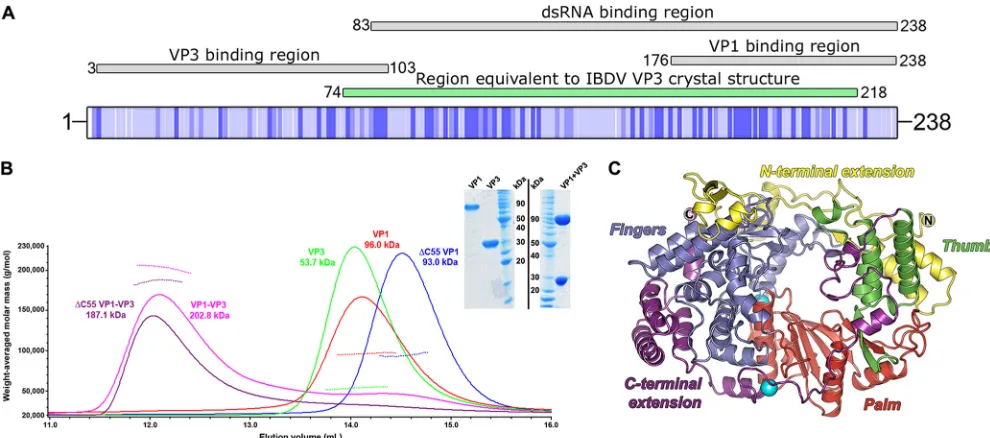
cell lysates were cleared by centrifugation (35,000⫻g, 8°C, 30 min). Cleared lysate was applied to a 1-ml HisTrap Ni2⫹affinity column (GE Healthcare), which was washed with 50 mM Tris, 500 mM NaCl, and 50 mM imidazole (pH 7.5) and eluted in 50 mM Tris, 500 mM NaCl, and 500 mM imidazole (pH 7.5). The eluate was further purified by size exclusion chromatography (SEC) using HiLoad 16/60 Superdex 75 or 200 columns (GE Healthcare) equilibrated in 20 mM Tris and 200 mM NaCl (pH 7.5) and an ÄKTA Purifier 10 UPC or ÄKTAxpress system (GE Healthcare). Fractions containing purified VP3 were pooled and incubated with 50l of rhinovirus 3C protease (starting concentration, 2 mg/ml) overnight at 4°C, to remove the N-terminal His6tag before being passed through a FIG 1Domain organization of VP3, VP1, and MALS analysis of the VP1-VP3 complex. (A) Schematic representation of the full-length IPNV strain Jasper VP3 polypeptide (UniProt identifier P05844), shaded from white (variable) to dark blue (conserved), based on the alignment in Fig. S1 in the supplemental material. Regions of IPNV VP3 reported to perform VP3 binding, dsRNA binding, and VP1 binding activities (24) are shown above the VP3 schematic, labeled, and shaded gray. The region of IPNV VP3 equivalent to the reported structure of IBDV VP3 (25) is labeled and shaded green. (B) SEC-MALS of birnavirus VP1 and VP3 proteins. SEC elution profiles (absorbance at 280 nm, solid lines) are shown for IPNV VP1,⌬C55 VP1, VP3, and VP1-VP3 complexes. The weight-averaged molar mass is shown across the elution profiles as a dotted line and labeled for each sample, the mass of VP3 being consistent with it forming a dimer in solution. The inset shows SimplyBlue (Invitrogen)-stained SDS-PAGE of purified VP1-VP3 complex and individual components (VP1 and VP3) prior to complex formation. Molecular size markers are labeled in kDa. (C) Cartoon representation of the IPNV VP1 structure (colored by domain, with N and C termini labeled). The two K⫹ions bound to VP1 are shown as oversized cyan spheres, the lower K⫹ion not having been observed in previous IPNV VP1 structures.
on November 7, 2019 by guest
http://jvi.asm.org/
[image:2.585.43.538.66.285.2]1-ml HisTrap Ni2⫹affinity column to remove the His
6-tagged 3C
pro-tease.
Purified, untagged VP3 at 1 mg/ml was mixed with either full-length or⌬C55 VP1 at 1 mg/ml and incubated for 1 h at 4°C to facilitate complex formation. The VP1-VP3 complex was purified by Ni2⫹affinity
chroma-tography followed by size exclusion chromachroma-tography, and fractions con-taining pure complex were pooled and concentrated in 30,000-molecular-weight-cutoff centrifugal concentrators (Millipore).
MALS experiments.Multiangle light scattering (MALS) experiments were performed immediately following analytical SEC by inline measure-ment of static light scattering (DAWN HELEOS II; Wyatt Technology), the differential refractive index (Optilab rEX; Wyatt Technology), and UV absorbance (Agilent 1200 UV; Agilent Technologies). Purified protein samples were injected onto an S200 10/300 size exclusion column (GE Healthcare) preequilibrated in 20 mM Tris (pH 7.5) and 200 mM NaCl at a flow rate of 0.5 ml/min. Data were analyzed using the ASTRA software package (Wyatt Technology).
Binding experiments.Peptide binding experiments were performed at 20°C in a PerkinElmer LS55 luminescence spectrometer with excita-tion/emission wavelengths of 480 nm/530 nm and slit widths of 15 nm/20 nm, respectively. The photomultiplier voltage was 800 V with the integra-tion time at 5 s per measurement. A peptide corresponding to the C-ter-minal 14 residues of VP3 with an N-terC-ter-minal fluorescein ([Fluorescein]S GSRFTPSGDDGEV; Designer Bioscience) was rehydrated and diluted in an 800-l volume of 10 mM Tris (pH 7.5) and 200 mM NaCl to a final concentration of 12.5 nM. VP1 (26.5M) was titrated into a continuously stirred cuvette containing the fluorescein-labeled peptide. Anisotropy was monitored continuously, and the values were recorded once they had stabilized for several consecutive readings. Dissociation constants (Kd) were calculated by fitting the titration data (corrected for dilution) to a one-site binding equilibrium model in Prism5 (GraphPad Software).
Competition assays were performed by equilibrating fluorescein-la-beled VP3 peptide (as described above) with 0.3M VP1 before adding full-length VP3 protein (134 to 669M) and monitoring the anisotropy as described above. TheKdfor VP3 was calculated using the method
described in reference28.
Crystallization and data collection.Crystallization screening of ei-ther full-length VP1 and VP3 or⌬C55 VP1 and VP3 purified complexes was performed at concentrations between 2 to 4 mg/ml and at two differ-ent temperatures (21°C and 4°C) using 96-well sitting drop plates (Greiner) containing 100 nl protein and 100 nl reservoir (29). A single crystal appeared between 155 to 220 days after setting up crystallization trials containing full-length VP1 and VP3 (at 3.1 mg/ml), incubated at 4°C. The reservoir solution contained 20% (wt/vol) polyethylene glycol 3350 and 0.2 M potassium fluoride, and the crystal was cryoprotected by soaking in mother liquor supplemented with 25% glycerol. The crystal belonged to space groupP21212, and diffraction data were collected at 100
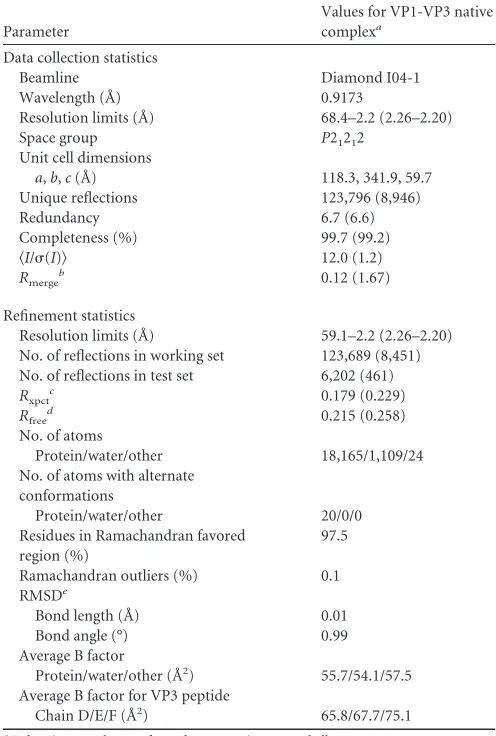
K at Diamond Light Source beamline I04-1 (Table 1).
Structure solution, refinement, and analysis.Diffraction data to a resolution of 2.2 Å were indexed and integrated using XDS (30) and scaled with SCALA (31) as implemented in the program xia2 (32). The structure was solved using molecular replacement with PHASER (33), taking the structure of IPNV VP1 (9), Protein Data Bank (PDB) identifier 2YI9, as a starting model. Manual model building was performed using COOT (34), and the structure was refined with BUSTER-TNT (35), imposing both noncrystallographic local structure similarity restraints (36), and TLS (translation/libration/screw) refinement, as implemented in the auto-BUSTER framework (37). Structure validation was performed with the tools in COOT and the MolProbity Web server (38). Refinement statistics are shown inTable 1. Molecular graphics were generated with PyMOL (39), sequence alignments were generated with Clustal W (40) and ESPript (41), and superpositions were performed with SHP (42). The VP1-VP3 crystal structure was analyzed using the protein interfaces, sur-faces, and assemblies service (PISA) at European Bioinformatics Institute (http://www.ebi.ac.uk/pdbe/prot_int/pistart.html) (43) and the Protein
Interactions Calculator (44). Structure factors and final refined coordi-nates have been deposited in the Protein Data Bank under the accession code 3ZED.
RNA polymerization activation/inhibition assay.The IPNV RdRP was expressed and purified (9), and the s⌬⫹CCCsingle-stranded RNA
(ssRNA) template (713 nucleotides) was generated as described previ-ously (45). VP1-catalyzed replication assays were incubated with 0- to 50-fold molar excess of the full-length VP3 protein or the synthetic VP3 peptide for 10 min on ice prior to the addition of ssRNA template and nucleotides (1 mM ATP, GTP, and 0.2 mM CTP, UTP supplemented with 0.3 pmol [␣32P]-UTP). Following 2 h of incubation at 37°C, the reactions
were stopped by the addition of 2⫻loading buffer (46), and the reaction products were analyzed by electrophoresis on a 0.8% (wt/vol) agarose gel (Tris-borate-EDTA [TBE]) (47). Signals were collected by autoradiography on BAS1500 image plates (Fujifilm) and scanned using a Fuji BAS-1500 phos-phorimager (Fujifilm). Densitometry was performed using AIDA Image An-alyzer software (version 4.50; Raytest Isotopenmessgeräte GmbH), measur-TABLE 1Data collection and refinement statistics
Parameter
Values for VP1-VP3 native complexa
Data collection statistics
Beamline Diamond I04-1
Wavelength (Å) 0.9173
Resolution limits (Å) 68.4–2.2 (2.26–2.20)
Space group P21212
Unit cell dimensions
a,b,c(Å) 118.3, 341.9, 59.7
Unique reflections 123,796 (8,946)
Redundancy 6.7 (6.6)
Completeness (%) 99.7 (99.2)
具I/(I)典 12.0 (1.2)
Rmerge
b 0.12 (1.67)
Refinement statistics
Resolution limits (Å) 59.1–2.2 (2.26–2.20) No. of reflections in working set 123,689 (8,451) No. of reflections in test set 6,202 (461)
Rxpctc 0.179 (0.229)
Rfreed 0.215 (0.258)
No. of atoms
Protein/water/other 18,165/1,109/24 No. of atoms with alternate
conformations
Protein/water/other 20/0/0
Residues in Ramachandran favored region (%)
97.5
Ramachandran outliers (%) 0.1 RMSDe
Bond length (Å) 0.01
Bond angle (°) 0.99
Average B factor
Protein/water/other (Å2) 55.7/54.1/57.5
Average B factor for VP3 peptide
Chain D/E/F (Å2) 65.8/67.7/75.1 a
Values in parentheses refer to the appropriate outer shell. bR
merge⫽ ⌺hkl⌺i|I(hkl;i)⫺ 具I(hkl)典|/⌺hkl⌺iI(hkl;i), whereI(hkl;i) is the intensity of an individual measurement of a reflection and具I(hkl)典is the average intensity of that reflection.
c
Rxpct⫽ ⌺hkl||Fobs|⫺|Fxpct||/⌺hkl|Fobs|, where |Fobs| and |Fxpct| are the observed structure factor amplitude and the expectation of the model structure factor amplitude, respectively.
dR
freeequals theRxpctof test set (5% of the data removed prior to refinement). e
RMSD, root mean square deviation from ideal geometry.
on November 7, 2019 by guest
http://jvi.asm.org/
[image:3.585.297.545.79.447.2]ing the total lane and specific band intensities in one-dimensional evaluation mode using lane and peak determination with automatic background sub-traction.
RESULTS
VP3 readily forms complexes with VP1.
Complexes of either
full-length VP1 and VP3 or
⌬
C55 VP1 and VP3 were prepared
by mixing individually purified proteins and repurifying by
Ni
2⫹affinity and size exclusion chromatography (
Fig. 1B
).
These complexes were characterized by SEC-MALS and were
observed to form higher molecular mass oligomers than their
individual components (
Fig. 1B
). Both full-length VP1 and
⌬
C55 VP1 formed complexes with VP3, suggesting that the
extreme C terminus of VP1 is not required for VP3 complex
formation. From the SEC-MALS results, the stoichiometry of
the purified VP3-VP1 complex is close to 2:2 (
Fig. 1B
). When
VP3 is present in excess, apparent mass is more consistent with
a 2:1 VP3-VP1 complex (data not shown), suggesting that VP1
and VP3 are in dynamic exchange.
The crystal structure of the complex between full-length VP1
and VP3 was solved by molecular replacement using the structure
of IPNV VP1 (
9
) as a search model and refined to a resolution of
2.2 Å with residual
R
xpct⫽
0.179 and
R
free⫽
0.215 (
Table 1
). The
final structure is of high stereochemical quality with 98% of the
residues occupying the favored region of the Ramachandran plot.
Three copies of VP1 are present in the asymmetric unit (AU) of
the crystal, and electron density was observed from residues 27 to
792 for each chain of VP1 (no density was observed for residues 1
to 26 and 793 to 845). Additional density, not attributable to VP1,
was observed bound to the canonical RdRP fingers domain in all
three copies of VP1. This density was unambiguously assigned as
the last 12 residues of the IPNV VP3 C terminus and modeled into
the density. The presence of three copies of the VP1-VP3 peptide
complex in the crystallographic AU corresponds to a solvent
con-tent of 46%, consiscon-tent with the resolution of the data obtained;
the lack of additional density beyond the peptide suggests that the
remainder of VP3 had degraded. In one copy of the complex,
continuous electron density is observed for the VP3 peptide
resi-dues 227 to 238; the other two copies of the peptide in the AU
contain a break in the peptide density for residues 234 to 237
(SGDD). Apart from this difference, the three copies of the
com-plex in the crystallographic asymmetric unit were essentially
iden-tical (average root mean square deviations [RMSD] over all the
C-
␣
s, 0.38 and 0.19 Å, between the copies of VP1 and VP3,
respec-tively). Electron density was also observed for a second K
⫹ion
bind-ing site, in addition to the sbind-ingle K
⫹ion observed bound in a previous
structure of IPNV VP1 (
9
). This new K
⫹ion binds on a
surface-exposed region of the C-terminal extension domain of VP1 (
Fig. 1C
).
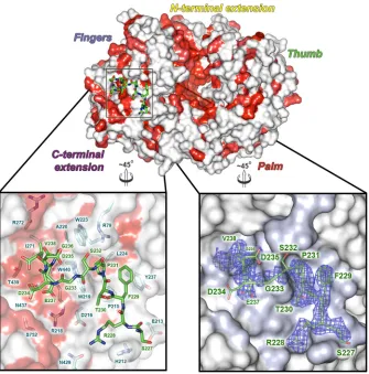
Structural basis for the VP1-VP3 interaction.
The VP3
pep-tide binds in an extended groove on the outside of the VP1 fingers
domain (residues 161 to 343 and 406 to 475) (
Fig. 2
) and is
an-chored at either end by a pair of charge-based and hydrophobic
interactions. At one end of the groove, VP3 residue R228 forms a
salt bridge with D216 of VP1 while F229 of VP3 stacks against the
side chain of VP1 Y237 (
Fig. 2
). At the C terminus of the VP3
peptide, E237 forms an ion pair with VP1 residue R218, and the
final residue of VP3 (V238) pokes into a hydrophobic pocket lined
by VP1 residues A220, I271, R272, and W440. The intervening
residues of the VP3 peptide (230-TPSGDDG-236) form mainly
backbone interactions with the VP1 groove. In total, some 800 Å
2of solvent-accessible area is buried from the VP3 peptide upon
binding VP1 (assessed by the PISA server) (
43
), a little over half of
the total surface area of the peptide. A superposition of unbound
and VP3-bound VP1 gives an RMSD of 0.5 Å, over 758 equivalent
C-
␣
atoms (
42
), with no major conformational changes in the
fingers domain of VP1 (0.3 Å RMSD, over 251 C-
␣
equivalences).
This suggests that the VP1 groove used to bind VP3 is preformed
on the surface of VP1, ready to bind VP3.
Characterization of VP1-VP3 interaction.
The structure
sug-gests that residues 227 to 238 of VP3 are key to interaction with
VP1. This peptide was thus synthesized with an N-terminal
fluo-rescein label, and fluorescence anisotropy measurements
con-firmed that it forms a tight complex with VP1,
K
d⫽
0.11 (
⫾
0.01)
M (
Fig. 3A
;
n
⫽
2). To investigate if this was the primary mode of
interaction of VP1/VP3, we then performed an experiment where
prebound labeled peptide competed with full-length VP3 (
Fig.
3B
). This showed that VP3 successfully outcompeted the peptide
at a sufficiently high concentration, indicating that the peptide
binding mode was similar to that of full-length VP3. An analysis
of the competition experiment suggests that the binding
con-stant for full-length VP3 is
⬃
0.36
M (
28
), a little weaker than
the peptide, presumably due to entropic effects and/or
confor-mational restraints in the context of the full protein. Clearly,
the binding seen in the crystal is likely to represent an authentic
picture of the binding of full-length VP3 to VP1; only the C
terminus of VP3 binds strongly to VP1, the remainder being
redundant.
VP1-VP3 complex formation does not alter the biochemical
activity of VP1.
VP1 in complex with full-length VP3 or a
syn-thetic VP3 peptide was characterized for
de novo
RNA synthesis
activity using a 713-nucleotide ssRNA template. Although
VP1-VP3 complex formation occurs readily, quantitative total lane
im-age densitometry analysis of VP1-VP3-catalyzed replication
as-says reveals no stimulation of RNA polymerization (
Fig. 3C
; see
also Table S1 in the supplemental material). By comparison to the
VP1 control reaction (
Fig. 3C
, lane 1), addition of VP3 yields a
0.9-to 1.2-fold change in 0.9-total activity (see Table S1), which is well
within the normal error range (
⫾
10%) for this assay. Further
saturating the reaction with VP3 leads to a 0.7-fold reduction in
total activity.
VP3 on its own is devoid of enzymatic activity, although the
full-length protein does show an affinity for RNA that is not
ex-hibited by the synthetic C-terminal peptide. VP1 also has RNA
binding affinity, forming high molecular weight RNA-protein
complexes during templated RNA synthesis (
Fig. 3C
, lane 1) (
9
).
This phenomenon is strongly enhanced by the addition of VP3
(
Fig. 3C
, lanes 2 to 12). Measuring specific band intensities reveals
that a mere 2-fold excess of VP3 yields a 7-fold increase in complex
formation, whereas a 34-fold increase can be obtained when
sat-urating the assay with a 50-fold excess of VP3 (see Table S1 in the
supplemental material). Although supplementing VP1-catalyzed
RNA polymerization reactions with an excess of full-length VP3
significantly enhances the appearance of RNA-protein complexes,
it occurs at the expense of free RNA products (
Fig. 3C
; see also
Table S1 in the supplemental material). The RNA sequestering
ability of the VP1-VP3 complex, and VP3 in particular, is clearly
demonstrated by the retardation in electrophoretic mobility seen
in the rightmost lanes of
Fig. 3C
.
on November 7, 2019 by guest
http://jvi.asm.org/
DISCUSSION
Studies of the two best-characterized birnaviruses, IPNV and
IBDV, have revealed that VP3 is a multifunctional protein with
different activities throughout the viral life cycle (
23
,
24
). In this
study, we attempted to determine the molecular details of the
interaction between IPNV VP3 with the RdRP polymerase VP1.
We have crystallized a VP1-VP3 complex and determined its
structure to a resolution of 2.2 Å. Although full-length dimeric
VP3 was used during crystallization of the VP1-VP3 complex, the
crystal packing is inconsistent with this being the species
crystal-lized. Electron density shows only the C-terminal 12 residues of
VP3 to be well ordered and bound into an extended groove on the
VP1 fingers domain. The long period of crystal growth (
⬃
6
months) suggests that proteolysis of the rest of VP3 had occurred.
Fluorescence anisotropy shows that the affinity of full-length VP1
for the VP3 C-terminal peptide is tight (
K
d,
⬃
0.1
M) and that
full-length VP3 binds almost as strongly and can compete with
labeled VP3 peptide for VP1 binding (
K
dfor VP3,
⬃
0.4
M),
indicating that they both bind the same surface of VP1. Using the
IPNV VP1-VP3 complex as a framework, we have modeled the
IBDV VP3 peptide onto the structure of IBDV polymerase.
With-out significant readjustment of the conformation of the VP3
pep-tide seen in IPNV, a satisfactory docking is obtained, which
con-serves the pattern of favorable hydrophobic and electrostatic
interactions seen between VP1 and VP3 in IPNV, with minimal
side chain clashes.
It is well established that IPNV VP1 and VP3 have a
consider-able affinity for RNA (
6
,
9
,
24
,
48
). Previous studies show that the
N-terminal part of VP3 is crucial for dsRNA binding (ssRNA
binding was not assayed) and that VP1-VP3 complex formation
does not require the presence of dsRNA (
24
). We have previously
shown that the N-terminal tail of VP1 interacts strongly with
ssRNA (
9
). The VP1-VP3 protein complex is thus capable of
in-teracting with both single- and double-stranded RNA. Our data
suggest a model whereby VP1-VP3 interactions are relevant for
the morphogenesis of mature birnavirus particles, with VP3
act-ing as a modular protein, havact-ing distinct regions responsible for
RNA binding, dimer formation, and VP1 attachment. This
sup-ports the notion that during viral infection, VP1 replicates the
genomic ssRNA precursor molecules and, in conjunction with
VP3, sequesters the viral genome, forming RNA-protein
com-plexes (as shown in reference
24
). Indeed VP1-VP3 complexes
FIG 2Structure of the VP1-VP3 complex. In the top panel, the IPNV VP1 polymerase is shown as a molecular surface, in the same view as inFig. 1C. VP1 sequence conservation across birnavirus species is mapped onto the surface and colored from white (variable) to red (conserved), based on the alignment in Fig. S2 in the supplemental material. The VP3 peptide (green sticks) is shown bound to the fingers domain of VP1 and boxed. The lower panels show expanded views of the VP3 peptide (residues labeled green) bound into the VP1 surface groove (shown semitransparent). In the left panel, residues from VP1 that bind VP3 are shown as sticks beneath the surface, colored, and labeled cyan. The right panel shows the experimental 2FO-FCelectron density, calculated prior to modeling ofthe VP3 peptide, as a blue mesh (0.9). The VP3 peptide is shown as sticks (green, with side chains labeled) to guide interpretation of the density.
on November 7, 2019 by guest
http://jvi.asm.org/
[image:5.585.126.461.65.404.2]have been detected prior to the formation of mature virus
parti-cles, indicating a role in promoting the assembly process (
24
).
These subassemblies act as scaffolds around which VP2 can
as-semble to form a precursor capsid, which undergoes maturation
to yield an infectious virion. This is similar to, but simpler than,
the viroplasm model proposed for rotavirus capsid assembly (
49
).
There is no indication that the genome would be packaged into a
preassembled capsid using an active packaging mechanism, such
as a hexameric packaging motor. In fact, the evidence showing
that the birnavirus virion contains a polyploid genome (
50
) argues
against this and suggests that the packaging mechanism is
stochastic.
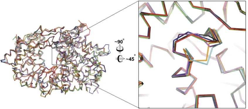
Our structural results are not in agreement with the suggestion
(based on inference from inconclusive electron density) that VP3
binds close to the polymerase active site, increasing activity by
stimulating movement of catalytic motif B (
26
). Indeed, in our
previous structure of isolated IPNV VP1 (
9
) and also in an
inde-pendent determination of the isolated IBDV VP1 structure (
10
),
motif B is already in the catalytically competent conformation
(
Fig. 4
). It is therefore unlikely that this movement is directly
stimulated by VP3 binding. To address this discrepancy, we
per-formed a detailed analysis of the effect of VP3 on the polymerase
activity of VP1 and found that, as expected from our structural
results, in contrast to the earlier report (
26
), VP3 attachment has
no effect on the activity of VP1.
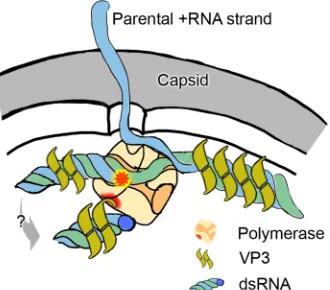
The model we propose of the role of VP3 in birnavirus
struc-ture is shown schematically in
Fig. 5
. Note that the site of
attach-ment to the polymerase is likely to orientate the polymerase within
the capsid in such a way as to facilitate polymerase activity,
re-quired for the active transcriptional activity observed in
birnavi-FIG 3Fluorescence anisotropy of VP1-VP3 interactions and polymerase activity assay. (A) Fluorescence anisotropy study of the binding of fluorescently labeled VP3 C-terminal peptide (residues 227 to 238) to VP1. (B) Fluorescence anisotropy competition assay between unlabeled VP3 protein and a preformed complex between VP1 and fluorescently labeled VP3 C-terminal peptide. (C) Autoradiograph of IPNV VP1-VP3 catalyzedde novoreplication reactions analyzed in a 0.8% agarose (TBE) gel. The leftmost lane includes a reference reaction (VP1 only) to which the VP1-VP3 reactions are compared. RNA polymerization activity is determined by image densitometry analysis of the total lane, whereas RNA-protein complex formation is assessed by measuring the intensity of both the unbound and bound dsRNA product.FIG 4Comparison of birnavirus VP1 B-loop motif between VP3 bound and unbound VP1 structures. The left panel shows birnavirus VP1 crystal structures superposed, in a 180° view around theyaxis, to the view inFig. 1C, looking toward the nucleotide entry tunnel. The B-loop (residues⬃468 to 477 in IPNV VP1) (see Fig. S2 in the supplemental material) motif is boxed. The right panel shows a zoom of the catalytic motif B; ribbons are colored as follows (PDB identifiers in parentheses): green, IPNV VP1-VP3 complex presented in this paper; red, IPNV VP1 apo-form (2YIB); blue, IBDV VP1 (2PGG); magenta, IBDV VP1 soaked with a VP3 peptide (2QJ1); black, IBDV VP1 cocrystallized with a VP3 peptide (2R70); orange, IBDV VP1 Mg2⫹-bound (2R72); and cyan, IBDV VP1 apo-form (2PUS). Note that the B loop for the two IBDV VP1 structures either soaked or cocrystallized with VP3 (magenta and black) is in the same conformation as the independently determined structures of unbound IPNV and IBDV VP1 (red and blue, respectively). Further, the structure presented in this study, VP3 bound to the VP1 fingers domain (distal to the nucleotide entry tunnel) (green), also exhibits this conformation, suggesting that VP3 binding is not the driving force for this conformational change of the B loop.
on November 7, 2019 by guest
http://jvi.asm.org/
[image:6.585.46.541.65.184.2] [image:6.585.82.505.453.639.2]rus capsids. In summary, not only does this structure of the
VP1-VP3 complex provide a detailed view of the precise molecular
determinants of the interaction between birnavirus VP1 and VP3
proteins for the first time, but it also provides a simple model for a
key stage in birnavirus morphogenesis.
ACKNOWLEDGMENTS
We thank Bert Janssen for helpful advice and the staff at beamline I04-1 at the Diamond synchrotron light source for technical support. We also thank Riitta Tarkiainen for her excellent technical assistance.
This work has been supported by Academy of Finland Academy Pro-fessor research funding (grants 255342 and 256518 to D.H.B.). S.C.G. is a Royal Commission for the Exhibition of 1851 Research Fellow. The Cam-bridge Institute for Medical Research (CIMR) is in receipt of a Wellcome Trust Strategic Award (079895). The work at Oxford is supported by the UK Medical Research Council and administrative support is provided by the Wellcome Trust (grant 075491/Z/04).
REFERENCES
1.Rodriguez Saint-Jean S, Borrego JJ, Perez-Prieto SI. 2003. Infectious pancreatic necrosis virus: biology, pathogenesis, and diagnostic methods. Adv. Virus Res.62:113–165.
2.Jackwood DJ, Saif YM, Hughes JH.1982. Characteristics and serologic studies of two serotypes of infectious bursal disease virus in turkeys. Avian Dis.26:871– 882.
3.Dobos P, Hill BJ, Hallett R, Kells DT, Becht H, Teninges D. 1979. Biophysical and biochemical characterization of five animal viruses with bisegmented double-stranded RNA genomes. J. Virol.32:593– 605. 4.Coulibaly F, Chevalier C, Gutsche I, Pous J, Navaza J, Bressanelli S,
Delmas B, Rey FA.2005. The birnavirus crystal structure reveals struc-tural relationships among icosahedral viruses. Cell120:761–772. 5.Dobos P.1993.In vitroguanylylation of infectious pancreatic necrosis
virus polypeptide VP1. Virology193:403– 413.
6.Calvert JG, Nagy E, Soler M, Dobos P.1991. Characterization of the VPg-dsRNA linkage of infectious pancreatic necrosis virus. J. Gen. Virol. 72(Pt 10):2563–2567.
7.Muller H, Nitschke R.1987. The two segments of the infectious bursal disease virus genome are circularized by a 90,000-Da protein. Virology 159:174 –177.
8.Gorbalenya AE, Pringle FM, Zeddam JL, Luke BT, Cameron CE, Kalmakoff J, Hanzlik TN, Gordon KH, Ward VK.2002. The palm subdomain-based active site is internally permuted in viral RNA-dependent RNA polymerases of an ancient lineage. J. Mol. Biol.324: 47– 62.
9.Graham SC, Sarin LP, Bahar MW, Myers RA, Stuart DI, Bamford DH, Grimes JM.2011. The N-terminus of the RNA polymerase from infec-tious pancreatic necrosis virus is the determinant of genome attachment. PLoS Pathog.7:e1002085. doi:10.1371/journal.ppat.1002085.
10. Pan J, Vakharia VN, Tao YJ.2007. The structure of a birnavirus poly-merase reveals a distinct active site topology. Proc. Natl. Acad. Sci. U. S. A. 104:7385–7390.
11. Lee J, Feldman AR, Delmas B, Paetzel M.2007. Crystal structure of the VP4 protease from infectious pancreatic necrosis virus reveals the acyl-enzyme complex for an intermolecular self-cleavage reaction. J. Biol. Chem.282:24928 –24937.
12. Birghan C, Mundt E, Gorbalenya AE.2000. A non-canonical lon pro-teinase lacking the ATPase domain employs the ser-Lys catalytic dyad to exercise broad control over the life cycle of a double-stranded RNA virus. EMBO J.19:114 –123.
13. Duncan R, Nagy E, Krell PJ, Dobos P.1987. Synthesis of the infectious pancreatic necrosis virus polyprotein, detection of a virus-encoded pro-tease, and fine structure mapping of genome segment A coding regions. J. Virol.61:3655–3664.
14. Da Costa B, Chevalier C, Henry C, Huet JC, Petit S, Lepault J, Boot H, Delmas B.2002. The capsid of infectious bursal disease virus contains several small peptides arising from the maturation process of pVP2. J. Virol.76:2393–2402.
15. Sanchez AB, Rodriguez JF.1999. Proteolytic processing in infectious bursal disease virus: identification of the polyprotein cleavage sites by site-directed mutagenesis. Virology262:190 –199.
16. Caston JR, Martinez-Torrecuadrada JL, Maraver A, Lombardo E, Ro-driguez JF, Casal JI, Carrascosa JL.2001. C terminus of infectious bursal disease virus major capsid protein VP2 is involved in definition of the T number for capsid assembly. J. Virol.75:10815–10828.
17. Bottcher B, Kiselev NA, Stel’Mashchuk VY, Perevozchikova NA, Bori-sov AV, Crowther RA.1997. Three-dimensional structure of infectious bursal disease virus determined by electron cryomicroscopy. J. Virol.71: 325–330.
18. Ona A, Luque D, Abaitua F, Maraver A, Caston JR, Rodriguez JF.2004. The C-terminal domain of the pVP2 precursor is essential for the interac-tion between VP2 and VP3, the capsid polypeptides of infectious bursal disease virus. Virology322:135–142.
19. Chevalier C, Lepault J, Da Costa B, Delmas B.2004. The last C-terminal residue of VP3, glutamic acid 257, controls capsid assembly of infectious bursal disease virus. J. Virol.78:3296 –3303.
20. Maraver A, Ona A, Abaitua F, Gonzalez D, Clemente R, Ruiz-Diaz JA, Caston JR, Pazos F, Rodriguez JF.2003. The oligomerization domain of VP3, the scaffolding protein of infectious bursal disease virus, plays a critical role in capsid assembly. J. Virol.77:6438 – 6449.
21. Kochan G, Gonzalez D, Rodriguez JF. 2003. Characterization of the RNA-binding activity of VP3, a major structural protein of infectious bursal disease virus. Arch. Virol.148:723–744.
22. Hjalmarsson A, Carlemalm E, Everitt E. 1999. Infectious pancreatic necrosis virus: identification of a VP3-containing ribonucleoprotein core structure and evidence for O-linked glycosylation of the capsid protein VP2. J. Virol.73:3484 –3490.
23. Tacken MG, Peeters BP, Thomas AA, Rottier PJ, Boot HJ. 2002. Infectious bursal disease virus capsid protein VP3 interacts both with VP1, the RNA-dependent RNA polymerase, and with viral double-stranded RNA. J. Virol.76:11301–11311.
24. Pedersen T, Skjesol A, Jorgensen JB.2007. VP3, a structural protein of infectious pancreatic necrosis virus, interacts with RNA-dependent RNA polymerase VP1 and with double-stranded RNA. J. Virol.81:6652– 6663. 25. Casanas A, Navarro A, Ferrer-Orta C, Gonzalez D, Rodriguez JF, Verdaguer N.2008. Structural insights into the multifunctional protein VP3 of birnaviruses. Structure16:29 –37.
26. Garriga D, Navarro A, Querol-Audi J, Abaitua F, Rodriguez JF, Ver-daguer N. 2007. Activation mechanism of a noncanonical RNA-dependent RNA polymerase. Proc. Natl. Acad. Sci. U. S. A.104:20540 – 20545.
27. Berrow NS, Alderton D, Owens RJ.2009. The precise engineering of expression vectors using high-throughput In-Fusion PCR cloning. Meth-ods Mol. Biol.498:75–90.
28. Rossi AM, Taylor CW.2011. Analysis of protein-ligand interactions by fluorescence polarization. Nat. Protoc.6:365–387.
29. Walter TS, Diprose JM, Mayo CJ, Siebold C, Pickford MG, Carter L, Sutton GC, Berrow NS, Brown J, Berry IM, Stewart-Jones GB, Grimes FIG 5The complex of IPNV VP1 and VP3 in the context of the viral capsid.
The 5=end of a dsRNA segment (plus and minus strands are colored blue and green, respectively) is covalently attached to the actively transcribing VP1 polymerase (orange). The parental plus strand is released as mRNA during the semiconservative replication. VP3 (mustard) associates with the dsRNA, as well as forming a specific complex with VP1 (orange).
on November 7, 2019 by guest
http://jvi.asm.org/
[image:7.585.83.248.68.213.2]JM, Stammers DK, Esnouf RM, Jones EY, Owens RJ, Stuart DI, Harlos K.2005. A procedure for setting up high-throughput nanolitre crystalli-zation experiments. Crystallicrystalli-zation workflow for initial screening, auto-mated storage, imaging and optimization. Acta Crystallogr. D Biol. Crys-tallogr.61:651– 657.
30. Kabsch W.1993. Automatic processing of rotation diffraction data from crystals of initially unknown symmetry and cell constants. J. Appl. Crys-tallogr.26:795– 800.
31. Evans P.2006. Scaling and assessment of data quality. Acta Crystallogr. D Biol. Crystallogr.62:72– 82.
32. Winter G.2010. xia2: an expert system for macromolecular crystallogra-phy data reduction. J. Appl. Crystallogr.43:186 –190.
33. McCoy AJ, Grosse-Kunstleve RW, Adams PD, Winn MD, Storoni LC, Read RJ.2007. Phaser crystallographic software. J. Appl. Crystallogr.40: 658 – 674.
34. Emsley P, Lohkamp B, Scott WG, Cowtan K.2010. Features and devel-opment of Coot. Acta Crystallogr. D Biol. Crystallogr.66:486 –501. 35. Blanc E, Roversi P, Vonrhein C, Flensburg C, Lea SM, Bricogne G.
2004. Refinement of severely incomplete structures with maximum like-lihood in BUSTER-TNT. Acta Crystallogr. D Biol. Crystallogr.60:2210 – 2221.
36. Smart O, Brandl M, Flensburg C, Keller P, Paciorek W, Vonrhein C, Womack T, Bricogne G.2008. Refinement with local structure similarity restraints (LSSR) enables exploitation of information from related struc-tures and facilitates use of NCS. Abstr. Annu. Meet. Am. Crystallogr. As-soc., abstr TP139, p 117.
37. Bricogne G, Blanc E, Brandl M, Flensburg C, Keller P, Paciorek P, Roversi P, Sharff A, Smart O, Vonrhein C, Womack T.2011. BUSTER version 2.11.1. Global Phasing Ltd., Cambridge, United Kingdom. 38. Chen VB, Arendall WB, III, Headd JJ, Keedy DA, Immormino RM,
Kapral GJ, Murray LW, Richardson JS, Richardson DC.2010. MolPro-bity: all-atom structure validation for macromolecular crystallography. Acta Crystallogr. D Biol. Crystallogr.66:12–21.
39. DeLano WL.2008. The PyMOL molecular graphics system. DeLano Sci-entific LLC, Palo Alto, CA.http://www.pymol.org.
40. Larkin MA, Blackshields G, Brown NP, Chenna R, McGettigan PA, McWilliam H, Valentin F, Wallace IM, Wilm A, Lopez R, Thompson JD, Gibson TJ, Higgins DG.2007. Clustal W and Clustal X version 2.0. Bioinformatics23:2947–2948.
41. Gouet P, Robert X, Courcelle E.2003. ESPript/ENDscript: extracting and rendering sequence and 3D information from atomic structures of proteins. Nucleic Acids Res.31:3320 –3323.
42. Stuart DI, Levine M, Muirhead H, Stammers DK.1979. Crystal struc-ture of cat muscle pyruvate kinase at a resolution of 2.6 A. J. Mol. Biol. 134:109 –142.
43. Krissinel E, Henrick K.2007. Inference of macromolecular assemblies from crystalline state. J. Mol. Biol.372:774 –797.
44. Tina KG, Bhadra R, Srinivasan N.2007. PIC: Protein Interactions Cal-culator. Nucleic Acids Res.35:W473–W476.
45. Wright S, Poranen MM, Bamford DH, Stuart DI, Grimes JM.2012. Noncatalytic ions direct the RNA-dependent RNA polymerase of bacterial double-stranded RNA virus6 fromde novoinitiation to elongation. J. Virol.86:2837–2849.
46. Pagratis N, Revel HR.1990. Detection of bacteriophage phi 6 minus-strand RNA and novel mRNA isoconformers synthesizedin vivoandin vitro,by strand-separating agarose gels. Virology177:273–280. 47. Sambrook J, Russell D.2001. Molecular cloning: a laboratory manual,
3rd ed. Cold Spring Harbor Laboratory Press, Cold Spring Harbor, NY. 48. Dobos P.1995. Protein-primed RNA synthesisin vitroby the
virion-associated RNA polymerase of infectious pancreatic necrosis virus. Virol-ogy208:19 –25.
49. Trask SD, McDonald SM, Patton JT.2012. Structural insights into the coupling of virion assembly and rotavirus replication. Nat. Rev. Micro-biol.10:165–177.
50. Luque D, Rivas G, Alfonso C, Carrascosa JL, Rodriguez JF, Caston JR. 2009. Infectious bursal disease virus is an icosahedral polyploid dsRNA virus. Proc. Natl. Acad. Sci. U. S. A.106:2148 –2152.
on November 7, 2019 by guest
http://jvi.asm.org/