0022-538X/10/$12.00 doi:10.1128/JVI.01326-09
Copyright © 2010, American Society for Microbiology. All Rights Reserved.
Porcine Reproductive and Respiratory Syndrome Virus Nonstructural
Protein 1

Modulates Host Innate Immune Response by
Antagonizing IRF3 Activation
䌤
Lalit K. Beura,
1Saumendra N. Sarkar,
2* Byungjoon Kwon,
1Sakthivel Subramaniam,
1Clinton Jones,
1Asit K. Pattnaik,
1and Fernando A. Osorio
1*
Nebraska Center for Virology and School of Veterinary and Biomedical Sciences, University of Nebraska, Lincoln, Nebraska 68583,1
and Molecular Virology Program, University of Pittsburgh Cancer Institute and Department of Microbiology and
Molecular Genetics, University of Pittsburgh School of Medicine, Pittsburgh, Pennsylvania 152132
Received 27 June 2009/Accepted 10 November 2009
Porcine reproductive and respiratory syndrome virus (PRRSV) infection of swine leads to a serious disease characterized by a delayed and defective adaptive immune response. It is hypothesized that a suboptimal innate immune response is responsible for the disease pathogenesis. In the study presented here we tested this hypothesis and identified several nonstructural proteins (NSPs) with innate immune evasion properties encoded by the PRRS viral genome. Four of the total ten PRRSV NSPs tested were found to have strong to moderate inhibitory effects on beta interferon (IFN-) promoter activation. The strongest inhibitory effect was exhibited by NSP1 followed by, NSP2, NSP11, and NSP4. We focused on NSP1␣and NSP1(self-cleavage products of NSP1 during virus infection) and NSP11, three NSPs with strong inhibitory activity. All of three proteins, when expressed stably in cell lines, strongly inhibited double-stranded RNA (dsRNA) signaling pathways. NSP1 was found to inhibit both IFN regulatory factor 3 (IRF3)- and NF-B-dependent gene induction by dsRNA and Sendai virus. Mechanistically, the dsRNA-induced phosphorylation and nuclear translocation of IRF3 were strongly inhibited by NSP1. Moreover, when tested in a porcine myelomonocytic cell line, NSP1 inhibited Sendai virus-mediated activation of porcine IFN-promoter activity. We propose that this NSP1-mediated subversion of the host innate immune response plays an important role in PRRSV pathogenesis.
Porcine reproductive and respiratory syndrome virus (PRRSV) is a member of familyArteriviridaewhich, along with
the Coronaviridae, are classified in the order Nidovirales(9).
PRRSV is an enveloped, single-stranded RNA virus with pos-itive-sense genome of ⬃15 kb. The genome has nine open reading frames (ORFs), namely, ORF1a, ORF1b, ORF2a, ORF2b, and ORF3-7. ORF1a and ORF1b are synthesized as a single polyprotein and later processed to 14 different nonstruc-tural proteins (NSPs), e.g., NSP1␣, NSP1, and NSP2 to NSP12 by the viral proteases. NSP1␣, NSP1, NSP2, and NSP4 are the viral proteases which carry out this function. The viral genome replication and transcription is carried out by NSP9, which encodes the viral RNA-dependent RNA polymerase, and NSP10, which encodes a helicase (5, 49).
The ailment caused by PRRSV is considered to be the most economically significant infectious disease of swine worldwide. It causes an annual loss of approximately $560 million in the United States (38). The clinical signs of PRRSV infection include late-term reproductive failure in sows and respiratory illness in growing pigs. Most cases of PRRSV infection in pigs are complicated by secondary opportunistic bacterial
infec-tions, which have been attributed to the immunosuppressive nature of the virus (13). Upon initial virus infection, viremia lasts for a few weeks, after which it resolves. However, the virus can be detected in secondary lymphoid organs for several months, indicating failure of host immune response to clear the virus (2).
The immune response to PRRSV infection is characterized by a delayed appearance of neutralizing antibodies (31), a short cell-mediated immune response (34, 59), and slow devel-opment of a virus-specific gamma interferon (IFN-␥) response (35). We and others have identified various factors that are likely to play multiple roles in delayed clearance of PRRSV from the host. These include weak innate immune response (1, 35), presence of decoy epitopes (39), and glycan shielding of envelope proteins (3). Previous studies have shown that very low or negligible levels of IFN-␣are produced upon PRRSV infection in pulmonary alveolar macrophages (PAMs) and PRRSV permissive monkey kidney cells (MARC-145)in vitro
(1, 36). IFN-␣production in the lungs of pigs acutely infected with PRRSV was either almost undetectable or 100- to 200-fold lower than that induced by porcine respiratory coronavi-rus (PRCV) (8, 57). PRRSV has also been found to suppress IFN-␣production by transmissible gastroenteritis corona virus (TGEV), a known inducer of IFNs in infected alveolar mac-rophages (1). At the same time, externally provided IFN-␣or IFN- have been able to reduce viral replication in cultured alveolar macrophages (1, 40). The virus was also found to inhibit the dsRNA-mediated upregulation of IFN-gene tran-scription (36). A microarray analysis of PAMs infected with
* Corresponding authors. Mailing address for F. A. Osorio: 111 MOLR Nebraska Center for Virology, UNL East Campus, Lincoln, NE 68583-0900. Phone: (402) 472-7809. Fax: (402) 472-3323. E-mail: fosorio@unl.edu. Mailing address for S. N. Sarkar: University of Pittsburgh Cancer Institute HCC 1.8, 5117 Centre Avenue, Pittsburgh, PA 15213. Phone: (412) 623-7720. Fax: (412) 623-7715. E-mail: saumen@pitt.edu.
䌤Published ahead of print on 18 November 2009.
1574
on November 8, 2019 by guest
http://jvi.asm.org/
Lelystad virus (European type PRRSV) showed no significant change in the IFN-␣ from the control at 12 h postinfection (17). Considering the broad role of IFNs in establishing an effective adaptive immune response, we hypothesize that the suboptimal induction of type I IFN may be one of the deter-mining factors in deficient development of acquired immunity. In the constant struggle to outsmart the host, viruses have developed strategies to evade and/or inhibit key elements of host immune response. Sometimes, a substantial part of the viral genome is dedicated toward suppressing IFN signaling pathways, IFN-stimulated gene (ISG) functions, or pathways for RNA processing and translation (7, 16). In the present study we sought to identify the PRRSV proteins that are re-sponsible for mediating the inhibition of IFN production. We observed that several PRRSV NSPs inhibit IFN regulatory factor 3 (IRF3)-mediated activation of IFN-promoter. De-tailed investigation of the mechanism of inhibition of one of those viral proteins, NSP1, revealed that it inhibits double-stranded RNA (dsRNA)-mediated IRF3 phosphorylation and nuclear translocation.
MATERIALS AND METHODS
Cells and viruses.HEK293-TLR3 (Invivogen), HeLa (American Type Culture Collection [ATCC]), HT1080 (ATCC), MARC-145 (obtained from Will Lae-greid, USMARC, USDA/ARS) (25), and ST (ATCC) cells were maintained in Dulbecco modified Eagle medium (DMEM) containing 10% fetal bovine serum and 50g of gentamicin (Sigma)/ml. HEK293-TLR3 and HT1080 cells stably expressing PRRSV NSP1␣, NSP1, and NSP11 were established by cotransfect-ing them with correspondcotransfect-ing PRRSV protein expression plasmid and pBabe-Puro. After selection with 1g of Puromycin (Mediatech)/ml, individual colonies were picked and screened for stable expression of PRRSV NSPs. For the swine macrophage experiments, mononuclear cells were isolated from blood of donor pigs by Ficoll density centrifugation using lymphocyte separation medium (Me-diatech) (32). These cells were then plated in a glass petri dish for 1 day, followed by washing with phosphate-buffered saline (PBS) to enrich the monocytes. The attached monocytes were cultured in RPMI 1640 medium supplemented with 10% BVDV-free fetal bovine serum in the presence of the growth factor mac-rophage colony-stimulating factor (5 ng/ml) (Sigma) for 7 days for maturation into macrophages. These monocyte-derived macrophages were then harvested by cell dissociation buffer (Gibco) and plated in appropriate tissue culture plate with RPMI 1640 and 10% fetal bovine serum (FBS) for virus infection. Porcine monocytic cell line 3D4/31 (ATCC) was maintained in RPMI 1640 medium supplemented with 10% BVDV-free fetal bovine serum and 50g of gentamicin (Sigma)/ml. Highly virulent North American PRRSV strain FL12 (56) was prop-agated in MARC-145 cells to prepare the virus stocks for the present study. TGEV (Purdue strain) (1) was obtained from the Veterinary Diagnostic Center (University of Nebraska) and propagated in ST cells to prepare the virus stocks for infection in macrophages. Sendai virus (SeV; Cantell strain) was obtained from Charles River Laboratories and was used as described below.
Reporter assays.IFN--CAT assays were performed in HeLa cells transfected with 1g of IFN--CAT reporter plasmid, 0.5g of IRF3 plasmid (Luwen Zhang, University of Nebraska), and 1g of indicated viral protein coding plasmid using Lipofectamine-2000 (Invitrogen) according to the manufacturer’s instructions. At 40 h posttransfection, the cells were lysed by freeze-thawing, and a chloramphenicol acetyltransferase (CAT) assay was performed as described earlier (44). CAT activity is expressed as the fold induction relative to the empty vector control. A minimum of three independent experiments was performed to confirm the results. For ISG56-luciferase reporter assays, HEK293-TLR3 cells were transfected with 0.4g of ISG56-luciferase reporter plasmid and 10 ng of pRL-TK (Promega), along with the indicated protein expression vectors using Fugene 6 (Roche Applied Science) according to the manufacturer’s protocol. For the NF-B luciferase assays, HEK293-TLR3 cells were transfected with 0.5
g of 5X-NF-B-luciferase reporter plasmid (48) and 25 ng of pRL-TK (Pro-mega), along with the indicated protein expression vector using Fugene 6 as described above. At 24 h posttransfection, the medium was replaced with fresh medium. At 40 h posttransfection, the cells were either treated with 10g of poly(I-C) (GE Healthcare)/ml or PBS for 6 h, and luciferase assays were per-formed by using a dual luciferase assay kit (Promega). For the porcine IFN-
-luciferase assay, 3D4/31 cells were transfected with 0.4g of porcine IFN--luc plasmid and 10 ng of pRL-TK (Promega), along with the indicated protein expression vectors using Ingenio electroporation solution (Miru Bio) in an Amaxa Nucleofector (Lonza) according to the manufacturer’s instructions. Then, at 40 h posttransfection, the cells were either mock infected or infected with SeV (Cantell strain) for 8 h, after which the cells were lysed, and luciferase assay was performed. Firefly luciferase activities were expressed in terms of the fold change with respect to controls after normalization with theRenilla lucif-erase activities.
Real-time PCR and enzyme-linked immunosorbent assay (ELISA). Total RNA was isolated from cells by using TRIzol (Invitrogen) reagent and cDNA was prepared by using Moloney murine leukemia virus reverse transcriptase (Invitrogen). This cDNA was used for quantification of the indicated mRNA copy number by using SmartCycler (Cepheid). Porcine IFN-␣, IFN-, and -ac-tin (internal control) real-time PCR was performed using a TaqMan probe based method. Human interleukin-8 (IL-8) and human ribosomal protein L32 (RPL32, internal control) copy numbers were calculated by Sybr-green based quantitative PCR. The following primer sets and, where indicated, probes were used in the real-time PCRs. For porcine IFN-␣, we used forward primer 5⬘-AGAGCCTCC TGCACCAGTTCT-3⬘, reverse primer 5⬘-CTGCATGACACAGGCTTCCA-3⬘, and probe 5⬘-ACTGGACTGGATCAGCAGCTCAGGGA-3⬘; for porcine -ac-tin, we used forward primer 5⬘-CTCCTTCCTGGGCATGGA-3⬘, reverse primer 5⬘-CCACGTCGCACTTCATGATC-3⬘, and probe 5⬘-TGCGGCATCCACGAG ACCACCT-3⬘; and for human IL-8, we used forward primer 5⬘-TAGCCAGG ATCCACAAGTCC-3⬘and reverse primer 5⬘ -GCTTCCACATGTCCTCACAA-3⬘. The primer and probe sequences for swine IFN-(17) and human RPL-32 (33) were as described earlier. IFN-␣, IFN-, and IL-8 mRNA levels in virus-infected or poly(I-C)-stimulated cells were expressed in copy numbers relative to mock-infected or unstimulated cells according to the method described earlier (51). The porcine IFN and human IL-8 mRNA levels were normalized across the experiments using porcine-actin and human RPL-32 levels, respectively. Por-cine IFN-␣ protein levels were measured by ELISA using F17 monoclonal antibody (MAb; PBL Biomedical Labs) and peroxidase-conjugated K9 MAb (PBL Biomedical Labs) as coating and detection antibodies, respectively, as described previously (29). Briefly, flat-bottom 96-well plates were coated over-night at 4°C with F17 at a concentration of 2g/l in bicarbonate coating buffer (pH 9.6). After blocking with 1% bovine serum albumin, for 1 h at 37°C, the plates were washed three times with 0.05% Tween 20 in PBS. Samples (100l) were added into each well, followed by incubation for 4 h at 37°C. After three washes, 100l of peroxidase-conjugated K9 at a concentration of 0.5g/ml was added to each well. After 2 h of incubation and three washes, 100l of tetra-methylbenzidine (TMB) substrate solution was added for 30 min, the reaction was stopped with 1 N HCl, and the optical density was measured at a 450-nm wavelength by an ELISA plate reader (Bio-Tek). Quantified recombinant por-cine IFN-␣(R&D Systems) was used as a standard, and IFN-␣concentrations were calculated based upon a standard curve.
Immunoblotting.For immunoblotting, cells were washed twice with PBS and then pelleted by centrifugation. In the case of isolation of a nuclear fraction, these pellets were processed as described previously (45). For whole-cell protein extraction, PBS-washed cell pellets were resuspended in lysis buffer (20 mM Tris [pH 7.5], 150 mM NaCl, 1 mM EDTA, 1 mM EGTA, 1% Triton X-100, 2.5 mM sodium pyrophosphate, 1 mM-glycerophosphate, 1 mM sodium orthovanadate, complete protease inhibitor [Roche]). The cell suspension was incubated on ice for 15 min, followed by clarification by centrifugation at 17,000⫻gat 4°C for 10 min. The total protein concentrations in the supernatants were quantified by using a Bradford assay kit (Bio-Rad). Equal amounts of total protein were separated by SDS-PAGE, transferred to polyvinylidene difluoride membrane, and probed with appropriate antibodies. Protein bands were detected by using an ECL detection system (Pierce).
Plasmids and antibodies.All PRRSV (FL-12 strain) NSPs were amplified by using pFL12 (56) as a template and the primers as listed in Table 1. Since the exact site of cleavage between NSP1␣and NSP1is not known, the putative cleavage site (nucleotides [nt] 678 to 692) was included in both expression constructs. The NSP1␣ and NSP1 expression constructs contain the FL12 (GenBank accession no. AY545985) coding sequence from nt 192 to 710 and nt 651 to 1340, respectively. They were cloned into a mammalian expression vector pIHA-FLAG, which was derived from pHyg-EGFP (Clontech) by removing the Hygro-EGFP part and inserting an oligonucleotide containing 5⬘FLAG epitope tag and a few restriction enzyme sites to enable the expression of an N-terminal FLAG-tagged gene. NSP1 was cloned in the same vector but with an N-terminal hemagglutinin (HA) tag. Porcine IFN--luciferase plasmid was con-structed by placing porcine IFN-promoter (from nt⫺281 to⫹19; the⫹1 position refers to the transcription start site of swine IFN- gene GenBank
on November 8, 2019 by guest
http://jvi.asm.org/
accession no. M86762.1) upstream of the firefly luciferase gene in a pGL3-Basic vector (Promega). pIFN--CAT (44), pISG56-luc (45), p5X-NF-B-luciferase, pEFBOS-N-RIG-I (52), pmyc-IKKε(53), pHA-TRIF (23), and pCMV2C-bICP0 (44) were described previously. Immunoblotting and indirect immunofluores-cence were performed with the following antibodies: anti-FLAG M2 MAb (Sigma), anti-HA MAb (Covance), anti--actin MAb (Santa Cruz Biotech), rabbit anti-IRF3 polyclonal antibody (pAb) (obtained from Michael David, Uni-versity of California at San Diego), rabbit anti-ISG56 pAb (19), anti-IRF3-p396 MAb (Cell Signaling), rabbit anti-DRBP76 (45), anti-tubulin MAb (Santa-Cruz Biotech), and peroxidase-conjugated goat anti-mouse and goat anti-rabbit pAbs (KPL). Protein bands were visualized by using ECL Plus Western blotting de-tection reagent (Pierce), followed by exposure to X-ray film.
Indirect immunofluorescence.Cells grown in coverslips were treated as indi-cated, washed with PBS twice, and then fixed with 4% paraformaldehyde for 20 min at room temperature. After two washes in PBS, the cells were then perme-abilized with 0.2% Triton X-100 for 15 min and blocked in PBS containing 3% bovine serum albumin, 5% glycine, 10% nonimmune horse serum, and 0.05% Tween 20 for 2 h at room temperature. The coverslips were then incubated with a rabbit polyclonal antiserum against human IRF3 (1:500) or with anti-HA tag MAb (1:1,000) in the same blocking buffer at 4°C overnight. After three PBS washes, coverslips were incubated with Alexa Fluor 568-labeled donkey anti-mouse (Invitrogen) and Alexa Fluor 488-labeled goat anti-rabbit (Invitrogen) antibodies at room temperature for 1 h. The coverslips were washed and treated with DAPI (4⬘,6⬘-diamidino-2-phenylindole) to view the nuclei (Sigma), followed by washing. The coverslips were then mounted in aqueous mounting medium (Sigma) and observed under Olympus FV500/IX81 inverted confocal micro-scope.
RESULTS
PRRSV inhibits type I IFN induction. PRRSV has been found to inhibit IFN induction (1, 29, 36). In order to confirm and characterize this feature of PRRSV in a well-known model, we used our well-characterized, infectious clone-de-rived PRRS virus strain, FL12 (27, 56), corresponding to a highly virulent North American PRRSV strain (NVSL no. 97-7895). The FL12 virus strain has been extensively used in our laboratory for studies on virulence and immune response
in infected animals (3, 11, 27, 56). The FL12 strain was used at a multiplicity of infection of 1.0 to infect macrophages derived from monocytes isolated from the peripheral blood mononu-clear cells of healthy pigs. Transmissible gastroenteritis virus (TGEV), a well-known inducer of type I IFN, was used as a positive control for type I IFN induction. As shown in Fig. 1, very little or no IFN-␣(Fig. 1A) or IFN-(Fig. 1B) mRNA was detected in the cells infected with PRRSV, whereas TGEV infection showed strong induction of IFN-␣and IFN-mRNA in the same types of cells at both 9 and 18 h postinfection (p.i.). This observation was confirmed at the protein levels by mea-suring porcine IFN-␣in infected cell culture supernatants by ELISA (Fig. 1C). Productive PRRSV infection in these cells was confirmed by an immunofluorescence assay conducted at 24 h p.i. using the monoclonal antibody SDOW-17, which is specific for PRRSV nucleocapsid protein (data not shown). Similar differences in the type I IFN induction were observed between PRRSV and TGEV infections in pulmonary alveolar macrophages obtained from lung lavage of adult animals (data not shown).
Several of the PRRSV NSPs exhibit strong to moderate inhibition of IFN- promoter activation. Viruses have been known to code for proteins that antagonize the type I IFN response (7). It has been hypothesized that the poor induction of type I IFN by PRRSV may be an active pathogenic process triggered by the virus when infecting the host (1). To investi-gate the role of PRRS viral proteins in active IFN antagonism, we focused on NSPs of PRRSV. NSPs comprise almost 80% of the PRRSV genome coding capacity. All of the PRRSV NSPs, except NSP6 (a very short peptide of 16 amino acids) derived from the viral genome (FL12 strain), were cloned as N-termi-nal tagged fusion protein in a mammalian expression vector and verified for expression by transient transfection and
im-TABLE 1. Primers used to generate individual NSP-expressing plasmids
Primer Sequence (5⬘–3⬘)a Genomic positionb
NSP1F ATATATCTCGAGATGTCTGGGATACTTGATCGG 192–212
NSP1␣R ATATATGCGGCCGCTTAAAAGTCTTCAGGCTTA 694–710
NSP1F ATATATGCTAGCATGTACCCATACGATGTTCCAGATTACGCTGGAGCAACTCATGTGC 651–666
NSP1R ATATATGCGGCCGCTTAACCGTACCATTTGTGACTGC 1321–1340
NSP2F ATATATACGCGTGCTGGAAAGAGAGCAAGGAA 1341–1360
NSP2R ATATATGCGGCCGCTTAGCCCAGTAACCTGCCAAGA 4261–4280
NSP3F ATATATCTCGAGGGGGCACGCTACATCTGGC 4281–4299
NSP3R ATATATGCGGCCGCTTACTCAAGAAGGGACCCAAGC 5599–5618
NSP4F ATATCTCGAGATGGGCGCTTTCAGAACTC 5619–5634
NSP4R TATATCTAGATCATTCCAGTTCGGGTTTG 6214–6230
NSP5F ATATATCTCGAGGGAGGCCTCTCCACCGTC 6231–6248
NSP5R ATATATGCGGCCGCTTACTCGGCAAAGTATCGC 6724–6740
SP7F ATATATCTCGAGTCGCTGACTGGTGCCCTC 6788–6806
NSP7R ATATATGCGGCCGCTTATTCCCATTGGTTTTTTTTGCTC 7543–7565
NSP8F ATATATCTCGAGGCTGCGAAGCTTTCCGTG 7566–7583
NSP8R ATATATGCGGCCGCTTACTTAGTCAGGCCCTGGA 7665–7683
NSP9F ATATATCTCGAGGGAGCAGTGTTTAAACTGCTAGC 7684–7706
NSP9R ATATATGCGGCCGCTTACTCATGATTGGATCTGAGTTTTTC 9595–9619
NSP10F ATATATCTCGAGGGGAAGAAGTCCAGAATGTG 9620–9639
NSP10R ATATATGCGGCCGCTTATTCCAGATCTGCACAAATG 10923–10942
NSP11F ATATCTCGAGATGTCGAGCTCCCCGCTC 10946–10960
NSP11R TATATCTAGATCATTCAAGTTGAAAATAGGC 11593–11611
NSP12F ATATATCTCGAGGGCCGCCATTTCACCTGGTA 11612–11631
NSP12R ATATATGCGGCCGCTTATCAATTCAGGCCTAAAGTTGGTTC 12049–12073
aThe restriction enzyme sites used for cloning are underlined. The N-terminal HA tag in the NSP1F primer is indicated in boldface. bThe genomic positions for the primers are based on GenBank accession number AY545985.
on November 8, 2019 by guest
http://jvi.asm.org/
[image:3.585.45.541.81.318.2]munofluorescence in HeLa cells. We were able to obtain sat-isfactory expression levels for 10 NSPs (data not shown). We failed to achieve high-level expression for NSP3, which may be due to its instability when expressed alone, since it has been most often reported to be part of polyprotein complex consist-ing of other NSPs in case of equine arteritis virus (EAV), the prototypic family member ofArteriviridae(41). Each of these NSP constructs was cotransfected into HeLa cells, along with IRF3 and human IFN--CAT constructs, and CAT assays were performed. As shown in Fig. 2A, IFN-promoter activation by IRF3 was suppressed to various degrees by NSP1, NSP2, NSP4, and NSP11. NSP1 exhibited the strongest inhibitory effect, followed by others (Fig. 2A). The bovine herpesvirus protein bICP0 was used as a positive control, which is known to inhibit IFN- promoter induction by IRF3 (44). This result suggested that several of the PRRSV NSPs are capable of suppressing IFN-promoter activation. To confirm this inhi-bition of IFN-promoter activation, we cotransfected increas-ing concentrations of each of the correspondincreas-ing NSP plasmids into HeLa cells by using the same assay. Figure 2B shows the dose-dependent inhibition of IFN-promoter activity with in-creasing concentrations of NSPs (Fig. 2B, bottom panel).
PRRSV NSP1 and NSP11 inhibit IRF3-mediated gene in-duction. Viral nucleic acids are detected by the host innate
[image:4.585.100.485.68.372.2]immune system using Toll-like receptors (TLR) and RIG-like receptors (RLR). One such unique nucleic acid is dsRNA, a common by-product or intermediate in viral genome replica-tion. In mammals, TLR3, retinoic acid inducible gene I (RIG-I), and melanoma differentiation-associated gene 5 (MDA5) are the three known sensors of dsRNA. Engagement of TLR3 or RLR with dsRNA induces signaling cascades, leading to the activation of multiple transcription factors such as NF-B and IRF3. These activated transcription factors induce the expres-sion of a set of cytokine genes, among which are type I IFNs, the hallmark of the antiviral response. IFNs signal via cell surface receptors and regulate many genes (IFN-stimulated genes [ISGs]) which sensitize cells for detection of invading pathogens, inhibit protein synthesis, and limit viral replication (46). We used dsRNA-mediated TLR3 activation and IFN induction as a model system to understand the mechanism of IFN antagonism by PRRSV NSPs. We decided to focus on NSP1 and NSP11 since they consistently exhibited the strong inhibition property (Fig. 2B). NSP1 is proteolytically processed during the infection by its own papainlike protease domain into NSP1␣ and NSP1. Therefore, the three proteins (NSP1␣, NSP1, and NSP11) were tested for their IFN antag-onistic activity using an ISG56-luciferase reporter assay. Un-like a complex promoter such as IFN-, theISG56promoter
FIG. 1. Inhibition of type I IFN production after PRRSV infection. Porcine monocyte-derived macrophages were mock infected or infected with PRRSV (FL-12) or TGEV at a multiplicity of infection of 1. Supernatants and cells were collected after 9 and 18 h p.i., respectively. Total RNA isolated from cells was reverse transcribed, and real-time PCR was performed for the detection of porcine IFN-␣(A) and IFN-(B) mRNA. The mRNA copy numbers were calculated after normalization with the-actin copy number and are expressed as percentages relative to the mock control. Bars show the average of mRNA copy numbers⫾the standard error of the mean (SEM) from three independent experiments using cells isolated from three different pigs. (C) Supernatants from the same experiments were used to detect secreted IFN-␣ by ELISA. Quantified recombinant porcine IFN-␣was used as a standard, and IFN-␣levels were calculated based upon a standard curve.
on November 8, 2019 by guest
http://jvi.asm.org/
has a simpler structure and can be activated by IRF3/IRF7. HEK293 cells stably expressing human TLR3 (HEK293-TLR3) were used in order to focus on the TLR3-IRF3 arm of the dsRNA signaling pathway. These cells were cotransfected with the NSP expression constructs, ISG56-luciferase, and
Re-nilla-luciferase vectors. At 40 h after transfection, the cells
were stimulated with dsRNA for 6 h, followed by a luciferase assay. As expected, all of the three proteins showed significant downregulation of dsRNA-mediated ISG56 promoter activa-tion compared to an empty vector control (Fig. 3A).
For a more direct confirmation of the inhibition of dsRNA signaling by these proteins, we established HEK293-TLR3 cell lines stably expressing these three PRRSV NSPs. Upon
treat-ment with dsRNA, these cell lines induced no or very low levels of endogenous ISG56 compared to the empty vector-express-ing cells (Fig. 3B, C, and D). A different dsRNA-responsive cell line, HT1080 (a human fibrosarcoma cell line), stably ex-pressing these viral proteins also showed similar results upon dsRNA treatment (data not shown). This indicates that the inhibitory effect observed is independent of the cell line being used. Taken together, our results from two different assays suggest that PRRSV NSP1␣, NSP1, and NSP11 are involved in the inhibition of IRF3-mediated IFN signaling.
PRRSV NSP1inhibits NF-B-mediated gene induction.In addition to IRF3, the TLR3 signaling pathway also activates NF-B. The next series of experiments were directed to
inves-FIG. 2. Specific PRRSV proteins are involved in downregulation of IFN-promoter. (A) HeLa cells were cotransfected with pIFN--CAT plasmid (1g), IRF3 expression plasmid (0.5g), and the viral nonstructural protein expression plasmids (1g) or bovine herpesvirus type 1 protein bICP0. Empty vector was added to the transfection mixture to keep the total DNA amount constant. Cell extracts collected at 40 h posttransfection were analyzed for CAT activity as described in Materials and Methods. The value of the CAT activity in the presence of empty vector was set at 100%, and those of the viral proteins were normalized accordingly. (B) Increasing amounts of indicated NSP coding plasmids were transfected, and CAT activities were measured as described earlier. The bars represent the means⫾the SEM from three independent experiments. The bottom panels show increasing levels of individual NSP expression upon transfection (actin was used as a loading control).
on November 8, 2019 by guest
http://jvi.asm.org/
[image:5.585.134.454.69.497.2]tigate the effect of these NSPs on NF-B-mediated gene in-duction by dsRNA. Cells were cotransfected with NSPs, along with 5X-NF-B-luciferase and Renilla luciferase reporters. Upon dsRNA treatment, the empty vector-transfected cells showed strong induction of luciferase activity compared to the untreated cells. Expression of NSP1 and NSP11 downregu-lated the induction of luciferase activity to the level of the untreated control (Fig. 4A). Interestingly, NSP1␣expression did not have a significant negative effect on NF-B promoter activity. This suggests differential inhibition of NSP1␣ com-pared to NSP1and NSP11. To confirm the negative effect of NSP1on NF-B signaling, we assessed the induction of en-dogenous IL-8 mRNA, a cytokine whose expression is princi-pally driven by NF-B. In accordance with the reporter assay result, IL-8 mRNA induction upon dsRNA treatment was sig-nificantly reduced in HEK293-TLR3 cells stably expressing NSP1compared to empty vector control cells (Fig. 4B).
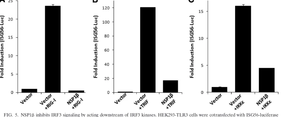
TRIF or IKKdo not rescue NSP1-mediated inhibition of IRF3 activation.Besides TLR3, cytoplasmic dsRNA is sensed by DExD/H box RNA helicases: RIG-I and MDA5 (24). A number of RNA viruses induce signaling through dsRNA re-ceptor RIG-I (30). Having established that PRRSV NSP1 inhibits TLR3-mediated IRF3 and NF-B signaling, we inves-tigated the effect of NSP1on RIG-I signaling pathways. We took advantage of expressing the N-terminal CARD domain of RIG-I, which constitutively activates the RIG-I signaling path-way (14), and tested the effect of NSP1. As shown in Fig. 5A, the constitutively active RIG-I, when coexpressed with ISG56-luciferase, induced strong luciferase activity compared to the vector control. However, in the presence of NSP1the induc-tion is completely inhibited showing that, in addiinduc-tion to the
TLR3 pathway, NSP1also inhibits the RIG-I-mediated sig-naling pathway.
Oftentimes, a very similar approach is used to identify the step(s) of the signaling pathway that is being inhibited (33). We used expression constructs for TRIF and IKKε, two major components of TLR3 signaling pathway, to examine whether the NSP1-mediated inhibition could be rescued. Our results show that IRF3 activity was induced by transfection of TRIF (Fig. 5B) and IKKε(Fig. 5C) in an ISG56-luciferase reporter assay. The presence of NSP1significantly downregulated the ISG-56 promoter activation induced by these molecules. These results indicate that part of the inhibitory activity of NSP1 affects the downstream signaling of IRF3 kinases, probably by directly affecting IRF3. Exogenous expression of TRIF could not recover activation of a NF-B-dependent promoter in the presence of NSP1either (data not shown).
NSP1inhibits dsRNA-induced IRF3 phosphorylation and nuclear translocation.In the resting state, IRF3 shuttles be-tween the cytoplasm and the nucleus. Activation by dsRNA causes its phosphorylation and translocation to the nucleus (58). The binding and recruitment of IRF3 on ISRE promoters is stabilized by CBP/p300, which prevents its export from the nucleus. To investigate the mechanism of NSP1-mediated inhibition of IRF3 activation, we assayed several hallmark steps of the IRF3 activation process, such as IRF3 phosphor-ylation (on Ser396) and nuclear translocation, upon dsRNA treatment. As expected, dsRNA treatment induced a marked increase in the level of phosphorylated IRF3 in the control cells. However, in NSP1-expressing cells, IRF3 phosphoryla-tion was reduced to the basal level (Fig. 6A). IRF3 nuclear translocation was similarly affected by NSP1expression. As
FIG. 3. Inhibition of IRF3-mediated gene induction by PRRSV proteins. (A) HEK-293TLR3 cells were cotransfected with the indicated viral NSP expression plasmids (1g) or empty vector, ISG56-luciferase plasmid (0.4g), and pRLTK (0.01g). At 40 h posttransfection the cells were treated with 10g of dsRNA/ml or PBS for 6 h and assayed for luciferase activity. The bars represent the average fold inductions⫾the SEM for luciferase activities compared to PBS-treated vector control. The panel below the bar graph shows the expression of the respective NSPs. The asterisk (*) at NSP1 lane indicates the detection of proteolytically processed NSP1 to NSP1␣, which carries the N-terminal FLAG tag. (B to D) HEK293-TLR3 cells stably expressing NSP1␣(B), NSP1(C), and NSP11 (D) (lanes 4 to 6) were stimulated with the indicated amount of dsRNA for 6 h, and the ISG56 level was measured by immunoblotting and compared to empty vector-transfected, puromycin-resistant control cells (lanes 1 to 3). Expressions of NSPs were detected by probing with anti-FLAG/HA antibody.
on November 8, 2019 by guest
http://jvi.asm.org/
shown in Fig. 6B, the level of IRF3 present in the nuclear fraction increased upon dsRNA treatment. However, in the presence of NSP1IRF3 failed to translocate to the nucleus. This was consistent with the previous result showing the re-duction of IRF3 phosphorylation in the presence of NSP1. This phenomenon was further verified by visualization of the
[image:7.585.84.502.67.292.2]subcellular location of IRF3 by indirect immunofluorescence. HEK293-TLR3 cells transfected with empty vector showed efficient IRF3 nuclear translocation 2 h after dsRNA treatment but, in the presence of NSP1, most of the IRF3 was retained in the cytoplasm (Fig. 6C) in a manner similar to empty vector-transfected cells without dsRNA treatment (top left panel).
FIG. 4. Inhibition of NF-B-mediated gene induction by PRRSV proteins. (A) HEK293-TLR3 cells were cotransfected with the indicated viral NSP expression plasmids or empty vector (1g), 5X NF-B-luciferase plasmid (0.5g), and pRLTK (0.025g). At 40 h posttransfection the cells were treated with dsRNA for 6 h and assayed for luciferase activity. The fold induction of the average luciferase activity compared to the untreated vector control from a representative experiment is shown. The immunoblot panel indicates the expression of individual NSPs in the luciferase assay. (B) IL-8 mRNA levels were measured by real-time PCR in HEK293-TLR3 cells stably expressing NSP1or vector control cells with or without dsRNA treatment. The mRNA copy numbers were calculated after normalizing them with the RPL32 (internal control) copy number and are expressed as percentages relative to the uninduced vector control.
FIG. 5. NSP1inhibits IRF3 signaling by acting downstream of IRF3 kinases. HEK293-TLR3 cells were cotransfected with ISG56-luciferase plasmid, NSP1expression plasmids, or empty vector. IRF3 signaling pathway was induced by including in the transfection constitutively active RIG-I (A), TRIF (B), and IKKε(C). Cells were lysed 40 h posttransfection and assayed for luciferase activity. Bars indicate the mean fold inductions⫾the SEM of luciferase activity compared to the vector control from three independent experiments.
on November 8, 2019 by guest
http://jvi.asm.org/
[image:7.585.44.541.482.688.2]These results suggest that NSP1 inhibits dsRNA-mediated phosphorylation of IRF3 and its nuclear translocation.
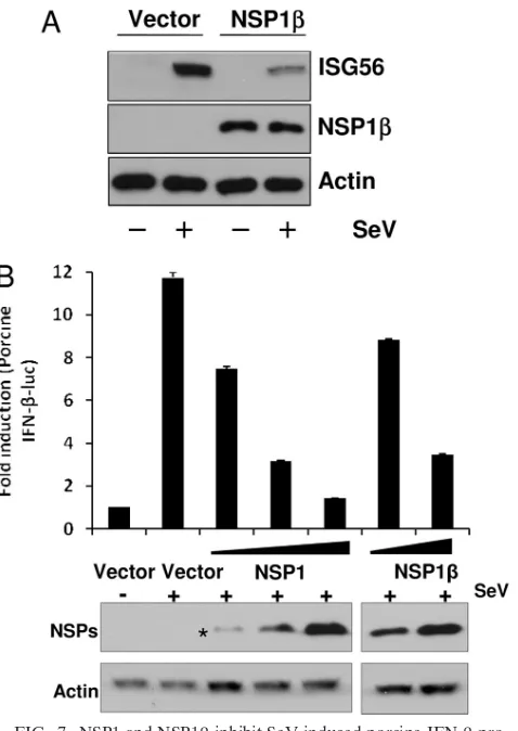
NSP1inhibits SeV-induced IFN-promoter activation.To establish the innate immune evasion property of NSP1in a physiological context, we tested SeV-mediated ISG56 induc-tion in NSP1-expressing cells. The innate immune response against SeV infection is primarily mediated by RIG-I (30). We tested SeV-mediated ISG56 induction in NSP1-expressing cells. HEK293-TLR3 cells stably expressing NSP1- or empty vector-transfected control cells were either mock infected or infected with SeV (40 HA unit/ml) for 8 h. As shown in Fig. 7A, the presence of NSP1significantly inhibited SeV-medi-ated ISG56 induction compared to control cells. Finally, to replicate the effect of NSP1on porcine IFN-induction, we
[image:8.585.137.453.68.472.2]used the porcine monocytic cell line 3D4/31. This myelomono-cytic cell line has been recently characterized to support PRRSV replication and infectious virus production upon transfection with PRRSV genomic RNA, but it lacks the nec-essary receptor in order to enable spread of virus from cell to cell (22). The 3D4/31 cells were transfected by electroporation with porcine IFN--luciferase reporter and NSP1 or NSP1. SeV infection of vector-transfected cells showed induction of porcine IFN--luciferase activity. However, in the presence of NSP1 or NSP1, porcine IFN--luciferase activity was inhib-ited in a dose-dependent manner (Fig. 7B). This result con-firms our findings that NSP1 plays a crucial role in innate immune evasion function of PRRSV in a physiologically rele-vant environment.
FIG. 6. NSP1inhibits dsRNA-induced IRF3 phosphorylation and nuclear translocation. (A) HEK293-TLR3 cells stably expressing NSP1 -or vect-or-transfected cells (vect-or) were either mock treated -or treated with dsRNA f-or 2 h. Phosph-orylated IRF3 (pIRF3) and total IRF3 levels were measured by immunoblotting. (B) Nuclear fractions (N.P) from HEK293-TLR3 cells stably expressing NSP1either mock treated or treated with dsRNA were blotted for IRF3. DRBP76 and tubulin are markers for nuclear and cytoplasmic fractions, respectively. WCE, whole-cell extract. (C) IRF3 subcellular localization was determined by confocal microscopy in HEK-293TLR3 cells stably transfected with empty vector (vector) or NSP1after dsRNA stimulation. For this purpose, cells were either treated or left untreated with 100g of dsRNA/ml for 2 h, followed by immunofluorescence for endogenous IRF3 (green) and NSP1(red) with rabbit anti-IRF3 and mouse anti-HA antibody, as indicated at the top of the panel. Position of nucleus is indicated by DAPI (blue) staining in the merge image.
on November 8, 2019 by guest
http://jvi.asm.org/
DISCUSSION
Viruses have developed a number of mechanisms to subvert or prevent IFN induction and response (7). In the case of PRRSV, both North American and European PRRSV strains have been shown to be poor inducers of IFNin vitroand in vivo. Infection of PAMs with the North American prototype PRRSV VR-2332 significantly reduced their ability to produce type I IFN (36, 37). Furthermore, the absence of IFN-␣ pro-duction in TGEV-infected swine alveolar macrophages, which had been previously infected with PRRSV, implicates an active inhibition of IFN production by PRRSV. In summary, all of the studies are consistent with the fact that PRRSV exerts an active suppression of type I IFN response. In agreement with
these, we have shown here that PRRSV infection of monocyte-derived macrophages showed inhibition of IFN-␣ and IFN- production both at the mRNA and the protein levels. Our data show that five PRRSV NSPs—the NSP1␣, NSP1, NSP2, NSP4, and NSP11—have roles in IFN antagonism and inhibit activation of the IFN-promoter.
In the present study we used TLR3- and RIG-I-mediated dsRNA signaling pathways as model systems to understand the mechanism of innate immune evasion by PRRSV. NSP1␣, NSP1, and NSP11, when stably expressed in HEK293-TLR3 cells, were able to inhibit upregulation of endogenous ISG56 by dsRNA. Moreover, stable expression of NSP1 inhibited endogenous ISG56 induction by SeV. Investigation of IRF3 phosphorylation status after dsRNA stimulation showed that NSP1 inhibits IRF3 (Ser396) phosphorylation and subse-quent nuclear translocation. Although the biochemical nature of this inhibition by PRRSV NSP1remains to be character-ized, there are several mechanisms by which different viruses have been shown to inhibit IRF3 activation. Certain paramyxo-viral V proteins interact with IKKε/TBK1 and function as a competitive inhibitor for IRF3 phosphorylation (33). The W protein of Nipah virus specifically interacts with karyopherin ␣3 and␣4 through its nuclear localization signal and prevents IRF3 translocation (47). It has been reported that NSP2 of EAV, the prototypic member ofArteriviridaefamily, antago-nizes host innate immune response by deconjugating ubiquitin and ISG15 from their cellular substrates (15). Therefore, it is possible that NSP1, a member of cysteine protease family that includes most deubiquitinases, might also use a similar strategy of deconjugating important ubiquitin modifications of IRF3 or RIG-I (6). All of these possible mechanisms of inhi-bition of dsRNA signaling by NSP1 are currently under in-vestigation.
NSP1 and NSP11 expression also inhibited transcription from an NF-B-responsive promoter, but NSP1␣ expression has no inhibitory effect. The suppressive role of NSP1 in NF-B-mediated signaling was further evident from the low level of IL-8 induction in dsRNA-treated HEK293-TLR3 cells stably expressing NSP1. This finding contradicts a previous report that found PRRSV infection of MARC-145 cells and alveolar macrophages resulting in the activation of the NF-B pathway through the degradation of IB (28). The disagree-ment might be due to the fact that the NF-B activation in that study was assayed at a late phase of virus infection (24 to 48 h after virus infection). The activation of NF-B is an immedi-ate-early event that occurs within minutes after exposure to a stimulus and results in a strong transcriptional stimulation of several cytokines (21). Considering that the PRRSV replica-tion cycle in macrophages is completed by 12 h, this effect may be a secondary effect of virus infection. Inhibition of NF-B pathway by PRRSV NSPs, especially during the early infection phase, would help the virus not only to subvert the host innate immune response but also to produce enough progeny virus before being released by apoptosis as proposed by others (10). PRRSV NSP1 contributes two accessory proteinases NSP1␣ and NSP1for the processing of replicase polyprotein. NSP1␣ and NSP1 both possess two papainlike cysteine protease (PCP) domains, PCP␣and PCP, which mediate the release of NSP1␣ and NSP1 from the polyproteins (12). Previous re-verse genetics experiments showed that PCP␣activity is
essen-FIG. 7. NSP1 and NSP1inhibit SeV-induced porcine IFN- pro-moter activation and human ISG56 induction. (A) HEK293-TLR3 cells stably transfected with empty vector (vector) or NSP1 were either mock infected or infected with SeV (40 HA units/ml) for 8 h. Endogenous ISG56 levels were detected by immunoblotting. NSP1 protein was detected by anti-HA antibody. (B) Porcine monocytic cells (3D4/31 cell line) were cotransfected with increasing concentrations of NSP1/NSP1expression plasmids or empty vector (vector), along with porcine IFN--luciferase plasmid (0.4g) and pRLTK (0.01g). At 40 h after the transfection cells were either mock infected or infected with 80 HA units of SeV/ml for 8 h and assayed for luciferase activity. Bars indicate the average fold induction of luciferase activities⫾the SEM compared to uninfected vector control cells. The bottom panel shows immunoblots for the level of each NSP expression in respective transfection. The asterisk indicates detection of N-terminally FLAG-tagged NSP1␣, the cleavage product of NSP1.
on November 8, 2019 by guest
http://jvi.asm.org/
[image:9.585.44.283.65.402.2]tial for subgenomic mRNA synthesis but dispensable for ge-nome replication. However, mutation of active-site residues of PCPresulted in no genome replication and a failure in virus recovery after electroporation ofin vitro-transcribed RNA, un-derscoring its important role (26). Whether the NSP1active site residues (Cys and His) are involved in its IFN antagonistic activity in the context of whole virus infection remains to be investigated. However, it may not be feasible to structurally separate proteolytic function of NSP1from its IFN antago-nistic function because of the general multifunctional nature of viral proteins. This multifunctional nature of NSP1 has been already established in EAV, and it has been found that the structural integrity of NSP1 is essential for transcription (54, 55). In such case, given the essential function of PRRSV NSP1in virus viability, it may not be possible to recover by reverse genetics a replication-capable virus that lacks anti-IFN activity.
The inadequate immunological response to PRRSV infec-tion is underscored by the fact that an infected animal exhibits prolonged viremia, persistent infection, and continuous virus shedding. The poor type I IFN production at the site of PRRSV infection (e.g., lungs) has long been considered the root cause behind defective or suboptimal initiation and elab-oration of the antigen-specific adaptive immune response (1, 35). Influenza virus carrying truncated nonstructural 1 (NS1) protein, which counteracts the host type I IFN response, is highly attenuated in mice, swine, and chicken models (42, 50). Another important example of correlation between anti-IFN activity and virulence is Ebola virus VP35 protein, which in-hibits IRF3 activation. Single amino acid mutation (R312A) of VP35 protein, which compromises its ability to inhibit IRF3 activation, renders the virus highly attenuated in mice (20). It is possible that the strong anti-IFN activity of the NSPs of PRRSV may have a direct effect in the paradoxical immune response observed in PRRSV infection and would explain the inability of the host to clear PRRSV. Such an important im-munomodulatory effect of these NSPs may also be the basis for the pathogenesis and virulence of this virus. On the other hand, pestiviruses, which include classical swine fever virus (CSFV) and bovine viral diarrhea virus, have been shown to induce proteasomal degradation of IRF3 by the viral autopro-tease Npro, which subsequently inhibits IFN-␣/ production (4, 18). A recent investigation into the correlation between the ability of CSFV for IRF3 degradation and viral virulence found that impairment of the IFN antagonistic activity did not give rise to an attenuated virus (43). The authors of that study suggest that the inhibition of type I IFN may have a role in longer persistence of the virus in pigs. Previous results from our laboratory indicated that viral NSP3-8 might play a major role in PRRSV virulence (27). Taken together with our find-ings reported here, this suggests that the pathogenesis of PRRSV may be a complex summation of several major effec-tors contributing to the overall pathogenesis of PRRSV. The NSP1 acts as a suppressor of innate immune response, and NSP3-8 determines the virulence and invasive capacity, which helps the virus to translocate and cross placenta and infect fetuses. Highly pathogenic viruses such as SARS-CoV and Ebola viruses encode multiple proteins targeting different steps in IFN signaling. This not only ensures complete inhibi-tion of the IFN response but also provides a fail-safe
mecha-nism for the virus in case a single protein became dysfunc-tional. Our current findings that five NSPs of PRRSV are involved in IFN antagonism, though of variable ability, point toward PRRSV’s pathogenic nature.
ACKNOWLEDGMENTS
This research has been supported by a grant from the USDA NRICGP (project 2008-00903 USDA-NRICGP).
The primary cells obtained ex vivo for these experiments were col-lected according animal protocols reviewed and approved by the In-stitutional Animal Care Committee of the University of Nebraska-Lincoln under protocol IACUC no. 07-10-048C.
We thank Luwen Zhang (UNL) and Michael David (UCSD) for providing IRF3 expression plasmid and IRF3 antibody, respectively. We thank Terry Fangman for help in confocal microscopy. The invalu-able help of Kazima Saira and Vu Hiep in the preparation and exe-cution of some of these experiments is greatly appreciated.
REFERENCES
1.Albina, E., C. Carrat, and B. Charley.1998. Interferon-alpha response to swine arterivirus (PoAV), the porcine reproductive and respiratory syn-drome virus. J. Interferon Cytokine Res.18:485–490.
2.Allende, R., W. W. Laegreid, G. F. Kutish, J. A. Galeota, R. W. Wills, and F. A. Osorio.2000. Porcine reproductive and respiratory syndrome virus: description of persistence in individual pigs upon experimental infection. J. Virol.74:10834–10837.
3.Ansari, I. H., B. Kwon, F. A. Osorio, and A. K. Pattnaik.2006. Influence of N-linked glycosylation of porcine reproductive and respiratory syndrome virus GP5 on virus infectivity, antigenicity, and ability to induce neutralizing antibodies. J. Virol.80:3994–4004.
4.Bauhofer, O., A. Summerfield, Y. Sakoda, J. D. Tratschin, M. A. Hofmann, and N. Ruggli.2007. Classical swine fever virus Npro interacts with inter-feron regulatory factor 3 and induces its proteasomal degradation. J. Virol. 81:3087–3096.
5.Bautista, E. M., K. S. Faaberg, D. Mickelson, and E. D. McGruder.2002. Functional properties of the predicted helicase of porcine reproductive and respiratory syndrome virus. Virology298:258–270.
6.Bhoj, V. G., and Z. J. Chen.2009. Ubiquitylation in innate and adaptive immunity. Nature458:430–437.
7.Bowie, A. G., and L. Unterholzner.2008. Viral evasion and subversion of pattern-recognition receptor signalling. Nat. Rev. Immunol.8:911–922. 8.Buddaert, W., K. Van Reeth, and M. Pensaert.1998. In vivo and in vitro
interferon (IFN) studies with the porcine reproductive and respiratory syn-drome virus (PRRSV). Adv. Exp. Med. Biol.440:461–467.
9.Cavanagh, D.1997.Nidovirales: a new order comprisingCoronaviridaeand Arteriviridae. Arch. Virol.142:629–633.
10.Costers, S., D. J. Lefebvre, P. L. Delputte, and H. J. Nauwynck.2008. Porcine reproductive and respiratory syndrome virus modulates apoptosis during replication in alveolar macrophages. Arch. Virol.153:1453–1465. 11.de Lima, M., B. Kwon, I. H. Ansari, A. K. Pattnaik, E. F. Flores, and F. A.
Osorio.2008. Development of a porcine reproductive and respiratory syn-drome virus differentiable (DIVA) strain through deletion of specific immu-nodominant epitopes. Vaccine26:3594–3600.
12.den Boon, J. A., K. S. Faaberg, J. J. Meulenberg, A. L. Wassenaar, P. G. Plagemann, A. E. Gorbalenya, and E. J. Snijder. 1995. Processing and evolution of the N-terminal region of the arterivirus replicase ORF1a pro-tein: identification of two papainlike cysteine proteases. J. Virol.69:4500– 4505.
13.Feng, W., S. M. Laster, M. Tompkins, T. Brown, J. S. Xu, C. Altier, W. Gomez, D. Benfield, and M. B. McCaw.2001. In utero infection by porcine reproductive and respiratory syndrome virus is sufficient to increase suscep-tibility of piglets to challenge byStreptococcus suistype II. J. Virol.75:4889– 4895.
14.Foy, E., K. Li, R. Sumpter, Jr., Y. M. Loo, C. L. Johnson, C. Wang, P. M. Fish, M. Yoneyama, T. Fujita, S. M. Lemon, and M. Gale, Jr.2005. Control of antiviral defenses through hepatitis C virus disruption of retinoic acid-inducible gene-I signaling. Proc. Natl. Acad. Sci. U. S. A. 102:2986–2991. 15.Frias-Staheli, N., N. V. Giannakopoulos, M. Kikkert, S. L. Taylor, A.
Brid-gen, J. Paragas, J. A. Richt, R. R. Rowland, C. S. Schmaljohn, D. J. Len-schow, E. J. Snijder, A. Garcia-Sastre, and H. W. t. Virgin.2007. Ovarian tumor domain-containing viral proteases evade ubiquitin- and ISG15-depen-dent innate immune responses. Cell Host Microbe2:404–416.
16.Garcia-Sastre, A., and C. A. Biron.2006. Type 1 interferons and the virus-host relationship: a lesson in detente. Science312:879–882.
17.Genini, S., P. L. Delputte, R. Malinverni, M. Cecere, A. Stella, H. J. Nauw-ynck, and E. Giuffra.2008. Genome-wide transcriptional response of pri-mary alveolar macrophages following infection with porcine reproductive and respiratory syndrome virus. J. Gen. Virol.89:2550–2564.
on November 8, 2019 by guest
http://jvi.asm.org/
18.Gil, L. H., I. H. Ansari, V. Vassilev, D. Liang, V. C. Lai, W. Zhong, Z. Hong, E. J. Dubovi, and R. O. Donis.2006. The amino-terminal domain of bovine viral diarrhea virus Npro protein is necessary for alpha/beta interferon an-tagonism. J. Virol.80:900–911.
19.Guo, J., K. L. Peters, and G. C. Sen.2000. Induction of the human protein P56 by interferon, double-stranded RNA, or virus infection. Virology267: 209–219.
20.Hartman, A. L., B. H. Bird, J. S. Towner, Z. A. Antoniadou, S. R. Zaki, and S. T. Nichol.2008. Inhibition of IRF-3 activation by VP35 is critical for the high level of virulence of Ebola virus. J. Virol.82:2699–2704.
21.Hiscott, J., H. Kwon, and P. Genin.2001. Hostile takeovers: viral appropri-ation of the NF-B pathway. J. Clin. Invest.107:143–151.
22.Huang, Y. W., Y. Fang, and X. J. Meng.2009. Identification and character-ization of a porcine monocytic cell line supporting porcine reproductive and respiratory syndrome virus (PRRSV) replication and progeny virion produc-tion by using an improved DNA-launched PRRSV reverse genetics system. Virus Res.145:1–8.
23.Jiang, Z., T. W. Mak, G. Sen, and X. Li.2004. Toll-like receptor 3-mediated activation of NF-B and IRF3 diverges at Toll-IL-1 receptor domain-con-taining adapter inducing IFN-beta. Proc. Natl. Acad. Sci. U. S. A.101:3533– 3538.
24.Kato, H., O. Takeuchi, S. Sato, M. Yoneyama, M. Yamamoto, K. Matsui, S. Uematsu, A. Jung, T. Kawai, K. J. Ishii, O. Yamaguchi, K. Otsu, T. Tsu-jimura, C. S. Koh, C. Reis e Sousa, Y. Matsuura, T. Fujita, and S. Akira. 2006. Differential roles of MDA5 and RIG-I helicases in the recognition of RNA viruses. Nature441:101–105.
25.Kim, H. S., J. Kwang, I. J. Yoon, H. S. Joo, and M. L. Frey.1993. Enhanced replication of porcine reproductive and respiratory syndrome (PRRS) virus in a homogeneous subpopulation of MA-104 cell line. Arch. Virol.133:477– 483.
26.Kroese, M. V., J. C. Zevenhoven-Dobbe, J. N. Bos-de Ruijter, B. P. Peeters, J. J. Meulenberg, L. A. Cornelissen, and E. J. Snijder.2008. The nsp1alpha and nsp1 papain-like autoproteinases are essential for porcine reproductive and respiratory syndrome virus RNA synthesis. J. Gen. Virol.89:494–499. 27.Kwon, B., I. H. Ansari, A. K. Pattnaik, and F. A. Osorio.2008. Identification
of virulence determinants of porcine reproductive and respiratory syndrome virus through construction of chimeric clones. Virology380:371–378. 28.Lee, S. M., and S. B. Kleiboeker.2005. Porcine arterivirus activates the
NF-B pathway through IB degradation. Virology342:47–59.
29.Lee, S. M., S. K. Schommer, and S. B. Kleiboeker.2004. Porcine reproduc-tive and respiratory syndrome virus field isolates differ in in vitro interferon phenotypes. Vet. Immunol. Immunopathol.102:217–231.
30.Loo, Y. M., J. Fornek, N. Crochet, G. Bajwa, O. Perwitasari, L. Martinez-Sobrido, S. Akira, M. A. Gill, A. Garcia-Sastre, M. G. Katze, and M. Gale, Jr.2008. Distinct RIG-I and MDA5 signaling by RNA viruses in innate immunity. J. Virol.82:335–345.
31.Lopez, O. J., and F. A. Osorio.2004. Role of neutralizing antibodies in PRRSV protective immunity. Vet. Immunol. Immunopathol.102:155–163. 32.Loving, C. L., S. L. Brockmeier, and R. E. Sacco.2007. Differential type I
interferon activation and susceptibility of dendritic cell populations to por-cine arterivirus. Immunology120:217–229.
33.Lu, L. L., M. Puri, C. M. Horvath, and G. C. Sen.2008. Select paramyxoviral V proteins inhibit IRF3 activation by acting as alternative substrates for inhibitor ofB kinase epsilon (IKKe)/TBK1. J. Biol. Chem. 283:14269– 14276.
34.Mateu, E., and I. Diaz.2008. The challenge of PRRS immunology. Vet. J. 177:345–351.
35.Meier, W. A., J. Galeota, F. A. Osorio, R. J. Husmann, W. M. Schnitzlein, and F. A. Zuckermann.2003. Gradual development of the interferon-gamma response of swine to porcine reproductive and respiratory syndrome virus infection or vaccination. Virology309:18–31.
36.Miller, L. C., W. W. Laegreid, J. L. Bono, C. G. Chitko-McKown, and J. M. Fox.2004. Interferon type I response in porcine reproductive and respiratory syndrome virus-infected MARC-145 cells. Arch. Virol.149:2453–2463. 37.Miller, L. C., K. M. Lager, and M. E. Kehrli, Jr.2009. Role of Toll-like
receptors in activation of porcine alveolar macrophages by porcine repro-ductive and respiratory syndrome virus. Clin. Vaccine Immunol.16:360–365. 38.Neumann, E. J., J. B. Kliebenstein, C. D. Johnson, J. W. Mabry, E. J. Bush, A. H. Seitzinger, A. L. Green, and J. J. Zimmerman.2005. Assessment of the economic impact of porcine reproductive and respiratory syndrome on swine production in the United States. J. Am. Vet. Med. Assoc.227:385–392. 39.Ostrowski, M., J. A. Galeota, A. M. Jar, K. B. Platt, F. A. Osorio, and O. J.
Lopez.2002. Identification of neutralizing and nonneutralizing epitopes in the porcine reproductive and respiratory syndrome virus GP5 ectodomain. J. Virol.76:4241–4250.
40.Overend, C., R. Mitchell, D. He, G. Rompato, M. J. Grubman, and A. E. Garmendia.2007. Recombinant swine beta interferon protects swine alveo-lar macrophages and MARC-145 cells from infection with porcine repro-ductive and respiratory syndrome virus. J. Gen. Virol.88:925–931. 41.Posthuma, C. C., K. W. Pedersen, Z. Lu, R. G. Joosten, N. Roos, J. C.
Zevenhoven-Dobbe, and E. J. Snijder.2008. Formation of the arterivirus replication/transcription complex: a key role for nonstructural protein 3 in the remodeling of intracellular membranes. J. Virol.82:4480–4491. 42.Richt, J. A., P. Lekcharoensuk, K. M. Lager, A. L. Vincent, C. M. Loiacono,
B. H. Janke, W. H. Wu, K. J. Yoon, R. J. Webby, A. Solorzano, and A. Garcia-Sastre.2006. Vaccination of pigs against swine influenza viruses by using an NS1-truncated modified live-virus vaccine. J. Virol.80:11009–11018. 43.Ruggli, N., A. Summerfield, A. R. Fiebach, L. Guzylack-Piriou, O. Bauhofer, C. G. Lamm, S. Waltersperger, K. Matsuno, L. Liu, M. Gerber, K. H. Choi, M. A. Hofmann, Y. Sakoda, and J. D. Tratschin.2009. Classical swine fever virus can remain virulent after specific elimination of the interferon regula-tory factor 3-degrading function of Npro. J. Virol.83:817–829.
44.Saira, K., Y. Zhou, and C. Jones.2007. The infected cell protein 0 encoded by bovine herpesvirus 1 (bICP0) induces degradation of interferon response factor 3 and, consequently, inhibits beta interferon promoter activity. J. Vi-rol.81:3077–3086.
45.Sarkar, S. N., K. L. Peters, C. P. Elco, S. Sakamoto, S. Pal, and G. C. Sen. 2004. Novel roles of TLR3 tyrosine phosphorylation and PI3 kinase in double-stranded RNA signaling. Nat. Struct. Mol. Biol.11:1060–1067. 46.Sarkar, S. N., and G. C. Sen.2004. Novel functions of proteins encoded by
viral stress-inducible genes. Pharmacol. Ther.103:245–259.
47.Shaw, M. L., W. B. Cardenas, D. Zamarin, P. Palese, and C. F. Basler.2005. Nuclear localization of the Nipah virus W protein allows for inhibition of both virus- and Toll-like receptor 3-triggered signaling pathways. J. Virol. 79:6078–6088.
48.Sizemore, N., N. Lerner, N. Dombrowski, H. Sakurai, and G. R. Stark.2002. Distinct roles of the IB kinase alpha and beta subunits in liberating nuclear factorB (NF-B) from IB and in phosphorylating the p65 subunit of NF-B. J. Biol. Chem.277:3863–3869.
49.Snijder, E. J., and J. J. Meulenberg.1998. The molecular biology of arteri-viruses. J. Gen. Virol.79(Pt. 5):961–979.
50.Steel, J., A. C. Lowen, L. Pena, M. Angel, A. Solorzano, R. Albrecht, D. R. Perez, A. Garcia-Sastre, and P. Palese.2009. Live attenuated influenza viruses containing NS1 truncations as vaccine candidates against H5N1 highly pathogenic avian influenza. J. Virol.83:1742–1753.
51.Stordeur, P., L. F. Poulin, L. Craciun, L. Zhou, L. Schandene, A. de Lava-reille, S. Goriely, and M. Goldman.2002. Cytokine mRNA quantification by real-time PCR. J. Immunol. Methods259:55–64.
52.Sumpter, R., Jr., Y. M. Loo, E. Foy, K. Li, M. Yoneyama, T. Fujita, S. M. Lemon, and M. Gale, Jr.2005. Regulating intracellular antiviral defense and permissiveness to hepatitis C virus RNA replication through a cellular RNA helicase, RIG-I. J. Virol.79:2689–2699.
53.Tenoever, B. R., S. L. Ng, M. A. Chua, S. M. McWhirter, A. Garcia-Sastre, and T. Maniatis.2007. Multiple functions of the IKK-related kinase IKKεin interferon-mediated antiviral immunity. Science315:1274–1278.
54.Tijms, M. A., D. D. Nedialkova, J. C. Zevenhoven-Dobbe, A. E. Gorbalenya, and E. J. Snijder.2007. Arterivirus subgenomic mRNA synthesis and virion biogenesis depend on the multifunctional nsp1 autoprotease. J. Virol.81: 10496–10505.
55.Tijms, M. A., L. C. van Dinten, A. E. Gorbalenya, and E. J. Snijder.2001. A zinc finger-containing papain-like protease couples subgenomic mRNA syn-thesis to genome translation in a positive-stranded RNA virus. Proc. Natl. Acad. Sci. U. S. A.98:1889–1894.
56.Truong, H. M., Z. Lu, G. F. Kutish, J. Galeota, F. A. Osorio, and A. K. Pattnaik.2004. A highly pathogenic porcine reproductive and respiratory syndrome virus generated from an infectious cDNA clone retains the in vivo virulence and transmissibility properties of the parental virus. Virology325: 308–319.
57.Van Reeth, K., G. Labarque, H. Nauwynck, and M. Pensaert.1999. Differ-ential production of proinflammatory cytokines in the pig lung during dif-ferent respiratory virus infections: correlations with pathogenicity. Res. Vet. Sci.67:47–52.
58.Weaver, B. K., K. P. Kumar, and N. C. Reich.1998. Interferon regulatory factor 3 and CREB-binding protein/p300 are subunits of double-stranded RNA-activated transcription factor DRAF1. Mol. Cell. Biol.18:1359–1368. 59.Xiao, Z., L. Batista, S. Dee, P. Halbur, and M. P. Murtaugh.2004. The level of virus-specific T-cell and macrophage recruitment in porcine reproductive and respiratory syndrome virus infection in pigs is independent of virus load. J. Virol.78:5923–5933.