Identical Consensus Sequence and Conserved Genomic Polymorphism
of Hepatitis E Virus during Controlled Interspecies Transmission
Jerome Bouquet,a,b,cJustine Cheval,dSophie Rogée,a,b,cNicole Pavio,a,b,cand Marc Eloita,b,c,d,e
UMR 1161 Virology, ANSES, Laboratoire de Santé Animale, Maisons-Alfort, Francea; UMR 1161 Virology, INRA, Maisons-Alfort, Franceb; UMR 1161 Virology, Ecole Nationale
Vétérinaire d’Alfort, Maisons-Alfort, Francec; Pathoquest, Paris, Franced; and Department of Virology, Institut Pasteur, Paris, Francee
High-throughput sequencing of bile and feces from two pigs experimentally infected with human hepatitis E virus (HEV)
of genotype 3f revealed the same full-length consensus sequence as in the human sample. Twenty-nine percent of
polymor-phic sites found in HEV from the human sample were conserved throughout the infection of the heterologous host. The
interspecies transmission of HEV quasispecies is the result of a genomic negative-selection pressure on random mutations
which can be deleterious to the viral population. HEV intrahost nucleotide diversity was found to be in the lower range of
other human RNA viruses but correlated with values found for zoonotic viruses. HEV transmission between humans and
pigs does not seem to be modulated by host-specific mutations, suggesting that adaptation is mainly regulated by
ecologi-cal drivers.
H
epatitis E virus (HEV) is a causative agent of acute hepatitis in
humans. The disease is usually self-limited but is a major
public health concern both in developing countries, where it
causes large waterborne epidemics, and in industrialized
coun-tries, where sporadic autochthonous cases of unclear origin are
reported. It is the only hepatitis virus that infects animals other
than primates, such as swine, wild boars, and deer (
28
). Direct
zoonotic transmissions through consumption of contaminated
food were observed in a few cases in a region where HEV is not
endemic (
20
,
38
).
HEV is a positive single-stranded RNA virus. It is the sole
member of the
Hepeviridae
family and the
Hepevirus
genus (
25
).
HEV isolates have been divided into at least four genotypes, two
putative genotypes, and 24 subtypes (
12
,
16
,
22
,
25
). Genotype 1
and 2 are present in humans only, while genotypes 3 and 4 can
infect both humans and animals (
28
). The 7.2-kb genome of HEV
is composed of three open reading frames (ORF). ORF1 encodes a
nonstructural polyprotein with six conserved domains and one
hypervariable region (
14
,
19
). ORF2 encodes the capsid protein,
and ORF3 encodes a phosphoprotein necessary for infection
in
vivo
(
9
).
Many RNA viruses circulate as a population of heterogeneous
but closely related genomes within the same individual. The
emer-gence of such quasispecies is the consequence of a high mutation
rate engendered by the activity of nonproofreading
RNA-depen-dent RNA polymerases (RdRP), coupled with a high replication
rate.
To date, the only description of the HEV quasispecies has
been obtained by restriction fragment length polymorphism
and sequencing of a 448-bp fragment from HEV genotype 1
(
10
). Since genotype 1 is restricted to humans, the results of
this study had limited significance for HEV population
vari-ability in other host species and for the existence of a putative
species barrier for genotypes 3 and 4 (
22
). Swine isolates of
genotype 3 and 4 HEV can infect primates, and human isolates
of genotype 3 and 4 HEV have been shown to replicate in pigs
(
1
,
8
,
11
,
23
,
24
). The objective of the present study was to
analyze the genomic diversity of full-length HEV genotype 3
during a single passage between the two different host species
using high-throughput sequencing (HTS) techniques and deep
genomic variability analysis.
MATERIALS AND METHODS
Experimental infection.Human fecal samples were collected from a French patient with no recent travel history outside France who had de-veloped an acute autochthonous hepatitis E of subtype 3f, according to the
classification of Lu et al. (22).
Two 3-month-old pigs were orally inoculated using industrially
ster-ilized pet food mixed with 1 g of human sample infected with 2⫻109
copies of HEV RNA. After inoculation, feces of pigs were collected every 2 days for 1 month, and bile samples were collected after a light surgical
procedure at 15 days postinfection (dpi) (Fig. 1). This experimental
pro-tocol was validated by the ethics committee (ComEth; saisine number 10-0041) from the National Veterinary School of Alfort, the National Agency for Safety, and University Paris 12.
Serological analyses.Serological analyses were conducted as
previ-ously described (32). Briefly, serum samples were tested with an anti-HEV
total immunoglobulin kit for human diagnosis (EIAgen HEV Ab Kit; Adaltis, Ingen, France), replacing the secondary antibody by a
peroxidase-conjugated rabbit polyclonal anti-pig IgG(H⫹L) (Abcam, France).
Sam-ples were considered positive when the optical density at 450 nm (OD450)
ratio of the sample to the cutoff value (equal to the value of the negative
control⫹0.350) was⬎1.
RNA extraction and quantitative RT-PCR.HEV load was estimated by real-time reverse transcription-PCR (RT-PCR). Total RNA was
ex-tracted from 200l of fecal samples in 10% phosphate-buffered saline
(PBS) or bile samples using a viral QiAmp kit (Qiagen, Courtaboeuf, France) according to the manufacturer’s instructions, and real-time
RT-PCR, as developed by Jothikumar et al., was performed on 2l of RNA
using a Quantitect RT-PCR probe (Qiagen, Courtaboeuf, France) (17). A
LightCycler apparatus (Roche Molecular Biochemicals, Meylan, France) was used for sample analysis. Standard quantification curves were calcu-lated with standard HEV RNA of subtype 3f. The standard plasmid was
Received17 November 2011Accepted16 March 2012
Published ahead of print28 March 2012
Address correspondence to Marc Eloit, marc.eloit@pasteur.fr.
Copyright © 2012, American Society for Microbiology. All Rights Reserved.
doi:10.1128/JVI.06843-11
on November 7, 2019 by guest
http://jvi.asm.org/
constructed by cloning a fragment corresponding to the genomic region from nucleotides (nt) 5190 to 5489 of a French swine HEV sequence of
genotype 3f (accession numberJF718793) into the NheI/XhoI-digested
pCDNA 3.1 (Life Technologies, Villebon sur Yvette, France)
Amplifica-tion and cloning were performed using forward (5=-NheI-CTGCATCGC
CCATGGGATCGC-3=) and reverse (5=-XhoI-CGCTGGGACTGGTCAC
GCC-3=) primers.
The HEV-positive human fecal sample, a pool of swine fecal samples collected at 16 dpi, and a pool of swine bile samples collected at 15 dpi were subjected to HTS.
Sequence-independent amplification.After DNase treatment for 2 h
at 37°C (0.33 U/l of sample; Qiagen, France), total nucleic acids were
extracted using a Nucleospin RNA virus kit (Macherey-Nagel, Germany) and then amplified without use of HEV-specific PCR primers, as
de-scribed previously (6). Briefly, bacteriophage⌽29 polymerase-based
mul-tiple-displacement amplification was preceded by a cDNA synthesis step performed with random hexamer primers. Ligation and whole-genome amplification were then performed with a QuantiTect whole-transcrip-tome kit (Qiagen, France) according to the manufacturer’s instructions.
High-throughput sequencing.Illumina GAII sequencing was sub-contracted to GATC (Constance, Germany). High-molecular-weight DNA (5 g), resulting from genomic RNAs as described above, was frag-mented into 200- to 350-nt fragments, to which adapters were ligated. Adapters included a nucleotide tag allowing for multiplexing of the three samples in one channel.
Data filtering and establishing consensus sequences.Illumina se-quencing data were processed by using a bioinformatic analysis pipeline as
described previously (6). Briefly, Illumina sequence reads were trimmed
of their low-quality score extremities, and host genome sequences (Homo
sapiensandSus scrofa) scanned with SOAPaligner (http://soap.genomics
.org) were discarded. A quick and very restrictive BLASTN study was also
performed to eliminate additional host reads. BLASTN and BLASTX were used to scan dedicated specialized viral, bacterial, and generalist databases
maintained locally (GenBank viral and bacterial databases) (6). Reads and
contigs matching HEV sequence were mapped over the closest sequence hit using relaxed alignment settings (length fraction, 0.5; similarity, 0.8) in the CLC Genomics Workbench (CLC bio, Cambridge, MA).
Validation of polymorphic sites and analysis of sequence diversity.
To eliminate overmutated reads generated by the technique, a new map-ping of reads matching HEV sequences was performed with SOAPaligner (http://soap.genomics.org) on the newly assembled HEV consensus se-quences, removing reads with more than two mismatches.
The error rate due to the amplification and sequencing processes was established by observing the variability of conserved genes from host spe-cies and bacteria present in the samples following the same filtering pro-cess as HEV sequences. The number of sequencing errors was plotted against the number of nucleotides mapped over a consensus sequence to uncover the error rate. A theoretical number of mutations generated by the technique was calculated for each nucleotide position of HEV quasi-species by multiplying the error rate with the coverage at each position and rounded to the immediate upper whole number. Polymorphic sites were validated when the observed number of a base (or gap) different from the consensus sequence was superior to the theoretical number of mutation errors.
Polymorphism parameter calculation.To define the intrahost diver-sity of HEV quasispecies, the genome-wide data of validated nucleotide sites were analyzed to measure the average nucleotide diversity and mean
diversity. Nucleotide diversity as developed by Nei and Li (27) was
calcu-lated as the average percentage of single nucleotide polymorphism (SNP) over the genome, whereas mean diversity corresponds to the percentage of the number of substitutions divided by the total number of nucleotides.
For each sample, a theoretical sequence containing all validated mu-tations was created. Selective pressure along the three ORFs was calculated
with the random effects likelihood method from Datamonkey (http:
//www.datamonkey.org), and the average ratio of nonsynonymous to synonymous changes (dN/dS) was calculated using an online calculation
tool (http://services.cbu.uib.no/tools/kaks). AdN/dSratio above 1
im-plies a positive or directional selection in which advantageous mutations are being fixed, and a ratio of less than 1 implies a negative or purifying selection, suggesting the removal of deleterious mutations. Finally, qua-sispecies complexity was calculated using normalized Shannon entropy
(Sn) as follows: Sn⫽ ⫺⌺i[pi· ln(pi)]/ln(N), whereNis the total number
of sequences analyzed, andpiis the frequency of each sequence in the
FIG 1Experimental infection of two pigs with human HEV. The HEV load of initial human fecal sample inoculated to pigs is plotted as a black square. Excretion of the virus in the feces of pigs is plotted as white squares for pig 1 and as gray squares for pig 2. The HEV load in the bile is plotted as white dots for pig 1 and as gray dots for pig 2. The presence of anti-HEV IgG in serum is indicated by black diamonds. Samples used in high-throughput sequencing are boxed in black. Times of inoculation, surgery, and euthanasia are indicated on the axis for days postinfection.
on November 7, 2019 by guest
http://jvi.asm.org/
[image:2.585.128.463.70.281.2]viral quasispecies. Sn varies from 0 (no complexity) to 1 (maximum complexity).
Detection of nonviable mutations.In order to have an estimate of the percentage of the nonviable HEV population, mutations creating internal stop codons and mutations changing an amino acid into a proline were considered. We assumed that any stop codon within one of the three ORFs would produce a nonviable virion. Prolines are known to disrupt second-ary structures and thus affect proper folding of proteins. ORF2 of HEV has
been fully characterized (31) as coding for the capsid protein. Any
muta-tions creating an additional proline in ORF2 would disrupt the structure of the capsid monomers, preventing its oligomerization and yielding non-viable virions. The range of frequency of these disruptive mutations was approximated from the highest mutation frequency observed in all sites to the sum of all frequencies, considering whether all disruptive mutations are situated on the same sequence or whether all disruptive sites are on different sequences.
Nucleotide sequence accession numbers.The consensus sequences for the full-length genomes of human HEV, swine HEV from feces, and swine HEV from bile were deposited in the GenBank under accession
numbersJN906974,JN906975, andJN906976, respectively.
RESULTS
Experimental infection.
Prior to inoculation, both pigs tested
se-ronegative and negative for HEV RNA (
Fig. 1
). Oral inoculation of
2
⫻
10
9copies of human HEV was successful, leading to virus
excretion in both pigs from 2 dpi and seroconversion at 21 dpi in
pig 2 (
Fig. 1
). Peak viral excretion reached 4
⫻
10
8copies of HEV
RNA/g of feces at 11 dpi. Shortly after the peak of excretion, light
surgery was performed to collect the bile of the two infected
ani-mals. Bile samples of pig 1 and pig 2 contained 2
⫻
10
6and 4
⫻
10
8copies of HEV RNA/ml of bile, respectively. Subsequent feces
samples of pig 1 and pig 2 collected at 16 dpi reached 8
⫻
10
7and
3
⫻
10
6copies of HEV RNA/g of feces, respectively (
Fig. 1
).
Generation of HEV consensus genomes from Illumina
se-quencing data.
Illumina sequencing generated around 27
⫻
10
6reads per sample. An average of 10% of the reads was discarded
after quality filtering and mapping over the host species genomes;
0.15% to 15% of these reads matched HEV sequences (
Table 1
).
For each of the three samples, numerous contigs were assembled,
and three consensus sequences were derived with 4.3
⫻
10
3to
3.5
⫻
10
6reads. HEV consensus genomes differed in length (
Table
1
), with sequences from human feces and pig feces being shorter
than the one from pig bile of 3 nt at the 5
=
untranslated region
(UTR) and 53 to 55 nt at the 3
=
UTR. These differences correspond
to a lower coverage of the extremities due to the trimming process
of each read.
The HEV consensus sequences of the three samples were 100%
identical. As expected, they were found to be of genotype 3,
sub-type 3f. HEV genome coverage ranged from 2 to 146,597 reads per
nucleotide position, depending on sample and genomic region
(
Fig. 2
). The pool of swine bile samples had the highest HEV load:
2.56
⫻
10
9copies of HEV RNA/ml after amplification compared
to 7.42
⫻
10
4and 6.44
⫻
10
6copies of HEV RNA/ml for the pool
of swine feces and the human sample, respectively (data not
shown). As a result, the pool of swine bile sample had the highest
genome coverage, with a mean coverage of 36,792 reads per
nu-cleotide position compared to 143 and 46 reads for swine feces and
human feces, respectively (
Table 1
).
HEV polymorphism parameters.
The number of sequencing
errors was plotted against the number of nucleotides mapped over
conserved genes of host species and bacteria. The error rate was
found to be 0.28% (
Fig. 3
). Because of the coverage differences, the
number of polymorphic sites above the error rate found in each
sample varied according to the coverage of HEV sequences in each
sample: 42 SNPs for the human sample, 172 SNPs for the pool of
pig feces, and 614 SNPs for the pool of pig bile, which represented
0.5%, 2.4%, and 8.3% of the genome, respectively (
Table 2
).
Mu-tations occurring at a frequency as little as 1/356 could be detected,
and the proportion of the HEV population displaying one
partic-ular SNP could be as high as 33% (
Fig. 4
).
This polymorphism is not constant along the genome (
Fig. 4
).
The HEV quasispecies from the pool of pig bile presented values of
intrahost nucleotide diversity higher for the 5
=
untranslated
re-gion (UTR) and hypervariable rere-gion than for the rere-gion coding
for the RdRP (1.4%, 0.093%, and 0.044%, respectively) (data not
shown).
[image:3.585.38.553.78.261.2]Mean diversity ranged from 0.03% for the human sample to
0.18% for the pool of pig bile, whereas intrahost nucleotide
diver-sity ranged from 0.028% to 0.07%. The type of mutations found in
the three samples gave an unusual rate of transition/transversion
of around 0.6. From 50 to 85% of SNPs resulted in
nonsynony-mous mutations, which represented 1.4% to 14.1% of the total
TABLE 1Properties of sequenced data from human and pig samples
Process and/or parameter
Value by sample type
Human Pig feces Pig bile
No. of reads
Total 28,726,064 27,146,966 25,022,058
After quality filtering 26,846,188 24,846,942 21,888,778
After host filtering 26,820,255 24,834,733 21,285,465
Matching HEV sequence 4,256 13,267 3,455,265
Assembling of HEV consensus sequences
No. of nucleotides mapped over the HEV genome 334,924 1,035,285 269,207,053
Length of consensus HEV genome (nt) 7,261 7,259 7,317
Depth coverage (mean no. of reads per nucleotide position) 46 143 36,792
Mapping of HEV quasispecies
No. of nucleotides mapped over the HEV genome 297,263 925,591 243,773,002
Length of consensus HEV genome (nt) 7,234 7,231 7,316
Depth coverage (mean no. of reads per nucleotide position) 41 128 33,316
Bouquet et al.
on November 7, 2019 by guest
http://jvi.asm.org/
length of the three combined ORFs (
Table 2
). Selective pressure
along the three ORFs was mainly neutral, with a few negatively
selected sites in ORF2; no positively selected sites (
dN
⬎
dS
) could
be detected (data not shown). The average genome-wide
dN
/
dS
ratio ranged from 0.91 to 0.51 (
Table 2
), suggestive of a negative
selection. Finally, genome-wide normalized Shannon entropy was
fairly low, ranging from 0.006 to 0.011 (
Table 2
).
A total of 5 to 32 sites with mutations creating internal stop
codons or additional prolines could be detected in the HEV
se-quences of the three samples. The frequency of these disruptive
mutations in the HEV population ranged from 2.7 to 20.7% (
Ta-ble 2
).
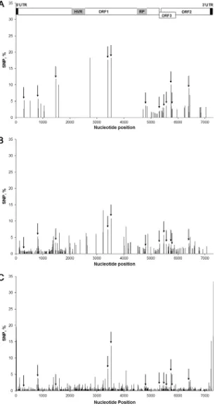
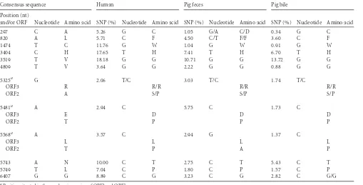
Conserved polymorphism.
Ninety-two SNPs were shared by
the sequences from the pool of pig bile or feces, but, more
impor-tantly, 22 polymorphic nucleotide positions were shared by the
sequences from human and pig bile; and 12 SNPs were shared by
the sequences of all three samples (
Fig. 4
and
Table 3
). Of these 12
SNPs, 6 were situated in ORF1, 3 were in the overlapping fragment
of ORF2 and ORF3, and 3 others were in ORF2 alone. Only two of
these mutations were transitions, and only two resulted in
synon-ymous amino acid changes. The average frequency of these 12
shared SNPs were, respectively, 1.6, 3.5, and 4 times higher than
the average SNP frequency in the human sample, the pig feces, and
the pig bile samples (
Tables 2
and
3
).
FIG 2Coverage of the HEV genome by high-throughput sequencing. A schematic representation of the HEV genome is shown at the top of the figure. ORFs are drawn to scale, and the UTR, the hypervariable region (HVR), and the region coding for the RNA polymerase (RP) are highlighted. Numbers of reads are projected along the genomic position. Black line, bile samples pooled from the two pigs; plain gray line, feces sample pooled from the two pigs; dotted gray line, human sample.
FIG 3Plot of the number of sequencing errors versus the number of nucleotides mapped over conserved genes of host species and bacteria from our HEV samples. The black line represents the linear regression resulting in an error rate of 0.28%.
on November 7, 2019 by guest
http://jvi.asm.org/
[image:4.585.125.463.68.279.2] [image:4.585.122.455.497.701.2]Comparison of polymorphism with other viruses.
The values
for intrahost nucleotide diversity (
) are consistent with those of
other viruses (
Fig. 5
). The average
of the full-length HEV
ge-nome varied according to the coverage of HEV sequences in each
sample and ranged from 0.028% in the human sample to 0.07% in
the pool of pig bile. These values are in the range of the values
obtained for zoonotic viruses, such as the West Nile virus (WNV)
(0.021% in birds to 0.034% in mosquitoes) (
15
), but are in the
lower range of viruses present in humans, such as the human
immunodeficiency virus (HIV) (range, 0.04 to 2.5%), or even five
times lower than values found for hepatitis C virus (HCV) (range,
0.04 to 4.1%) (
33
,
34
).
DISCUSSION
Zoonotic transmissions between humans and swine have been
highly suspected since partial HEV sequences from both hosts can
share more than 99% identity (
4
) and since experimental
cross-infection of subtype 3a HEV in pigs and primates leads to
produc-tive HEV infections (
24
). Subtype 3f HEV is the most common
subtype in France and Europe (
4
,
22
,
40
) and has been shown to be
circulating actively between humans and swine (
4
). This subtype
was selected to study the effect of an interspecies transmission on
the genomic adaptation of HEV in its full-length consensus
se-quence and its quasispecies.
The oral route mimics natural infection of this enterically
transmitted disease but has been shown to be less efficient than
intravenous or intrahepatic routes (
18
). In the present study, oral
exposure of pigs to human subtype 3f HEV led to a productive
HEV infection. A previous observation that oral exposure to
hu-man genotype 1 did not give rise to infection may thus have been
related to the restriction of genotype 1 to humans rather than to
the route of inoculation (
2
,
21
).
Surprisingly, no nucleotide mutations could be found over the
full-length consensus sequence amplified after the interspecies
transmission, which demonstrates a clear adaptation of genotype
3 HEV to both humans and swine. Additionally, 29% (12/42) of
polymorphic sites of HEV from the human sample were
effec-tively infectious and found to be excreted in the feces of pigs at 16
dpi. As demonstrated in other studies, this spectrum of mutations
does not necessarily increase fitness of one virion but might rather
lead to increased infectivity and zoonotic potential through the
diversity of HEV quasispecies population (
7
,
39
).
Conversely, not all polymorphic sites could be transmitted
since a large number of mutations were deleterious. At least 2 to
20.7% of HEV quasispecies population have been found to be
nonviable. These results represent the lowest range of nonviable
sequences since no mutations other than stop codons or proline
were considered. Sanjuan et al. estimated that up to 40% of
ran-dom mutations in RNA viruses are lethal (
35
).
The ratio of transitions/transversions observed in the present
study is indicative of whether or not the mutations observed are
random. In phylogeny, a bias toward a ratio of 1 is commonly
observed since transitions seem favored over transversions,
possi-bly as a result of the underlying chemistry of mutation. In the
present study, a ratio closer to 0.5 has been observed, suggesting
that mutations seem to occur at random. It is then possible to infer
that the development of HEV quasispecies occurs at random,
re-sulting in a high proportion of deleterious mutants, as stated by
Sanjuan et al. (
35
). HEV quasispecies is then purified of its
dele-terious mutations, as shown by the negative
dN
/
dS
ratio.
As Belshaw et al. discussed, mutations and substitutions occur
at different tempos and at different biological levels (
3
).
Substitu-tions are defined as mutaSubstitu-tions which are fixed in a population. The
present study dealt with nonfixed mutations undetectable at the
level of the consensus sequence but observed at the level of
the quasispecies and therefore expressed as the average percentage
of SNPs. Nonetheless, a correlation in the variation of the
muta-tion rate along the genome observed in this study could be made
with a previous report studying the substitution rate along HEV
genomes. Variation in the substitution rate along the genome of
HEV has been predicted previously as being lower for the region
encoding the RdRP (8.4
⫻
10
⫺4substitutions per site per year)
than for the complete genome (1.51
⫻
10
⫺3substitutions per site
per year) (
30
). In the present study, the mutation rate was also
observed to be significantly lower in the RdRP (0.044%) than in
other parts of the genome (up to 1.4%), which may be explained
by higher functional constraints on this coding region.
In the end, HEV quasispecies resulted in a low-diversity and
low-complexity population compared to other human RNA
vi-ruses such as HIV or HCV (
33
,
34
). HEV intrahost nucleotide
diversity is closer to what has been found for the zoonotic virus
WNV. Indeed, viruses that need to infect diverse hosts to produce
a full viral cycle, like arboviruses, are subjected to higher
con-straints and thus evolve more slowly than other RNA viruses (
15
).
In addition to being a useful tool for discovering new
patho-gens (
5
,
36
,
37
), HTS is also of great interest in delineating the
quasispecies of viruses since the use of specific PCRs to amplify
subgenomic regions of the virus, which could introduces bias, is
avoided. But great care should be put into the handling of
poly-TABLE 2Statistics on polymorphism of HEV from human and pig samples
Sample source
No. of SNPs (% of the genome) Avg SNP frequency (%) Mean diversity
(%)a (%)b Ts/Tvc
No. of variable amino acid
sites (%) dN/dSd Sne
No. of sites with deleterious mutationsf Frequency of deleterious mutations (%)
Human 42 (0.5) 4.93 0.03 0.028 0.35 36 (1.4) 0.91 0.006 5 7-19.6
Pig feces 172 (2.4) 1.17 0.19 0.047 0.67 110 (4.3) 0.72 0.008 6 8.1-17
Pig bile 614 (8.3) 0.83 0.18 0.070 0.74 355 (14.1) 0.51 0.011 32 2.7-20.7
aCalculated as the percentage of the number of substitutions divided by the total number of nucleotides. b
is the nucleotide diversity calculated as the average percentage of SNPs over the genome (27).
cTransition/transversion ratio. d
Ratio of nonsynonymous to synonymous mutations.
eShannon entropy. f
Internal stop codons and disruptive proline. Bouquet et al.
on November 7, 2019 by guest
http://jvi.asm.org/
[image:5.585.39.545.79.156.2]FIG 4SNPs along the HEV genome. A schematic representation of the HEV genome is aligned at the top of the figure (see the legend ofFig. 2). Bars indicate the percentage of validated SNPs along the HEV genome sequenced from the human sample (A), the feces of pigs (B), and the bile of pigs (C). Black arrows indicate the position of SNPs shared by the three samples.
on November 7, 2019 by guest
http://jvi.asm.org/
[image:6.585.137.453.67.644.2]morphic data since various biases are reported for HTS techniques
(
13
,
26
).
Sequence coverage depth is, for example, critical when
differ-ent samples are compared. Variations of polymorphism observed
in this study between the three samples should not be considered
as properties of HEV quasispecies in different hosts or sampling
points but as a consequence of the differences in nucleotide
cov-erage. A higher sequence coverage contains more information and
is therefore more accurate in detecting low-frequency variants.
The lower number of SNPs and the smaller intrahost nucleotide
diversity observed for HEV from the human sample than from the
pig samples are only the results of its lower coverage.
Interestingly, the mutation error rate for Illumina GaIIx
calcu-lated in this study as being 0.28% was the same as previously
reported (
26
). A number of insertions/deletions were found in the
HEV sequences, all of which fell under the mutation error rate.
The insertion/deletion rate generated by the amplification and
high-throughput sequencing processes could be calculated as
be-ing 5.7
⫻
10
⫺7(data not shown), which is lower than what has
been previously established (4
⫻
10
⫺6) (
26
). This
insertion/dele-tion rate was very likely reduced by the second mapping of HEV
sequences, which removed all reads containing more than two
mismatches.
Here is presented the first report on the use of HTS for the
study of full-length genomes of HEV and, more generally, on the
use of HTS to analyze viral variability upon interspecies
transmis-sion. The observation that the full-length consensus sequence of
HEV is conserved in spite of a change of host demonstrates the
absence of a species barrier and the clear adaptation of genotype 3f
HEV to both hosts. Moreover, this study confirms that HEV exists
as a quasispecies in the
in vivo
setting and that genetic variability
extends throughout its genome. Finally, major SNPs were
con-served during the interspecies transmission. These results may
suggest that transmission of swine HEV to humans would result in
the absence of adaptation and in a productive HEV infection.
In conclusion, the transmission of human HEV to pigs did not
seem associated with a restriction in genetic diversity, most likely
because HEV infection of either host does not impact its viral
cycle. According to the typology of zoonosis proposed by Pepin
et al. (
29
), the transmission of some zoonotic agents can be
[image:7.585.40.549.77.336.2]gov-erned only by ecological drivers. In this case, all viral genotypes
circulating in the reservoir are already competent for transmission
in the new host. Founder effects or adaptative fine-tuning in the
new host could explain the variability of the strains. These results
suggest that HEV could belong to this category of viruses.
TABLE 3Single nucleotide positions shared by HEV sequences from human and pig
Consensus sequence Human Pig feces Pig bile
Position (nt)
and/or ORF Nucleotide Amino acid SNP (%) Nucleotide Amino acid SNP (%) Nucleotide Amino acid SNP (%) Nucleotide Amino acid
297 C A 5.26 G C 1.05 G/A C/D 0.34 G C
820 A L 5.71 C F 4.50 C/T F/F 3.60 C F
1474 T C 11.76 G W 1.04 G W 0.91 G W
3404 C H 17.65 T H 7.41 T H 6.70 T H
3519 T V 18.18 G G 10.71 G G 13.72 G G
4809 T V 3.64 G G 2.22 G G 0.88 G G
5325a G 2.06 T/C 3.03 T/C 1.74 T/C
ORF3 R R/R R/R R/R
ORF2 A S/P S/P S/P
5481a A 2.94 C 5.75 C 1.73 C
ORF3 E D D D
ORF2 T P P P
5568a A 3.57 C 2.94 G 1.37 C
ORF3 L L L L
ORF2 T P A P
5743 A N 10.00 C T 2.75 C T 5.43 C T
5749 T L 7.04 C P 1.80 C P 1.57 C P
6407 G G 8.89 C G 3.23 C G 2.82 C G/G
aPosition situated in the overlapping region of ORF2 and ORF3.
FIG 5Viral intrahost nucleotide diversity () of viruses. Values ofare displayed as percentages on a logarithmic number line for HEV alongside the West Nile
virus (WNV), human immunodeficiency virus (HIV), and hepatitis C virus (HCV) (15,33,34).
Bouquet et al.
on November 7, 2019 by guest
http://jvi.asm.org/
[image:7.585.115.471.641.703.2]ACKNOWLEDGMENTS
J.B. was supported by a Ph.D. grant from the ANSES.
We thank Elizabeth Nicand and Sophie Tessé from the National Ref-erence Center for HEV, HIA Val de Grace, Paris, France, for providing the human fecal sample used for the experimental infection. We thank Kevin Pariente from the Institut Pasteur for technical assistance. We also thank Thomas Lilin, Francis Moreau, and Benoit Lécuelle from the research center for molecular biology, ENVA, Maisons-Alfort, France, for the an-imal care and expertise they provided for the experimental infection. Fi-nally, we warmly thank Jennifer Richardson for editing the English ver-sion of the manuscript.
REFERENCES
1.Arankalle VA, Chobe LP, Chadha MS.2006. Type-IV Indian swine HEV
infects rhesus monkeys. J. Viral Hepat.13:742–745.
2.Balayan MS, Usmanov RK, Zamyatina NA, Djumalieva DI, Karas FR.
1990. Brief report: experimental hepatitis E infection in domestic pigs. J.
Med. Virol.32:58 –59.
3.Belshaw R, Sanjuán R, Pybus OG.2011. Viral mutation and substitution:
units and levels. Curr. Opin. Virol.1:430 – 435.
4.Bouquet J, et al.2011. Close similarity between sequences of hepatitis E virus recovered from humans and swine, France, 2008 –2009. Emerg.
In-fect. Dis.17:2018 –2025.
5.Buckwalter MR, et al.2011. Identification of a novel neuropathogenic
Theiler’s murine encephalomyelitis virus. J. Virol.85:6893– 6905.
6.Cheval J, et al.2011. Evaluation of high-throughput sequencing for iden-tifying known and unknown viruses in biological samples. J. Clin.
Micro-biol.49:3268 –3275.
7.Coffey LL, Vignuzzi M. 2011. Host alternation of chikungunya virus increases fitness while restricting population diversity and adaptability to
novel selective pressures. J. Virol.85:1025–1035.
8.Feagins AR, Opriessnig T, Huang YW, Halbur PG, Meng XJ. 2008.
Cross-species infection of specific-pathogen-free pigs by a genotype 4
strain of human hepatitis E virus. J. Med. Virol.80:1379 –1386.
9.Graff J, et al.2005. The open reading frame 3 gene of hepatitis E virus
contains acis-reactive element and encodes a protein required for
infec-tion of macaques. J. Virol.79:6680 – 6689.
10. Grandadam M, et al.2004. Evidence for hepatitis E virus quasispecies. J.
Gen. Virol.85:3189 –3194.
11. Halbur PG, et al.2001. Comparative pathogenesis of infection of pigs with hepatitis E viruses recovered from a pig and a human. J. Clin.
Micro-biol.39:918 –923.
12. Haqshenas G, Shivaprasad HL, Woolcock PR, Read DH, Meng XJ.
2001. Genetic identification and characterization of a novel virus related to human hepatitis E virus from chickens with hepatitis-splenomegaly
syndrome in the United States. J. Gen. Virol.82:2449 –2462.
13. Harismendy O, et al.2009. Evaluation of next generation sequencing
platforms for population targeted sequencing studies. Genome Biol.10:
R32.http://genomebiology.com/content/10/3/R32.
14. Huang CC, et al.1992. Molecular cloning and sequencing of the Mexico
isolate of hepatitis E virus (HEV). Virology191:550 –558.
15. Jerzak G, Bernard KA, Kramer LD, Ebel GD.2005. Genetic variation in West Nile virus from naturally infected mosquitoes and birds suggests
quasispecies structure and strong purifying selection. J. Gen. Virol.86:
2175–2183.
16. Johne R, et al.2010. Novel hepatitis E virus genotype in Norway rats,
Germany. Emerg. Infect. Dis.16:1452–1455.
17. Jothikumar N, Cromeans TL, Robertson BH, Meng XJ, Hill VR.2006.
A broadly reactive one-step real-time RT-PCR assay for rapid and
sensi-tive detection of hepatitis E virus. J. Virol. Methods131:65–71.
18. Kasorndorkbua C, et al.2004. Routes of transmission of swine hepatitis
E virus in pigs. J. Clin. Microbiol.42:5047–5052.
19. Koonin EV, et al.1992. Computer-assisted assignment of functional domains in the nonstructural polyprotein of hepatitis E virus: delineation of an additional group of positive-strand RNA plant and animal viruses.
Proc. Natl. Acad. Sci. U. S. A.89:8259 – 8263.
20. Li T-C, et al.2005. Hepatitis E virus transmission from wild boar meat.
Emerg. Infect. Dis.11:1958 –1960.
21. Lu L, et al.2004. Complete sequence of a Kyrgyzstan swine hepatitis E virus (HEV) isolated from a piglet thought to be experimentally infected
with human HEV. J. Med. Virol.74:556 –562.
22. Lu L, Li C, Hagedorn CH.2006. Phylogenetic analysis of global hepatitis E virus sequences: genetic diversity, subtypes and zoonosis. Rev. Med.
Virol.16:5–36.
23. Meng XJ, et al. 1998. Experimental infection of pigs with the newly identified swine hepatitis E virus (swine HEV), but not with human strains
of HEV. Arch. Virol.143:1405–1415.
24. Meng XJ, et al.1998. Genetic and experimental evidence for cross-species
infection by swine hepatitis E virus. J. Virol.72:9714 –9721.
25. Meng XJ, et al.2011.Hepeviridae, p 991–998.InKing AMQ, et al (ed), Virus taxonomy. Ninth report of the International Committee on Taxon-omy of Viruses. Elsevier, London, United Kingdom.
26. Minoche AE, Dohm JC, Himmelbauer H.2011. Evaluation of genomic
high-throughput sequencing data generated on Illumina HiSeq and
Ge-nome Analyzer systems. GeGe-nome Biol.12:R112.http://genomebiology
.com/content/12/11/R112.
27. Nei M, Li WH.1979. Mathematical model for studying genetic variation
in terms of restriction endonucleases. Proc. Natl. Acad. Sci. U. S. A.76:
5269 –5273.
28. Pavio N, Meng X-J, Renou C.2010. Zoonotic hepatitis E: animal
reser-voirs and emerging risks. Vet. Res.41:46.
29. Pepin KM, Lass S, Pulliam JRC, Read AF, Lloyd-Smith JO. 2010.
Identifying genetic markers of adaptation for surveillance of viral host
jumps. Nat. Rev. Microbiol.8:802– 813.
30. Purdy MA, Khudyakov YE.2010. Evolutionary history and population
dynamics of hepatitis e virus. PLoS One5:e14376.
31. Robinson RA, et al.1998. Structural characterization of recombinant hepatitis E virus ORF2 proteins in baculovirus-infected insect cells.
Pro-tein Expr. Purif.12:75– 84.
32. Rose N, et al.2011. High prevalence of Hepatitis E virus in French
domestic pigs. Comp. Immunol. Microbiol. Infect. Dis.34:419 – 427.
33. Sakai A, Kaneko S, Honda M, Matsushita E, Kobayashi K. 1999.
Quasispecies of hepatitis C virus in serum and in three different parts of
the liver of patients with chronic hepatitis. Hepatology30:556 –561.
34. Salazar-Gonzalez JF, et al.2009. Genetic identity, biological phenotype, and evolutionary pathways of transmitted/founder viruses in acute and
early HIV-1 infection. J. Exp. Med.206:1273–1289.
35. Sanjuán R, Moya A, Elena SF.2004. The distribution of fitness effects caused by single-nucleotide substitutions in an RNA virus. Proc. Natl.
Acad. Sci. U. S. A.101:8396 – 8401.
36. Sauvage V, et al.2011. Identification of the first human gyrovirus, a virus
related to chicken anemia virus. J. Virol.85:7948 –7950.
37. Sauvage V, et al.2011. Human polyomavirus related to African green
monkey lymphotropic polyomavirus. Emerg. Infect. Dis.17:1364 –1370.
38. Tei S, Kitajima N, Takahashi K, Mishiro S.2003. Zoonotic transmission
of hepatitis E virus from deer to human beings. Lancet362:371–373.
39. Vignuzzi M, Stone JK, Arnold JJ, Cameron CE, Andino R. 2006.
Quasispecies diversity determines pathogenesis through cooperative
in-teractions in a viral population. Nature439:344 –348.
40. Widén F, et al.2011. Molecular epidemiology of hepatitis E virus in
humans, pigs and wild boars in Sweden. Epidemiol. Infect.139:361–371.
on November 7, 2019 by guest
http://jvi.asm.org/