0022-538X/08/$08.00⫹0 doi:10.1128/JVI.02198-07
Copyright © 2008, American Society for Microbiology. All Rights Reserved.
Epstein-Barr Virus Immediate-Early Protein Zta Co-Opts Mitochondrial
Single-Stranded DNA Binding Protein To Promote Viral
and Inhibit Mitochondrial DNA Replication
䌤
Andreas Wiedmer,
1Pu Wang,
1Jing Zhou,
1Andrew J. Rennekamp,
1Valeria Tiranti,
2Massimo Zeviani,
2and Paul M. Lieberman
1*
The Wistar Institute, Philadelphia, Pennsylvania 19104,1and IRCCS Foundation Neurological Institute C. Besta, Milano 20126, Italy2 Received 8 October 2007/Accepted 30 January 2008
Disruption of cellular metabolic processes and usurpation of host proteins are hallmarks of herpesvirus lytic infection. Epstein-Barr virus (EBV) lytic replication is initiated by the immediate-early protein Zta. Zta is a multifunctional DNA binding protein that stimulates viral gene transcription, nucleates a replication complex at the viral origin of lytic replication, and inhibits cell cycle proliferation. To better understand these functions and identify cellular collaborators of Zta, we purified an epitope-tagged version of Zta in cells capable of supporting lytic replication. FLAG-tagged Zta was purified from a nuclear fraction using FLAG antibody immunopurification and peptide elution. Zta-associated proteins were isolated by sodium dodecyl sulfate-polyacrylamide gel electrophoresis and identified by mass spectrometry. The Zta-associated proteins included members of the HSP70 family and various single-stranded DNA and RNA binding proteins. The nuclear replication protein A subunits (RPA70 and RPA32) and the human mitochondrial single-stranded DNA binding protein (mtSSB) were confirmed by Western blotting to be specifically enriched in the FLAG-Zta immunopurified complex. mtSSB coimmunoprecipitated with endogenous Zta during reactivation of EBV-positive Burkitt lymphoma and lymphoblastoid cell lines. Small interfering RNA depletion of mtSSB reduced Zta-induced lytic replication of EBV but had only a modest effect on transcription activation function. A point mutation in the Zta DNA binding domain (C189S), which is known to reduce lytic cycle replication, eliminated mtSSB association with Zta. The predominantly mitochondrial localization of mtSSB was shifted to partly nuclear localization in cells expressing Zta. Mitochondrial DNA synthesis and genome copy number were reduced by Zta-induced EBV lytic replication. We conclude that Zta interaction with mtSSB serves the dual function of facilitating viral and blocking mitochondrial DNA replication.
Epstein-Barr virus (EBV) is a human lymphotropic gamma-herpesvirus that infects over 90% of the adult population worldwide (reviewed in references 24 and 41). Despite its prev-alence among healthy individuals, EBV is a potent growth-transforming virus that has been linked to several epithelial and lymphoid cell malignancies, including Burkitt’s lymphoma, nasopharyngeal carcinoma, Hodgkin’s disease, gastric carci-noma, posttransplant lymphoproliferative disease, and AIDS-associated non-Hodgkin’s lymphomas (26, 53). Upon primary infection of B lymphocytes, EBV establishes a highly successful latency program that drives B-cell proliferation and ultimately evades immune detection (48, 49). Productive lytic infection occurs in terminally differentiating plasma B cells and in some epithelial cells. Long-term survival of the virus requires a com-plex interplay between latent and lytic infections, and both life cycles contribute to virus-associated disease.
Lytic cycle replication and associated gene products may contribute directly and indirectly to EBV pathogenesis. Lytic infection is observed at high rates in patients with oral hairy leukoplakia and EBV-positive gastric carcinoma (19, 22, 29). Elevated levels of EBV lytic antigens are a prognostic risk factor for nasopharyngeal carcinoma in regions of endemicity
(13). Lytic cycle gene expression or replication is also required for lymphomagenesis in SCID mouse models (20, 21). EBV encodes numerous viral gene products during lytic infection that have the potential to alter cell physiology and host im-mune response, but the precise role of these gene products in viral disease has not been characterized completely.
EBV encodes two immediate-early proteins, Zta and Rta, that are essential for lytic replication (10, 11). Viruses lacking Zta are incapable of lytic cycle gene expression or DNA rep-lication, indicating that Zta is essential for virus viability (16). Zta (also referred to as BZLF1, ZEBRA, and EB1) is a mem-ber of the basic leucine zipper (b-zip) family of DNA binding proteins with sequence similarity to C/EBP, c-Jun, and c-Fos (27). Despite Zta’s simple structure, it has multiple functions in the EBV life cycle. Zta binds multiple recognition sites, including AP-1 and C/EBP recognition sites, and activates transcription of both viral and cellular genes (8, 23, 34, 35). Zta functions as a DNA-bound transcription activator that can recruit cellular general transcription factors and coactivators to target promoters through an amino-terminal activation do-main (14). Zta also functions as a lytic cycle replication factor by recruiting viral replication proteins to the origin of lytic replication (OriLyt) (32, 33, 45). Zta has a profound effect on cellular gene expression and cell cycle progression. It activates the transcription of transforming growth factor(TGF-) (8) and fatty acid synthase (31) cellular genes through direct in-teraction with promoter sequences. Zta can also bind to
sev-* Corresponding author. Mailing address: The Wistar Institute, 3601 Spruce St., Philadelphia, PA 19104. Phone: (215) 898-9491. Fax: (215) 898-0663. E-mail: [email protected].
䌤Published ahead of print on 27 February 2008.
4647
on November 8, 2019 by guest
http://jvi.asm.org/
eral key regulatory proteins, including p53, NF-B, and c-Myb, and can disrupt PML-associated nuclear domain 10 (ND10/ PODs) (1, 4). Zta also induces a cell cycle arrest through a mechanism that requires its b-zip domain, independent of its transcription activation function (7, 9, 42).
In an effort to better understand the multiple functions of Zta during lytic infection, we isolated Zta as a multiprotein complex from cells undergoing lytic replication. We identified several cellular proteins that associate with Zta, including two proteins typically associated with mitochondrial functions. We present evidence that the mitochondrial single-stranded DNA binding protein is a Zta-associated protein that facilitates EBV lytic replication and serves as a target for EBV to subvert mitochondrial DNA replication.
MATERIALS AND METHODS
Cells and plasmids.ZKO-293 cells (a gift from H. J. Delecluse) are 293 cells transformed with hygromycin-resistant EBV bacmid with a deletion of the BZLF1 gene and were grown in RPMI medium with 10% fetal bovine serum
(FBS) and 100g/ml hygromycin. MutuI and LCL cells are EBV-positive
B-lymphoblastoid cells and were grown in RPMI medium supplemented with 10% FBS. D98/HR1 cells (a generous gift from S. Kenney) are an EBV-positive adherent cell line generated by the fusion of the P3HR1 Burkitt lymphoma line and HeLa cells and were grown in Dulbecco modified Eagle medium with 10% FBS. 293 cells were grown in Dulbecco modified Eagle medium with 10% FBS.
3⫻FLAG-Zta was generated by inserting Zta cDNA as a HindIII-BamHI
frag-ment into a p3⫻FLAG-myc-CMV24 vector (Sigma) for mammalian cell
expres-sion. FLAG-Zta-C189S was described previously (50). mtSSB was cloned into the ApaI-HindIII of pRc-CMV and was a gift from V. Tiranti and M. Zeviani.
Protein purification.ZKO-293 cells were grown on 50 dishes (25 cm) and then
transfected with 500g (10g/plate) expression plasmid for Zta
(pCMV-FLAG-Zta) or control vector (pFLAG3⫻-CMV) by Lipofectamine. Cells were
har-vested 48 h posttransfection by cell scraping, washed two times with ice-cold phosphate-buffered saline (PBS), transferred to hypotonic buffer A (10 mH HEPES [pH 7.9], 10 mM KCl, 1 mM EDTA, 1 mM dithiothreitol [DTT], 1 mM phenylmethylsulfonyl fluoride [PMSF], and protease inhibitor cocktail II), and subjected to 10 strokes of a Dounce homogenizer to isolate nuclei. The nuclei were pelleted by centrifugation at 5,000 rpm for 10 min in a Sorvall SS34 rotor. Nuclei were then resuspended in buffer B (10 mM HEPES [pH 7.9], 400 mM NaCl, 10% glycerol, 1 mM EDTA, 1 mM DTT, 1 mM PMSF, and protease inhibitor cocktail) using a Dounce homogenizer (B pestle) to resuspend nuclei. Nuclei were stirred for 30 min in buffer B and then pelleted by centrifugation at 25,000 rpm for 30 min in a Sorvall SS34 rotor. The supernatant was designated the soluble nuclear fraction. The residual nuclear pellet was then resuspended in buffer E (10 mM Tris [pH 7.5], 10% glycerol, 400 mM NaCl, 0.05% Ipegal, 1 mM DTT, 1 mM PMSF, and protease inhibitor cocktail). The nuclear pellet was subjected to sonication for 10 min in a Bronson sonicator until the bulk of the pellet was solubilized. The insoluble residual material was pelleted by centrifu-gation at 10,000 rpm for 10 min, and the remaining solubilized nuclear pellet was used for immunopurification using FLAG-agarose (Sigma). The extract was
incubated with FLAG-agarose beads (10l/mg of extract) overnight (⬃16 h) at
4°C and then subjected to four washes with buffer E and one wash with buffer C. The FLAG-agarose was then incubated with a 1-column volume of buffer C containing 1 mg/ml of FLAG peptide. Eluted material was either subjected to Western blot analysis or concentrated by trichloroacetic acid (TCA) precipita-tion and subjected to sodium dodecyl sulfate-polyacrylamide gel electrophoresis (SDS-PAGE) and colloidal blue staining for analysis by mass spectrometry.
Immunoprecipitation (IP) assays.Cell lysates were generated by incubating cells in NET buffer (10 mM Tris [pH 7.5], 150 mM NaCl, 0.05% NP-40, 1 mM PMSF, and protease inhibitor cocktail) at 4°C for 30 min with gentle agita-tion. The nuclei were pelleted at 15,000 rpm in a Microfuge centrifuge at 4°C. The supernatant was subjected to a preclearing with protein-A or protein-G Sepharose for 4 h and then incubated with primary antibody for 4 h, and then protein-A (rabbit antibodies) or protein-G (mouse antibodies) Sepharose was added for 1 h. The immune complex was washed three times with NET buffer and one time with PBS and then was eluted by SDS-PAGE and heated at 95°C for 5 min.
IF assays.D98/HR1 cells were plated on glass slides and then transfected with Zta expression vector and assayed by immunofluorescence (IF) as described
previously (14). Forty-eight hours posttransfection, the cells were fixed with paraformaldehyde, washed with PBS prior to the addition of primary antibody (1:500 to 1:1,000), washed two times with PBS containing 0.05% Ipegal, and again washed two times with PBS. Fluorescein isothiocyanate-linked anti-rabbit and Texas Red-linked anti-mouse antibodies were used as fluorescence-tagged secondary antibodies.
siRNA depletion. Small interfering RNAs (siRNAs) for mtSSB were pur-chased from Dharmacon as a Smartpool (no. L-021365-01) and as a single siRNA targeting the following sequence: GUACUUCGUCAGUUUGUA AUU. siRNAs were transfected using Dharmacon transfection reagent, as rec-ommended by the manufacturer.
BrdU incorporation assay. Biomodeoxyuridine (BrdU) incorporation into DNA was performed and quantified essentially as described previously (6). Cells
(5⫻105
) were pulse-labeled with 50 mM BrdU for 30 min. DNA was then extracted by incubating cell pellets in DNA lysis buffer (50 mM Tris [pH 8.0], 1 M NaCl, 10 mM EDTA, 0.5% SDS, 0.2-mg/ml protease K, and 0.3-mg/ml final concentration of sonicated salmon sperm DNA) at 50°C for 2 h, extracted with phenol-chloroform, and precipitated with ethanol. The DNA was dissolved in
500 ml Tris-EDTA and sonicated to an average length of⬃700 bp. DNA was
then heat denatured at 95°C for 5 min and cooled rapidly on ice; 50l was kept
as input. A total of 50l 10⫻IP buffer (100 mM NaPO4 [pH 7.0], 1.4 M NaCl,
0.5% Triton X-100) was added to the DNA to make to a final 1⫻concentration.
Then 4l anti-BrdU (stock 25g/ml; BD Pharmingen) was incubated with the
solution for 60 min at room temperature with rotation. Then 3.5g (10l) rabbit
anti-mouse immunoglobulin G (IgG) (Sigma) was added to each tube and ro-tated for 30 min at room temperature. The immune-complexed DNA was pre-cipitated by centrifugation for 5 min at 14,000 rpm and washed two times in 750
l 1⫻IP buffer. The pellet was then resuspended in 200l lysis buffer II (10 mM
EDTA; 50 mM Tris-HCl [pH 8.8], 0.5% SDS, and 0.25 mg/ml proteinase K) for
1 h at 50°C, and then an additional 100l lysis buffer II was added and the DNA
was incubated at 37°C overnight. DNA was purified by phenol-chloroform ex-traction twice and precipitated with ethanol in the presence of glycogen. Real-time PCR was done to compare the levels of BrdU incorporation on different regions with specific primers for the EBV genome OriLyt region (GCCCGTTG GGTTTCATTAAG; CCAAATCTCGCGGACCTCTA), mitochondrial genomic DNA (CACCATTAGCACCCAAAGCT; ACATAGCGGTTGTTGATGGG), or nuclear DNA using cellular actin (GCCATGGTTGTG CCATTACA; GGCCAG GTTCTCTTTTTATTTCTG).
EBV genome copy number assay.Cells (⬃1⫻106
cells per sample) were
collected and resuspended in 100l chromatin IP SDS lysis buffer (1% SDS, 10
mM EDTA, 50 mM Tris, pH 8.0.). After brief sonication, chromatin IP dilution buffer (0.01% SDS, 1.1% Triton X-100, 1.2 mM EDTA, 16.7 mM Tris [pH 8.0], and 167 mM NaCl) was added to 1 ml and then incubated with proteinase K for
2 to 3 h at 50°C. A total of 300l was removed and subjected to
phenol-choloroform extraction and ethanol precipitation. Precipitated DNA was then assayed by real-time PCR using primers for the dyad symmetry region of EBV and for cellular actin DNA using a standard curve method for quantification of each DNA. The relative copy number of EBV was determined by dividing the copy number of double-stranded DNA by that of actin DNA.
Mitochondrial DNA analysis.Total genomic DNA was isolated using SDS lysis buffer and proteinase K for 2 to 3 h at 50°C. DNA was recovered by phenol-chloroform extraction and ethanol precipitation. RNA was then removed by RNase (DNase-free grade). Purified DNA was then digested with XmaI and analyzed by 0.7% agarose gel electrophoresis and Southern blotting. The probe for mitochondrial DNA was amplified with primers oPL2146 (CCACAACTCA ACGGCTACATAG) and oPL2147 (CACTCATAGGCCAGACTTAGGG). The same primers were used for quantitative PCR analysis of mitochondrial
DNA using real-time PCR. CellularAluDNA was identified using a single
primer, oPL1824 (CGGAGTCTCGCTCTGTCGCCCAGGCTGGAGTGCAG TGGCGCGA). EBV DNA was identified using the OriLyt sequence amplified by the primers oPL1682 (GCCCGTTGGGTTTCATTAAG) and oPL1244 (CC AGGAAGTGGCGAGCAT). PCR products were labeled with digoxigenin us-ing the PCR DIG probe synthesis kit (Roche, Inc.) or a DIG OligoLabelus-ing kit (Roche, Inc.) according to the manufacturer’s protocol.
Additional methods.EBV lytic replication was measured by real-time PCR of viral DNA relative to cellular actin DNA as described previously (50). Electro-phoretic mobility shift assays (EMSA), luciferase assays, and reporter plasmids were described previously (50). The oligonucleotide used for single-strand DNA
binding in EMSAs and glutathioneS-transferase (GST) pull-down assays was
derived from the OriLyt upstream element (TCTCTGTGTAATACTTTAAGG TTTGCTCAGGAG).
on November 8, 2019 by guest
http://jvi.asm.org/
RESULTS
Identification of Zta-associated proteins.To identify cellu-lar proteins that interact with Zta under conditions known to cause lytic replication, we transiently expressed epitope-tagged Zta in 293 cells carrying a stable EBV genome lacking Zta (ZKO-293 cells). Zta with an amino-terminal 3⫻FLAG epitope was expressed to high levels in over 80% of the cell population, and robust activation of lytic antigens EA-D and Rta were detected by Western blot analysis (data not shown). Nuclear extracts were prepared from transfected cells. We found that most of Zta remained in the nuclear pellet (data not shown). The nuclear pellet was redissolved in buffer containing nonionic detergent (0.05% Ipegal) and then subjected to son-ication to disrupt the chromatin-bound forms of Zta. The resolubilized Zta was then subjected to immuno-affinity puri-fication using anti-FLAG agarose and FLAG-peptide elution. Untransfected control cells were treated identically. The pep-tide-eluted proteins were then assayed by SDS-PAGE and visualized by colloidal blue staining (Fig. 1A). The major bands unique to FLAG-Zta were excised and identified by mass spec-trometry using liquid chromatography-tandem mass spectrom-etry. We identified several prominent proteins, including three members of the HSP70 family (HSP70, mortalin, and GSAS), RNA binding proteins (hnRNPC and PABPC4), histone sub-units, and several single-stranded DNA binding proteins (Ku70, RPA1, RPA3, and mtSSB) (Fig. 1B). The single-stranded DNA binding proteins RPA1, RPA3, and mtSSB were confirmed by Western blot analysis to be highly specific for FLAG-Zta, relative to untransfected control extracts (Fig. 1C).
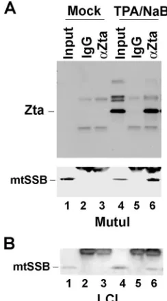
Endogenous Zta associates with mtSSB.We were intrigued by the fact that mitochondrial proteins mtSSB and HSP70 member mortalin were associated with Zta when isolated from nuclear pellet fractions. We therefore asked whether mtSSB associates with endogenous Zta in EBV-infected B-cell lines during reactivation (Fig. 2). Nuclear extracts were generated from an EBV-positive Burkitt lymphoma cell line (MutuI) and from an EBV-transformed lymphoblastoid cell line (LCL)
[image:3.585.126.461.66.242.2]be-fore and after reactivation was induced by phorbol ester (TPA) and sodium butyrate (NaB). The lysates were subject to IP with antibody specific for Zta or control IgG and then were assayed by Western blotting with antibody to Zta (Fig. 2A, top panel) or mtSSB (Fig. 2A, bottom panel). As expected, Zta was highly induced after treatment with TPA and NaB and was efficiently precipitated with Zta antibody (Fig. 2A, top panel). We also found that mtSSB was efficiently coimmunoprecipitated with
FIG. 1. Identification of Zta-associated proteins. (A) FLAG-Zta or vector control CMV-FLAG (FLAG-Control) was transfected into ZKO-293 cells and purified using anti-FLAG agarose. Purified proteins were analyzed by SDS-PAGE and colloidal blue staining. M, molecular weight markers. (B) The major polypeptide species enriched in FLAG-Zta were excised and identified by liquid chromatography-tandem mass spec-trometry, and the NCBI accession number and number of hits are indicated. (C) Western blot analysis of FLAG-purified proteins from FLAG-Zta or control transfected cells (FLAG-Control) with antibodies to RPA70, RPA32, mtSSB, and Zta, as indicated.
FIG. 2. Endogenous Zta interacts with mtSSB. (A) EBV-positive MutuI cells were untreated or treated with TPA and NaB to induce lytic reactivation. At 48 h postinduction, cell lysates were prepared for IP with anti-Zta antibody or control IgG. Immunoprecipitates were analyzed by Western blot analysis with antibodies to Zta (top panel) or mtSSB (bottom panel). (B) EBV-positive LCLs were treated and im-munoprecipitated as described above for panel A and assayed by Western bloting of mtSSB.
on November 8, 2019 by guest
http://jvi.asm.org/
[image:3.585.358.480.429.648.2]Zta from extracts where lytic infection was induced (lane 6). A similar co-IP of mtSSB with Zta was observed in EBV-positive LCLs (Fig. 2B).
mtSSB depletion reduces lytic replication.To determine if the association between mtSSB and Zta was functionally rele-vant, we depleted mtSSB by siRNA transfection in ZKO-293 cells. Depletion of mtSSB was monitored by Western blot analysis and found to be reduced by⬃70% (Fig. 3A). Deple-tion of mtSSB had no detectable effect on cell viability or proliferation (data not shown). Viral replication was then as-sayed by real-time PCR comparing EBV DNA (OriLyt) to cellular actin DNA. Transfection of Zta resulted in an⬃ 42-fold stimulation of viral DNA in ZKO-293 cells cotransfected with control siRNA (Fig. 3B, si-Control). In contrast, Zta
in-duced an⬃19-fold amplification of viral DNA in cells cotrans-fected with mtSSB-specific siRNA (Fig. 3B, si-mtSSB). Viral early antigen EA-D was assayed by Western blot analysis in these transfected cells (Fig. 3C). We found that mtSSB-specific siRNA caused a slight reduction in EA-D expression relative to control siRNA, consistent with a reduction in lytic replica-tion. However, the relatively small reduction in EA-D expres-sion suggests that mtSSB was not contributing significantly to the transcription activation function of Zta.
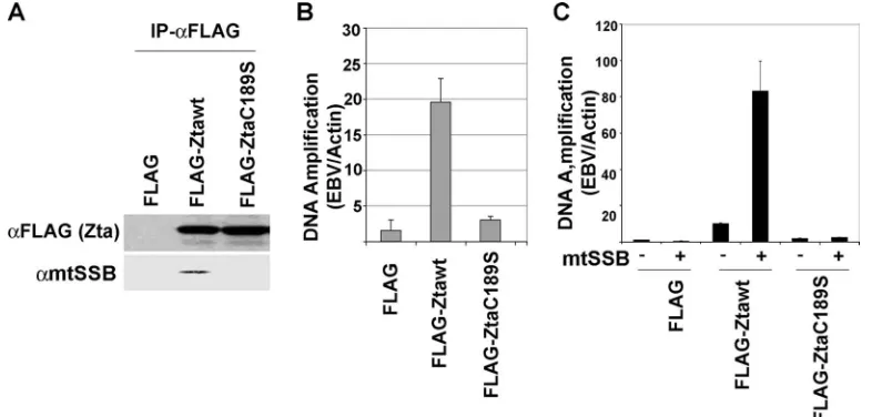
Genetic and functional evidence for mtSSB function in EBV lytic replication. The role of mtSSB in lytic replication was further investigated by genetic analysis. A cysteine substitution mutation in the Zta basic region (C189S) has been character-ized previously (51). Zta-C189S is defective for viral
[image:4.585.124.458.67.232.2]reactiva-FIG. 4. Replication-defective Zta fails to interact with mtSSB. (A) FLAG-Zta-wt or FLAG-Zta-C189S was expressed in ZKO-293 cells and immunoprecipitated with anti-FLAG antibody. Immunoprecipitates were analyzed by Western blotting for FLAG-Zta (top panel) and for mtSSB (bottom panel). (B) The transfected ZKO-293 cells represented in panel A were analyzed for EBV lytic replication using real-time PCR analysis with primers specific for the EBV genome relative to cellular actin. (C) ZKO-293 cells were transfected with expression plasmids for Zta, mtSSB, or their respective control vectors, as indicated. EBV lytic replication was assayed by real-time PCR analysis of EBV DNA relative to cellular DNA. FIG. 3. Inhibition of EBV lytic replication by siRNA depletion of mtSSB. (A) ZKO-293 cells were untransfected (lane 1) or transfected with mtSSB-specific siRNA (si-mtSSB) (lane 2) or the siRNA control (si-Control) (lane 3) and assayed by Western blotting for the expression of mtSSB. (B) ZKO-293 cells were transfected with si-mtSSB or si-Control with either Zta or control expression vectors and assayed 48 h posttransfection for the EBV DNA copy number relative to that of cellular actin using real-time PCR. The error bars represent the standard deviations for three independent transfections. (C) The ZKO-293 cells represented in panel B were assayed by Western blotting for EA-D and the cellular loading control, PCNA.
on November 8, 2019 by guest
http://jvi.asm.org/
[image:4.585.96.489.487.675.2]tion but can otherwise stimulate transcription of viral promot-ers on transiently transfected DNA. The molecular basis for this defect was shown to be the inability of Zta-C189S to recognize C/EBP binding sites, but additional deficiencies may also contribute to the inability to activate lytic replication. We therefore compared the abilities of Zta-wild type (wt) and Zta-C189S to coimmunoprecipitate with mtSSB (Fig. 4A). We found that mtSSB immunoprecipitated efficiently with Zta-wt but was not detectable in the immunoprecipitates with Zta-C189S. The levels of replication of Zta-wt and -C189S in these ZKO-293 cells were assayed (Fig. 4B). Consistent with previ-ous findings, Zta-wt induced lytic replication (⬃20-fold), while Zta-C189S was incapable of stimulating lytic replication more than twofold. When mtSSB was cotransfected with Zta-wt, we found a superactivation of lytic replication (an additional four-fold increase). In contrast, mtSSB had no effect on viral lytic replication in the absence of Zta or with Zta-C189S. These findings suggest that mtSSB requires C189S for stable interac-tion with Zta and that its overexpression enhances lytic repli-cation.
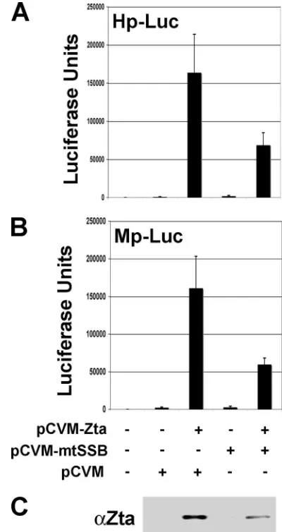
To determine if some of these effects on replication were a consequence of changes in Zta transcription function, we as-sayed Zta transcription activation function directly. Zta tran-scription activity was measured using luciferase reporter plas-mids in EBV-negative 293 cells (Fig. 5). We tested the effect of mtSSB on Zta transcription activation of the BMLF1 (Mp-Luc) and BHLF1 (Hp-(Mp-Luc) gene promoters. We found that the addition of mtSSB inhibited Zta transcription activation of both Mp-Luc and Hp-Luc approximately threefold. This inhi-bition was partly explained by a reduction in Zta expression levels when mtSSB was contransfected (Fig. 5C). Nevertheless, these findings suggest that mtSSB does not significantly en-hance and may even inhibit Zta transcription activity. Thus, the stimulation of lytic replication by mtSSB is not an indirect result of Zta transcription activation.
Single-stranded DNA enhances the interaction between mtSSB and Zta. To test the possibility that single-stranded DNA may contribute to the interaction between mtSSB and Zta, we examined the interaction with proteins expressed and purified fromEscherichia coli. GST-mtSSB and GST proteins were purified to near homogeneity (Fig. 6A). These proteins were incubated with highly purified Zta-wt or Zta-C189S and then assayed for their ability to bind using the GST pull-down assay with glutathione Sepharose beads (Fig. 6B). We found that Zta-wt bound to GST-mtSSB weakly, but detectably, in the absence of any exogenous DNA (Fig. 6B, top panel, lane 6). Addition of 1 pmol of a 33-bp single-stranded oligonucle-otide derived from the EBV OriLyt region greatly facilitated the interaction between mtSSB and Zta-wt (Fig. 6B, top panel, lane 3). No interaction between Zta and GST was observed. Similarly, Zta-C189S did not interact with mtSSB in the ab-sence or preab-sence of single-strand oligonucleotide DNA (Fig. 6B, bottom panel). This suggests that the defect in Zta-C189S is in the ability to interact with mtSSB and single-strand DNA. To examine the single-stranded DNA binding properties of Zta more directly, we assayed Zta-wt and Zta-C189S for their abilities to bind either double-stranded DNA (Fig. 6C) or single-stranded DNA (Fig. 6D) using EMSA. We found that the Zta-wt and -C189S binding abilities to a double-stranded AP1 consensus binding site were indistinguishable (Fig. 6C).
mtSSB did not bind to double-stranded DNA and had no effect on Zta-wt or -C189S binding to double-stranded DNA. In contrast, mtSSB bound to a single-stranded 33-bp oligonucle-otide probe derived from the EBV OriLyt region (Fig. 6D). Furthermore, Zta-wt bound significantly more (approximately fivefold) single-stranded DNA than did Zta-C189S. The addi-tion of mtSSB stimulated Zta-wt and -C189S binding to single-stranded DNA, but this stimulation was only additive and not synergistic (Fig. 6D). These findings indicate that Zta-wt has significantly higher single-stranded DNA binding capacity than does the C189S mutant and that this difference accounts for its ability to interact preferentially with mtSSB.
[image:5.585.317.517.69.446.2]Zta induces nuclear localization of mtSSB.Since mtSSB and Zta are thought to be in different subcellular compartments, we examined the localization of these two proteins in intact
FIG. 5. mtSSB inhibits Zta expression and transcription activation. (A) Zta, mtSSB, or control expression vectors were transfected into EBV-negative 293 cells along with reporter plasmid for BMLF1-luciferase. Luciferase activity was assayed at 48 h posttransfection. Error bars rep-resent the standard deviations for at least three independent transfections. (B) Conditions are the same as described above for panel A, except with the BHLF1-luciferase reporter plasmid. (C) Western blot of Zta ex-pressed in the cells whose transfection in representative experiments is shown in panels A and B.
on November 8, 2019 by guest
http://jvi.asm.org/
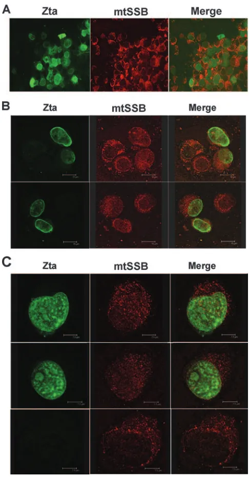
cells using indirect IF and confocal microscopy (Fig. 7). FLAG-Zta was transfected into EBV-positive adherent cell line D98/HR1 and then visualized with a mouse monoclonal antibody to FLAG, and mtSSB was visualized with a rabbit polyclonal antibody to mtSSB. We found that mtSSB was pre-dominantly cytoplasmic in untransfected cells, with some punc-tate staining likely representing mitochondria. Zta was ex-pressed predominantly in the nuclear compartment, although some cytoplasmic staining was observed. At low resolution, it was apparent that a percentage of Zta and mtSSB colocalized, mostly at perinuclear and nuclear patches (Fig. 7A). At a
higher resolution using confocal microscopy, we observed an increase in mtSSB in the nuclei of Zta-transfected cells and substantial colocalization within the nuclear compartment (Fig. 7B and C). Based on these representative images, we conclude that Zta induces mtSSB to enter the nuclear com-partment and that some Zta protein colocalizes with mtSSB in the cytoplasm.
[image:6.585.299.542.66.531.2]Lytic infection inhibits mitochondrial DNA replication.The relocalization of mtSSB to the nuclear compartment suggests
[image:6.585.46.281.68.445.2]FIG. 6. Single-stranded DNA facilitates Zta binding to mtSSB. (A) GST-mtSSB and GST proteins were purified and analyzed by Coomassie blue staining of SDS-PAGE gels. M, molecular weight markers. (B) GST or GST-mtSSB was assayed for binding to bacteri-ally purified Zta-wt or Zta-C189S. Input and bound Zta proteins were assayed by immunoblotting (IB) with anti-Zta antibody. Single-stranded oligonucleotide DNA (SSDNA) from EBV OriLyt was in-cluded in reactions indicated by “⫹.” (C) EMSA with DS DNA probe from EBV OriLyt incubated with GST, GST-mtSSB, wt, or Zta-C189S as described above each lane. (D) EMSA with single-stranded DNA probe from EBV OriLyt incubated with the proteins depicted in panel C, as indicated above each lane. “Z” indicates Zta binding, and “S” indicates mtSSB binding. Quantification by PhosphorImager anal-ysis is shown below each EMSA result in panels C and D.
FIG. 7. Colocalization of mtSSB with nuclear Zta. EBV-positive adherent D98/HR1 cells were transfected with Zta and assayed by indirect IF with mouse monoclonal antibody to Zta (green) and rabbit polyclonal antibody to mtSSB (red). (A) Low-resolution (⫻40) mag-nification. (B) Confocal microscopy at magnification of⫻40. (C) Con-focal microscopy at magnification of⫻60. The bottom panel shows the predominantly cytoplasmic localization of mtSSB in cells not express-ing Zta.
on November 8, 2019 by guest
http://jvi.asm.org/
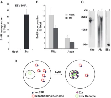
that mitochondrial DNA replication or genome stability may be altered by Zta and EBV lytic replication. To test the effect of Zta expression on DNA replication, we assayed BrdU in-corporation for EBV, cellular DNA, and mitochondrial DNA before and after Zta expression in ZKO-293 cells (Fig. 8). Cells were transfected with Zta or control expression plasmid and then pulse-labeled with BrdU at 48 h posttransfection. BrdU incorporation in DNA was then determined by IP with BrdU-specific antibody and real-time PCR analysis of immunopre-cipitated DNA relative to the total input DNA for each primer pair. We found that Zta induced an ⬃200-fold increase in BrdU incorporation in EBV genome DNA (Fig. 8A). In con-trast, Zta expression reduced mitochondrial BrdU incorpora-tion to⬃61% and cellular actin BrdU incorporation to⬃57% of that of mock-transfected cells (Fig. 8B). To determine if Zta affected the copy number or stability of mitochondrial ge-nomes, we assayed total cellular DNA 48 h after transfection with Zta into ZKO-293 cells (Fig. 8C). We found that Zta transfection dramatically reduced mitochondrial DNA copy number (Fig. 8C, left panel). Zta also reduced and altered the
mobility of cellularAlurepeat DNA (Fig. 8C, middle panel). As expected, Zta increased the copy number of EBV genome DNA (Fig. 8C, right panel). These data indicate that Zta in-hibits mitochondrial DNA replication and decreases the mito-chondrial genome copy number while activating viral lytic rep-lication (Fig. 8D).
DISCUSSION
[image:7.585.105.480.68.400.2]Our results indicate that mtSSB can be isolated in a stable complex with Zta during lytic cycle replication of EBV. mtSSB was isolated by biochemical purification of FLAG-tagged Zta expressed in ZKO-293 cells (Fig. 1). FLAG-Zta protein effi-ciently activates transcription and lytic replication in ZKO-293 cells and therefore is likely to associate with functionally sig-nificant cellular proteins. mtSSB associated with endogenous Zta in lymphoblastoid cells after lytic induction with TPA and NaB (Fig. 2). The functional significance of these interactions was explored by several methods. siRNA depletion of mtSSB inhibited EBV lytic replication but had a modest effect on
FIG. 8. Repression of mitochondrial DNA replication and copy number by EBV lytic replication. (A) ZKO-293 cells were transfected with Zta or control expression vector and then pulse-labeled with BrdU for 30 min. BrdU-labeled DNA was precipitated with anti-BrdU antibody and analyzed by real-time PCR for EBV DNA relative to total (non-BrdU plus BrdU-labeled) DNA. (B) The same BrdU-immunoprecipitated DNA from the experiment described above for panel A was assayed by real-time PCR for mitochondrial DNA (Mito) or cellular actin DNA. Error bars represent standard deviations for three independent IPs. (C) Total cellular DNA was isolated from Zta-transfected or untransfected controls, digested with XmaI, and analyzed by Southern blotting with a mitochondrion-specific probe (Mito) (left panel), anAlurepeat DNA probe (Alu) (middle panel), or an EBV OriLyt DNA probe (right panel). (D) Model depicting the corresponding loss of mitochondrial DNA replication and copy number as EBV undergoes lytic replication.
on November 8, 2019 by guest
http://jvi.asm.org/
transcription activation of early viral antigens (Fig. 3). A point mutation in Zta (C189S) that compromises replication func-tion disrupted the interacfunc-tion of Zta with mtSSB (Fig. 4). Overexpression of mtSSB stimulated lytic replication with Zta-wt but had no effect on Zta-C189S (Fig. 4). In contrast, mtSSB expression inhibited transcription activation of two vi-ral promoters fused to reporter luciferase genes (Fig. 5). This suggests that mtSSB may stimulate the replication function but inhibit transcription activation. Based on these findings, we propose that mtSSB plays a role in switching Zta from a tran-scription activator to a replication protein during lytic cycle reactivation (Fig. 8D).
The role of mtSSB in EBV lytic replication is not completely understood. mtSSB is a single-stranded DNA binding protein with properties similar toE. coliSSB (12, 30). mtSSB is a key component of the mitochondrial DNA replication machinery and is an essential gene in several organisms for which this question has been examined (36, 46). LikeE. coliSSB, mtSSB stabilizes single-strand formation and stimulates homologous strand exchange by RecA (15). Mitochondrial DNA replica-tion initiates through a complex mechanism involving RNA transcription-mediated priming and strand invasion to form a stable triple-strand structure referred to as an R-loop (38, 47). The precise role for mtSSB in the formation and resolution of these structures at mitochondrial origins of DNA replication is not completely elucidated.
The molecular mechanism of EBV lytic replication and the role of cellular factors in this process are not yet known. Some studies have implicated cellular proteins, like topoisomerases, as essential for herpesvirus lytic replication (5, 37). At EBV OriLyt, several cellular DNA binding proteins have been im-plicated in the replication function (3, 54). The DNA structure of the downstream element at OriLyt is non-B-DNA and readily unwound (39). Recent studies also indicate that numer-ous RNA transcripts initiate within OriLyt (55). In studies with the related gammaherpesvirus, Kaposi’s sarcoma-associated herpesvirus, RNA transcription initiation within OriLyt was found to be essential for efficient lytic replication (52). Studies with cytomegalovirus have found that RNA transcripts and RNA hybrids exist at the lytic origin, and these structures may reflect complex transcription-related events during the initia-tion of lytic replicainitia-tion (25, 40). Our experiments do not ad-dress whether mtSSB plays a similar role in replication initia-tion at EBV OriLyt as it does at mitochondrial DNA origins or whether RNA transcripts contribute to EBV replication. How-ever, the positive contribution of mtSSB to EBV lytic replica-tion suggests that EBV and other herpesvirus lytic origins may share some common features with mitochondrial DNA repli-cation origins.
A second major finding from these studies is that Zta-in-duced lytic replication led to a loss of mitochondrial DNA replication. Zta is known to inhibit cell cycle progression and nuclear DNA synthesis, but it has not been demonstrated that the EBV lytic cycle blocks mitochondrial DNA replication. The inhibition of mitochondrial DNA replication may be ex-pected based on our understanding of herpesvirus replication and its tendency to shut off many host-cell metabolic pathways. Recent studies have found that herpes simplex virus lytic in-fection leads to a nucleolytic degradation of mitochondrial DNA through the action of the virus-encoded nuclease UL12.5
(44). A Kaposi’s sarcoma-associated herpesvirus orthologue of UL12.5 was found to be important for the degradation of host mRNA, although this function was separable from the DNase functional domain (17). More recent studies indicate that EBV orthologue BGLF5 can also eliminate host cell immune func-tion by selective degradafunc-tion of cellular mRNA (18, 43). The relationship between DNA and RNA nuclease activities and their potential roles in the disruption of mitochondrial DNA stability remains unclear. We found that EBV lytic gene ex-pression led to a loss of BrdU incorporation and a loss of mitochondrial DNA copy number (Fig. 8). We did not detect any clear evidence of degraded mitochondrial genomes, but we cannot exclude that a nuclease-based mechanism for the deg-radation of mitochondrial DNA also exists in EBV. The inhi-bition of mitochondrial DNA synthesis may be achieved through multiple mechanisms. Consistent with the findings re-ported here is the report that Zta expression alters mitochon-drial morphology (28). We did not observe a significant loss of mitochondrial DNA copy number after Zta transfection in EBV-negative cells (data not shown), so it is likely that addi-tional virus-encoded lytic genes contribute to the loss of mito-chondrial DNA synthesis and genome copy number. Future studies will be required to determine if the EBV orthologue of HSV UL12.5 also contributes to the loss of mitochondrial DNA copy number or if EBV has acquired a different mech-anism for achieving the same function. The interaction of Zta with mtSSB suggests that EBV has acquired a novel mecha-nism for disrupting cellular mitochondrial function and pirated this protein for efficient replication of its own genome.
ACKNOWLEDGMENTS
We thank H.-J. Delecluse for ZKO-293 cells and M. Wold for antibodies to RPA.
This work was supported by grants from the NIH (CA86678) to P.M.L., the Wistar Cancer Center (NCI), and the PA Settlement for Tobacco Research. A.J.R. was supported by a predoctoral fellowship (UPenn tumor virology training grant 1T32 CA115299).
REFERENCES
1.Adamson, A. L., and S. Kenney.2001. Epstein-Barr virus immediate-early protein BZLF1 is SUMO-1 modified and disrupts promyelocytic leukemia
bodies. J. Virol.75:2388–2399.
2. Reference deleted.
3.Baumann, M., R. Feederle, E. Kremmer, and W. Hammerschmidt.1999. Cellular transcription factors recruit viral replication proteins to activate the
Epstein-Barr virus origin of lytic DNA replication, oriLyt. EMBO J. 18:
6095–6105.
4.Bell, P., P. M. Lieberman, and G. G. Maul.2000. Lytic but not latent replication of Epstein-Barr virus is associated with PML and induces
sequen-tial release of nuclear domain 10 proteins. J. Virol.74:11800–11810.
5.Boehmer, P. E., and I. R. Lehman.1997. Herpes simplex virus DNA
repli-cation. Annu. Rev. Biochem.66:347–384.
6.Carroll, S. M., J. Trotter, and G. M. Wahl.1991. Replication timing control can be maintained in extrachromosomally amplified genes. Mol. Cell. Biol.
11:4779–4785.
7.Cayrol, C., and E. Flemington.1996. G0/G1 growth arrest mediated by a region encompassing the basic leucine zipper (bZIP) domain of the
Epstein-Barr virus transactivator Zta. J. Biol. Chem.271:31799–31802.
8.Cayrol, C., and E. K. Flemington.1995. Identification of cellular target genes of the Epstein-Barr virus transactivator Zta: activation of transforming
growth factorigh3 (TGF-igh3) and TGF-1. J. Virol.69:4206–4212.
9.Cayrol, C., and E. K. Flemington.1996. The Epstein-Barr virus bZIP tran-scription factor Zta causes G0/G1 cell cycle arrest through induction of
cyclin-dependent kinase inhibitors. EMBO J.15:2748–2759.
10.Chevallier, G. A., E. Manet, P. Chavrier, C. Mosnier, J. Daillie, and A. Sergeant.1986. Both Epstein-Barr virus (EBV)-encoded trans-acting fac-tors, EB1 and EB2, are required to activate transcription from an EBV early
promoter. EMBO J.5:3243–3249.
11.Countryman, J., and G. Miller. 1985. Activation of expression of latent
on November 8, 2019 by guest
http://jvi.asm.org/
Epstein-Barr herpesvirus after gene transfer with a small cloned subfragment
of heterogeneous viral DNA. Proc. Natl. Acad. Sci. USA82:4085–4089.
12.Curth, U., C. Urbanke, J. Greipel, H. Gerberding, V. Tiranti, and M. Zeviani.
1994. Single-stranded-DNA-binding proteins from human mitochondria and Escherichia coli have analogous physicochemical properties. Eur. J. Biochem.
221:435–443.
13.Dardari, R., M. Khyatti, A. Benider, H. Jouhadi, A. Kahlain, C. Cochet, A. Mansouri, B. El Gueddari, A. Benslimane, and I. Joab.2000. Antibodies to the Epstein-Barr virus transactivator protein (ZEBRA) as a valuable biomarker in young patients with nasopharyngeal carcinoma. Int. J. Cancer
86:71–75.
14.Deng, Z., C.-J. Chen, D. Zerby, H.-J. Delecluse, and P. M. Lieberman.2001. Identification of acidic and aromatic residues in the Zta activation domain
essential for Epstein-Barr virus reactivation. J. Virol.75:10334–10347.
15.Edmondson, A. C., D. Song, L. A. Alvarez, M. K. Wall, D. Almond, D. A. McClellan, A. Maxwell, and B. L. Nielsen.2005. Characterization of a mi-tochondrially targeted single-stranded DNA-binding protein in Arabidopsis
thaliana. Mol. Genet. Genomics273:115–122.
16.Feederle, R., M. Kost, M. Baumann, A. Janz, E. Drouet, W. Hammer-schmidt, and H. J. Delecluse.2000. The Epstein-Barr virus lytic program is controlled by the co-operative functions of two transactivators. EMBO J.
19:3080–3089.
17.Glaunsinger, B., L. Chavez, and D. Ganem.2005. The exonuclease and host shutoff functions of the SOX protein of Kaposi’s sarcoma-associated
her-pesvirus are genetically separable. J. Virol.79:7396–7401.
18.Glaunsinger, B. A., and D. E. Ganem.2006. Messenger RNA turnover and
its regulation in herpesviral infection. Adv. Virus Res.66:337–394.
19.Greenspan, J. S., D. Greenspan, E. T. Lennette, D. I. Abrams, M. A. Conant, V. Petersen, and U. K. Freese.1985. Replication of Epstein-Barr virus within the epithelial cells of oral “hairy” leukoplakia, an AIDS-associated lesion.
N. Engl. J. Med.313:1564–1571.
20.Hong, G. K., M. L. Gulley, W. H. Feng, H. J. Delecluse, E. Holley-Guthrie, and S. C. Kenney.2005. Epstein-Barr virus lytic infection contributes to
lymphoproliferative disease in a SCID mouse model. J. Virol.79:13993–
14003.
21.Hong, G. K., P. Kumar, L. Wang, B. Damania, M. L. Gulley, H. J. Delecluse, P. J. Polverini, and S. C. Kenney.2005. Epstein-Barr virus lytic infection is required for efficient production of the angiogenesis factor vascular
endo-thelial growth factor in lymphoblastoid cell lines. J. Virol.79:13984–13992.
22.Hoshikawa, Y., Y. Satoh, M. Murakami, M. Maeta, N. Kaibara, H. Ito, T. Kurata, and T. Sairenji.2002. Evidence of lytic infection of Epstein-Barr
virus (EBV) in EBV-positive gastric carcinoma. J. Med. Virol.66:351–359.
23.Kenney, S., J. Kamine, E. Holley-Guthrie, J.-C. Lin, E.-C. Mar, and J. Pagano.1989. The Epstein-Barr virus (EBV) BZLF1 immediate-early gene product differentially affects latent versus productive EBV promoters. J.
Vi-rol.63:1729–1736.
24.Kieff, E., and A. B. Rickinson.2007. Epstein-Barr virus and its replication, p.
2603–2654.InD. M. Knipe and P. M. Howley (ed.), Fields virology, 5th ed.,
vol. 2. Lippincott Williams & Wilkins, Philadelphia, PA.
25.Kiehl, A., L. Huang, D. Franchi, and D. G. Anders.2003. Multiple 5⬘ends of human cytomegalovirus UL57 transcripts identify a complex,
cycloheximide-resistant promoter region that activates oriLyt. Virology314:410–422.
26.Klein, E., L. L. Kis, and G. Klein.2007. Epstein-Barr virus infection in humans: from harmless to life endangering virus-lymphocyte interactions.
Oncogene26:1297–1305.
27.Kouzarides, T., G. Packham, A. Cook, and P. J. Farrell.1991. The BZLF1 protein of EBV has a coiled coil dimerisation domain without a heptad leucine repeat but with homology to the C/EBP leucine zipper. Oncogene
6:195–204.
28.LaJeunesse, D. R., K. Brooks, and A. L. Adamson.2005. Epstein-Barr virus immediate-early proteins BZLF1 and BRLF1 alter mitochondrial
morphol-ogy during lytic replication. Biochem. Biophys. Res. Commun.333:438–442.
29.Lau, R., J. Middeldorp, and P. J. Farrell.1993. Epstein-Barr virus gene
expression in oral hairy leukoplakia. Virology195:463–474.
30.Li, K., and R. S. Williams.1997. Tetramerization and single-stranded DNA binding properties of native and mutated forms of murine mitochondrial
single-stranded DNA-binding proteins. J. Biol. Chem.272:8686–8694.
31.Li, Y., J. Webster-Cyriaque, C. C. Tomlinson, M. Yohe, and S. Kenney.2004. Fatty acid synthase expression is induced by the Epstein-Barr virus immedi-ate-early protein BRLF1 and is required for lytic viral gene expression.
J. Virol.78:4197–4206.
32.Liao, G., J. Huang, E. D. Fixman, and S. D. Hayward.2005. The Epstein-Barr virus replication protein BBLF2/3 provides an origin-tethering function through interaction with the zinc finger DNA binding protein ZBRK1 and
the KAP-1 corepressor. J. Virol.79:245–256.
33.Liao, G., F. Y. Wu, and S. D. Hayward.2001. Interaction with the Epstein-Barr virus helicase targets Zta to DNA replication compartments. J. Virol.
75:8792–8802.
34.Lieberman, P. M., and A. J. Berk.1990. In vitro transcriptional activation, dimerization, and DNA-binding specificity of the Epstein-Barr virus Zta
protein. J. Virol.64:2560–2568.
35.Lieberman, P. M., J. M. Hardwick, J. Sample, G. S. Hayward, and S. D. Hayward.1990. The Zta transactivator involved in induction of lytic cycle gene expression in Epstein-Barr virus-infected lymphocytes binds to both AP-1 and ZRE sites in target promoter and enhancer regions. J. Virol.
64:1143–1155.
36.Maier, D., C. L. Farr, B. Poeck, A. Alahari, M. Vogel, S. Fischer, L. S. Kaguni, and S. Schneuwly.2001. Mitochondrial single-stranded DNA-bind-ing protein is required for mitochondrial DNA replication and development
in Drosophila melanogaster. Mol. Biol. Cell12:821–830.
37.Nimonkar, A. V., and P. E. Boehmer.2003. Reconstitution of recombination-dependent DNA synthesis in herpes simplex virus 1. Proc. Natl. Acad. Sci.
USA100:10201–10206.
38.Ohsato, T., T. Muta, A. Fukuoh, H. Shinagawa, N. Hamasaki, and D. Kang.
1999. R-loop in the replication origin of human mitochondrial DNA is resolved by RecG, a Holliday junction-specific helicase. Biochem. Biophys.
Res. Commun.255:1–5.
39.Portes-Sentis, S., A. Sergeant, and H. Gruffat.1997. A particular DNA structure is required for the function of a cis-acting component of the
Epstein-Barr virus OriLyt origin of replication. Nucleic Acids Res.25:1347–
1354.
40.Prichard, M. N., S. Jairath, M. E. Penfold, S. St. Jeor, M. C. Bohlman, and G. S. Pari.1998. Identification of persistent RNA-DNA hybrid structures
within the origin of replication of human cytomegalovirus. J. Virol.72:6997–
7004.
41.Rickinson, A. B., and E. Kieff.2007. Epstein-Barr virus, p. 2655–2700.In
D. M. Knipe and P. M. Howley (ed.), Fields virology, 5th ed., vol. 2. Lip-pincott Williams & Wilkins, Philadelphia, PA.
42.Rodriguez, A., E. J. Jung, Q. Yin, C. Cayrol, and E. K. Flemington.2001. Role of c-myc regulation in Zta-mediated induction of the cyclin-dependent
kinase inhibitors p21 and p27 and cell growth arrest. Virology284:159–169.
43.Rowe, M., B. Glaunsinger, D. van Leeuwen, J. Zuo, D. Sweetman, D. Ganem, J. Middeldorp, E. J. Wiertz, and M. E. Ressing.2007. Host shutoff during productive Epstein-Barr virus infection is mediated by BGLF5 and may
contribute to immune evasion. Proc. Natl. Acad. Sci. USA104:3366–3371.
44.Saffran, H. A., J. M. Pare, J. A. Corcoran, S. K. Weller, and J. R. Smiley.
2007. Herpes simplex virus eliminates host mitochondrial DNA. EMBO
Rep.8:188–193.
45.Sarisky, R. T., Z. Gao, P. M. Lieberman, E. D. Fixman, G. S. Hayward, and S. D. Hayward.1996. A replication function associated with the activation
domain of the Epstein-Barr virus Zta transactivator. J. Virol.70:8340–8347.
46.Sugimoto, T., C. Mori, T. Takanami, Y. Sasagawa, R. Saito, E. Ichiishi, and A. Higashitani.2007. Caenorhabditis elegans par2.1/mtssb-1 is essential for mitochondrial DNA replication and its defect causes comprehensive tran-scriptional alterations including a hypoxia response. Exp. Cell Res. [Epub ahead of print.] doi:10.1016/j.yexcr.2007.08.015.
47.Takamatsu, C., S. Umeda, T. Ohsato, T. Ohno, Y. Abe, A. Fukuoh, H. Shinagawa, N. Hamasaki, and D. Kang.2002. Regulation of mitochondrial D-loops by transcription factor A and single-stranded DNA-binding protein.
EMBO Rep.3:451–456.
48.Thorley-Lawson, D. A.2001. Epstein-Barr virus: exploiting the immune
sys-tem. Nat. Rev. Immunol.1:75–82.
49.Thorley-Lawson, D. A., and A. Gross.2004. Persistence of the Epstein-Barr
virus and the origins of associated lymphomas. N. Engl. J. Med.350:1328–
1337.
50.Wang, P., L. Day, J. Dheekollu, and P. M. Lieberman. 2005. A redox-sensitive cysteine in Zta is required for Epstein-Barr virus lytic cycle DNA
replication. J. Virol.79:13298–13309.
51.Wang, P., L. Day, and P. M. Lieberman.2006. Multivalent sequence recog-nition by Epstein-Barr virus Zta requires cysteine 171 and an extension of
the canonical B-ZIP domain. J. Virol.80:10942–10949.
52.Wang, Y., Q. Tang, G. G. Maul, and Y. Yuan.2006. Kaposi’s
sarcoma-associated herpesvirusori-Lyt-dependent DNA replication: dual role of
rep-lication and transcription activator. J. Virol.80:12171–12186.
53.Williams, H., and D. H. Crawford.2006. Epstein-Barr virus: the impact of
scientific advances on clinical practice. Blood107:862–869.
54.Wu, F. Y., S. E. Wang, H. Chen, L. Wang, S. D. Hayward, and G. S. Hayward.
2004. CCAAT/enhancer binding protein␣binds to the Epstein-Barr virus
(EBV) ZTA protein through oligomeric interactions and contributes to cooperative transcriptional activation of the ZTA promoter through direct binding to the ZII and ZIIIB motifs during induction of the EBV lytic cycle.
J. Virol.78:4847–4865.
55.Xue, S. A., and B. E. Griffin.2007. Complexities associated with expression of Epstein-Barr virus (EBV) lytic origins of DNA replication. Nucleic Acids
Res.35:3391–3406.