0022-538X/08/$08.00⫹0 doi:10.1128/JVI.00008-08
Copyright © 2008, American Society for Microbiology. All Rights Reserved.
West Nile Virus Entry Requires Cholesterol-Rich Membrane
Microdomains and Is Independent of
␣
v

3 Integrin
䌤
†
Guruprasad R. Medigeshi,* Alec J. Hirsch, Daniel N. Streblow,
Janko Nikolich-Zugich, and Jay A. Nelson
Vaccine and Gene Therapy Institute, Oregon Health and Science University, 505 N.W. 185th Avenue, Beaverton, Oregon 97006
Received 2 January 2008/Accepted 18 March 2008
West Nile virus (WNV) has been the leading cause of viral encephalitis in the United States since 1999. The endocytic processes involved in the internalization of infectious WNV by various cell types are not well characterized, and the involvement of cholesterol-rich membrane microdomains, or lipid rafts, in the life cycle of WNV has not been investigated previously. In this study, we found that the depletion of cellular cholesterol levels by brief treatment with methyl--cyclodextrin resulted in a 100-fold reduction of the titers of infectious WNV released into the culture supernatant, as well as a reduction in the number of WNV genome copies in the cholesterol-depleted cells. The addition of exogenous cholesterol to cholesterol-depleted cells reversed this effect. Cholesterol depletion postinfection did not affect WNV growth, suggesting that the effect occurs at the level of WNV entry. We also showed that while WNV entry did not require␣v3 integrin and focal adhesion kinase, WNV particles failed to be internalized by cholesterol-depleted cells. Finally, we showed the colocal-ization of the WNV envelope protein and cholera toxin B, which is internalized in a lipid raft-dependent pathway, in microdomain clusters at the plasma membrane. These data suggest that WNV utilizes lipid rafts during initial stages of internalization and that the lipid rafts may contain a factor(s) that may enhance WNV endocytosis.
West Nile virus (WNV) belongs to the familyFlaviviridae
and the genusFlavivirus, which includes pathogens of global concern such as dengue virus (DENV), Japanese encephalitis virus, and yellow fever virus. Severe forms of WNV disease manifest as neurological symptoms such as meningitis, enceph-alitis, and poliomyelitis. WNV is a single-stranded positive-sense RNA virus. The 11-kb WNV RNA genome encodes a single polyprotein that is cleaved by the host and viral pro-teases into three structural (capsid [C], membrane [M], and envelope [E]) and seven nonstructural (NS1, NS2A, NS2B, NS3, NS4A, NS4B, and NS5) proteins (3). WNV entry into the host cells is mediated by clathrin-coated vesicles (6, 19). Virus delivery into acidic endosomes leads to the uncoating of the virion, releasing viral RNA into the cytoplasm, followed by RNA replication via minus-strand RNA intermediates (3). The mechanism of WNV entry and the relative roles of different endocytic pathways in WNV internalization have not been well characterized.
Lipid rafts are membrane microdomains enriched with cho-lesterol and glycosphingolipids (10). They perform crucial roles in the establishment of cell polarity and act as platforms mediating signal transduction and protein trafficking. Proteins with posttranslational modifications, such as a glycosylphos-phatidylinositol anchor or palmitoylation, are associated with lipid rafts and are responsible for the functions ascribed to
lipid rafts. Some of the raft-associated proteins include Src family kinases, receptor tyrosine kinases, G protein-coupled receptors, integrins, microtubules, and cytoskeletal and inter-mediate filament proteins (12). Many viruses utilize raft-me-diated trafficking pathways for entering or exiting cells. While some animal viruses like human immunodeficiency virus type 1, coxsackievirus, simian virus 40, and severe acute respiratory syndrome coronavirus depend on lipid rafts for binding to and entry into the host, others like rotavirus, Newcastle disease virus, influenza virus, and Ebola and Marburg viruses utilize raft-mediated pathways for assembly and egress (4, 22, 26, 27). The involvement of lipid rafts in the life cycle of WNV has not been investigated. A previous study with DENV has indicated a role for lipid rafts in the flaviviral life cycle (29). However, the exact step at which lipid rafts play a role and the mecha-nism involved remain uncharacterized. In the present study, we provide evidence to support the role of lipid rafts in WNV entry. Our results provide insights into the endocytic pathway involved in WNV internalization, which will further our un-derstanding of the cellular factors involved in WNV endocy-tosis.
MATERIALS AND METHODS
Cell culture and virus strains.WHO-certified Vero cells were grown in Dul-becco’s modified Eagle’s medium (DMEM) containing 10% Fetalplex serum (Gemini Bio-Products), penicillin, streptomycin, and glutamine. SK-N-MC neu-roblastoma and HeLa cells were grown in DMEM containing 10% fetal bovine serum with penicillin, streptomycin, and glutamine. CS-1 hamster melanoma cells were grown in suspension in RPMI 1640 medium containing 10% fetal bovine serum with penicillin, streptomycin, and glutamine (11, 37). Wild-type and3 integrin-deficient mouse embryonic fibroblasts (MEFs) were grown as described previously (16, 30). Focal adhesion kinase-deficient (FAK⫺/⫺) and FAK⫹/⫹cells were grown as described previously (33, 35). The WNV strain (NY385-99) used in this study has been described before (25). The infection of * Corresponding author. Mailing address: Vaccine and Gene
Ther-apy Institute, Oregon Health and Science University, 505 N.W. 185th Ave., Beaverton, OR 97006. Phone: (503) 2434. Fax: (503) 494-6862. E-mail: [email protected].
† Supplemental material for this article may be found at http://jvi .asm.org/.
䌤Published ahead of print on 2 April 2008.
5212
on November 8, 2019 by guest
http://jvi.asm.org/
cells with WNV and the determination of viral titers by plaque assays were performed as described previously (15, 25). Vesicular stomatitis virus (VSV) Indiana (strain ATCC VR-158) was purchased from the ATCC. VSV infections and plaque assays were performed in the same manner as described for WNV except that VSV plaque assay preparations were fixed and stained 48 h postin-fection (hpi).
Cholesterol depletion and repletion.Cells grown in 12-well plates were washed three times with phosphate-buffered saline (PBS), and DMEM containing 2% delipidated serum (Biomeda) with or without the following concentrations of methyl--cyclodextrin (MBCD; Sigma) was added to the cells: 7.5 mM for 1 h for Vero cells, 10 mM for 30 min for HeLa cells, and 5 mM for 30 min for SK-N-MC cells. Plates were incubated at 37°C on a rocker for the indicated periods. After treatment, the cells were washed three times with PBS and infected with WNV in the above-mentioned medium without MBCD. Cholesterol replenishment was done as described in earlier studies (7, 9, 28). Briefly, cholesterol was depleted as described above, and cells were washed three times with PBS and incubated with 400g of water-soluble cholesterol (Sigma)/ml in the above-mentioned medium with 2 mM MBCD for 1 h. Cells were washed again three times with PBS and infected with WNV as described above. The level of viability of untreated or MBCD-treated cells was found to be⬎90%, as analyzed by trypan blue exclu-sion. MBCD treatment did not affect cell proliferation, as evaluated by cell counting before and after a 48-h treatment (data not shown).
Quantitative real-time RT-PCR.Cells were collected in 0.5 to 1 ml of TRIzol reagent (Invitrogen), and RNA was prepared according to the instructions of the reagent manufacturer. One hundred nanograms of RNA was used for determin-ing WNV genome copy numbers by analysis with a one-step reverse transcrip-tion-PCR (RT-PCR) system (Applied Biosystems) as described previously (25). Genome copy numbers were normalized to GAPDH (glyceraldehyde-3-phos-phate dehydrogenase) or-actin gene copy numbers by using the comparative cycle threshold values determined in parallel (25). An Applied Biosystems in-ventoried assay (assay identification no. Hs00168352_m1) was used to estimate the copy numbers of 3-hydroxy-methylglutaryl coenzyme A reductase (HMGCR) mRNA at the time points indicated in Fig. 3 by one-step RT-PCR using RNA isolated from SK-N-MC cells infected with 10 PFU of WNV/cell.
Virus attachment. Vero cells (3⫻105) were grown in 12-well plates and treated with MBCD as described above. After a 1-h treatment with MBCD, cells were shifted onto ice and washed three times with cold PBS. Cells on ice were infected with WNV diluted in cold DMEM containing 2% delipidated serum at a multiplicity of infection (MOI) of 10 PFU/cell (200l of virus medium/well). Cells were incubated at 4°C for 1 h with intermittent mixing. After incubation, virus medium was removed and cells were washed three times with cold PBS. Cells were collected in 500l of TRIzol reagent per well for RNA isolation and WNV genome copy number determination as described above.
Immunofluorescence microscopy.Cells were grown on 12-mm coverslips in 24-well plates. MBCD treatment and WNV infection were performed as de-scribed above. At 24 hpi, cells were washed twice with PBS, fixed in 4% para-formaldehyde (PFA) for 20 min, permeabilized in PBS containing 0.1% Triton X-100 and 0.2% bovine serum albumin, and blocked with normal goat serum in the same buffer for 10 min. Samples were incubated with anti-E antibodies (purified from D1-4G2 hybridomas from the ATCC) for 1 h, followed by rho-damine or fluorescein isothiocyanate (FITC)-conjugated goat anti-mouse immu-noglobulin G (IgG) secondary antibody (Invitrogen).
CTB and WNV endocytosis.For cholera toxin B (CTB) endocytosis, cells were washed twice with PBS and incubated for 30 min at 4°C with DMEM containing 0.5% bovine serum albumin with or without 10g of FITC-conjugated CTB (Invitrogen)/ml and with or without purified WNV at an MOI of 10 PFU/cell. After attachment at 4°C, cells were transferred to 37°C for 10 min to allow the endocytosis of CTB and WNV. Cells were washed twice with PBS, fixed with PFA, and processed for immunofluorescence analysis as described above.
In WNV endocytosis experiments, virus attachment was permitted as de-scribed above and virus internalization was allowed for 30 min. Cells were fixed and stained as described above with monoclonal anti-E and anti-early endosome antigen 1 (anti-EEA1) antibodies (BD Biosciences), followed by isotype-specific Alexa dye-conjugated goat anti-mouse secondary antibodies (Alexa 488-IgG2a [E] and Alexa 594-IgG1 [EEA1], respectively; Invitrogen).
After final washes, coverslips were mounted onto glass slides in Prolong antifade reagent with DAPI (4⬘,6⬘-diamidino-2-phenylindole) to stain the nuclei (Invitrogen). Images were acquired with a DeltaVision real-time image station (Applied Precision Instruments), a wide-field deconvolution microscope system using SoftWoRx as the software application. This system has normal band-pass filters and a Nikon CoolSnap HQ camera. Images were acquired with the 60⫻, 1.45-numerical-aperture lens objective at 0.2m for Z-stacks in red (Alexa 594; excitation wavelength, 594 nm; emission wavelength, 617 nm), green (Alexa 488;
excitation wavelength, 488 nm; emission wavelength, 538 nm), and blue (DAPI; excitation wavelength, 360 nm; emission wavelength, 460 nm). These stacks were then deconvolved using nine iterations. All images were captured under identical settings. Color levels were adjusted using Adobe Photoshop with the same template file for all images. For a projection of six stacks in the middle, see Fig. 2. For a single Z-stack image, see Fig. 5 and 6.
Western blots.Total protein was isolated from the TRIzol organic phase after RNA isolation per the instructions of the TRIzol manufacturer (Invitrogen). Equal amounts of protein were loaded onto a 10% sodium dodecyl sulfate-polyacrylamide gel, transferred onto polyvinylidene difluoride (Immobilon-Mil-lipore), and probed with rabbit polyclonal antibody against NS5 (a kind gift from Ian Lipkin) and monoclonal anti--actin antibodies (Sigma), followed by horse-radish peroxidase-conjugated anti-rabbit or anti-mouse antibodies (GE Health-care). Blots were visualized by using a SuperSignal West Pico chemiluminescent substrate according to the protocol of the manufacturer (Pierce).
Statistical analyses.GraphPad Prism software was used for graphical repre-sentations and statistical analyses.Pvalues were obtained from a two-tailed, unpaired Studentttest.
RESULTS
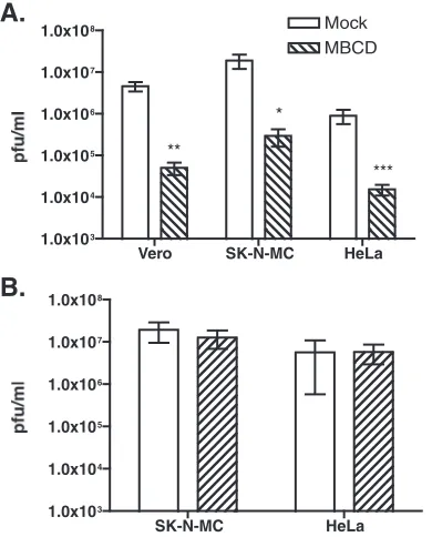
The depletion of cholesterol leads to a decrease in WNV titers in the supernatant.In order to characterize the role of cholesterol-rich membrane microdomains, or lipid rafts, in the life cycle of WNV, we treated Vero, SK-N-MC, and HeLa cells with MBCD, which is widely used to sequester cholesterol from the plasma membrane, prior to infection with WNV and measured viral titers in the culture supernatant 22 hpi. As shown in Fig. 1A, we observed a 100-fold reduction in the viral titers in various cell types treated with MBCD, suggesting that the lipid rafts play an essential role in the life cycle of WNV and that this effect is not cell type specific. The concentrations of MBCD and the periods of MBCD treatment used in our study have been shown to be nontoxic for many cell lines (39), and we have verified the same by trypan blue exclusion and found that⬃90% of the cells were viable under these condi-tions (data not shown).
The acute depletion of cholesterol by MBCD has been shown previously to transiently inhibit clathrin-mediated en-docytic pathways (31). To rule out the possibility that the reduction in viral titers was due to a gross disruption in the membrane architecture and the inhibition of all cellular uptake pathways upon cholesterol depletion, we treated SK-N-MC and HeLa cells with MBCD as described above and infected the cells with VSV, which enters cells via clathrin-dependent endocytosis (36). We measured viral titers in the supernatants 16 hpi. Under our conditions of cholesterol depletion of these cells, MBCD treatment did not affect VSV titers (Fig. 1B). These data suggest a specific requirement for lipid rafts for normal WNV replication.
Cholesterol-depleted cells have reduced levels of WNV RNA.
Cholesterol-rich membrane microdomains are present not only within the plasma membrane but also in the membranes of intracellular organelles and are involved in protein traffick-ing from the plasma membrane and in the secretory pathway. The depletion of plasma membrane cholesterol by MBCD may cause the depletion of cholesterol stores at the endoplasmic reticulum as the cell attempts to restore a cholesterol balance. Therefore, the disruption of lipid rafts by MBCD may have an effect on WNV entry, RNA replication, assembly, or egress. To identify the stage(s) at which lipid rafts play a role in the WNV life cycle, we pretreated Vero cells with MBCD and infected them with WNV as described above. Twenty-four hours
VOL. 82, 2008 LIPID RAFTS MEDIATE WEST NILE VIRUS ENTRY 5213
on November 8, 2019 by guest
http://jvi.asm.org/
postinfection, cells were fixed and analyzed by immunoflu-orescence with anti-E antibodies. Simultaneously, lysates from mock- or WNV-infected cells were prepared for Western blot analysis. As shown in Fig. 2A, MBCD-treated cell samples had fewer infected cells than mock-treated samples, as seen by staining with WNV E antibody. Western blot analysis of the lysates also showed smaller amounts of WNV NS5 protein in MBCD-treated cells than in mock-treated cells (Fig. 2B). Sim-ilar results were obtained for SK-N-MC cells (data not shown). We next determined the WNV RNA copy numbers in mock-or MBCD-treated, WNV-infected cells 24 hpi by quantitative RT-PCR (qRT-PCR). As shown in Fig. 2C, we found 100-fold-lower levels of WNV-RNA copies in cells treated with MBCD than in the mock-treated cells. These results indicated that lipid rafts either play a direct role in RNA replication or function at an earlier stage in the virus life cycle, possibly during virus entry.
Cholesterol depletion postinfection does not affect WNV ti-ters.A recent study has indicated a role for cholesterol and the cholesterol biosynthetic pathway in the replication of the RNA of Kunjin virus (a nonpathogenic Australian variant of WNV) (21). We determined if cholesterol depletion affects postentry events in the WNV life cycle by infecting cells with WNV and treating them with MBCD postinfection. Cells were treated with MBCD 6 hpi as described above, and the viral titers in the
culture supernatants were determined by plaque assays at 6 and 16 h post-MBCD treatment (12 and 22 hpi, respectively). As shown in Fig. 3A, cholesterol depletion by MBCD postin-fection had no effect on viral titers. Similar results were ob-tained when cells were treated with MBCD 3 hpi (data not shown), suggesting that the effect of MBCD is at a step prior to WNV RNA replication. Since it is impossible to completely deplete cellular cholesterol without affecting viability, it is pos-sible that the residual cholesterol present in the cells was suf-ficient to participate in WNV replication. Alternatively, it is possible that cellular cholesterol levels were replenished by de novo synthesis upon the removal of the drug, allowing for cholesterol participation in viral RNA replication, assembly, or egress.
[image:3.585.297.540.68.396.2]To determine if cholesterol biosynthesis is upregulated dur-ing WNV infection, SK-N-MC cells were infected with WNV
FIG. 1. Effect of cholesterol depletion on WNV and VSV titers. (A) Vero, SK-N-MC, and HeLa cells were treated with MBCD and infected with WNV (at MOIs of 5 PFU/cell for Vero cells and 10 PFU/cell for SK-N-MC and HeLa cells) as described in Materials and Methods. WNV titers in the culture supernatants at 22 hpi were de-termined by plaque assays on Vero cells. Error bars represent standard deviations (SD;n⫽4, 3, and 5, respectively).*,**, and***indicate
Pvalues of⬍0.0003,⬍0.009, and⬍0.0001, respectively. (B) SK-N-MC and HeLa cells were treated with MBCD and infected with VSV (MOI of 5 PFU/cell) as described in Material and Methods. VSV titers in the culture supernatants at 16 hpi were determined by plaque assays on Vero cells. Error bars represent SD (n⫽6).
FIG. 2. Cholesterol depletion results in reduced infection and RNA replication. Vero cells grown on coverslips were treated with MBCD and infected with WNV (MOI of 5 PFU/cell). (A) Twenty-four hours postinfection, cells were fixed with PFA and stained with WNV E antibodies, followed by Alexa 594-conjugated secondary antibodies. The nuclei were stained by DAPI. Scale bars, 20m. (B) Lysates from parallel samples were processed for Western analysis to detect WNV NS5. Results for-actin are shown as a loading control.⫹, present;⫺, absent. (C) Vero cells were treated with MBCD and infected with WNV as described above, and 22 hpi, RNA was isolated from cells and WNV genome copy numbers were determined by qRT-PCR as de-scribed in Materials and Methods. Results representative of three experiments performed with two or more replicates are shown. Error bars represent SD (n⫽6).
on November 8, 2019 by guest
http://jvi.asm.org/
[image:3.585.67.262.69.311.2]and cells were collected 8, 24, and 48 hpi for RNA isolation. The levels of mRNA for HMGCR, an enzyme involved in cholesterol biosynthesis, were measured by qRT-PCR. We saw a modest induction of HMGCR in WNV-infected cells at early time points, and by 48 h, HMGCR levels were similar to those in uninfected controls (Fig. 3B). In agreement with our results, Mackenzie et al. also observed much less induction of HMGCR in cells infected with WNV strain NY99 than in those infected with Kunjin virus (21). This finding suggests that, unlike the Kunjin virus, WNV does not significantly in-duce cholesterol biosynthesis and that de novo synthesis of cholesterol may not be required for WNV RNA replication.
WNV entry is dependent on lipid rafts and independent of
␣v3 integrin.We next tested directly if cholesterol depletion by MBCD affects WNV entry. Vero cells were pretreated with MBCD and infected with WNV as before. After 1 h of incu-bation at 37°C, cells were treated with trypsin to release any attached viral particles (see the supplemental material) that failed to be internalized and washed extensively with PBS. RNA was isolated from the cells, and the relative number of internalized WNV genome copies was determined by qRT PCR. We observed an 85% reduction in the amount of inter-nalized WNV RNA in MBCD-treated cells compared to that
in mock-treated samples (Fig. 4A). These results demonstrate the requirement for cholesterol-rich membrane microdomains in the internalization of WNV. To further determine that the observed effect was due to cholesterol depletion and not due to a pleiotropic effect of MBCD on other membrane components, we supplemented cholesterol-depleted cells with exogenous cholesterol in the same experiment. As shown in Fig. 4A, the addition of cholesterol to the cholesterol-depleted cells com-pletely eliminated the defects in WNV entry and restored normal viral titers in the culture supernatants (Fig. 4B). These data further indicate that the plasma membrane cholesterol plays an important role in WNV entry.
WNV has been proposed to enter cells by endocytosis in clathrin-coated vesicles. ␣v3 integrin was identified previ-ously as a receptor for WNV, and it was shown that the inter-nalization of WNV leads to the activation of FAK (5, 6). We tested the requirement for␣v3 integrin and FAK in WNV infection by infecting wild-type MEFs and MEFs lacking3 integrin or FAK and found no difference in the viral titers in the culture supernatants of infected cells between the control and knockout MEFs (Fig. 5A and B), suggesting that WNV is capable of infecting and replicating in cells lacking␣v3
[image:4.585.330.512.68.335.2]inte-FIG. 3. Cholesterol depletion postinfection does not affect WNV growth. (A) Vero cells were infected at an MOI of 5 PFU of WNV/cell. Six hours postinfection, cells were treated with MBCD as described above, and cell growth continued until the indicated time points. Viral titers were determined by plaque assays as described in Materials and Methods. Results representative of two independent experiments per-formed in triplicate are shown. Error bars represent SD (n⫽ 3). (B) SK-N-MC cells were infected at an MOI of 10 PFU of WNV/cell, and at the indicated time points, cells were harvested for RNA isola-tion. Copy numbers of HMGCR mRNA were estimated by TaqMan gene expression analysis primer-probe sets as described in Materials and Methods.-actin cycle threshold values were used for normaliza-tion. Error bars represent SD (n⫽6).
FIG. 4. Cholesterol depletion affects WNV entry, and the effect can be eliminated by cholesterol substitution. (A) Vero cells were treated with MBCD as described above. For cholesterol (Chol) reconstitution, MBCD-treated cells were incubated in the presence of medium con-taining 400g of water-soluble cholesterol/ml in 2 mM MBCD for 1 h. After extensive washing, cells were infected at an MOI of 5 PFU/cell for 1 h at 37°C. RNA was isolated from cells, and WNV genome copy numbers were determined by qRT-PCR as described in Materials and Methods. Error bars represent SD (n⫽ 10). (B) Vero cells were treated as described in the legend to panel A, and viral titers in the culture supernatants 24 hpi were determined by plaque assays. Error bars represent SD (n⫽ 3). Results representative of at least two independent experiments performed with triplicates are shown.
VOL. 82, 2008 LIPID RAFTS MEDIATE WEST NILE VIRUS ENTRY 5215
on November 8, 2019 by guest
http://jvi.asm.org/
[image:4.585.70.254.68.329.2]grin and FAK. We further infected hamster melanoma CS-1 cells that have been shown previously to lack␣v3 integrin and
␣v5 integrin complexes at the plasma membrane (11, 37) with WNV and measured viral titers in the supernatant 24 hpi. WNV efficiently infected CS-1 cells, and the titers in the su-pernatant were comparable to those in the susu-pernatants of the other cell lines used in this study (Fig. 5C). These results suggest that WNV can utilize an alternate receptor in the absence of␣v3 or␣v5 integrin and that these integrins are not essential for WNV infection.
WNV particles fail to be internalized in cholesterol-depleted cells.We next examined if the effect of MBCD on WNV entry is due to the inefficient attachment of the virus at the cell surface. MBCD-treated cells were incubated with the virus at 4°C for 1 h to allow for binding, after which the unbound virus
[image:5.585.42.285.71.412.2]was removed and the amount of bound virus was measured by determining the WNV genome copy numbers by qRT-PCR. As shown in Fig. 6A, cholesterol depletion did not affect the number of attached virions at the cell surface, suggesting that virus attachment does not require lipid rafts. We further ex-amined the endocytosis of WNV by infecting Vero cells at an MOI of 10 PFU/cell. Cells were incubated with the virus at 4°C to allow virus attachment and then shifted to 37°C for 30 min to allow internalization, after which the cells were subjected to fixation and immunofluorescence staining with antibodies spe-cific for WNV E and EEA1, a marker for early endosomes. As shown in Fig. 6B, most of the internalized WNV particles were in EEA1-positive early endosomes in control cells, whereas no
FIG. 5.␣v3 integrins are not required for WNV entry. (A) Wild-type (WT) and3 integrin⫺/⫺MEFs were infected with WNV (MOI
of 3 PFU/cell) as described in Materials and Methods. WNV titers in the culture supernatants at 20 hpi were determined by plaque assays on Vero cells. Error bars represent SD (n⫽6). (B) FAK⫹/⫹and FAK⫺/⫺
[image:5.585.311.531.71.414.2]cells were infected with WNV (MOI of 3 PFU/cell), and 18 to 24 hpi, WNV titers in the culture supernatants were determined by plaque assays on Vero cells. Error bars represent SD (n⫽6). (C) CS-1 cells were infected with WNV (MOI of 3 PFU/cell), and 24 hpi, WNV titers in the culture supernatants were determined by plaque assays on Vero cells. Error bars represent SD (n⫽6). Results representative of at least two independent experiments performed with two or more rep-licates are shown. ND, not detected.
FIG. 6. Effect of cholesterol depletion on WNV attachment and internalization. (A) Vero cells were treated with MBCD for 1 h, infected on ice at an MOI of 10 PFU/cell, and incubated at 4°C for 1 h as described in Materials and Methods. Cells were collected in TRIzol for RNA isolation and WNV RNA copy number determination. Re-sults from two independent experiments performed in duplicate are shown. Error bars represent SD (n⫽ 4). (B) Vero cells grown on coverslips were infected at an MOI of 10 PFU of WNV/cell and incubated for 30 min on ice. Cells were shifted to 37°C for 30 min, after which they were fixed in 4% PFA and stained with WNV E and EEA1 antibodies, followed by Alexa 488- and Alexa 594-conjugated second-ary antibodies. The nuclei were stained by DAPI. RGB overlay images are shown. Arrows indicate WNV particles in EEA1-positive endo-somes in mock-treated cells and the sparse peripheral distribution of WNV particles in MBCD-treated cells. UI, uninfected cells; scale bars, 10m.
on November 8, 2019 by guest
http://jvi.asm.org/
intracellular WNV particles could be detected in MBCD-treated cells. A few of the WNV particles that were present in the cholesterol-depleted cells were at the cell peripheries. These data further confirm our qRT-PCR and viral titer data and demonstrate that cholesterol depletion affects WNV in-ternalization.
WNV E and CTB colocalize during entry.To investigate if WNV entry is mediated by cholesterol-rich membrane mi-crodomains, we examined the internalization of WNV and FITC-conjugated CTB by Vero cells. The CTB entry pathway has been well-characterized, and CTB has been shown previ-ously to enter cells in a lipid raft-dependent manner by binding to its receptor GM1 ganglioside (13). Vero cells were infected with WNV at an MOI of 10 PFU/cell and incubated with 10g of FITC-CTB/ml at 4°C for 30 min to allow for attachment. Cells were then shifted to 37°C for 10 min, washed extensively, fixed, and analyzed by immunofluorescence microscopy using anti-E antibodies. We observed discrete areas of colocalization of WNV E and CTB at the plasma membrane (Fig. 7). These data suggest that both WNV E and CTB associate with lipid rafts at the plasma membrane and that lipid rafts are required for the initial stages of WNV internalization.
DISCUSSION
In this study, we showed that cholesterol-rich membrane microdomains at the plasma membrane were important for normal WNV growth. The depletion of plasma membrane cholesterol from cultures of various cell lines by MBCD af-fected WNV titers in the culture supernatants. We observed a reduction in (i) the number of WNV-positive cells, as detected by immunofluorescence analysis, (ii) the amounts of viral pro-teins, and (iii) the levels of WNV RNA in cholesterol-depleted
cells. The observed effects were due to a block in WNV entry into the cells. WNV particles in cholesterol-depleted cells were undetectable in our internalization assays, further suggesting that WNV failed to bind to its receptor or an attachment factor in these cell samples. Additionally, WNV particles colocalized with CTB, which utilizes lipid rafts for internalization, in mi-crodomain clusters at the plasma membrane. Together, our data suggest that WNV utilizes lipid rafts for entry by binding either to a receptor present in the lipid rafts or to an accessory protein (an attachment factor or a signaling molecule) in the lipid rafts that promotes WNV binding to the receptor and WNV internalization.
We also showed that WNV entry was ␣v3 integrin inde-pendent and that FAK was nonessential for WNV growth. Previous studies have used a lab strain of WNV (Sarafend) to study WNV endocytosis and have identified␣v3 integrin as a receptor for WNV. WNV Sarafend is a nonpathogenic strain whose source, year of isolation, and passage history are not known. Phylogenetic analyses based on sequences of E and NS5 proteins and 5⬘ ends of the 3⬘ untranslated regions of multiple WNV isolates have been used to classify WNV Sarafend as a lineage II WNV strain. WNV Sarafend also falls into a separate antigenic group, as anti-E monoclonal antibod-ies recognizing all other WNV isolates fail to recognize WNV Sarafend (32). It is possible that this virus differs from the infectious WNV strain NY99 that we used in our study in its receptor usage. This theory is further supported by the fact that while WNV Sarafend is reportedly unable to infect CS-1 cells that lack␣v3 integrin (5), the infection of these cells with WNV NY99 resulted in high viral titers comparable to those in Vero cells. While our studies were in progress, Mackenzie et al. reported that infection with Kunjin virus (a nonpathogenic
FIG. 7. WNV E localizes with CTB at the plasma membrane. Vero cells grown on coverslips were infected at an MOI of 10 PFU of WNV/cell and incubated with 10g of FITC-conjugated CTB/ml for 30 min on ice. Cells were shifted to 37°C for 10 min, after which they were fixed in 4% PFA and stained with WNV E antibodies, followed by Alexa 594-conjugated secondary antibodies. The nuclei were stained by DAPI. The inset shows a magnification of the marked region of interest in the merged image. Scale bars, 10m.
VOL. 82, 2008 LIPID RAFTS MEDIATE WEST NILE VIRUS ENTRY 5217
on November 8, 2019 by guest
http://jvi.asm.org/
Australian variant of WNV) induces cholesterol biosynthesis and that the viral replication is inhibited by drugs that inhibit cholesterol biosynthesis (lovastatin) but not by drugs that de-plete cellular cholesterol (MBCD) (21). We have shown that, unlike Kunjin virus, WNV does not significantly induce cho-lesterol biosynthesis and that chocho-lesterol depletion affects WNV at the level of entry. In all the experiments described by Mackenzie et al., MBCD or other drugs were added 90 min postaddition of virus and were left on the cells for the duration of the experiment. Therefore, any effect of cholesterol on WNV entry would not be observed under their experimental conditions. They found that cholesterol depletion by MBCD postentry has no effect on Kunjin virus RNA replication or protein expression, but a roughly 10-fold reduction in viral titers was observed upon prolonged MBCD treatment, sug-gesting a possible role for cholesterol in later stages of the viral life cycle, such as assembly and egress. In our experiments, we added MBCD for 30 min at 3 or 6 hpi and then removed the drug (we found prolonged exposure to result in significant toxicity), and we have not observed an effect on viral genome copies or titers. We cannot rule out the possibility of the presence of residual intracellular cholesterol in the cells suffi-cient to participate in WNV replication. Alternatively, it is possible that cellular cholesterol levels were replenished by de novo synthesis upon the removal of the drug, allowing for cholesterol participation in viral replication, assembly, or egress.
Our results do not dispute the role of clathrin in WNV endocytosis (6, 19) but suggest that the lipid rafts are also required for WNV internalization. Several viruses from the familyFlaviviridaehave been shown previously to exploit lipid rafts for entry (DENV, hepatitis C virus [HCV], and bovine viral diarrhea virus) or replication (Kunjin virus and HCV) (1, 17, 18, 21, 29). The HCV receptors tetraspanin CD81 and scavenger receptor B type I localize at specialized plasma membrane domains enriched with cholesterol. CD81-mediated HCV entry was shown previously to require plasma membrane cholesterol (17). Similarly, Hsp90 and Hsp70 have been posed to form a receptor complex for DENV, and these pro-teins were shown previously to be associated with the lipid rafts (29). In all these studies, the depletion of plasma membrane cholesterol by MBCD led to a reduction in the viral titers in culture supernatants. Furthermore, membrane fusion studies involving fusion between the liposomes and flavivirus or alpha-virus virions have shown that cholesterol plays a crucial role in promoting fusion, suggesting that the involvement of choles-terol-rich membrane microdomains in viral entry may be a general feature of the entry pathways of flaviviruses and the closely related alphaviruses (20, 34).
In addition to entry, viruses utilize lipid rafts at various other stages of the life cycle. HCV and human immunodeficiency virus have been shown previously to modify the compositions of the lipid rafts so as to facilitate viral replication (2, 23). Lipid rafts act as signaling platforms due to the association of sig-naling proteins, such as various growth factor receptors, Src family kinases, and GTPases, in these membrane microdo-mains (12). Therefore, hijacking lipid raft machinery by using one of the lipid raft-associated proteins as a receptor or an attachment factor would enable the virus to gain access to various components of signaling and protein-trafficking
ma-chinery in the lipid rafts for later stages of viral replication (for an example with poliovirus, see reference 8). Further studies are required to examine how lipid rafts facilitate WNV growth and if WNV, like HCV, modifies host cell lipid raft composi-tions at late stages of virus replication. There is increasing evidence that a number of viruses utilize caveolin-independent, cholesterol- and clathrdependent endocytic pathways for in-ternalization (14, 24, 38). Understanding the relative roles of clathrin-dependent and clathrin-independent endocytic pro-cesses in WNV internalization warrants further investigation. Identifying other proteins of the endocytic pathway that are involved in WNV internalization will provide further insights into the mechanism of WNV entry and the pathway involved. Our results demonstrate the importance of plasma membrane cholesterol for WNV entry. This observation provides new opportunities to further understand the mechanisms of WNV entry and replication and also to develop reagents that can block WNV entry and prevent WNV disease in the susceptible host.
ACKNOWLEDGMENTS
We thank Richard Hynes, David Cheresh, and David Schlaepfer for providing the3 integrin⫺/⫺MEFs, CS-1 cells, and FAK⫺/⫺MEFs,
respectively. We thank members of the Johnson lab for various re-agents and members of the Nelson lab for useful discussions. We thank Peter Mason for many useful suggestions and for critical reading of the manuscript.
This project has been funded in whole or in part with federal funds from the National Institute of Allergy and Infectious Diseases, National Institutes of Health, Department of Health and Human Services, under contract no. HHSN266200500027C and by National Institutes of Health grant AI 61527 (to J.A.N.).
We declare no conflict of interest.
REFERENCES
1.Aizaki, H., K. S. Choi, M. Liu, Y. J. Li, and M. M. Lai.2006. Polypyrimidine-tract-binding protein is a component of the HCV RNA replication complex and necessary for RNA synthesis. J. Biomed. Sci.13:469–480.
2.Brugger, B., B. Glass, P. Haberkant, I. Leibrecht, F. T. Wieland, and H.-G. Krausslich.2006. The HIV lipidome: a raft with an unusual composition. Proc. Natl. Acad. Sci. USA103:2641–2646.
3.Burke, D. S., and T. P. Monath.2001. Flaviviruses, p. 1043–1125.InD. M. Knipe and P. M. Howley (ed.), Fields virology, 4th ed. Lippincott Williams & Wilkins, Philadelphia, PA.
4.Chazal, N., and D. Gerlier.2003. Virus entry, assembly, budding, and mem-brane rafts. Microbiol. Mol. Biol. Rev.67:226–237.
5.Chu, J. J., and M. L. Ng.2004. Interaction of West Nile virus with␣v3 integrin mediates virus entry into cells. J. Biol. Chem.279:54533–54541. 6.Chu, J. J. H., and M. L. Ng.2004. Infectious entry of West Nile virus occurs
through a clathrin-mediated endocytic pathway. J. Virol.78:10543–10555. 7.Chung, C.-S., C.-Y. Huang, and W. Chang.2005. Vaccinia virus penetration
requires cholesterol and results in specific viral envelope proteins associated with lipid rafts. J. Virol.79:1623–1634.
8.Coyne, C. B., K. S. Kim, and J. M. Bergelson.2007. Poliovirus entry into human brain microvascular cells requires receptor-induced activation of SHP-2. EMBO J.26:4016–4028.
9.Danthi, P., and M. Chow.2004. Cholesterol removal by methyl- -cyclodex-trin inhibits poliovirus entry. J. Virol.78:33–41.
10.Edidin, M.2003. The state of lipid rafts: from model membranes to cells. Annu. Rev. Biophys. Biomol. Struct.32:257–283.
11.Filardo, E. J., P. C. Brooks, S. L. Deming, C. Damsky, and D. A. Cheresh. 1995. Requirement of the NPXY motif in the integrin beta 3 subunit cyto-plasmic tail for melanoma cell migration in vitro and in vivo. J. Cell Biol. 130:441–450.
12.Foster, L. J., C. L. De Hoog, and M. Mann.2003. Unbiased quantitative proteomics of lipid rafts reveals high specificity for signaling factors. Proc. Natl. Acad. Sci. USA100:5813–5818.
13.Fujinaga, Y., A. A. Wolf, C. Rodighiero, H. Wheeler, B. Tsai, L. Allen, M. G. Jobling, T. Rapoport, R. K. Holmes, and W. I. Lencer.2003. Gangliosides that associate with lipid rafts mediate transport of cholera and related toxins from the plasma membrane to endoplasmic reticulum. Mol. Biol. Cell14: 4783–4793.
on November 8, 2019 by guest
http://jvi.asm.org/
14.Hambleton, S., S. P. Steinberg, M. D. Gershon, and A. A. Gershon.2007. Cholesterol dependence of varicella-zoster virion entry into target cells. J. Virol.81:7548–7558.
15.Hirsch, A. J., G. R. Medigeshi, H. L. Meyers, V. DeFilippis, K. Fruh, T. Briese, W. I. Lipkin, and J. A. Nelson.2005. The Src family kinase c-Yes is required for maturation of West Nile virus particles. J. Virol.79:11943– 11951.
16.Hodivala-Dilke, K. M., K. P. McHugh, D. A. Tsakiris, H. Rayburn, D. Crowley, M. Ullman-Cullere, F. P. Ross, B. S. Coller, S. Teitelbaum, and R. O. Hynes.1999.3-Integrin-deficient mice are a model for Glanzmann thrombasthenia showing placental defects and reduced survival. J. Clin. Investig.103:229–238.
17.Kapadia, S. B., H. Barth, T. Baumert, J. A. McKeating, and F. V. Chisari. 2007. Initiation of hepatitis C virus infection is dependent on cholesterol and cooperativity between CD81 and scavenger receptor B type I. J. Virol. 81:374–383.
18.Krey, T., H.-J. Thiel, and T. Rumenapf.2005. Acid-resistant bovine pestivi-rus requires activation for pH-triggered fusion during entry. J. Virol.79: 4191–4200.
19.Krishnan, M. N., B. Sukumaran, U. Pal, H. Agaisse, J. L. Murray, T. W. Hodge, and E. Fikrig.2007. Rab 5 is required for the cellular entry of dengue and West Nile viruses. J. Virol.81:4881–4885.
20.Lu, Y. E., T. Cassese, and M. Kielian.1999. The cholesterol requirement for Sindbis virus entry and exit and characterization of a spike protein region involved in cholesterol dependence. J. Virol.73:4272–4278.
21.Mackenzie, J. M., A. A. Khromykh, and R. G. Parton.2007. Cholesterol manipulation by West Nile virus perturbs the cellular immune response. Cell Host Microbe2:229–239.
22.Manes, S., G. del Real, and A. C. Martinez.2003. Pathogens: raft hijackers. Nat. Rev. Immunol.3:557–568.
23.Mannova, P., R. Fang, H. Wang, B. Deng, M. W. McIntosh, S. M. Hanash, and L. Beretta.2006. Modification of host lipid raft proteome upon hepatitis C virus replication. Mol. Cell. Proteomics5:2319–2325.
24.Martin-Acebes, M. A., M. Gonzalez-Magaldi, K. Sandvig, F. Sobrino, and R. Armas-Portela.2007. Productive entry of type C foot-and-mouth disease virus into susceptible cultured cells requires clathrin and is dependent on the presence of plasma membrane cholesterol. Virology369:105–118. 25.Medigeshi, G. R., A. M. Lancaster, A. J. Hirsch, T. Briese, W. I. Lipkin, V.
DeFilippis, K. Fruh, P. W. Mason, J. Nikolich-Zugich, and J. A. Nelson. 2007. West Nile virus infection activates the unfolded protein response, leading to CHOP induction and apoptosis. J. Virol.81:10849–10860. 26.Ono, A., and E. O. Freed.2005. Role of lipid rafts in virus replication. Adv.
Virus Res.64:311–358.
27.Pelkmans, L.2005. Secrets of caveolae- and lipid raft-mediated endocytosis revealed by mammalian viruses. Biochim. Biophys. Acta1746:295–304. 28.Popik, W., T. M. Alce, and W.-C. Au.2002. Human immunodeficiency virus
type 1 uses lipid raft-colocalized CD4 and chemokine receptors for produc-tive entry into CD4⫹T cells. J. Virol.76:4709–4722.
29.Reyes-del Valle, J., S. Chavez-Salinas, F. Medina, and R. M. del Angel.2005. Heat shock protein 90 and heat shock protein 70 are components of dengue virus receptor complex in human cells. J. Virol.79:4557–4567.
30.Reynolds, L. E., F. J. Conti, M. Lucas, R. Grose, S. Robinson, M. Stone, G. Saunders, C. Dickson, R. O. Hynes, A. Lacy-Hulbert, and K. Hodivala-Dilke. 2005. Accelerated re-epithelialization in3-integrin-deficient-mice is associ-ated with enhanced TGF-1 signaling. Nat. Med.11:167–174.
31.Rodal, S. K., G. Skretting, O. Garred, F. Vilhardt, B. van Deurs, and K. Sandvig.1999. Extraction of cholesterol with methyl-beta-cyclodextrin per-turbs formation of clathrin-coated endocytic vesicles. Mol. Biol. Cell10:961– 974.
32.Scherret, J. H., M. Poidinger, J. S. Mackenzie, A. K. Broom, V. Deubel, W. I. Lipkin, T. Briese, E. A. Gould, and R. A. Hall.2001. The relationships between West Nile and Kunjin viruses. Emerg. Infect. Dis.7:697–705. 33.Sieg, D. J., D. Ilic, K. C. Jones, C. H. Damsky, T. Hunter, and D. D.
Schlaepfer.1998. Pyk2 and Src-family protein-tyrosine kinases compensate for the loss of FAK in fibronectin-stimulated signaling events but Pyk2 does not fully function to enhance FAK⫺cell migration. EMBO J.17:5933–5947. 34.Stiasny, K., C. Koessl, and F. X. Heinz.2003. Involvement of lipids in different steps of the flavivirus fusion mechanism. J. Virol.77:7856–7862. 35.Streblow, D. N., J. Vomaske, P. Smith, R. Melnychuk, L. Hall, D. Pancheva,
M. Smit, P. Casarosa, D. D. Schlaepfer, and J. A. Nelson.2003. Human cytomegalovirus chemokine receptor US28-induced smooth muscle cell mi-gration is mediated by focal adhesion kinase and Src. J. Biol. Chem.278: 50456–50465.
36.Sun, X., V. K. Yau, B. J. Briggs, and G. R. Whittaker. 2005. Role of clathrin-mediated endocytosis during vesicular stomatitis virus entry into host cells. Virology338:53–60.
37.Thomas, L., P. W. Chan, S. Chang, and C. Damsky. 1993. 5-Bromo-2-deoxyuridine regulates invasiveness and expression of integrins and matrix-degrading proteinases in a differentiated hamster melanoma cell. J. Cell Sci. 105 (Pt 1):191–201.
38.Vela, E. M., L. Zhang, T. M. Colpitts, R. A. Davey, and J. F. Aronson.2007. Arenavirus entry occurs through a cholesterol-dependent, non-caveolar, clathrin-mediated endocytic mechanism. Virology369:1–11.
39.Zidovetzki, R., and I. Levitan.2007. Use of cyclodextrins to manipulate plasma membrane cholesterol content: evidence, misconceptions and control strategies. Biochim. Biophys. Acta1768:1311–1324.
VOL. 82, 2008 LIPID RAFTS MEDIATE WEST NILE VIRUS ENTRY 5219