7 E-mail: roberto.arrigoni@unimib.it
Key words: COI, evolution, histone H3, Lobophyllia, Pacific Ocean, rDNA, Symphyllia, systematics, taxonomic revision
Abstract
Novel micromorphological characters in combination with mo-lecular studies have led to an extensive revision of the taxonomy and systematics of scleractinian corals. In the present work, we investigate the macro- and micromorphology and the phyloge-netic position of the genera Australomussa and Parascolymia, two monotypic genera ascribed to the family Lobophylliidae. The molecular phylogeny of both genera was addressed using three markers, the partial mitochondrial COI gene and the nuclear his-tone H3 and the ribosomal ITS region. Based on molecular data,
Australomussa and Parascolymia belong to the Lobophylliidae
and they cluster together with the genera Lobophyllia and
Sym-phyllia within the same clade. While A. rowleyensis and P. vitien-sis are closely related based on the three gene regions examined,
their macro and micromorphology suggest that these species are distinct, differing in several characters, such as continuity and thickness of the costosepta, the number of septa, septal tooth height, spacing, and shape, and the distribution and shape of gran-ules. Thus, we revise the taxonomic status of the genus
Australo-mussa as a junior synonym of Parascolymia.
Contents
Introduction ... 195
Material and methods ... 197
Sampling ... 197
Morphological analyses ... 197
Molecular analyses ... 199
Results ... 203
Macromorphology ... 203
Micromorphology ... 204
Molecular analyses ... 205
Discussion ... 207
Morphology of P. rowleyensis and P. vitiensis and consequences for taxonomy ... 207
Molecular phylogeny of P. rowleyensis and P. vitiensis . 209 Utility of the examined molecular markers ... 209
Acknowledgements ... 210
References ... 210
Appendix ... 214
Introduction
196 Arrigoni et al. – Taxonomy of Australomussa and Parascolymia
The stony coral family Lobophylliidae Dai and Horng, 2009 has recently been studied by several au-thors using an integrated morpho-molecular ap-proach, and is undergoing several taxonomic chang-es, although this process is far from complete. For example, Indo-Pacific taxa traditionally ascribed to the Mussidae Ortmann, 1890 have been moved to the Lobophylliidae as a result of the molecular work by Fukami et al. (2004, 2008), and the finding of a deep divergence between Indo-Pacific and Atlantic species based on morphological characters (Budd and Stolar-ski, 2009; Budd et al., 2012). The family Lobophyl-liidae is now comprised of the genera Lobophyllia de Blainville, 1830, Acanthastrea Milne Edwards and Haime, 1848, Cynarina Brüggemann, 1877,
Echino-phyllia Klunzinger, 1879, HomoEchino-phyllia Brüggemann,
1877, Micromussa Veron, 2000, Moseleya Quelch, 1884, Oxypora Saville Kent, 1871, Parascolymia Wells, 1964 and Symphyllia Milne Edwards and Haime, 1848 (Budd et al., 2012). Also included in the family are two genera that have not been examined at a molecular level, namely Echinomorpha Veron, 2000 and Australomussa Veron, 1985, hence their phylogenetic placement is unresolved.
The macromorphology (budding, colony form, size and shape of corallites, numbers of septal cy-cles), the micromorphology (shapes and distributions of septal teeth and granules), and the microstructure (arrangement of calcification centres and thickening deposits within costosepta) of the lobophylliid genera
Acanthastrea, Cynarina, Echinophyllia, Homophyl-lia, Lobo phylHomophyl-lia, Micromussa, Oxypora, Parascoly-mia, and Symphyllia were examined by Budd and
Stolarski (2009) and Budd et al. (2012). They con-cluded that the shape and distribution of septal teeth and granules, the area between teeth, and the devel-opment of thickening deposits are informative char-acters for distinguishing the Lobophylliidae from representatives of the other coral families. Arrigoni
et al. (2014) presented a comprehensive molecular
phylogeny that shows that the Lobophylliidae is a monophyletic family comprising nine main molecu-lar clades (clades A-I), and that several genera are not monophyletic. The authors also showed that the mon-ospecific genus Parascolymia belongs to clade I
(sen-su Arrigoni et al., 2014) together with all the species
of Lobophyllia and Symphyllia for which molecular data is available, including the two type species
Lo-bophyllia corymbosa (Forskål, 1775) and Symphyllia radians Milne Edwards and Haime, 1849. The
au-thors did not, however, undertake any formal
taxo-nomic revision of the status of the genus
Para-scolymia.
Australomussa rowleyensis Veron, 1985 was
de-scribed from Western Australia and ade-scribed to the Mussidae. It is a colonial and zooxanthellate sclerac-tinian coral, characterized by flattened, helmet- or dome-shaped coralla, valleys approximately 20mm wide, with very thick walls and a well-developed columella (Veron, 1985). In the original description of A. rowleyensis, Veron (1985) stated that this genus showed ‘little resemblances to any other genus’ with the exception of Parascolymia and Symphyllia, and ‘its closest affinities are probably with the former’. The author referred only to the macromorphology of the coralla for the comparison of Australomussa with
Parascolymia and Symphyllia and did not consider
any micromorphological characters. Budd and Sto-larski (2009) and Budd et al. (2012) showed that the majority of macromorphological characters tradition-ally used in the taxonomy and systematics of Lobo-phylliidae and Mussidae exhibit homoplasy. In con-trast, novel micromorphological characters separate these two families and are useful for the description and formalization of species. Nevertheless, while the micromorphology of P. vitiensis (Brüggemann, 1877) was described by Budd and Stolarski (2009) and Budd et al. (2012), A. rowleyensis was not analysed in these studies.
The known distribution of A. rowleyensis includes the Western Pacific region known as the Coral Trian-gle (for definition see Hoeksema, 2007; Veron et al., 2009) and partially overlaps with the distribution of
P. vitiensis which is absent from Western Australia
but extends to the west in the Indian Ocean and to the east in the central Pacific (Veron, 2000).
Australo-mussa rowleyensis and P. vitiensis have very
differ-ent histories of nomenclature. Perhaps due to its re-cent description and rarity (Veron, 1985), A.
rowley-ensis has always been described as A. rowleyrowley-ensis
despite its morphological similarity to Parascolymia and Symphyllia (Veron, 1985, 2000). Conversely, P.
vitiensis has a long history of nomenclatural
confu-sion. It was originally ascribed to Litophyllia Milne Edwards and Haime, 1857 (Gardiner, 1899; Cross-land, 1952) and later described as Protolobophyllia
japonica Yabe and Sugiyama, 1935. In agreement
with Matthai (1928), Wells (1937) and Vaughan and Wells (1943) considered Scolymia Haime, 1852 and
Protolobophyllia Yabe and Sugiyama, 1935 as junior
the geographical separation. Finally, Budd et al. (2012) restored the distinction between
Parascoly-mia (Indo-Pacific) and ScolyParascoly-mia (Atlantic) based on
molecular and micromorphological analyses (Fukami
et al., 2004, 2008; Budd and Stolarski, 2009).
Although P. vitiensis is generally monocentric, it
can also form polystomatous coralla (Chevalier, 1975; Veron and Pichon, 1980: figs 416-417) (Figs 1B-D, 2F-I). The macro-morphologic observation of a large series of mono- to polystomatous specimens of P. vitiensis from Papua New Guinea and New Cal-edonia and the similarity of the larger specimens with
A. rowleyensis prompted the detailed study of the
morphological affinities and molecular relationship between these two species and the two monospecific genera they are currently ascribed to.
Here we selected three DNA regions, the barcod-ing region of cytochrome oxydase subunit I gene, the nuclear ribosomal ITS region, and the nuclear histone H3 for molecular analysis of these species. The for-mer two molecular loci have been extensively used in phylogenetic studies of scleractinian corals (Fukami
et al., 2008; Gittenberger et al., 2011; Huang et al.,
2011; Benzoni et al., 2011, 2014) and, moreover, the most comprehensive phylogeny reconstruction of the Lobophylliidae to date is based on these two markers (Arrigoni et al., 2014). The latter locus was revealed to be informative for a broad-based phylogeny of the Merulinidae Verrill, 1865 (Huang et al., 2011, 2014b), a family closely related to the Lobophylliidae (Fuka-mi et al., 2008; Arrigoni et al., 2012). Several phylo-genetic studies of scleractinian corals achieved a well-resolved phylogeny using a concatenated spe-cies-tree, combining mitochondrial and nuclear mo-lecular markers (Huang et al., 2009, 2011; Souter, 2010; Gittenberger et al., 2011; Benzoni et al., 2012a; Richards et al., 2013; Arri goni et al., 2014). This kind of approach is a powerful way to obtain a robust phy-logeny, resolving all key nodes and yielding good resolution at species level.
Material and methods
Sampling
Specimens of Parascolymia vitiensis for this study were sampled in New Caledonia, Papua New Guinea, and Eastern Australia, while samples of
Australomus-sa rowleyensis were collected in the Kimberley,
North-West Australia (S1). Coral specimens were photographed and collected while SCUBA diving from 2 to 35 meters depth. Digital images of living corals in the field were taken with a Canon Powershot G9 in an Ikelite underwater housing system in New Caledonia and Papua New Guinea (Figs 1 and S5), and with an Olympus XZ1 in a PT-050 underwater housing in Australia (Figs 1 and S5). Coral specimens were collected, tagged, and preserved in 95% ethanol for further molecular analysis. After the sampling of fixed tissues for DNA extraction, each corallum was immersed in sodium hypochlorite for 48 hours to re-move all soft parts, rinsed in freshwater and dried for microscope observation. Specimens were identified at the species level based on skeletal morphology us-ing a Leica M80 microscope followus-ing the descrip-tions and illustradescrip-tions by Chevalier (1975), Veron and Pichon (1980), and Veron (1985).
Morphological analyses
198 Arrigoni et al. – Taxonomy of Australomussa and Parascolymia
Fig. 1. Parascolymia vitiensis (A-D) and P. rowleyensis (previously Australomussa) (E-G) in situ: A) IRD HS2984 (monostomatous);
[image:4.538.80.462.62.641.2]Microscopy (SEM) was used to analyze the shape and distribution of granules on septal faces and the interarea of teeth on representative specimens of P.
vitiensis (UNIMIB PFB151) and one of A. rowleyen-sis (WAM Z65789). Specimens were mounted using
silver glue, sputter-coated with conductive gold film and examined using a Vega Tescan Scanning Electron Microscopy at the SEM Laboratory, University of Milano-Bicocca.
For a glossary of skeletal terms we refer to Budd et
al. (2012).
Abbreviations:
CC1 IRD CoralCal1 Expedition, Côte Oubliée, New Caledonia, 2007
CC4 IRD CoralCal4 Expedition, New Caledo-nia, IRD, 2012
CCAP IRD CoralCap Expedition, New Caledo-nia, 2007
Cs cycle of costosepta
IRD Institut de Recherche pour le Développe-ment, Nouméa, New Caledonia
NIUGINI Niugini Biodiversity Expedition, Papua New Guinea, 2012
RMNH Naturalis Biodiversity Center (former Rijksmuseum van Natuurlijke Historie), Leiden, the Netherlands
S cycle of septa
UNIMIB Università di Milano-Bicocca, Milan, Italy WAM Western Australian Museum, Perth,
Australia
In the list of examined material for IRD specimens the station number (ST) is provided, when available, after the sampling locality. Station numbers can be searched in the IRD online database LagPlon (http:// lagplon.ird.nc/consultv2_5/rechSimple.faces) where additional details on the reef habitat, GPS coordi-nates, and a map of each station can be found.
I gene (COI) from mitochondrial DNA, (2) a ~350 bp portion of the nuclear histone H3, and (3) a ~800 pb portion of the ITS region, including the 3’ end of 18S, the entire ITS1, 5.8S, and ITS2, and the 5’ end of the 28S, as nuclear loci. COI was amplified using MCOIF - MCOIR primers (Fukami et al., 2004) and the pro-tocol by Benzoni et al. (2011), the histone H3 using H3F - H3R primers (Colgan et al., 1998), and the ITS region using ITS4 (Takabayashi et al., 1998) - A18S (White et al., 1990) primers and the protocol by Ben-zoni et al. (2011), or alternately using 1S and 2SS primers (Wei et al., 2003) and the protocol by Kitano
et al. (2014). Sequencing was carried out by
Genom-ics and Bioscience and Technology Co., Ltd, Xizhi City, Taipei County, Taiwan. Sequences obtained in this study have been deposited in EMBL, and acces-sion numbers are listed in S1.
Sequences were viewed, edited and assembled us-ing CodonCode Aligner 4.2.5 (CodonCode Corpora-tion, Dedham, MA, USA) and manually checked us-ing BioEdit 7.2.5 (Hall, 1999). Alignments of the four separated datasets (three single gene trees and one concatenated) were carried out using the E-INS-i option in MAFFT 7.110 (Katoh et al., 2002; Katoh and Standley, 2013) under default parameters.
Plesi-astrea versipora (Lamarck, 1816) and several species
200 Arrigoni et al. – Taxonomy of Australomussa and Parascolymia
Fig. 2. Corallum morphology in Parascolymia vitiensis: A) IRD HS3255; B) IRD HS2955; C) detail of the same specimen in B showing
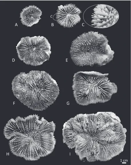
[image:6.538.41.499.61.637.2]Fig. 3. Corallum morphology in Parascolymia rowleyensis (previously Australomussa): A) WAM Z65785; B) WAM Z65788; C) WAM
[image:7.538.39.501.60.581.2]202 Arrigoni et al. – Taxonomy of Australomussa and Parascolymia
Fig. 4. Comparison of the macromorphology of Parascolymia vitiensis (A-C) and P. rowleyensis (D-E): A) septa in the monocentric
specimen IRD HS2964; B) top view of the costosepta in the polycentric specimen UNIMIB PFB057; C) side view of the same portion of the specimen in B; D) peripheral calices in specimen WAM Z65786; E) top view of the costosepta in the same specimen as D; F) side view of the same portion of the specimen in E. Red arrows indicate the position of the columella in adjacent corallites, red brackets placed perpendicularly to the costosepta show the number of costosepta intercepted by a 1 cm transect.
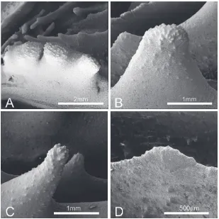
Fig. 5. SEM images of radial elements of Parascolymia rowleyensis (previously Australomussa) (WAM Z65789: A-B)
[image:8.538.40.348.363.671.2]Gascuel, 2003), respectively. The best-fit substitution model for each locus was determined using the Akai-ke Information Criterion (AIC) as implemented in MrModeltest 2.3 (Nylander, 2004) in conjunction with PAUP4.0b10 (Swofford, 2003). As most suitable mod-els AIC selected the General Time-Reversible (GTR) model with a proportion of sites being invariable (+I) and the remainder following a gamma distribution (+I) for COI and rDNA, and the Kimura (K80) model with a proportion of invariable sites (+I) for histone H3. The Maximum Likelihood (ML) tree was calculat-ed with PhyML and a total of 500 bootstrap replicates were performed to assess the robustness of each clade. Four independent Markov Chain Monte Carlo (MCMC) runs were conducted for 1.4 × 10⁷
genera-tions for COI dataset (1.7 × 10⁷generations for histone
H3 and 4 × 10⁷generations for ITS region) with trees
sampled every 100 generation for each analysis. The 25% first trees were discarded as burn-in, and posteri-or probabilities were estimated from the remaining trees in each run (10,500 remaining trees for COI, 12,750 for histone H3, and 30,000 for ITS region). To determine if the runs had achieved stationarity, we visualized log-likelihood scores and model parameter values across each run using Tracer 1.5 (Rambaut and Drummond, 2007). Finally, the three single gene data-sets were concatenated in a single partitioned align-ment and the phylogeny was reconstructed using Bayesian Inference and Maximum Likelihood analy-ses. Four independent Markov Chain Monte Carlo (MCMC) runs were conducted for 2.2 × 10⁷
genera-tions with trees sampled every 100 generation and the 25% first trees were discarded as burn-in. The ML tree
was built in PhyML and a total of 500 bootstrap repli-cates were performed to assess the robustness of each clade. Branches with >70% bootstrap support values and >0.90 posterior probabilities are considered sig-nificantly supported.
Results
Macromorphology
[image:9.538.46.502.63.205.2]In P. vitiensis coralla can be solitary (Figs 1A, 2A-E) or colonial (Figs 1B-D, 2F-I) and formed by intracali-cular and extracaliintracali-cular budding (e. g. Fig. 2I). In co-lonial coralla, as a result of circumoral budding, coral-lites are highly polymorphic (Fig. 2G-I) and corallite integration is uni- or multiserial. Corallum shape is generally flattened or concave (Figs 1-2). Calice or valley width is larger than 2.5cm (Fig. 2) and variable. In some specimens the central part of the calice can have a shallow depression (Fig. 2D-F, H). Continuity of costosepta is mostly confluent in di-tricentric cor-alla (Fig. 2F-G), but becomes mostly not confluent in polycentric coralla (Fig. 2I; Veron and Pichon, 1980: Fig. 417). There are six cycles of septa in the calices, rarely seven (Fig. 2E; Chevalier, 1975), those of the sixth are free. Septa spacing is large, with 4-5 septa per 5mm (Fig. 4A-B). Relative costosepta thickness be-tween Cs1 and Cs2 versus Cs3 is unequal (Fig. 5C-D). In polycentric coralla linkage between centres of adja-cent corallites within series is lamellar (Fig. 4B-C). Columella trabecular and spongy (indicated by arrows in Fig. 4B-C) and its size relative to calice width is less
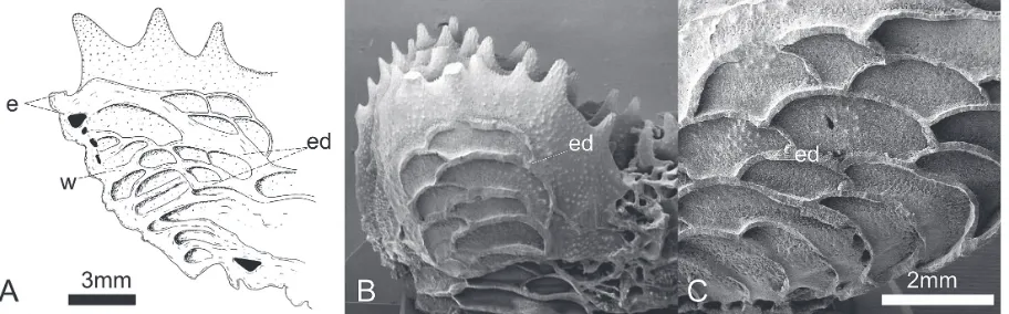
Fig. 6. Vesicular endotheca: A) longitudinal section of the periphery of a calice of Parascolymia vitiensis (modified from Chevalier,
204 Arrigoni et al. – Taxonomy of Australomussa and Parascolymia
than 1/5 (Fig. 2). The endotheca is vesicular (Fig. 6A).
In A. rowleyensis coralla are flattened or massive
and ‘helmet- or dome-shaped’ (Fig. 1E-H). Coralla are colonial as a result of primary circumoral budding and both intra and extracalicular budding occur (Fig. 3D-E). Corallites display polymorphism in smaller colonies where the central corallite is still larger as in the paratype WAM 173-84 (Veron, 1985: Fig. 25) and corallite integration is uni- or multiserial. Calice or valley width is large according to the character state in Budd et al. (2012) but smaller than 2.5cm (Fig. 4D-F). Calices at the periphery of the coralla can be inclined and the part of their calice which is not adja-cent to other calices can be wide (Fig. 3A-B, 6D). Continuity of costosepta is mostly confluent (Fig. 3A-E, 4E-F). There are four cycles of septa (Fig. 3D), those of the fourth are free. Septa spacing is large, with five septa per 5mm (Fig. 4D-E). Relative cos-tosepta thickness between Cs1 and Cs2 versus Cs3 is slightly unequal (Fig. 5A-B). Linkage between cen-tres of adjacent corallites within a series is lamellar (Fig. 4D-F). Columella are trabecular and spongy
(in-dicated by arrows in Fig. 4E-F) and the size relative to calice width less than or equal to 1/4 of calice width (Figs 3D-E, 4D-F). The endotheca is vesicular (Fig. 6B-C).
Micromorphology
[image:10.538.39.348.60.369.2]In P. vitiensis tooth base at mid-septum is elliptical in shape and parallel to the direction of the septum (Fig. 5C-D). Tooth tips are irregular and overall mainly lo-bate (Figs 5D, 7A-B, D). Teeth on S1 are 1mm or higher (Figs 4C, 7) and their spacing is very wide, with adjacent teeth more than 2mm apart. Tooth shape and size is very variable within and between septa (Fig. 7) as also noted by previous authors (Chevalier, 1975; Veron and Pichon, 1980) with some teeth becoming round in section towards the tip and having and overall pointed, or spiniform Chevalier (1975), shape (Fig. 7C). Granulation on the side of septa is weak and granules are enveloped by thicken-ing deposits (Fig. 7A-B). The inter-area structure is generally smooth (Fig. 5D) or with palisade. Tooth shape between Cs3 and Cs1 is unequal (Fig. 5C-D).
Fig. 7. SEM of Parascolymia vitiensis
In A. rowleyensis tooth base at mid-septum is
el-liptical in shape and parallel to the direction of the septum (Fig. 5A-B). Tooth tips are irregular and lo-bate (Figs 5B, 8). Teeth on S1 range between 0.8-0.9 mm (Figs 4F, 8) and their spacing is wide, with adja-cent teeth between 1 and 2mm apart. Granulation on the side of septa is strong and granules are scattered (Figs 5B, 8A). The inter-area structure has a palisade structure (Fig. 8A). Tooth shape between Cs3 and Cs1 is equal (Fig. 5B). In general, in this species tooth shape is not very variable within and between septa (Fig. 5A-B) especially when compared to the variability described in P. vitiensis.
Molecular analyses
The final alignment of COI data consisted of 580 bp, of which 48 were parsimony informative sites, with a total of 84 mutations. The aligned histone H3 matrix was 318 bp long with 86 parsimony informative sites and 122 mutations. The total alignment of ITS region was composed by 951 bp, 160 parsimony informative sites and 294 mutations. No intra-individual
poly-morphisms or double peaks were observed in the chromatograms of the two nuclear loci, thereby avoiding the need to clone the amplified fragments. The phylogeny reconstruction of the combined mo-lecular data is in Fig. 9, while the three single gene trees are in the Supplementary Information (Figs S2-S4). Phylogenetic analyses under BI and ML criteria yielded congruent results, with no contrasting sig-nals. Bayesian topologies with significant branch support indicated by ML bootstrapping support (MLs) and Bayesian posterior probability scores (BIs) are reported in Figs 9 and S2-S4.
The phylogram based on the concatenated (COI, histone H3, and ITS) molecular dataset shows high ML and BI supports at all key nodes (Fig. 9). Clade I sensu Arrigoni et al. (2014) contains all species of
Lobophyl-lia and SymphylLobophyl-lia analyzed so far and all our
[image:11.538.40.347.61.370.2]sequenc-es of A. rowleyensis and P. vitiensis. The latter two spe-cies group together in a strongly supported lineage (MLs = 100 and BIs = 0.9) and their genetic boundaries remain unclear being indistinguishable from each other with these molecular markers. The average genetic dis-tance of A. rowleyensis from P. vitiensis is 1.1 ± 0.2%,
Fig. 8. SEM of Parascolymia rowleyen-sis (previously Australomussa) (WAM
206 Arrigoni et al. – Taxonomy of Australomussa and Parascolymia
Fig. 9. Phylogenetic position of Parascolymia vitiensis and P. rowleyensis (previously Australomussa) and their relationships within the
[image:12.538.52.477.63.622.2]The Bayesian COI topology (Fig. S2) indicates that all newly obtained sequences of A. rowleyensis and P.
vitiensis are nested together with the genera Lobo-phyllia and SymLobo-phyllia within clade I (MLs = 94%
and BIs = 0.93). While the two species are not mono-phyletic and they occur together in two main groups within clade I, .the mitochondrial phylogenetic recon-struction is similar to that of the nuclear histone H3 (Fig. S3). Again, all newly obtained sequences of A.
rowleyensis and P. vitiensis form clade I sensu
Arrig-oni et al. (2014) (MLs = 95% and BIs = -) together with several species of Lobophyllia and Symphyllia. Clade I is composed of 10 species represented by a total of 28 sequences, of which 26 share the same hap-lotype and they are thus identical, while the remaining two sequences differ from the others by only one bp substitution. Moreover, all of the Merulinidae sub-clades defined by Budd and Stolarski (2011) and Huang et al. (2011) are recovered with the exception of D/E. Interestingly, also in the family Lobophyllii-dae, all of the molecular clades defined by Arrigoni et
al. (2014) based on COI and rDNA molecular
mark-ers, except F, are supported in our BI and ML analy-ses. The Bayesian topology obtained from the ITS region alignment is similar to both COI and histone H3ones, but has a higher resolution at species level with significant supports for the majority of key nodes (Fig. S4). Again, all our sequences of A. rowleyensis and P. vitiensis are found together in a strongly sup-ported group (MLs = 90 and BIs = 1) within clade I (Fig. S4). A similar situation is apparent for L.
hemp-richii and S. agaricia which occur in a strongly
sup-ported monophyletic group. The other Lobophyllia and Symphyllia species within clade I, i.e. L. costata,
L. diminuta Veron, 1985, L. flabelliformis Veron,
2000, L. robusta, S. erythraea (Klunzinger, 1879), S.
radians, S. recta, and S. valenciennesii, are recovered
as monophyletic lineages, while the only specimen of
Acanthastrea ishigakiensis Veron, 1990 is closely
re-lated to S. recta.
Australomussa and we formally consider Australo-mussa as a junior synonym of Parascolymia.
Morphology of P. rowleyensis and P. vitiensis and con-sequences for taxonomy
The lack of genetic resolution between P. vitiensis and
P. rowleyensis in all our molecular analyses might
sug-gest that these two species are in fact synonyms. The skeleton morphology, however, indicates that although the two species share some macro- and micromorpho-logic character, they are morphomicromorpho-logically distinct and they have a different state for 10 of the 21 characters used by Budd et al. (2012) (in bold in Table 1). Veron (1985) stated that Australomussa ‘differs from
Sym-phyllia in having an initial central corallite which buds
daughter corallites extracalicularly, in lacking meander-ing valleys (which some Symphyllia ecomorphs also lack) and in having widely separated series of centres without a true common wall between them’. However, he provided no detailed information on the morphologic characters that differentiate Australomussa from
Paras-colymia (=SParas-colymia). Our observations of the macro-
208 Arrigoni et al. – Taxonomy of Australomussa and Parascolymia
(Table 1 in bold). We propose therefore that these mor-phological differences are sufficient to distinguish two species despite the fact that the unresolved genetic boundaries based on multiple markers strongly argue against retaining the species as distinct. Thus we for-mally consider Australomussa as a junior synonym of
Parascolymia and retain P. vitiensis and P. rowleyensis
as separate sister species.
In P. vitiensis the teeth in different septal cycles
dif-fer significantly in shape as already discussed by Ve-ron and Pichon (1980) and Budd and Stolarski (2009). In P. rowleyensis the teeth in different septal cycles do not differ significantly in shape as described by Veron (1985) in the species original description. The type specimen of P. rowleyensis displays an obvious varia-bility of thickening of costosepta between specimens as remarked by Veron (1985). However, the variability of shape and size of septal dentation is far more re-duced in this species than in P. vitiensis. One of the specimens of P. rowleyensis in the series we exam-ined, Z65786, has relatively thin septa and costosepta
and is similar in this respect to the paratype WAM 173-84 (Veron, 1985: Fig. 25). The remainder have a simi-lar thickness of costosepta to the holotype WAM 171-84 (Veron, 1985: Fig. 23). However, none of the speci-mens we examined in this study has radial elements as thick as paratype WAM 172-84 (Veron, 1985: Fig. 24). The thickness of radial elements of this paratype comes close to that of the radial elements of higher cycles of some P. vitiensis. Nevertheless, the number of septal cycles, and the relative thickness of septa from different cycles, as well as the size of the denta-tion of the septa fall within the range of P. rowleyensis rather than in that of P. vitiensis.
In some genera of lobophylliids (e.g. Lobophyllia,
Symphyllia, Parascolymia), the teeth in different
[image:14.538.42.499.393.671.2]sep-tal cycles differ significantly in shape while in other genera (e.g. Acanthastrea and Homophyllia) such dif-ferentiation is not observed (Budd and Stolarski, 2009). Our results confirm that the size and shape of septal teeth of P. vitiensis is highly variable within and between septa of the same specimen (Chevalier, 1975;
Table 1. Macromorphology and micromorphology of Parascolymia vitiensis and P. rowleyensis (previously Australomussa).
Explana-tion of characters, their ID numbers (in brackets) and state names are from Budd et al. (2012).* = character examined on polycentric coralla; Csn= number of cycle of costosepta; Sn = number of cycle of septa. Names of characters which have different states in the two species in bold.
Character P. vitiensis P. rowleyensis (previously Australomussa)
Intracalicular budding (1) Present * Present
Extracalicular budding (2) Present * Present
Circumoral budding and associated Present * Present
corallite polymorphism (3)
Corallite integration (4) Uni or multiserial * Uni or multiserial
Calice or valley width (7) Large, >2.5cm Large, <2.5cm
Continuity of costosepta (9) Mostly not confluent * Mostly confluent
Number of septa (10) 4 cycles 6-7 cycles
Free septa (11) Present Present
Septa spacing (per 5mm) (12) Wide, <6 Wide, <6
Relative costosepta thickness Unequal Slightly unequal
(Cs1andCs2 -) vs- Cs3) (13
Corallite centres linkage (14) Discontinuous by lamellar linkage Discontinuous by lamellar linkage Columella structure (15) Trabecular spongy Trabecular spongy
Columella size relative to calice width (16) Small, <1/4 Small to medium, ≤1/4
Endotheca (19) Abundant/vesicular Abundant/vesicular
Tooth base (mid-septum) (35) Elliptical parallel Elliptical parallel
Tooth tips (38) Irregular lobate Irregular lobate
Tooth height (S1) (39) High, and ≥ 1mm High, but <1mm
Tooth spacing (S1) (40) Very wide, >2mm Wide, 1-2mm
Granules shape and distribution (43) Weak enveloped by thickening depositis Strong scattered
Interarea structure (44) Smooth and palisade Palisade
Cs3/Cs1 tooth shape (45) Unequal Equal
Microm
orphology
sis (for P. rowleyensis see Figs 4D-F, 5A-B, 8). Molecular phylogeny of P. rowleyensis and P. vitiensis
Our multi-locus molecular analyses showed that P.
rowleyensis belongs to the family Lobophylliidae (Fig.
9), as proposed by Dai and Horng (2009) and Budd et
al. (2012) based on the macromorphology of the
colo-ny and on traditional taxonomy (Veron, 1985, 1992, 2000). Moreover, the species, traditionally ascribed to the monotypic genus Australomussa, does not occur in a distinct molecular clade, rather it is nested within the well-supported clade I sensu Arrigoni at al. (2014), which comprises the genera Lobophyllia, Symphyllia, and Parascolymia (Fig. 9).
Parascolymia rowleyensis and P. vitiensis could not
be separated in any single gene tree or the concatenat-ed phylogeny (Figs 9, S2-S4) and the intraspecific and interspecific divergences within and between the two species completely overlap. The lack of genetic varia-tion suggests that these two nominal species could be just one species or that lineage sorting is incomplete because the two species have a recent common ances-tor. The former explanation is unlikely because P.
rowleyensis and P. vitiensis differ in several
micro-morphological characters (Table 1) and, therefore, it is more likely these two species have not completely di-verged although divergence time estimates are not available. An alternative hypothesis is hybridization between the two species, as reported for other genera (Diekmann et al., 2001; van Oppen et al., 2002; Vollmer and Palumbi, 2004; Richards et al., 2008). However, the lack of intra-individual polymorphism in nuclear sequences of both species and the absence of intermediate morphologies challenges this hypothesis.
Utility of the examined molecular markers
The three single gene trees gave congruent phylogeny reconstructions (Figs S2-S4), however higher
resolu-thy et al. (2013) detected four deeply divergent line-ages corresponding to four particular geographic re-gions. COI can also be informative when combined or compared in multi-marker analyses (Fukami et al., 2008; Forsman et al., 2009; Huang et al., 2011; Ben-zoni et al., 2011, 2012a; Gittenberger et al., 2011) (Figs 4, S2). This mitochondrial region does however resolve the majority of the inner nodes, i.e. older rela-tionships, within the family Lobophylliidae (this study and Arrigoni et al., 2012, 2014), Fungiidae Dana, 1846 (Gittenberger et al., 2011), and Poritidae Gray, 1842 (Kitano et al., 2014). In our phylogenetic reconstruc-tion based on this mtDNA region, P. vitiensis and P.
rowleyensis are nested within clade I (sensu Arrigoni et al., 2014) but they appear to be polyphyletic (Fig.
S2). The intra-specific variability of P. vitiensis (0.9 ± 0.2%) and P. rowleyensis (0.9 ± 0.2%) overlaps the inter-specific distance between the two species (0.9 ± 0.2%) and the last value is comparable to the mean closest congeneric inter-specific distances among An-thozoa (0.71 ± 0.15%) found by Huang et al. (2008). The nuclear histone H3 gene has been extensively used in phylogenetic studies of arthropods (Colgan et
al., 1998; Maxmen et al., 2003), annelids (Novo et al.,
210 Arrigoni et al. – Taxonomy of Australomussa and Parascolymia
The ITS region has been extensively used to resolve species boundaries in scleractinian corals (Diekmann
et al., 2001; Forsmann et al., 2009; Benzoni et al., 2010,
2012b, 2014; Flot et al., 2011; Gittenberger et al., 2011; Stefani et al., 2011; Schmidt-Roachet al., 2012; Arrig-oni et al., 2012, 2014; Keshavmurthy et al., 2013; Ki-tano et al., 2013, 2014). Despite the phylogenetic utili-ty of this marker being questioned because of its unique pattern of secondary structure in the genus Acropora Oken, 1815 (van Oppen et al., 2002; Vollmer and Palumbi, 2004; Chen et al., 2004; Wei et al., 2006), it is currently accepted and considered as the most suitable molecular locus to resolve phylogenetic relationships among closely related species. Here, the ITS region re-solved the majority of lobophylliid species (Fig. S4), except for species in clade E (Arrigoni et al., 2014). Within clade I (sensu Arrigoni et al., 2014) the majority of species included were monophyletic, with the nota-ble exception of P. rowleyensis and P. vitiensis. There-fore, these results confirmed the usefulness of this marker in phylogentic studies and we strongly encour-age its application for the delimitation of species boundaries in scleractinian corals until new highly var-iable markers are discovered.
In conclusion, this study demonstrated that compre-hensive studies conducted both at molecular and mi-cromorphological levels are and will be essential to evaluate the evolutionary relationships of scleractinian corals and their taxonomy. We strongly believe that different disciplines, such as morphology, molecular systematics, ecology, and reproduction, should be used for taxonomical studies to reach a more complete and comprehensive approach towards the understanding of coral species diversity and biogeography.
Acknowledgments
Collection during the CoralCal1 and CoralCal4 campaigns (IRD Noumea) was possible thanks to C Payri, J Butscher, A Arnaud, F Folcher, JL Menou, and the R/V Alis Captain R Proner and crew. We are grateful to E Karsenti (EMBL) and É Bourgois (Tara Expeditions) and the OCEANS consortium for sampling during the Tara Oceans expedition. We thank the commitment of the following people and sponsors who made this singular expe-dition possible: CNRS, EMBL, Genoscope/CEA, VIB, Stazione Zoologica Anton Dohrn, UNIMIB, ANR (projects POSEIDON/ ANR-09-BLAN-0348, BIOMARKS/ANR-08-BDVA-003, PROMETHEUS/ANR-09-GENM-031, and TARA-GIRUS/ ANR-09-PCS-GENM-218), EU FP7 (MicroB3/No.287589), FWO, BIO5, Biosphere 2, agnès b., the Veolia Environment Foundation, Region Bretagne, World Courier, Illumina, Cap L’Orient, the EDF Foundation EDF Diversiterre, FRB, the
Prince Albert II de Monaco Foundation, Etienne Bourgois, the Tara schooner and its captain and crew. Tara Oceans would not exist without continuous support from 23 institutes (http:// oceans.taraexpeditions.org). This article is contribution number 18 of the Tara Oceans Expedition 2009-2012. The Niugini Bio-diversity Expedition and P Bouchet (MNHN) are acknowledged for specimens from PNG. F Benzoni is deeply grateful to C Payri and B Dreyfus (IRD) for supporting her participation to this campaign. We thank CC Wallace (MTQ) and BW Hoekse-ma (RMNH) for museum support. R Arrigoni gratefully ac-knowledges the National Science Council of Taiwan (NSC) for his participation to Summer Program in Taiwan 2013 and the financial support of the European Commission’s Research Infra-structure Action via the Synthesys Program for his visit to Natu-ralis Biodiversity Center (Leiden). Z Richards was supported in fieldwork and write-up phased by Woodside Energy and the Woodside Collection (Kimberley) project. Coral collection by AH Baird was funded by the ARC Centre of Excellence for Coral Reef Studies. The authors are grateful to the three anony-mous reviewers for their help and constructive comments.
References
Arrigoni R, Stefani F, Pichon M, Galli P, Benzoni F. 2012. Mo-lecular phylogeny of the Robust clade (Faviidae, Mussidae, Merulinidae, and Pectiniidae): An Indian Ocean perspective.
Molecular Phylogenetics and Evolution 65: 183-193.
Arrigoni R, Terraneo TI, Galli P, Benzoni F. 2014. Lobophyllii-dae (Cnidaria, Scleractinia) reshuffled: pervasive non-mono-phyly at genus level. Molecular Phylogenetics and Evolution 73: 60-64.
Benzoni F. 2006. Psammocora albopicta sp. nov., a new species of scleractinian coral from the Indo-West Pacific (Sclerac-tinia; Siderastreidae). Zootaxa 1358: 49-57.
Benzoni F, Stefani F, Pichon M, Galli P. 2010. The name game: morpho-molecular species boundaries in the genus
Psammo-cora (Cnidaria, Scleractinia). Zoological Journal of the Lin-nean Society 160: 421-456.
Benzoni F, Arrigoni R, Stefani F, Pichon M. 2011. Phylogeny of the coral genus Plesiastrea (Cnidaria, Scleractinia).
Contri-butions to Zoology 80: 231-249.
Benzoni F, Arrigoni R, Stefani F, Stolarski J. 2012a.Systematics of the coral genus Craterastrea (Cnidaria, Anthozoa, Scler-actinia) and description of a new family through combined morphological and molecular analyses. Systematics and
Bio-diversity 10: 417-433.
Benzoni F, Arrigoni R, Stefani F, Reijnen BT, Montano S, Hoek-sema BW. 2012b. Phylogenetic position and taxonomy of
Cycloseris explanulata and C. wellsi (Scleractinia:
Fungii-dae): lost mushroom corals find their way home.
Contribu-tions to Zoology 81: 125-146.
Benzoni F, Arrigoni R, Waheed Z, Stefani F, Hoeksema BW. 2014. Phylogenetic relationships and revision of the genus
Blastomussa (Cnidaria: Anthozoa: Scleractinia) with
de-scription of a new species. Raffles Bulletin of Zoology 62: 358-378.
Brüggemann F. 1877. Notes on stony corals in the British Mu-seum. III. A revision of recent solitary Mussaceae. The
esis of the evolutionary history of scleractinian corals.
Mo-lecular Phylogenetics and Evolution 23: 137-149.
Chen CC, Chang CC, Wei NV, Chen CH, Lein YT, Dai CF, Wal-lace C. 2004. Secondary structure and phylogenetic utility of ribosomal internal spacer 2 (ITS2) in Scleractinian corals.
Zoological Studies 43: 759-771.
Chevalier JP. 1975. Les Scléractiniaires de la Mélanésie Fran-çaise (Nouvelle-Calédonie, Iles Chesterfield, Iles Loyauté, Nouvelles Hébrides). Expédition Francaise Sur les Récifs Coralliens de la Nouvelle-Calédonie, Deuxieme Partie 7: 1-407.
Colgan DJ, McLauchlan A, Wilson GDF, Livingston SP, Edge-combe GD, Macaranas J, Cassis G, Gray MR. 1998. Histone H3 and U2 snRNA DNA sequences and arthropod molecular evolution. Australian Journal of Zoology 46: 419-437. Colgan DJ, Ponder WF, Eggler PE. 2000. Gastropod
evolution-ary rates and phylogenetic relationships assessed using par-tial 28S rDNA and histone H3 sequences. Zoologica Scripta 29: 29-63.
Crossland C. 1952. Madreporaria, Hydrocorallinae, Heliopora and Tubipora. Great Barrier Reef Exped. 1928-29.
Cata-logue of the Madreporarian Corals in the British Museum (Natural History) 6: 85-257.
Dai CF, Horng S. 2009. Scleractinia fauna of Taiwan II. The robust group. Taipei: National Taiwan University. pp. 1-162. Diekmann OE, Bak RPM, Stam WT, Olsen JL. 2001. Molecular
genetic evidence for probable reticulate speciation in the coral genus Madracis from a Caribbean fringing reef slope.
Marine Biology 139: 221-233.
Flot JF, Blanchot J, Charpy L, Cruaud C, Licuanan WY, Nakano Y, Payri C, Tillier S. 2011. Incongruence between morpho-types and genetically delimited species in the coral genus
Stylophora: phenotypic plasticity, morphological
conver-gence, morphological stasis or interspecific hybridization?
BMC Ecology 11: 22.
Forsman ZH, Barshis DJ, Hunter CL, Toonen RJ. 2009. Shape-shifting corals: molecular markers show morphology is evo-lutionary plastic in Porites. BMC Evoevo-lutionary Biology 9: 45. Fukami H, Budd AF, Paulay G, Solé-Cava A, Chen CA, Iwao K,
Knowlton N. 2004. Conventional taxonomy obscures deep divergence between Pacific and Atlantic corals. Nature 427: 832-835.
Fukami H, Chen CA, Budd AF, Collins A, Wallace C, Chuang YY, Chen C, Dai CF, Iwao K, Sheppard C, Knowlton N. 2008. Mitochondrial and nuclear genes suggest that stony corals are monophyletic but most families of stony corals are not (Order Scleractinia, Class Anthozoa, Phylum Cnidaria).
PLoS ONE 3: e3222.
Hellberg M. 2006. No variation and low synonymous substitu-tion rates in coral mtDNA despite high nuclear variasubstitu-tion.
BMC Evolutionary Biology 6: 24.
Hoeksema BW. 2007. Delineation of the Indo-Malayan Centre of Maximum Marine Biodiversity: The Coral Triangle. Pp. 117-178 in: Renema W, ed., Biogeography, Time and Place: Distributions, Barriers and Islands.
Hoeksema BW. 2014. The “Fungia patella group” (Scleractinia, Fungiidae) revisited with a description of the mini mush-room coral Cycloseris boschmai sp. n. Zookeys 371: 57-84. Huang D, Meier R, Todd PA, Chou LM. 2008. Slow
mitochon-drial COI sequence evolution at the base of the metazoan tree and its implications for DNA barcoding. Journal of
Molecu-lar Evolution 66: 167-174.
Huang D, Licuanan WY, Baird AH, Fukami H. 2011. Cleaning up the “Bigmessidae”: molecular phylogeny of scleractinian corals from Faviidae, Merulinidae, Pectiniidae, and Trachy-phylliidae. BMC Evolutionary Biology 11: 37.
Huang D, Benzoni F, Fukami H, Knowlton N, Smith ND, Budd AF. 2014a. Taxonomic classification of the reef coral fami-lies Merulinidae, Montastraeidae, and Diploastraeidae (Cnidaria: Anthozoa: Scleractinia). Zoological Journal of the
Linnean Society 171: 277-355.
Huang D, Benzoni F, Arrigoni R, Baird AH, Berumen ML, Bouw-meester J, Chou LM, Fukami H, Licuanan WY, Lovell ER, Meier R, Todd PA, Budd AF. 2014b. Towards a phylogenetic classification of reef corals: the Indo-Pacific genera
Meruli-na, Goniastrea and Scapophyllia (Scleractinia, Merulinidae). Zoologica Scripta 43: 531-548.
Katoh K, Misawa K, Kuma K, Miyata T. 2002. MAFFT: a novel method for rapid multiple sequence alignment based on fast Fourier transform. Nucleic Acids Research 30: 3059-3066.
Katoh K, Standley DM. 2013. MAFFT multiple sequence align-ment software version 7: improvealign-ments in performance and usability. Molecular Biology and Evolution 30: 772-780. Keshavmurthy S, Yang SY, Alamaru A, Chuang YY, Pichon M,
Obura DO, Silvia S, De Palmas S, Stefani F, Benzoni F, Mac-Donald A, Noreen AME, Chen C, Wallace CC, Pillay R, Denis V, Amri AY, Reimer JD, Mezaki T, Sheppard C, Loya Y, Abelson A, Mohammed MS, Baker AC, Mostafavi PG, Suharsono BA, Chen CA. 2013. DNA barcoding reveals the coral “laboratory-rat”, Stylophora pistillata encompasses multiple identities. Scientific Reports 3: 1520.
212 Arrigoni et al. – Taxonomy of Australomussa and Parascolymia Kitahara MV, Stolarski J, Cairns SD, Benzoni F, Stake JL, Miller
DJ. 2012. The first modern solitary Agariciidae (Anthozoa, Scleractinia) revealed by molecular and microstructural analysis. Invertebrate Systematics 26: 303-315.
Kitahara MV, Cairns SD, Stolarski J, Miller DJ. 2013. Deltocy-athiidae, an early diverging family of Robust corals (Antho-zoa, Scleractinia). Zoologica Scripta 42: 201-212.
Kitano YF, Obuchi M, Uyeno D, MiyazakiK, Fukami H. 2013. Phylogenetic and taxonomic status of the coral Goniopora
stokesi and related species (Scleractinia: Poritidae) in Japan
based on molecular and morphological data. Zoological
Studies 52: 1-16.
Kitano YF, Benzoni F, Arrigoni R, Shirayama Y, Wallace CC, Fukami H. 2014. A phylogeny of the family Poritidae (Cnidaria, Scleractinia) based on molecular and morphologi-cal analyses. PLoS ONE 9: e98406.
Kozub D, Khmelik V, Shapoval J, Chentsov V, Yatsenko S, Litovchenko B, Starikh V. 2000-2012. Helicon Focus 5.3. Elicon Soft Ltd.
Librado P, Rozas J. 2009. DnaSP v5: a software for comprehen-sive analysis of DNA polymorphism data. Bioinformatics 25: 1451-1452.
Matthai G. 1928. A monograph of the recent meandroid Astraei-dae. Catalogue of the Madreporarian Corals in the British
Museum (Natural History) 7: 1-288.
Maxmen AB, King BF, Cutler EB, Giribet G. 2003. Evolutionary relationships within the protostome phylum Sipuncula: a mo-lecular analysis of ribosomal genes and histone H3 sequence data. Molecular Phylogenetics and Evolution 27: 489-503. Maxson R, Cohn R, Kedes L, Mohun T. 1983. Expression and
organization of histone genes. Annual Review of Genetics 17: 239-277.
Novo M, Almodóvar A, Fernández R, Giribet G, Díaz Cosín DJ. 2011. Understanding the biogeography of a group of earth-worms in the Mediterranean basin - The phylogenetic puzzle of Hormogastridae (Clitellata: Oligochaeta). Molecular
Phy-logenetics and Evolution 61: 125-135.
Nylander JAA. 2004. MrModeltest v2. Program distributed by the author. Evolutionary Biology Centre, Uppsala University. Oppen MJ van, Willis BL, Van Rheede T, Miller DJ. 2002.
Spawning times, reproductive compatibilities and genetic structuring in the Acropora aspera group: evidence for natu-ral hybridization and semi-permeable species boundaries in corals. Molecular Ecology 11: 1363-1376.
Pola M, Gosliner TM. 2010. The first molecular phylogeny of cladobranchian opisthobranchs (Mollusca, Gastropoda, Nudibranchia). Molecular Phylogenetics and Evolution 56: 931-941.
Rambaut A, Drummond AJ. 2007. Tracer v1.4. http://beast.bio. ed.ac.uk/Tracer
Richards ZT, van Oppen MJH, Wallace CC, Willis BL, Miller DJ. 2008. Some rare Indo-Pacific coral species are probable hybrids. PLoS ONE 3: e3240.
Richards ZT, Wallace CC, Miller DJ. 2013. Molecular phyloge-netics of geographically restricted Acropora species: Impli-cations for conservation. Molecular Phylogenetics and
Evo-lution 69: 837-851.
Romano SL, Palumbi SR. 1996. Evolution of scleractinian cor-als inferred from molecular systematic. Science 271: 640-642. Ronquist F, Huelsenbeck JP. 2003. MrBayes 3: Bayesian phylo-genetic inference under mixed models. Bioinformatics 19: 1572-1574.
Schmidt-Roach S, Lundgren P, Miller KJ, Gerlach G, Noreen A, Andreakis N. 2012. Assessing hidden speciesdiversity in the coral Pocillopora damicornis from Eastern Australia. Coral
Reefs 32: 1-12.
Schmidt-Roach S, Miller KJ, Andreadkis N. 2013. Pocillopora
aliciae: a new species of scleractinian coral (Scleractinia,
Pocilloporidae) from subtropical Eastern Australia. Zootaxa 3626: 576-582.
Schmidt-Roach S, Miller KJ, Lundgren P, Andreakis N. 2014. With eyes wide open: a revision of species within and close-ly related to the Pocillopora damicornis species complex (Scleractinia; Pocilloporidae) using morphology and genet-ics. Zoological Journal of the Linnean Society 170: 1-33. Shearer TL, Coffroth A. 2008. Barcoding corals: limited by
in-terspecific divergence, not intraspecific variation. Molecular
Ecology 8: 247-255.
Souter P. 2010. Hidden genetic diversity in a key model species of coral. Marine Biology 157: 875-885.
Stefani F, Benzoni F, Pichon M, Cancelliere C, Galli P. 2008. A multidisciplinary approach to the definition of species bound-aries in branching species of the coral genus Psammocora (Cnidaria, Scleractinia). Zoologica Scripta 37: 71-91. Stefani F, Benzoni F, Yang SY, Pichon M, Galli P, Chen CA.
2011. Comparison of morphological and genetic analyses reveals cryptic divergence and morphological plasticity in
Stylophora (Cnidaria, Scleractinia). Coral Reefs 30:
1033-1049.
Stolarski J, Kitahara MV, Miller DJ, Cairns SD, Mazur M, Mei-bom A. 2011. The ancient evolutionary origins of Sclerac-tinia revealed by azooxanthellate corals. BMC Evolutionary
Biology 11: 316.
Swofford DL. 2003. PAUP. Phylogenetic Analysis Using Parsi-mony (and other methods). Version 4. Sinauer Associates, Sunderland, Massachusetts.
Takabayashi M, Carter DA, Loh WKW, Hoegh-Guldberg O. 1998. A corals pecific primer for PCR amplifications of the internal transcribed spacer region in ribosomal DNA.
Mo-lecular Ecology 7: 928-930.
Tamura K, Peterson D, Peterson N, Stecher G, Nei M, Kumar S. 2011. MEGA5: molecular evolutionary genetics analysis us-ing Maximum Likelihood, Evolutionary Distance, and Max-imum Parsimony method. Molecular Biology and Evolution 28: 2731-2739.
Vaughan TW, Wells JW. 1943. Revision of the suborders, fami-lies, and genera of the Scleractinia. Geological Society of
America, Special Paper 44: 1-363.
Veron JEN. 1985. New Scleractinia from Australian coral reefs.
Records of the Western Australian Museum 12: 147-183.
Veron JEN. 1992. Hermatypic corals of Japan. Australian
Insti-tute of Marine Science, Townsville.
Veron JEN. 2000. Corals of the world. Australian Institute of
Marine Science, Townsville.
Veron JEN, Pichon M. 1980. Scleractinia of Eastern Australia. Part 3, Families Agariciidae, Siderastreidae, Fungiidae, Ocu-linidae, MeruOcu-linidae, Mussidae, Pectiniidae, Caryophyllii-dae, Dendrophyliidae. Australian Institute of Marine Science
Monograph Series 4: 1-422.
Studies 45: 404-418. Editor: R.W.M. van Soest
On-line Supplementary Information
S1. List of the material examined in this study. For each specimen we list code, identification, molecular clade
within the Lobophylliidae, sampling locality, collector, and COI, histone H3, and ITS region sequences used for the phylogenetic reconstructions.
S2. Phylogenetic position of Parascolymia vitiensis and P. rowleyensis (previously Australomussa) within the
fam-ily Lobophylliidae based on partial mitochondrial COI gene. Bayesian topology is shown. Numbers associated with branches indicate Maximum Likelihood bootstrap (>70%) support (left) and Bayesian posterior probabilities (>0.9) (right). Clades within Lobophylliidae are coloured and labelled A to I according to Arrigoni et al. (2014).
S3. Phylogenetic position of Parascolymia vitiensis and P. rowleyensis (previously Australomussa) within the
fam-ily Lobophylliidae based on nuclear histone H3. Bayesian topology is shown. Numbers associated with branches indicate Maximum Likelihood bootstrap (>70%) support (left) and Bayesian posterior probabilities (>0.9) (right). Clades within Lobophylliidae are coloured and labelled A to I according to Arrigoni et al. (2014).
S4. Phylogenetic relationships between Parascolymia vitiensis and P. rowleyensis (previously Australomussa)
with-in the family Lobophylliidae based on nuclear ITS region. Bayesian topology is shown. Numbers associated with branches indicate Maximum Likelihood bootstrap (>70%) support (left) and Bayesian posterior probabilities (>0.9) (right). Clades within Lobophylliidae are coloured and labelled A to I according to Arrigoni et al. (2014).
S5. In situ photos of the specimens of Parascolymia vitiensis analyzed in this study: A) 6816, B) 6830; C) IRD
214 Arrigoni et al. – Taxonomy of Australomussa and Parascolymia
Appendix
Based on the aforementioned molecular data and mor-phologic observations discussed above,
Australomus-sa is considered a junior synonym of Parascolymia
and A. rowleyensisis is hereafter formally moved to this genus.
Family Lobophylliidae Dai and Horng, 2009 Genus Parascolymia Wells, 1964
Type species. Scolymia vitiensis Brüggemann, 1877:
304
Revised diagnosis. Corallum attached, monocentric or
polycentric by intracalicular and extracalicular bud-ding. Corallite can display polymorphism. Corallite integration uni or multiserial. Calice or valley width is large (see Budd et al., 2012). Continuity of costosepta mostly confluent in policentric coralla. Septa of the last cycle free. Septa spacing large. Relative costosep-ta thickness between Cs1 and Cs2 versus Cs3 unequal or slightly unequal. In polycentric coralla linkage be-tween centres of adjacent corallites within series is la-mellar. Columella trabecular and spongy. Endotheca vesicular. Septal tooth base at mid-septum is elliptical in shape and parallel to the direction of the septum. Tooth tips irregular and lobate. Teeth on S1 high and their spacing is wide. The inter-area structure is gener-ally smooth or with palisade. Tooth shape between Cs3 and Cs1 equal or unequal.
Parascolymia vitiensis (Brüggemann, 1877)
Scolymia vitiensis Brüggemann, 1877, p. 304; Veron
2000, p. 68, figs 1-7; Dai and Horng, 2009, p. 71, figs 1-2; Turak and DeVantier, 2011, p. 174.
Scolymia cf vitiensis Veron and Pichon, 1980, pp.
244-250, figs 410, 411, 413-417.
Parascolymia vitiensis (Brüggemann, 1877) Budd and
Stolarski, 2009, figs 2, 4, 6-7, 9, 11; Budd et al., 2012, fig 4; Arrigoni et al., 2014, fig 2.
Holotype: (NHMUK 1862.2.4.49) from Fiji, dry
spec-imen.
Parascolymia rowleyensis (Veron, 1985)
Australomussa rowleyensis Veron, 1985, p.171, figs
23-25; Veron, 2000, p. 80, figs 1-5; Dai and Horng, 2009, p. 69, figs 1-2; Turak and DeVantier, 2011, p. 175.
Holotype: (WAM Z907) from Legendre Island,
Damp-ier Archipelago, Western Australia, dry specimen.
Paratypes: (WAM 172-84) from Mermaid Reef,
Row-ley Shoals, Western Australia, dry specimen, (WAM 183-84) from Phuket Peninsula, western Thailand, dry specimen.
Examined material
Parascolymia vitiensis (Brüggemann, 1877)
Australia – (AIMS monograph coral collection, Coll.
M. Pichon and J.E.N. Veron): MTQ G43171 Esk Is-land, Palm Islands, QLD (18°46’S; 146°31’E), 1-22m; MTQ G43207 Hook Island, Whitsunday Islands, QLD (20°04’S; 148°57’E), 2-8m; (Coll. A. Baird): 6830 Great Barrier Reef, Orpheus Island, Little Pioneer Bay (18°36’S, 146°29’ E), 23/05/2013; 6816 Great Barrier Reef, Orpheus Island, Little Pioneer Bay (18°36’S, 146°29’ E), 23/05/2013; Papua New Guinea – (NI-UGINI, Coll. F. Benzoni): UNIMIB PFB031, site PCT50, 10/11/2012; UNIMIB PFB032, PCT50, 10/11/2012; UNIMIB PFB052, PCT44 Kranget Island (-5,18927; 145,8273), 11/11/2012; UNIMIB PFB053, PCT44 Kranget Island (-5,18927; 145,8273), 11/11/2012; UNIMIB PFB054, PCT44 Kranget Island (-5,18927; 145,8273), 11/11/2012; UNIMIB PFB055, PCT44 Kranget Island (-5,18927; 145,8273), 11/11/2012; UN-IMIB PFB056 PCT44 Kranget Island (-5,18927; 145,8273), 11/11/2012; UNIMIB PFB057, PCT44 Kranget Island (-5,18927; 145,8273), 11/11/2012; UN-IMIB PFB151, PCT29, Paeowa Island (-5,1745; 145,8334), 13/11/2012; PFB152, PCT29 Paeowa Island (-5,1745; 145,8334), 13/11/2012; New Caledonia – (CC1, Coll. F. Benzoni and G. Lasne): IRD HS1440, ST1069, 19/03/2007; IRD HS1443, ST1069, 19/03/2007; IRD HS1452, ST1069, 19/03/2007; IRD HS1456, ST1069, 19/03/2007 - (CCAP, Coll. F. Ben-zoni and G. Lasne): IRD HS1722, ST1117, 30/10/2007; IRD HS1740, ST1119, 31/10/2007; IRD HS1796, ST1121, 01/11/2007; IRD HS1812, ST1123, 02/11/2007- (CC4, Coll. F. Benzoni): IRD HS2955, ST1452, 06/04/2012; IRD HS2964, ST1453, 06/04/2012; IRD HS2984, ST1455, 07/04/2012; IRD HS2985, ST1455, 07/04/2012; IRD HS3139, ST 1469, 16/04/2012; IRD HS3255, ST 1479, 22/04/2012.
Parascolymia rowleyensis (Veron, 1985)
Australia – (Woodside Collection (Kimberley)