progeny viruses, called OME and OME⌬2A, respectively. In their ability to induce a cytopathic effect (CPE), the strains ranked as OME⌬2A < OMEⱌPV1(M)OM. These results suggest that 2Aprois not essential for full-lengthdcPV to form progeny virus and that it contributes to the efficient viral replication and/or induction of a CPE. To clarify whether 2Aprois essential for P1-null (lacking the entire coding sequence for capsid proteins) PV, the RNA replication activity of P1-null PV (pOM⌬P1) or P1-null PV without 2Apro (pOM⌬P1⌬2A) or without both 2Aproand 2B (pOM⌬P1⌬2A⌬2B) was examined. The RNAs of pOM⌬P1 and pOM⌬P1⌬2A could replicate and form progeny viruses under atranssupply of P1 protein, whereas the RNA of pOM⌬P1⌬2A⌬2B could not. These results suggest that 2Aprois not needed for the replication of P1-null PV, although it is important for PV RNA replication and inducing a CPE. To know whether a 2Apro-deficient PV can be used as a vector, a P1-null PV containing the enhanced green fluorescent protein (EGFP) coding sequence with or without 2Apro was examined. It expressed fluorescent protein. This result suggests that 2Apro-deficient PV can express foreign genes.
Poliomyelitis is an acute disease of the central nervous sys-tem caused by the poliovirus (PV), a human enterovirus that belongs to the Picornaviridae family. Humans are the only natural hosts of PV. In humans, an infection is initiated by oral ingestion of the virus followed by multiplication in the alimen-tary mucosa (7, 38), from where the virus spreads through the bloodstream. Viremia is considered essential for leading to paralytic poliomyelitis in humans.
PV is a nonenveloped particle that consists of a positive single-stranded RNA genome and 60 copies each of four cap-sid proteins and occurs in three serologically distinct types, type 1, type 2, and type 3. The genome, composed of approx-imately 7,500 nucleotides (nt), is polyadenylylated and co-valently linked at the 5⬘ end to a small protein, VPg (31, 40, 44). The RNA alone is infectious; cells transfected with the RNA produce infectious progeny virions. The polyprotein is cotranslationally cleaved by virus-specific proteinases to form viral capsid proteins (VP0, VP1, and VP3) and noncapsid proteins (2A, 2B, 2C, 3A, 3B, 3C, and 3D). VP0 is further cleaved into VP2 and VP4 during the formation of virions. 2Apro, 3Cpro, and 3CDpro are viral proteinases involved in
processing specific to PV polyproteins (26). The translation of
the viral mRNA is controlled by an internal ribosomal entry site (IRES), a 400-nt RNA segment of the viral genome that precedes the open reading frame (ORF) (33, 34). An IRES element with a similar function exists in the genomic RNA of the encephalomyocarditis virus (EMCV) (19, 20). Molla et al. (29) inserted the type 2 EMCV IRES element into the ORF of PV (at the P1*P2 junction), thereby generating a virus carrying a dicistronic (dc) RNA genome. In thisdcvirus, viral proteins are produced by proteolytic processing of two distinct polypro-teins, P1 and P2-P3, specifying the capsid proteins and the nonstructural proteins, respectively.
PV defective interfering particles (DIs) have been isolated from laboratory-propagated viral populations (12, 13, 21, 28) and from manipulated cloned infectious cDNAs (16) and, in all cases, retain translational as well as replication competence (28, 32). It has been reported that foreign gene sequences could be substituted into the PV P1 region without affecting the replication of the RNA (4, 37) as long as the translational reading frame was maintained (16). Since the replicons do not encode capsid proteins, they were encapsidated when trans-fected into cells previously intrans-fected with a recombinant vac-cinia virus (VV-P1) which expresses the PV capsid precursor, P1 (36). Serial passage of these replicons in the presence of VV-P1 resulted in increasing titers of encapsidated replicons, allowing the generation of stocks of the recombinant PV vec-tors (36).
In addition to cleaving viral polyproteins, 2Aprois known to
cleave several cellular proteins. Cleavage of the eukaryotic translation initiation factor eIF4G by 2Apro inhibits the
cap-dependent translation of cellular mRNA without affecting the translation of viral RNA (15, 24). Independent of the shutoff of
* Corresponding author. Present address: Cancer Stem Cell Project, National Cancer Center Research Institute, 5-1-1 Tsukiji, Chuo-ku, Tokyo, 104-0045, Japan. Phone: 3547-5201, ext. 4701. Fax: 81-3-3547-5123. E-mail: [email protected].
# Present address: Institute of Microbial Chemistry, Gotanda, 3-14-23 Kamiosaki, Shinagawa-ku, Tokyo 141-0021, Japan.
䌤Published ahead of print on 14 April 2010.
† The authors have paid a fee to allow immediate free access to this article.
5947
on November 8, 2019 by guest
host protein synthesis, 2Aproalso stimulated the translation of
PV RNA (17). The stimulation of translation was later shown to be mediated, at least in part, by the C-terminal cleavage product of eIF4G that is generated by 2Apro(8, 18, 45).
There-fore, 2Apro enhances viral protein synthesis in infected cells
both by inhibiting host cell protein synthesis and by stimulating the translation of viral RNA. Several genetic studies suggest 2Apro to also have an essential role in PV RNA replication.
The replication of a subgenomic RNA replicon which con-tained a deletion mutation in 2Aprowas severely inhibited in
transfected cells (14). Interestingly, the replication of this RNA could be rescued intranswhen 2Aprowas provided by a
wild-type helper RNA (14). In addition, studies using adcPV RNA, where theciscleavage function of 2Apro was not required to
cleave the viral polyprotein, suggested that 2Aproactivity was
essential for efficient viral RNA replication (30). The C-termi-nal region of 2Apro has also been implicated in viral RNA
replication (27). 2Apro also targets nuclear factors, including
several transcription factors (42) and a structural component of small nuclear ribonucleoproteins (snRNPs), gemin-3, which is implicated in the removal of eukaryotic introns mediated by the spliceosome machinery (3). PV 2Aproinduces alterations in
the nuclear pore complex, which inhibits the nuclear export of U snRNA, rRNA, and mRNA but not tRNA. The inhibition of trafficking of de novo-synthesized mRNAs occurs early after 2Aproexpression, suggesting that this protease could prevent
host responses to viral infections (11).
PV induces an apoptotic response when its growth is mark-edly suppressed, for example, in the presence of guanidine hydrochloride. A temperature-sensitive (ts) mutant of PV also had suppressed viral growth and induced an apoptotic re-sponse. In contrast, a productive infection in these apoptotic cells was accompanied by a canonical necrotic cytopathic effect (CPE) (1, 2, 41). The viral infection triggers an apoptotic pathway involving the consecutive activation of caspase-9 and caspase-3. The productive viral infection suppresses the imple-mentation of this apoptotic program, at least in part, by aber-rant processing and degradation of procaspase-9 (6).
Here, we show that a 2Apro-deficient full-length dc PV is
replication competent, although the efficiency of its replication is decreased. Moreover, a 2Apro-deficient P1-null (lacking the
entire coding sequence for the capsid proteins) PV with or without enhanced green fluorescent protein (EGFP) can also produce progeny virions under atrans supply of P1 protein. Therefore, 2Aprois not needed for the replication of PV with
or without the P1 coding region in the viral genome, although 2Aproplays important roles in PV RNA replication and
induc-ing a CPE.
MATERIALS AND METHODS
Cells, viruses, and antibodies.Monolayers of HeLa and African green monkey kidney (AGMK) cells were grown in Dulbecco’s modified Eagle’s medium (DMEM; Invitrogen) supplemented with 5% newborn calf serum (NCS;
Mit-subishi Kasei), 0.11% NaHCO3(Wako Pure Chemical Industries Ltd.), and 0.1
mg/ml kanamycin sulfate (Meiji Seika Kaisha, Ltd.) at 37°C under 5% CO2and
used for the preparation of viruses, transfection with infectious cDNA clones, and plaque assays.
The virulent type 1 PV strain Mahoney [PV1(M)OM], derived from an infec-tious cDNA clone, pOM1 (39), and the type 3 PV strain Leon were employed in this study. The viruses recovered from the cells transfected with the RNAs of
pOM⌬0.8, pOM⌬1.8, pOM⌬P1, pOM⌬P1⌬2A, pOM-EGFP⌬P1,
pOM-EGFP⌬P1⌬2A, pOME, and pOME⌬2A were designated OM⌬0.8, OM⌬1.8,
OM⌬P1, OM⌬P1⌬2A, OM-EGFP⌬P1, OM-EGFP⌬P1⌬2A, OME, and
OME⌬2A, respectively. For providing the PV P1 capsid precursor for
P1-defec-tive genomes, a recombinant vaccinia virus (VV-P1) was used (5).
Filtrated ascites fluid of an anti-Mahoney mouse monoclonal antibody (7m008) and an anti-Leon mouse monoclonal antibody (Thai p34-120) were used for the neutralizing assay.
Construction of recombinant cDNAs.pOM⌬0.8 was constructed with a dele-tion of 816 nucleotides in pOM1 from nucleotide (nt) 1663 to nt 2478 (Fig. 1).
Similarly, pOM⌬1.8 had a 1,782-nucleotide deletion in pOM1 from nt 1175 to nt
2956, pOM⌬P1 had a 2,628-nucleotide deletion in pOM1 from nt 746 to nt 3373,
pOM⌬P1⌬2A had a 3,072-nucleotide deletion in pOM1 from nt 746 to nt 3817,
and pOM⌬P1⌬2A⌬2B had a 3,367-nucleotide deletion in pOM1 from nt 746 to
nt 4112. All these plasmids except pOM⌬1.8 were constructed by PCR using
KOD Plus DNA polymerase (Toyobo). pOM⌬1.8 was constructed from pOM1
digested by NruI (nt 1172) and SnaBI (nt 2954) and self-ligated.
pOM-EGFP⌬P1 was constructed by PCR, subcloning into pBluescript II
KS(⫹), and recombination. Briefly, the fragment from nt 1 to nt 735 of pOM1
which had an XhoI restriction site downstream of nt 735 was digested by KpnI
and XhoI and inserted into the equivalent sites of pBluescript II KS(⫹). This
plasmid was designated pBS(1). A fragment amplified from pEGFP-N1
(Clon-tech) by PCR using a sense primer (MunI⬎EGFP; 5⬘-CCCAATTGTATCATA
ATGGTGAGCAAGGCG-3⬘) and an anti-sense primer (EGFP⬍SmaI; 5⬘-TCC
CCCGGGCTTGTACAGCTCGT-3⬘) was digested by MunI and SmaI and
inserted into the equivalent sites of pBS(1). This plasmid was designated pBS(2). A fragment from nt 3365 to nt 4252 of pOM1 which had a SmaI restriction site just upstream of nt 3365 was digested by SmaI and SpeI (nt 3982 of pOM1) and inserted into the equivalent sites of pBS(2). This plasmid was designated pBS(3). pBS(3) was digested by KpnI and inserted into the equivalent sites of pOM1 (nt
66 and nt 3660). pOM-EGFP⌬P1⌬2A was similarly constructed except for the
final recombination sites (KpnI [nt 66 of pOM1] and SpeI [nt 3982 of pOM1]). pOME was constructed by PCR from pOM1 and a plasmid which contains the IRES of EMCV. A fragment containing the PV IRES and P1 coding region with a termination codon which had an EcoRI restriction site just downstream of the termination codon was digested by Bpu1102I (nt 285 of pOM1) and EcoRI (fragment 1). The other fragment contained an EMCV IRES-related region (nt 214 to nt 852 of EMCV cDNA) which had an EcoRI restriction site just upstream of nt 214 of EMCV cDNA and a SmaI restriction site downstream of nt 852 of
EMCV cDNA. The sequence of the junction was 5⬘-(EMCV IRES)-ATG GCC
ACA ACC ATG GAA CCC GGG-(SmaI restriction site)-3⬘. This fragment was
digested by EcoRI and SmaI (fragment 2), and fragment 1 and fragment 2 were
inserted between the Bpu1102I and SmaI sites of pBluescript II SK(⫹). This
plasmid was designated pBS(4). pBS(4) was digested by Bpu1102I and SmaI (fragment 3). A fragment containing the PV P2-P3 coding region which had a
SmaI restriction site just upstream of the sequence required for PV 2Apro
digestion between PV P1 and 2Aprowas digested by SmaI and BglII (nt 5601 of
pOM1) (fragment 4). The sequence of the junction between the SmaI restriction
site and the 2A coding sequence was 5⬘-(SmaI restriction site) ACC TAC (2Apro
coding sequence)-3⬘. Fragments 3 and 4 were inserted between the Bpu1102I
and BglII sites of pOM1, and the final plasmid was designated pOME.
pOME⌬2A was constructed similarly to pOME. The sequence of the junction
between the SmaI restriction site and 2B coding sequence was 5⬘-(SmaI
recog-nition site) GAA GCC ATG GAA CAA (2B coding sequence)-3⬘.
Nucleotide sequences derived from PCR fragments were analyzed using a BigDye terminator cycle sequencing kit (version 3 or 3.1) (Applied Biosystems) with an ABI Prism 310 genetic analyzer (Applied Biosystems) or ABI Prism 3100-Avant genetic analyzer (Applied Biosystems).
RNA transfection. RNA transcripts were synthesized from PvuI-linearized cDNAs using an AmpliScribe T7 high-yield transcription kit (Epicentre Biotech-nologies) and digested with RNase-free DNase I. AGMK cells on a 6-cm dish
(Falcon) were transfected with 1 to 3g of RNA by a DEAE-dextran method
(16). The cultures were harvested at 42 h [for PV1(M)OM and OME] or 75 h
(for OME⌬2A) after the transfection. OME⌬2A was serially passaged through
AGMK cells two times.
Reverse transcription (RT)-PCR. RNA was extracted from PV1(M)OM,
OME, OME⌬2A, OM-EGFP⌬P1, and OM-EGFP⌬P1⌬2A with chloroform
containing phenol and isoamyl alcohol and reverse transcribed with Superscript
II transcriptase (Invitrogen) using an anti-sense primer (PV5699⬍5719 or
PV4784⬍4805). PCR was then performed with KOD Plus (Toyobo) using a
sense primer [PV(M)2955⬎2923] and an anti-sense primer [PV(M)4232⬍4252]
for PV1(M)OM, OME, and OME⌬2A or using a sense primer (PV577⬎597)
and an anti-sense primer (PV4512⬍4431) for OM-EGFP⌬P1 and
OM-EGFP⌬P1⌬2A. The PCR products were analyzed by agarose gel electrophoresis.
on November 8, 2019 by guest
http://jvi.asm.org/
For sequencing of the products, DNA was extracted from the gels using GenElute Minus ethidium bromide spin columns (Sigma).
Slot blot analysis.Monolayers of HeLa cells (4.4⫻105cells/well) in 6-well
plates were transfected with thein vitro-synthesized RNA from each plasmid by
the DEAE-dextran method or infected with the viruses. At the time points indicated below, cytoplasmic RNA was extracted from the transfected cells using
Isogen (Nippon Gene Co., Ltd.) dissolved in 40l of H2O. One-fourth was
[image:3.585.76.502.64.609.2]mixed with 30l of denaturation buffer (66% [vol/vol] formamide and 7.8%
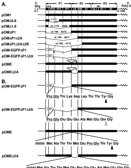
FIG. 1. PV genomic constructions. (A) Construction of PV and recombinant PVs. (B) Junctional amino acid sequences of pOM-EGFP⌬P1, pOM-EGFP⌬P1⌬2A, pOME, and pOME⌬2A. The nucleic acid sequence (CCCGGG) recognized by SmaI, corresponding to the amino acid sequence Pro Gly, is shown by underlines. The inserted EMCV IRES are shown by the striped horizontal bars. Closed triangles indicate the sites of cleavage by 2Apro, and open
triangles indicate the sites of cleavage by 3Cproor 3CDpro. Nucleotide numbers of the deleted fragments are shown at the deletion positions.
on November 8, 2019 by guest
[vol/vol] formaldehyde) in 3-(N-morpholino)propanesulfonic acid (MOPS) buffer (26 mM MOPS [pH 7.0], 6.5 mM sodium acetate, and 1.3 mM EDTA) and
denatured at 65°C for 5 min, followed by chilling on ice. An equal volume of 20⫻
SSC (333 mM NaCl and 333 mM sodium citrate tribasic dehydrate) was added. In each slot of a slot blotting apparatus (Bio-Dot SF; Bio-Rad Laboratories,
Inc.), 10l of RNA solution was applied, and the RNA was immobilized on a
nylon filter (Hybond-N; GE Healthcare UK Ltd.) and cross-linked by a UV cross-linker (UV Stratalinker1800; Stratagene). The filters were hybridized to a labeled cDNA corresponding to nt 1 to nt 742 of the viral RNA (43) or to exon 7 of glycerol-3-phosphate dehydrogenase (GAPDH) mRNA using AlkPhos Di-rect (GE Healthcare UK Ltd.) according to the manufacturer’s instructions. The probes were detected by chemiluminescence using CDP-Star (GE Healthcare UK Ltd.).
Encapsidation of PV replicons, serial passaging, and purification.The encap-sidation and serial passaging of PV replicons using VV-P1 have been described previously (5), and basically the same method was adopted here. Briefly, HeLa S3 cells were infected with 5 PFU of VV-P1, which expresses the PV capsid precursor protein P1, per cell. At 2 h postinfection, the cells were transfected by
the DEAE-dextran method within vitro-transcribed RNA. The cultures were
harvested at 24 h posttransfection by three successive freeze-thaws, sonicated,
and clarified by low-speed centrifugation at 14,000⫻gfor 20 min. For serial
passage of the encapsidated replicons and generation of virus stocks, HeLa S3 cells were first infected with 10 to 20 PFU of VV-P1 per cell. At 2 h postinfection, the cells were infected with passage 1 encapsidated replicons. The cultures were harvested at 16 h after PV infection by three successive freeze-thaws, sonicated,
and clarified by low-speed centrifugation at 14,000⫻gfor 20 min. The
super-natant was then stored at⫺80°C or used immediately for additional passages by
the same procedure.
For purifying the P1-null PV particles, the supernatant was lysed with 1% sodium dodecyl sulfate and centrifuged in a Beckman type 45 rotor at 35,000 rpm for 2.5 h. The supernatant was discarded, and the pellet was washed under the same conditions in 0.1 M phosphate buffer (pH 7.35) for an additional 2.5 h. The pellet was then resuspended in serum-free DMEM, filtrated, and stored at
⫺80°C.
Titration of viruses.The numbers of PFU in AGMK cells were determined by the plaque assay, and the numbers of infectious units (IU) were determined by counting fluorescence-positive cells. Units of viral RNA (U) were adopted for the infection with P1-null viruses. For the measurement of PFU, AGMK cells on 6-cm dishes were inoculated with the viral suspension and then incubated at 37°C for 2 to 5 days for the observation of plaques. To measure the units of viral RNA
of OM⌬0.8, OM⌬1.8, OM⌬P1, and OM⌬P1⌬2A, viral RNA was extracted from
the suspensions with chloroform containing phenol and isoamyl alcohol. The units were measured using a LightCycler system (Roche Diagnostics) according
to the manufacturer’s instructions. As 1.1⫻108U/ml was equivalent to 2.1⫻109
PFU/ml for PV1(M)OM, we adopted 5.2⫻101U/cell, which was supposed to be
equivalent to 1,000 PFU/cell (multiplicity of infection [MOI] of 1,000) of
PV1(M)OM for the infection with OM⌬0.8, OM⌬1.8, OM⌬P1, and
OM⌬P1⌬2A. For the measurement of IU of OM-EGFP⌬P1 and
OM-EGFP⌬P1⌬2A, fluorescence-positive cells were counted under an inverted
flu-orescence microscope (DM6000B; Leica Microsystems) at 24 h postinfection in HeLa cells. The amount of virus leading to one fluorescence-positive cell was defined as 1 IU. Comparing the units of RNA with the infectious units of
fluorescence-positive cells, 5.3⫻109
U/ml was equivalent to 7.1⫻109
IU/ml for
an OM-EGFP⌬P1 stock and 4.2⫻109
U/ml was equivalent to 1.1⫻109
IU/ml
for an OM-EGFP⌬P1⌬2A stock. Based on the data, we considered that the units
of RNA were not significantly different from the infectious units for
OM-EGFP⌬P1 and OM-EGFP⌬P1⌬2A.
Observation of CPE.HeLa S3 cells in 16-well Lab-Tek chamber slides (Nalge Nunc International K.K.) were infected with PVs. Eight, 18, or 24 h after the incubation with PVs at 37°C, the cells were observed under an inverted micro-scope.
Neutralization assay.The viral suspension (50l) was mixed with filtrated
ascites fluid (50l) containing the antibody and incubated at 37°C for 1 h. The
virus-antibody mixture was overlaid on HeLa S3 cells in 16-well chamber slides and incubated at room temperature for 20 min and then at 37°C for 30 min. The mixture was then replaced with DMEM supplemented with 5% NCS. The cells were observed 18 h after the incubation at 37°C under an inverted microscope.
TUNEL assay. A direct terminal deoxynucleotidyltransferase-mediated
dUTP-biotin nick end labeling (TUNEL) assay was performed using anin situ
cell death detection kit (Roche Diagnostics), and cells were observed under a confocal laser scanning microscope (LSM510; Carl Zeiss MicroImaging Co.
Ltd.). Actinomycin D (10g/ml) was added for an apoptosis-positive control.
RESULTS
2Apro
is not required for full-lengthdcPV to form progeny virus.2Aprois known to be cytotoxic, and a 2Apro-deficient PV
vector might be desirable. To examine whether 2Aprois needed
to form progeny virus, cDNAs of the full-lengthdcpOME and pOME⌬2A with the backbone of the PV type 1 Mahoney strain (pOM1) (Fig. 1A and B) were constructed and the ability to form progeny viruses was examined. HeLa cells were transfected with the RNA of pOM1, pOME, and pOME⌬2A and examined for a CPE. RNA derived from pOM1, pOME, and pOME⌬2A was not degraded as assessed by gel electro-phoresis (data not shown). All these RNAs induced a CPE that led to cell death, whereas no CPE was observed in the mock-transfected cells (data not shown). The supernatants also in-duced a CPE in HeLa cells. These results show that pOM1, pOME, and pOME⌬2A can form progeny viruses, called PV1(M)OM, OME, and OME⌬2A, respectively. This suggests that 2Aprois not essential to form progeny virus for full-length
dcPV.
To confirm that the OME and OME⌬2A suspensions do not contain IRES deletion revertants, the sizes of the fragments encompassing VP1 and 2B were examined by PCR (data not shown). The PV1(M)OM, OME, and OME⌬2A suspensions produced appropriate fragments, that is, 1.3-kb, 1.9-kb, and 1.5-kb fragments, respectively. The sequences of the PCR frag-ments were analyzed, and they were not mutated (data not shown). These results mean that IRES deletion revertants were not detected in the OME and OME⌬2A suspensions under the experimental conditions and that it is highly proba-ble that these suspensions do not contain IRES deletion re-vertants. This suggests that OME and OME⌬2A can prolifer-ate by themselves.
To ascertain that the CPE was actually induced by PV, a neutralization assay was performed using a monoclonal anti-body for the virulent type 1 Mahoney strain (7m008) and a monoclonal antibody for the virulent type 3 Leon strain (Thai p34-120). HeLa cells were infected with PV1(M)OM, OME, OME⌬2A, or Leon at an MOI of 10 in the presence or absence of 7m008 or Thai p34-120 and examined for a CPE (Fig. 2). In the absence of the antibodies, all the strains induced a CPE. In the presence of 7m008, only Leon caused a CPE. In contrast, in the presence of Thai p34-120, all the strains except Leon had a CPE. These results suggest that PV1(M)OM, OME, and OME⌬2A are type 1 strains, and the CPE was induced by PV. To define the characteristics of the viral strains, a plaque assay was performed using AGMK cells (Fig. 3). PV1(M)OM produced large plaques, OME moderate-sized plaques, and OME⌬2A the smallest plaques. The extremely small plaques of OME⌬2A may relate to its slow replication rate.
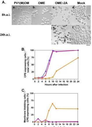
To compare these strains further, HeLa cells were infected at an MOI of 10 and cell morphology was observed 8 or 24 h after the infection (Fig. 4). All the viruses had a CPE (Fig. 4A). No CPE was observed in the mock-infected cells. To quantify the rate of CPE-expressing cells, the percentages of round-shaped cells, detached cells, and cells with membrane blebbing among all cells were analyzed, and the kinetics are shown in Fig. 4B. OME⌬2A had a lesser CPE than did OME or PV1(M)OM. OME exhibited a similar level of CPE to PV1(M)OM, although OME had slightly slower kinetics than
on November 8, 2019 by guest
http://jvi.asm.org/
PV1(M)OM. The results were reproducible. These results sug-gest that in speed of viral replication and/or the ability to induce CPE, the strains rank as OME⌬2A ⬍ OME ⱌ PV1(M)OM. They also suggest that 2Aprocontributes to
effi-cient viral replication and/or the induction of a CPE. When the cells were infected at a higher MOI (MOI of 50), the kinetics were essentially unchanged (data not shown). This result sug-gests that an MOI of 10 was high enough to assess the kinetics of CPE expression. Only OME⌬2A induced morphological changes typical of apoptosis, namely, cell membrane blebbing, in a significant proportion of cells (Fig.4Ac, Ae, and 4Cs). This result indicates that OME⌬2A may induce apoptosis.
2Apro
-deficient full-lengthdcPV induces apoptosis.To con-firm that OME⌬2A induces apoptosis, a direct TUNEL assay was performed in cells infected with or without PV1(M)OM or OME⌬2A or treated with actinomycin D, which induces apop-tosis (Fig. 5). Uninfected samples and PV1(M)OM-infected samples contained few fluorescence-positive cells. On the other hand, actinomycin D-treated samples and OME⌬ 2A-infected samples included many fluorescence-positive cells. These results suggest that OME⌬2A induces apoptosis. This raises the possibility that 2Aprois important to prevent typical
apoptosis in PV-infected cells.
P1-null PV RNA deficient in the 2Apro
coding region can replicate in cells.It has been reported that the PV P1 coding region is not required for RNA replication and the formation of progeny virus in P1-expressing cells (4, 37). To further define which parts of the PV genome are necessary for RNA replication, deletion mutants were prepared (Fig. 1A) and RNA replication activity was examined by slot blotting (Fig. 6A). RNAs derived from pOM1, pOM⌬0.8, pOM⌬1.8, pOM⌬P1, pOM⌬P1⌬2A, and pOM⌬P1⌬2A⌬2B were not de-graded as assessed by gel electrophoresis (data not shown). The RNAs were introduced into HeLa cells, and the cells were collected 2 and 8 h after the transfection. Cell lysates were used for slot blotting. When a probe for PV IRES RNA was used, all the RNAs except for pOM⌬P1⌬2A⌬2B RNA were in-creased at 8 h compared to the levels at 2 h after the transfec-tion. The amounts of GAPDH RNA hardly changed up to 8 h after the transfection. The results were reproducible. These results suggest that the 2Aprocoding region is not needed for
the RNA replication of P1-null PV and that 2B is necessary for the PV replicon activity.
The 2Apro
[image:5.585.134.450.67.247.2]coding region is not required for P1-null PV to form progeny virus in P1-expressing cells.Next, the ability to form progeny virus was examined. The RNAs of pOM1,
FIG. 2. Neutralization assay of PV1(M)OM, OME, OME⌬2A, and Leon with anti-PV type 1 or type 2 monoclonal antibody. HeLa cells were mock infected (e, j, and o) or infected with PV1(M)OM (a, f, and k), OME (b, g, and l), OME⌬2A (c, h, and m), or Leon (d, i, and n) in the absence (k to o) or presence of an anti-PV type 1 Mahoney monoclonal antibody (7m008, a to e) or anti-PV type 2 Leon monoclonal antibody (Thai p34-120, f to j). Eighteen hours after the infection, the CPE was observed under a microscope. Bar, 50m.
FIG. 3. Plaque phenotypes of PV1(M)OM, OME, and OME⌬2A. AGMK cells were infected with PV1(M)OM (a), OME (b), and OME⌬2A (c). The cells were fixed 2 days after the infection with PV1(M)OM, and OME and 4 days after the infection with OME⌬2A.
on November 8, 2019 by guest
[image:5.585.135.449.594.703.2]pOM⌬0.8, pOM⌬1.8, pOM⌬P1, pOM⌬P1⌬2A, and pOM⌬ P1⌬2A⌬2B were introduced into P1-expressing cells, and the supernatant was recovered after freezing and thawing. HeLa cells were covered with medium containing the supernatants and examined for a CPE 24 h later (data not shown). All the supernatants except those of pOM⌬P1⌬2A⌬2B RNA and mock transfectants had a CPE. These results suggest that the RNAs of pOM⌬0.8, pOM⌬1.8, pOM⌬P1, and pOM⌬P1⌬2A can form progeny viruses, OM⌬0.8, OM⌬1.8, OM⌬P1, and OM⌬P1⌬2A, respectively, whereas the pOM⌬P1⌬2A⌬2B RNA cannot. The quality and sizes of the viral RNA genomes were confirmed by agarose gel electrophoresis (data not shown). To
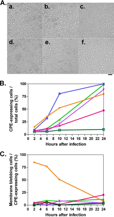
adjust the titers among these viruses, the copy numbers of the PV RNA strands in the virus-containing supernatants were examined by quantitative real-time PCR and the units of viral RNA were determined. To examine the CPE expression, HeLa cells were infected at 5.2⫻101U/cell and examined for a CPE up to 24 h
[image:6.585.135.452.68.507.2]after the infection (Fig. 7), and the rates of round cells, detached cells, and cells with membrane blebbing among all cells were analyzed (Fig. 7B). Similar to the previous results, all the viruses had a CPE. P1-null PV had slower kinetics than PV1(M)OM. OM⌬0.8, OM⌬1.8, and OM⌬P1 showed similar numbers of CPE-expressing cells, although the kinetics of OM⌬P1 until 10 h after the infection was faster than that of other P1-null PVs.
FIG. 4. CPE in cells infected with or without PV1(M)OM, OME, and OME⌬2A. (A) HeLa cells were mock infected (d and f) or infected with PV1(M)OM (a), OME (b), or OME⌬2A (c and e) at an MOI of 10. Eight (a to d) or 24 h (e and f) after the infection (h.a.i.), morphological changes were observed under a microscope. Bar, 50m. (B) Percentages of CPE expression among all cells. (C) Rates of membrane blebbing among CPE-expressing cells. Average rates in two to three microscopic fields in one experiment were plotted. The rates were examined 0, 2, 4, 6, 8, 10, 12, and 24 h after the infection. Blue lines with squares indicate results for PV1(M)OM, pink lines with triangles indicate results for OME, and orange lines with circles indicate results for OME⌬2A.
on November 8, 2019 by guest
http://jvi.asm.org/
OM⌬P1⌬2A reproducibly had a lower rate of CPE than other P1-null PVs until 24 h after the infection. This result suggests that OM⌬P1⌬2A is less able to induce a CPE or else takes longer and that 2Aproplays an important role in inducing a CPE. OM⌬P1,
significantly, started to cause membrane blebbing typical of apop-tosis from 2 h after the infection, and the proportion of CPE-expressing cells was relatively high at early time points (Fig. 7C). This result indicates the possibility that OM⌬P1 induces apopto-sis. Compared with other P1-null PVs, OM⌬P1⌬2A showed at least about a two-times-higher rate of membrane blebbing among CPE-expressing cells at 24 h after infection (OM⌬0.8, 12%; OM⌬1.8, 4.7%; OM⌬P1, 7.9%; and OM⌬P1⌬2A, 20%) (Fig. 7C). This indicates that OM⌬P1⌬2A may induce apoptosis at a relatively low efficiency.
2Apro
coding region-deficient P1-null PV vector expresses foreign genes.Because OM⌬P1⌬2A can form progeny virus, 2Apromay not be essential for P1-null PV to express foreign
genes. To examine whether OM⌬P1 and OM⌬P1⌬2A can express foreign genes, an EGFP coding sequence was inserted into the region of pOM⌬P1 and pOM⌬P1⌬2A with P1 deleted (Fig. 1A and B). The new constructs were designated pOM-EGFP⌬P1 and pOM-EGFP⌬P1⌬2A, respectively. RNAs derived from pOM1, pOM⌬P1, pOM⌬P1⌬2A, pOM⌬P1⌬2A⌬2B, pOM-EGFP⌬P1, and pOM-EGFP⌬P1⌬2A were not degraded as assessed by gel electrophoresis (data not shown). The RNAs were introduced into HeLa cells, and the cells collected 2 and 8 h after the transfection. The cell lysate was used for slot blotting (Fig. 6B). When a probe for PV IRES RNA was used, all the RNAs except for pOM⌬P1⌬2A⌬2B RNA increased at 8 h after the transfection compared to the levels at 2 h. pOM-EGFP⌬P1 RNA increased more than pOM-EGFP⌬P1⌬2A RNA. The amounts of GAPDH RNA hardly changed up to 8 h after the transfection. The results were reproducible. These results suggest that the 2Aprocoding region is not required for the RNA
[image:7.585.133.448.64.278.2]repli-FIG. 6. Slot blot analysis of cells after transfection with or without synthesized viral RNA or after infection with or without viruses. (A) HeLa cells were transfected with or without (Mock) RNAs of pOM1, pOM⌬0.8, pOM⌬1.8, pOM⌬P1, pOM⌬P1⌬2A, or pOM⌬P1⌬2A⌬2B, and cell lysates were collected 2 and 8 h later. The RNAs were detected with probes for the PV IRES sequence or GAPDH sequence. (B) A similar experiment was performed using the RNAs of pOM1, pOM⌬P1, pOM⌬P1⌬2A, pOM⌬P1⌬2A⌬2B, pOM-EGFP⌬P1, and pOM-EGFP⌬P1⌬2A. (C) HeLa cells were infected with or without (Mock) PV1(M)OM, OM⌬0.8, OM⌬1.8, OM⌬P1, or OM⌬P1⌬2A, and cell lysates were collected 2 and 8 h later. The RNAs were detected with probes for the PV IRES sequence or GAPDH sequence. Relative amounts of PV RNA corrected to the amount of GAPDH RNA are shown.
FIG. 5. Apoptosis in cells infected with OME⌬2A, detected by the TUNEL assay. HeLa cells were mock infected (d, h, and l), infected with PV1(M)OM (a, e, and i) or OME⌬2A (b, f, and j), or treated without (d, h, and l) or with (c, g, and k) actinomycin D (Act D). Seven hours after the infection with PV1(M)OM, 8 h after the treatment with actinomycin D, and 21 h after the infection with OME⌬2A, the cells were TUNEL stained. TUNEL-positive cells were fluorescent. Fluorescence (FL) images are shown at the top (a to d), bright-field (BF) images are shown in the middle (e to h), and merged (FL⫹BF) images are shown at the bottom (i to l). Bar, 50m.
on November 8, 2019 by guest
[image:7.585.91.235.362.592.2]cation of P1-null PV with EGFP inserted, although deletion of the 2Aprocoding region results in a lower rate of RNA
replica-tion. When a probe for PV IRES RNA was used, the RNAs of pOM⌬P1 and pOM⌬P1⌬2A reproducibly increased to levels
sim-ilar to the RNAs of pOM-EGFP⌬P1 and pOM-EGFP⌬P1⌬2A, respectively. These results suggest that the insertion of EGFP does not significantly affect PV RNA replication. Next, the RNAs of pOM-EGFP⌬P1 and pOM-EGFP⌬P1⌬2A were introduced into P1-expressing cells and the supernatants were recovered af-ter freezing and thawing. The viral particles, OM-EGFP⌬P1 and OM-EGFP⌬P1⌬2A, respectively, proliferated in P1-expressing cells and were then purified. HeLa cells were covered with the purified virus at an MOI of 100, and the fluorescence of EGFP was observed under the fluorescence microscope 24 h after the infection (Fig. 8). Fluorescence-positive cells were observed in the OM-EGFP⌬P1- and OM-EGFP⌬P1⌬2A-infected samples, whereas no positive cells were observed in the mock-infected sample. These results suggest that OM⌬P1 and OM⌬P1⌬2A can express EGFP and that 2Aprois not essential for P1-null PV to
express EGFP.
In their ability to express EGFP 24 h after the infection, the strains ranked reproducibly as OM-EGFP⌬P1⌬2A ⬍ OM-EGFP⌬P1 (the average rates of fluorescence positivity among all cells in three microscopic fields in one experiment were 23% for OM-EGFP⌬P1 and 9.7% for OM-EGFP⌬P1⌬2A). This suggests that the defect in 2Aprodecreases the expression of
EGFP and/or the speed of viral replication. OM-EGFP⌬P1⌬2A had a CPE, as did OM-EGFP⌬P1, but only OM-EGFP⌬P1⌬2A reproducibly induced morphological changes typical of apoptosis in a significant proportion of the CPE-expressing cells (the aver-age rates of membrane blebbing among CPE-expressing cells were 10% for OM-EGFP⌬P1 and 37% for OM-EGFP⌬P1⌬2A). The result implies that 2Apro masks apoptosis in a significant
proportion of cells. In the speed with which they induced a CPE, the strains reproducibly ranked as OM-EGFP⌬P1 ⱌ OM-EGFP⌬P1⌬2A (the average rates of CPE expression among all cells were 27% for OM-EGFP⌬P1 and 26% for OM-EGFP⌬P1⌬2A). This suggests that 2Aprohas little or no effect on
inducing a CPE. The quality and sizes of the RNA genomes of OM-EGFP⌬P1 and OM-EGFP⌬P1⌬2A were confirmed by Northern blotting (data not shown). The genomic stability of OM-EGFP⌬P1 and OM-EGFP⌬P1⌬2A was examined by gen-eral RT-PCR, but no deletion was detected even after 20 passages (data not shown). These results suggest that both OM-EGFP⌬P1 and OM-EGFP⌬P1⌬2A are genetically stable for at least 20 pas-sages.
DISCUSSION
2Aprohas been thought important for PV replication. Here,
we reveal that 2Apro is not required for PV replication and
2Apro-deficient PV with or without the capsid coding region
can produce progeny viruses. This shows the possibility of realizing a vector that expresses a longer foreign gene and is less toxic.
Molla et al. reported thatdc PV cDNA without the 2Apro
coding region (pT7PVE2B) does not produce a viable virus (30). In contrast, RNA of pOME⌬2A, which has a structure similar to that of pT7PVE2B, resulted in productive although inefficient replication. This may be due to a difference in the junctional sequences between P1 and 2B. In the case of pT7PVE2B, 2B is translated directly from the second EMCV IRES and methionine is added to the N terminus of 2B. On the other hand, in the case of pOME⌬2A, the additional
N-termi-FIG. 7. CPE in the cells infected with PV1(M)OM, OM⌬0.8, OM⌬1.8, OM⌬P1, and OM⌬P1⌬2A. (A) HeLa cells were mock infected (f) or infected with PV1(M)OM (a), OM⌬0.8 (b), OM⌬1.8 (c), OM⌬P1 (d), or OM⌬P1⌬2A (e) at an MOI of 1,000, and cell morphology was observed 24 h later under a microscope. Bar, 50m. (B) Percentages of CPE expression among all cells. (C) Rates of membrane blebbing among CPE-expressing cells. Average rates in two to four microscopic fields in one experiment were plotted. The rates were examined at 2, 6, 10, and 24 h after the infection. Blue lines with filled squares indicate results for PV1(M)OM, green lines with triangles indicate results for OM⌬0.8, violet lines with inverted triangles indicate results for OM⌬1.8, orange lines with rhombuses indicate results for OM⌬P1, pink lines with circles indicate results for OM⌬P1⌬2A, and moss-green lines with open squares indicate results for mock-infected cells.
on November 8, 2019 by guest
http://jvi.asm.org/
[image:8.585.43.283.70.526.2]nal sequences of 2B can be processed by 3Cpro or 3CDpro.
Moreover, pT7PVE2B contains EMCV IRES up to AUG834, whereas pOME⌬2A contains it up to⫹18 nt downstream of AUG834. Ribosomal initiation complexes attach directly to AUG834, and initiation does not involve scanning (22, 35). The interaction positions for ribosomal initiation complexes are up to⫹17 nt downstream of AUG834(23). It is possible that the difference leads to modification of the 2B activity and/or rela-tively low efficiency of the initiation of the second cistron translation.
OME⌬2A caused apoptosis, whereas neither PV1(M)OM nor OME did. Our results show the possibility that OM⌬P1, OM⌬P1⌬2A, and OM-EGFP⌬P1⌬2A induce apoptosis in a significant proportion of cells. Calandria et al. reported that independently expressed 2Aproand 3Cproinduced apoptosis by
mechanisms involving caspase activation (10). On the other hand, Burgon et al. reported that a 2A N32D mutation inde-pendently caused cells to die by apoptosis much earlier than wild-type-infected cells (9). This is consistent with the hypoth-esis that a wild-type function of the 2Aproprotein is to inhibit
apoptosis and cause a canonical necrotic CPE late in infection, perhaps directly or indirectly leading to the aberrant cleavage of procaspase-9, and that this activity is abrogated by the 2A N32D mutation. Together with our results, it seems likely that the apoptotic cell death induced in the cells infected with 2A-deficient or P1-deficient PV occurs via caspase-dependent apoptosis and that the expression of apoptosis depends on a
subtle balance of relating proteins, such as 2Apro, 3Cpro, and
procaspase-9, at each time point after the infection. It is highly possible that the balance of viral proteins differs depending on whether the virus is monocistronic or dicistronic. 2Aproactivity
may differ fundamentally depending on whether this protein is expressed individually or in the context of the viral infection. It has been reported that PV was replication competent upon the replacement of its P1 region with a foreign gene in P1-expressing cells (4, 37). In these cases, an insert of about 2.9 kilobases (carcinoembryonic antigen [CEA]) was the longest. DIs with a 1,212-base deletion at the longest in the P1 region have been detected (25). We revealed that PV can produce progeny viruses with the entire P1 and 2Apro coding region
deleted in P1-expressing cells. This construct lacks the longest region. We confirmed that PV can express EGFP without the P1 and 2Aprocoding regions and that the insertion of EGFP
does not significantly affect viral RNA replication. Moreover, these viruses are genetically stable. These results raise the possibility that a longer foreign gene than CEA can be ex-pressed using the PV vector.
OM⌬P1⌬2A had a lesser CPE than OM⌬0.8, OM⌬1.8, OM⌬P1, and PV1(M)OM at 5.2⫻101U/cell, which was
sup-posed to be equivalent to an MOI of 1,000 of PV1(M)OM. To correct the units of viral RNA genomes as infectious units, HeLa cells were infected with OM⌬0.8, OM⌬1.8, OM⌬P1, and OM⌬P1⌬2A at 5.2 ⫻ 101 U/cell or with PV1(M)OM at an
[image:9.585.132.452.66.360.2]MOI of 10, the cells were collected 2 and 8 h later, and cell
FIG. 8. P1-null PV vector with or without the 2Aprocoding region expresses EGFP. HeLa cells were mock infected (c, f, and i) or infected with
OM-EGFP⌬P1 (a, d, and g) or OM-EGFP⌬P1⌬2A (b, e, and h) and observed 24 h later under a fluorescence microscope. Upper panels show fluorescence (FL) images (a to c), panels in the middle show bright-field (BF) images (d to f), and lower panels show merged (FL⫹BF) images
(g to i). Bar, 100m.
on November 8, 2019 by guest
lysates were used for slot blotting (Fig. 6C). When a probe for PV IRES RNA was used, all the P1-null viruses had increased at almost the same rates at 8 h compared to the levels at 2 h after the infection. PV1(M)OM increased much more than P1-null viruses. These results suggest that 5.2⫻101U/cell for
P1-null viruses results in similar RNA replication activities under these conditions. The viral titers/cell might not be high enough to infect all the cells because the cells infected at 5.2⫻ 101 U/cell contained more RNA than those infected at 5.2
U/cell (data not shown); it is likely that the units of the RNA genomes of OM⌬0.8, OM⌬1.8, OM⌬P1, and OM⌬P1⌬2A were almost proportional to these RNA replication activities. Consequently, it was confirmed that OM⌬P1⌬2A is less toxic or takes a longer time to have a CPE even though its RNA replication activity is similar to those of OM⌬0.8, OM⌬1.8, and OM⌬P1.
Regarding the slot blot analysis of the cells transfected with synthesized viral RNA, the RNAs of pOM⌬P1⌬2A and pOM-EGFP⌬P1⌬2A showed less RNA replication activity than those of pOM⌬P1 and pOM-EGFP⌬P1, respectively (Fig. 6B). It seems likely that the defect in 2Apro suppresses the RNA
replication activity. In terms of the relevant effect of 2Apro,
viral replication speed, and/or the ability to induce a CPE, the ranking was OME⌬2A⬍OME and OM⌬P1⌬2A⬍OM⌬P1, whereas OM-EGFP⌬P1ⱌOM-EGFP⌬P1⌬2A. In the ability to express EGFP, the ranking was OM-EGFP⌬P1⌬2A⬍ OM-EGFP⌬P1. These results may be also because the defect in 2Apro suppresses the viral replication activity and/or the
ex-pression of foreign mRNA.
ACKNOWLEDGMENTS
We are grateful to T. Matano, N. Kamoshita, A. Yanagiya, and N. Matsuda for suggestions and discussions. We also thank A. Ohmura for technical support and E. Suzuki for help in preparing the manu-script. We are grateful to C. D. Morrow for generously providing VV-P1.
This work was supported in part by Grants-in-Aid for Advanced Medical Science Research by Ministry of Education, Culture, Sports, Science and Technology (MEXT), a Grant-in-Aid for Scientific Re-search on Priority Areas, a Grant-in-Aid for Scientific ReRe-search (S), a Grant-in-Aid for Scientific Research on Priority Areas, a Grant-in-Aid for Young Scientists (B), a Health Labor Sciences Research Grant, special coordination funds for promoting Science and Technology, contracted research allowance “Research and Development in a New Converting Field Based on Nanotechnology and Materials Science” by MEXT, and The Naito Foundation.
REFERENCES
1.Agol, V. I., G. A. Belov, K. Bienz, D. Egger, M. S. Kolesnikova, N. T. Raikhlin, L. I. Romanova, E. A. Smirnova, and E. A. Tolskaya.1998. Two types of death of poliovirus-infected cells: caspase involvement in the
apop-tosis but not cytopathic effect. Virology252:343–353.
2.Agol, V. I., G. A. Belov, K. Bienz, D. Egger, M. S. Kolesnikova, L. I. Ro-manova, L. V. Sladkova, and E. A. Tolskaya.2000. Competing death pro-grams in poliovirus-infected cells: commitment switch in the middle of the
infectious cycle. J. Virol.74:5534–5541.
3.Almstead, L. L., and P. Sarnow.2007. Inhibition of U snRNP assembly by a
virus-encoded proteinase. Genes Dev.21:1086–1097.
4.Ansardi, D. C., Z. Moldoveanu, D. C. Porter, D. E. Walker, R. M. Conry, A. F. LoBuglio, S. McPherson, and C. D. Morrow.1994. Characterization of
poliovirus replicons encoding carcinoembryonic antigen. Cancer Res. 54:
6359–6364.
5.Ansardi, D. C., D. C. Porter, and C. D. Morrow.1993. Complementation of a poliovirus defective genome by a recombinant vaccinia virus which
pro-vides poliovirus P1 capsid precursor in trans. J. Virol.67:3684–3690.
6.Belov, G. A., L. I. Romanova, E. A. Tolskaya, M. S. Kolesnikova, Y. A. Lazebnik, and V. I. Agol.2003. The major apoptotic pathway activated and
suppressed by poliovirus. J. Virol.77:45–56.
7.Bodian, D.1955. Emerging concept of poliomyelitis infection. Science122:
105–108.
8.Borman, A. M., R. Kirchweger, E. Ziegler, R. E. Rhoads, T. Skern, and K. M. Kean.1997. elF4G and its proteolytic cleavage products: effect on initiation of protein synthesis from capped, uncapped, and IRES-containing mRNAs.
RNA3:186–196.
9.Burgon, T. B., J. A. Jenkins, S. B. Deitz, J. F. Spagnolo, and K. Kirkegaard.
2009. Bypass suppression of small-plaque phenotypes by a mutation in
po-liovirus 2A that enhances apoptosis. J. Virol.83:10129–10139.
10.Calandria, C., A. Irurzun, A. Barco, and L. Carrasco.2004. Individual expression of poliovirus 2Apro and 3Cpro induces activation of caspase-3
and PARP cleavage in HeLa cells. Virus Res.104:39–49.
11.Castello, A., J. M. Izquierdo, E. Welnowska, and L. Carrasco.2009. RNA nuclear export is blocked by poliovirus 2A protease and is concomitant with
nucleoporin cleavage. J. Cell Sci.122:3799–3809.
12.Cole, C. N.1975. Defective interfering (di) particles of poliovirus. Prog. Med.
Virol.20:180–207.
13.Cole, C. N., D. Smoler, E. Wimmer, and D. Baltimore.1971. Defective interfering particles of poliovirus. I. Isolation and physical properties. J.
Vi-rol.7:478–485.
14.Collis, P. S., B. J. O’Donnell, D. J. Barton, J. A. Rogers, and J. B. Flanegan.
1992. Replication of poliovirus RNA and subgenomic RNA transcripts in
transfected cells. J. Virol.66:6480–6488.
15.Etchison, D., S. C. Milburn, I. Edery, N. Sonenberg, and J. W. Hershey.
1982. Inhibition of HeLa cell protein synthesis following poliovirus infection correlates with the proteolysis of a 220,000-dalton polypeptide associated with eucaryotic initiation factor 3 and a cap binding protein complex. J. Biol.
Chem.257:14806–14810.
16.Hagino-Yamagishi, K., and A. Nomoto.1989. In vitro construction of
polio-virus defective interfering particles. J. Virol.63:5386–5392.
17.Hambidge, S. J., and P. Sarnow.1992. Translational enhancement of the
poliovirus 5⬘noncoding region mediated by virus-encoded polypeptide 2A.
Proc. Natl. Acad. Sci. U. S. A.89:10272–10276.
18.Hunt, S. L., T. Skern, H. D. Liebig, E. Kuechler, and R. J. Jackson.1999. Rhinovirus 2A proteinase mediated stimulation of rhinovirus RNA transla-tion is additive to the stimulatransla-tion effected by cellular RNA binding proteins.
Virus Res.62:119–128.
19.Jang, S. K., M. V. Davies, R. J. Kaufman, and E. Wimmer.1989. Initiation
of protein synthesis by internal entry of ribosomes into the 5⬘nontranslated
region of encephalomyocarditis virus RNA in vivo. J. Virol.63:1651–1660.
20.Jang, S. K., H. G. Krausslich, M. J. Nicklin, G. M. Duke, A. C. Palmenberg, and E. Wimmer.1988. A segment of the 5⬘nontranslated region of encepha-lomyocarditis virus RNA directs internal entry of ribosomes during in vitro
translation. J. Virol.62:2636–2643.
21.Kajigaya, S., H. Arakawa, S. Kuge, T. Koi, N. Imura, and A. Nomoto.1985. Isolation and characterization of defective-interfering particles of poliovirus
Sabin 1 strain. Virology142:307–316.
22.Kaminski, A., M. T. Howell, and R. J. Jackson.1990. Initiation of encepha-lomyocarditis virus RNA translation: the authentic initiation site is not
se-lected by a scanning mechanism. EMBO J.9:3753–3759.
23.Kolupaeva, V. G., I. B. Lomakin, T. V. Pestova, and C. U. Hellen.2003. Eukaryotic initiation factors 4G and 4A mediate conformational changes downstream of the initiation codon of the encephalomyocarditis virus
inter-nal ribosomal entry site. Mol. Cell. Biol.23:687–698.
24.Kuechler, E., J. Seipelt, H. D. Liebig, and W. Sommergruber.2002. Picor-navirus proteinase-mediated shutoff of host cell translation: direct cleavage
of a cellular initiation factor, p. 301–311.InB. L. Semler and E. Wimmer
(ed.), Molecular biology of picornaviruses. ASM Press, Washington, DC. 25.Kuge, S., I. Saito, and A. Nomoto.1986. Primary structure of poliovirus
defective-interfering particle genomes and possible generation mechanisms
of the particles. J. Mol. Biol.192:473–487.
26.Lawson, M. A., and B. L. Semler.1990. Picornavirus protein processing— enzymes, substrates, and genetic regulation. Curr. Top. Microbiol. Immunol.
161:49–87.
27.Li, X., H. H. Lu, S. Mueller, and E. Wimmer.2001. The C-terminal residues of poliovirus proteinase 2A(pro) are critical for viral RNA replication but
not for cis- or trans-proteolytic cleavage. J. Gen. Virol.82:397–408.
28.Lundquist, R. E., M. Sullivan, and J. V. Maizel, Jr.1979. Characterization of
a new isolate of poliovirus defective interfering particles. Cell18:759–769.
29.Molla, A., S. K. Jang, A. V. Paul, Q. Reuer, and E. Wimmer.1992. Cardio-viral internal ribosomal entry site is functional in a genetically engineered
dicistronic poliovirus. Nature356:255–257.
30.Molla, A., A. V. Paul, M. Schmid, S. K. Jang, and E. Wimmer.1993. Studies
on dicistronic polioviruses implicate viral proteinase 2Aproin RNA
replica-tion. Virology196:739–747.
31.Nomoto, A., B. Detjen, R. Pozzatti, and E. Wimmer.1977. The location of the polio genome protein in viral RNAs and its implication for RNA synthesis.
Nature268:208–213.
32.Omata, T., H. Horie, S. Kuge, N. Imura, and A. Nomoto.1986. Mapping and sequencing of RNAs without recourse to molecular cloning: application to RNAs of the Sabin 1 strain of poliovirus and its defective interfering
parti-cles. J. Biochem.99:207–217.