DNA-Binding Activity of Adeno-Associated Virus Rep Is Required for
Inverted Terminal Repeat-Dependent Complex Formation
with Herpes Simplex Virus ICP8
Martin Alex,a* Stefan Weger,aMario Mietzsch,aHeiko Slanina,a* Toni Cathomen,a,band Regine Heilbronna
Institute of Virology, Campus Benjamin Franklin, Charité—Medical School, Berlin, Germany,aand Institute of Experimental Hematology, Hannover Medical School,
Hannover, Germanyb
Herpes simplex virus (HSV) helper functions for (AAV) replication comprise HSV ICP8 and helicase-primase UL5/UL52/UL8.
Here we show that N-terminal amino acids of AAV Rep78 that contact the Rep-binding site within the AAV inverted terminal
repeat (ITR) are required for ternary-complex formation with infected-cell protein 8 (ICP8) on AAV single-strand DNA (ssDNA)
in vitro
and for colocalization in nuclear replication domains
in vivo
. Our data suggest that HSV-dependent AAV replication is
initiated by Rep contacting the AAV ITR and by cooperative binding of ICP8 on AAV ssDNA.
A
subset of six out of seven herpes simplex virus
(HSV)-encoded replication functions was shown to provide helper
activity for productive replication of adeno-associated virus
(AAV). Four of these, the single-strand-DNA (ssDNA)-binding
protein infected-cell protein 8 (ICP8) (UL29) and the
heterotri-meric helicase-primase complex UL5/UL8/UL52, constitute the
minimal four-protein complex of helper functions for AAV DNA
replication (32). Further analysis showed that the helicase UL5
and the primase UL52 are primarily needed as structural
compo-nents of replicative structures, able to recruit AAV Rep and the
AAV genome for the initiation of AAV DNA replication (10, 27).
AAV type 2 contains a 4.7-kb linear single-stranded DNA
ge-nome flanked by 145-bp inverted terminal repeats (ITR), which
comprise the origins of replication where AAV Rep78 and its
C-terminal variant Rep68 bind to the Rep-binding site (RBS). By
means of its ATP-dependent helicase activity, Rep unwinds the
ITR, and its endonuclease activity leads to nicking of the adjacent
terminal resolution site (3, 5, 14, 28). The role of Rep78/68 as
ori
-binding proteins and initiators of AAV DNA replication was
analyzed previously using HSV as a helper virus. In coinfection
experiments, Rep78 was found to colocalize to HSV ICP8 in
nu-clear replication compartments in a manner dependent on
ITR-flanked single-stranded AAV genomes. Furthermore, direct AAV
ssDNA-dependent interaction of purified ICP8 and Rep78 was
shown
in vitro
(10). ICP8 displays high-affinity, cooperative
bind-ing to ssDNA (1, 7, 20). Rep78/68 also displays some
ssDNA-binding activity (16, 19, 36) but preferentially binds to the
double-stranded RBS within the hairpin-shaped AAV ITR (12). We have
shown before that ssDNA devoid of AAV ITRs displayed severely
reduced ternary-complex formation with wild-type Rep78/68 and
ICP8 (10).
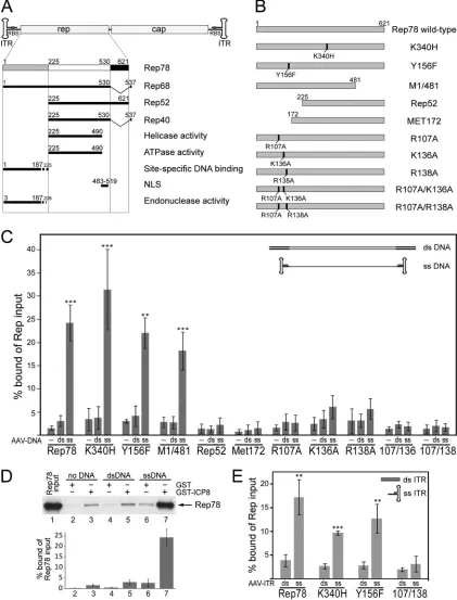
In this study, we aimed to identify the Rep domain(s) (Fig. 1A
and B) responsible for the interaction with ICP8 on the AAV
ge-nome. Ternary-complex formation between Rep78, HSV ICP8,
and AAV DNA was analyzed by
in vitro
pulldown assays with
purified glutathione
S
-transferase (GST)-tagged ICP8 and
in
vitro
-translated
35S-labeled Rep proteins as described before (10).
Plasmid-excised, full-length AAV wild-type genomes were gel
pu-rified and either used directly as linear dsDNA templates or heat
denatured to adopt ssDNA conformation. Subsequent
snap-cooling on ice ensured reassociation of the hairpin-shaped ITRs
flanking the single-stranded AAV genome (9). Rep78 and ICP8
interact to a low degree in the absence of DNA (Fig. 1C; Fig. 1D,
lane 3) without enhancement upon addition of double-stranded
full-length AAV DNA (Fig. 1D, lane 5). In contrast,
single-stranded AAV genomes significantly enhanced the interaction
be-tween Rep78 and ICP8 up to 10-fold (Fig. 1C; Fig. 1D, lane 7).
In order to delineate the domain(s) involved in
ternary-complex formation, a series of Rep mutants (Fig. 1B) were
as-sessed. As shown in Fig. 1C, ssDNA-dependent complex
forma-tion was maintained with the single-amino-acid exchange mutant
RepK340H. RepK340H is impaired in ATP-binding precluding
DNA helicase activity
in vitro
(3, 4, 35) and AAV DNA replication
in vivo
(2, 17). The ability of RepK340H to bind to the
hairpin-structured AAV ITR (4, 23, 24, 33) and its site-specific
endonu-clease activity are retained (5). Likewise, Rep78 mutants with
de-letions of the C terminus encompassing the nuclear localization
signal (M1/481) retained the ability to interact with ICP8 and
AAV ssDNA
in vitro
(Fig. 1C). In contrast, N-terminal-deletion
mutants of Rep, shown to be replication negative
in vivo
(13, 17),
were entirely deficient for ternary-complex formation
in vitro
(Fig. 1C, Rep52 and Met172). The N terminus of Rep78/68
com-prises the DNA-binding domain that mediates site-specific
bind-ing to the RBS within the AAV ITR and also the domain for
en-donuclease activity (Fig. 1A). In addition, enen-donuclease requires
binding to the RBS to exert its activity (5). To differentiate
be-tween the two activities, the exclusively endonuclease-deficient
mutant RepY156F (5) was generated, which proved to retain
Received20 September 2011Accepted16 December 2011
Published ahead of print28 December 2011
Address correspondence to Regine Heilbronn, [email protected].
* Present address: Martin Alex, Medical Clinic for Oncology and Hematology, CCM, Charité Medical School, Berlin, Germany; Heiko Slanina, Institute of Hygiene and Microbiology, University of Würzburg, Germany.
M.A. and S.W. contributed equally to this article.
Copyright © 2012, American Society for Microbiology. All Rights Reserved.
doi:10.1128/JVI.06364-11
on November 7, 2019 by guest
http://jvi.asm.org/
FIG 1Rep78 domains involved in AAV ssDNA-dependent interaction with HSV ICP8. (A) Individual domains of Rep78 are shown as rectangles. The N terminus unique to Rep78 and Rep68 (amino acids 1 to 225) is gray, the central region common to all four Rep proteins is white, and the C terminus specific to Rep78 and Rep52 derived from unspliced mRNAs is black. Domains involved in functional activities are represented by solid black lines. Approximate amino acid positions are shown as boundary marks as compiled from the literature. The respective references are given in the text. (B) Plasmid constructs for Rep78 and mutants thereof. The mutated amino acids and the amino acid positions are indicated. (C) AAV DNA-dependent interaction of AAV Rep78 with HSV ICP8 was analyzed byin vitroGST pulldown assays.In vitro-translated35S-labeled Rep78 and its mutants were incubated with GST-ICP8. Assays were performed either in the absence of DNA, in the presence of double-stranded wild-type AAV-2 DNA, or in the presence of heat-denatured, single-stranded wild-type AAV-2 DNA. Bound Rep proteins were analyzed by autoradiography in sodium dodecyl sulfate-polyacrylamide gel electrophoresis (SDS-PAGE) gels. The results of phos-phorimager quantifications in three to five independent experiments are given as means⫾standard deviations. Significances were calculated for ssDNA versus dsDNA by Student’sttest. Significance levels are indicated as follows: ***,P⬍0.001; **,P⬍0.01; *,P⬍0.05. (D) Representative autoradiogram and results of five independentin vitropulldown experiments either with GST alone or with wild-type Rep78, performed as described for panel C. The lane labeled “Rep78input” was loaded with 50% of the amount of Rep included in the binding assay. (E) Results of an experiment performed as described for panel C with AAV plasmid-excised dsITR or hairpin-structured ssITR (nt 1 to 181).
on November 7, 2019 by guest
http://jvi.asm.org/
[image:2.585.81.502.36.589.2]ternary-complex formation (Fig. 1C). Rep mutants with exclusive
ITR-binding defects were designed as follows. Based on crystal
structure analysis of the N terminus of AAV-5 Rep78 bound to the
RBS of the AAV-5 ITR (12), the critical amino acids of AAV-2
Rep78 that contact the ITR were aligned to positions R107, R136,
and R138 (11, 12). These amino acid positions were mutated
in-dividually or in combination, as shown in Fig. 1B. In pulldown
assays, all R-to-A exchanges at position 107 (R107A, R107A/
K136A, and R107A/R138A) led to a complete loss and the
muta-tions K136A and R138A to a severe reduction in ternary-complex
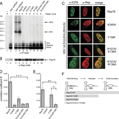
FIG 2In vivoanalysis of AAV DNA-dependent nuclear colocalization of Rep78 mutants and ICP8. (A) Analysis of AAV DNA replication by Rep78 and mutants thereof. HeLa cells were transfected with AAV-2 constructs expressing Rep78 or mutants thereof, as described in the text (data not shown for RepY156F). Sixteen hours later, cells were infected with helper virus (Ad-2) and were harvested 24 h postinfection. Low-molecular-weight DNA was prepared by Hirt extraction and digested with DpnI. Equal amounts of DNA were separated on agarose gels, and Southern blots were hybridized with a32P-labeled DNA probe spanning the AAV cap. Replicated AAV DNA was visualized by autoradiography. RF1 and RF2, monomeric and dimeric replicative forms. Similar results were obtained with helper HSV (data not shown). (B) Rep expression was analyzed by Western blot analysis of cell extracts processed in parallel to the Southern blot in panel A. Rep expression was detected by the anti-Rep monoclonal antibody 303.9. (C) AAV DNA-dependent nuclear colocalization of Rep78 and ICP8. BHK cells grown on coverslips were cotransfected with AAV plasmids expressing Flag-tagged Rep78 wild-type protein or mutants, together with plasmids for the minimal set of HSV helper genes, the primase/helicase complex UL5/UL8/UL52 and the ssDNA binding protein ICP8 (UL29). Cells were fixed and permeabilized by formaldehyde-Triton treatment 40 h later and stained with antibodies against HSV ICP8 and the Flag tag of AAV Rep, followed by fluorophore-labeled secondary antibodies, as described previously (27). Cells were analyzed by confocal microscopy with a Zeiss LSM 510 microscope. The images represent cross sections of 0.8m. Anti-ICP8 reactivity is displayed in green, while anti-Flag (Rep) reactivity yields red. Merged foci (yellow) indicate colocalization. (D) Quantification of AAV DNA-dependent nuclear colocalization of Rep78 and ICP8 displayed in panel C. To determine the extent of colocalization, the correlation coefficient (R) of green (ICP8) and red (Rep) fluorescence above a fixed background value was determined with the help of the Zeiss LSM 510 software for a total of 15 cells for each individual construct, and values are means⫾standard errors of the means (*,P⬍0.05; **,P⬍0.01; ***,P⬍0.001). (E) Quantification of colocalization of Rep78 and ICP8 in U2OS cells transduced with a monomeric rAAV subtype 2 vector at an MOI of 1⫻104genomic particles/cell as a source of AAV ssDNA. Cells had been transfected with plasmids expressing Rep and the minimal HSV helper genes. Colocalization was quantified in six individual cells per construct, as described for panel D. (F) The initial steps of Rep-dependent initiation of AAV DNA replication at the AAV ITR are displayed in consecutive order: DNA binding of Rep to the RBS followed by Rep-dependent helicase activity, leading to ssDNA strands, followed by the Rep-dependent endonuclease step. The degree to which Rep78wt and the mutants support these activities are indicated by arrows.
AAV Rep Domains Interacting with HSV ICP8 on the AAV-ITR
on November 7, 2019 by guest
http://jvi.asm.org/
[image:3.585.98.485.66.454.2]formation (Fig. 1C). To further narrow down the presumed site of
interaction, the experiment was repeated with the isolated AAV
ITR (nucleotides [nt] 1 to 181) in a double-stranded
tion, or a hairpin-structured, partially single-stranded
conforma-tion (Fig. 1E). Similar to the entire AAV-2 genome, Rep78wt,
RepK340H, and RepY156F retained the capacity for
ssDNA-dependent complex formation with ICP8, whereas the RBS
binding-deficient mutant RepR107A/R138A lost this activity.
To-gether, these data show that the ability of Rep78 to directly contact
the AAV ITR is required for ternary-complex formation with
ICP8.
To analyze whether
in vitro
ternary-complex formation is
re-flected
in vivo
by the ability of AAV-Rep and HSV-ICP8 to
colo-calize in nuclear replication domains, their distribution was
ana-lyzed by confocal microscopy. We had previously demonstrated
colocalization of Rep and ICP8 upon coinfection of wild-type
AAV and HSV (10) and upon cotransfection of plasmids coding
for wild-type AAV-2 and for the minimal set of HSV helper
pro-teins, consisting of ICP8 and the helicase-primase complex UL5/
UL8/UL52 (27). Full-length AAV-2 plasmids were generated
that expressed Flag-tagged versions of Rep78wt, RepK340H,
RepY156F, or N-terminal amino acid exchange mutants. In the
presence of helper virus, the plasmids mediated comparable Rep
expression but, with the exception of Rep78wt, had lost DNA
replication properties (Fig. 2A and B). Subcellular localization of
Rep and ICP8 was quantified by confocal microscopy 40 h after
cotransfection of expression plasmids for Rep and the four HSV
helper functions as described before (27). When transfected alone,
Rep and all mutants thereof displayed a homogenous nuclear
dis-tribution pattern (data not shown). In the presence of HSV
repli-cation proteins, Rep78wt followed the punctate distribution
pat-tern of HSV replication foci and colocalized to ICP8 (Fig. 2C and
D), as described before (27). In contrast, the DNA
binding-deficient mutants RepR107A/K136A and RepR107A/R138A
hardly ever colocalized to ICP8 above threshold levels (Fig. 2C and
D). The helicase-deficient mutant RepK340H, despite its ability to
form the ternary complex
in vitro
, never colocalized to ICP8,
whereas the helicase-proficient mutant RepY156F occasionally
colocalized to ICP8 in HSV replication foci (Fig. 2C and D).
To test
in vivo
colocalization on authentic AAV ssDNA, U2OS
cells were infected with recombinant AAV (rAAV) vectors at a
multiplicity of infection (MOI) of 1
⫻
10
4genomic particles/cell
after cotransfection with plasmids for Rep and the four HSV
helper genes. The data displayed in Fig. 2E confirmed the drop in
colocalization to ICP8 of the N-terminal mutant RepR107A/
R136A compared to that of Rep78wt (
P
⬍
0.01). In contrast,
RepY156F showed a high and significant degree of colocalization
to ICP8 (
P
⬍
0.05) just slightly below that of Rep78wt, whereas
RepK340H never colocalized with either plasmid- or
virus-derived AAV template DNA (Fig. 2D and E). Although both
RepK340H and RepY156F can bind to the AAV ITR, only
RepY156F unwinds it to expose ssDNA (5), but it cannot proceed
with DNA replication due to its endonuclease defect (Fig. 2F).
Obviously, the unwound AAV ITR exposes sufficient ssDNA for
ICP8 binding. RepK340H, which is entirely defective in ITR
un-winding only
in vitro
, binds to ssDNA templates (Fig. 1) but is
unable to generate ssDNA for
in vivo
ternary-complex formation.
In summary, colocalization of Rep and ICP8 in nuclear
repli-cation domains depends on the ability of Rep to bind and unwind
the AAV ITR, generating ssDNA regions. The
in vitro
data show
that neither helicase nor endonuclease activities of Rep are needed
as such for ternary-complex formation as long as AAV ssDNA is
present. In addition, the reduced
in vivo
colocalization of ICP8
and certain Rep78 mutants likely reflects their inability to support
AAV DNA replication to generate and amplify sufficient AAV
ssDNA templates (Fig. 2F).
HSV ICP8 is characterized by highly cooperative and DNA
sequence-independent ssDNA-binding (21) with an apparent
binding constant (
K
␣) for monomeric ICP8 on ssDNA in the
range of 1
⫻
10
7M
⫺1(1, 7, 20). Electron microscopy confirmed
the highly cooperative but unspecific nature of ICP8 binding to
ssDNA (18, 25) whereas Rep molecules on the AAV ssDNA
ge-nome bind exclusively to the hairpin-shaped ITR that contains the
RBS (14, 15, 34, 37). For Rep78/68 DNA-binding affinities to the
isolated RBS of up to 8
⫻
10
10M
⫺1were calculated (3, 8, 22), while
Rep68 also binds to unrelated ssDNA (16), though with a lower
binding affinity, around 2
⫻
10
8M
⫺1(19, 36). Obviously, the
affinity of Rep for the AAV ITR is roughly 2 orders of magnitude
higher than its affinity for ssDNA. These data, taken together with
the above findings, suggest that Rep78/68 serves as the driving
force for ternary-complex formation by binding to the RBS within
the ITR. Due to the high degree of cooperativity, ICP8 quickly
covers available ssDNA regions. We cannot say at present whether
the two processes are coupled or take place independently. Rep
domains interacting with heterologous proteins have been
exclu-sively mapped to the C terminus (6, 8, 26, 29–31), and the ICP8
domain for interactions with heterologous proteins was described
as separate from that engaged in cooperative ssDNA binding (21).
In an alternative scenario, formation of the ternary complex could
therefore be initiated by protein-protein interaction of ICP8 and
Rep with subsequent binding to the AAV genome. Our data
ex-tend previous evidence that Rep78 serves as origin-binding
pro-tein on the AAV ITR (27). In analogy to the HSV
ori
-binding
protein (UL9), AAV Rep78 binds and unwinds the AAV ITR and
recruits ICP8, the HSV helicase-primase complex, and additional
replication factors to initiate AAV DNA replication.
ACKNOWLEDGMENTS
We thank E. Hammer for experienced technical help, J. Richter for expert confocal microscopy service, and C. Stutika for critical reading of the manuscript.
The initial phase of the study was supported by grants from the Deut-sche Forschungsgemeinschaft, DFG-SFB506.
REFERENCES
1.Boehmer PE, Craigie MC, Stow ND, Lehman IR.1994. Association of origin binding protein and single strand DNA-binding protein, ICP8, during herpes simplex virus type 1 DNA replication in vivo. J. Biol. Chem.
269:29329 –29334.
2.Chejanovsky N, Carter BJ.1990. Mutation of a consensus purine nucle-otide binding site in the adeno-associated virusrepgene generates a dom-inant negative phenotype for DNA replication. J. Virol.64:1764 –1770. 3.Chiorini JA, et al.1994. Sequence requirements for stable binding and
function of Rep68 on the adeno-associated virus type 2 inverted terminal repeats. J. Virol.68:7448 –7457.
4.Davis MD, Wonderling RS, Walker SL, Owens RA.1999. Analysis of the effects of charge cluster mutations in adeno-associated virus Rep68 pro-tein in vitro. J. Virol.73:2084 –2093.
5.Davis MD, Wu J, Owens RA. 2000. Mutational analysis of adeno-associated virus type 2 Rep68 protein endonuclease activity on partially single-stranded substrates. J. Virol.74:2936 –2942.
6.Di Pasquale G, Stacey SN.1998. Adeno-associated virus Rep78 protein interacts with protein kinase A and its homolog PRKX and inhibits CREB-dependent transcriptional activation. J. Virol.72:7916 –7925.
Alex et al.
on November 7, 2019 by guest
http://jvi.asm.org/
7.Dudas KC, Ruyechan WT.1998. Identification of a region of the herpes simplex virus single-stranded DNA-binding protein involved in coopera-tive binding. J. Virol.72:257–265.
8.Han SI, et al.2004. Rep68 protein of adeno-associated virus type 2 inter-acts with 14-3-3 proteins depending on phosphorylation at serine 535. Virology320:144 –155.
9.Heilbronn R, Bürkle A, Stephan S, zur Hausen H.1990. The adeno-associated virusrepgene suppresses herpes simplex virus-induced DNA-amplification. J. Virol.64:3012–3018.
10. Heilbronn R, et al.2003. ssDNA-dependent colocalization of adeno-associated virus Rep and herpes simplex virus ICP8 in nuclear replication domains. Nucleic Acids Res.31:6206 – 6213.
11. Hickman AB, Ronning DR, Kotin RM, Dyda F.2002. Structural unity among viral origin binding proteins: crystal structure of the nuclease do-main of adeno-associated virus Rep. Mol. Cell10:327–337.
12. Hickman AB, Ronning DR, Perez ZN, Kotin RM, Dyda F.2004. The nuclease domain of adeno-associated virus rep coordinates replication initiation using two distinct DNA recognition interfaces. Mol. Cell13: 403– 414.
13. Hörer M, et al.1995. Mutational analysis of adeno-associated virus Rep. protein-mediated inhibition of heterologous and homologous promoters. J. Virol.69:5485–5496.
14. Im D-S, Muzyczka N.1990. The AAV origin-binding protein Rep68 is an ATP-dependent site-specific endonuclease with helicase activity. Cell61: 447– 457.
15. Im D-S, Muzyczka N.1989. Factors that bind to adeno-associated virus terminal repeats. J. Virol.63:3095–3104.
16. Im D-S, Muzyczka N.1992. Partial purification of adeno-associated virus Rep78, Rep52, and Rep40 and their biochemical characterization. J. Virol.
66:1119 –1128.
17. Kleinschmidt JA, Möhler M, Weindler F, Heilbronn R.1995. Sequence elements of the adeno-associated virusrep-gene required for suppression of herpes-simplex virus induced DNA amplification. Virology206:254 – 262.
18. Makhov AM, Boehmer PE, Lehman IR, Griffith JD.1996. Visualization of the unwinding of long DNA chains by the herpes simplex virus type 1 UL9 protein and ICP8. J. Mol. Biol.258:789 –799.
19. Mansilla-Soto J, et al.2009. DNA structure modulates the oligomeriza-tion properties of the AAV initiator protein Rep68. PLoS Pathog.
5:e1000513.
20. Mapelli M, Mühleisen M, Persico G, van der Zandt H, Tucker PA.2000. The 60-residue C-terminal region of the single-stranded DNA binding protein of herpes simplex virus type 1 is required for cooperative DNA binding. J. Virol.74:8812– 8822.
21. Mapelli M, Panjikar S, Tucker PA.2005. The crystal structure of the herpes simplex virus 1 ssDNA-binding protein suggests the structural ba-sis for flexible, cooperative single-stranded DNA binding. J. Biol. Chem.
280:2990 –2997.
22. McCarty DM, Ryan JH, Zolotukhin S, Zhou X, Muzyczka N.1994. Interaction of the adeno-associated virus Rep protein with a sequence
within the A palindrome of the viral terminal repeat. J. Virol.68:4998 – 5006.
23.Owens RA, Trempe JP, Chejanovsky N, Carter BJ. 1991. Adeno-associated virus rep proteins produced in insect and mammalian expres-sion systems: wild-type and dominant-negative mutant proteins bind to the viral replication origin. Virology184:14 –22.
24. Owens RA, Weitzman MD, Kyöstiö SRM, Carter BJ.1993. Identifica-tion of a DNA-binding domain in the amino terminus of adeno-associated virus Rep proteins. J. Virol.67:997–1005.
25. Ruyechan WT, Weir AC.1984. Interaction with nucleic acids and stim-ulation of the viral DNA polymerase by the herpes simplex virus type 1 major DNA-binding protein. J. Virol.52:727–733.
26. Schmidt M, Chiorini JA, Afione S, Kotin R.2002. Adeno-associated virus type 2 Rep78 inhibition of PKA and PRKX: fine mapping and anal-ysis of mechanism. J. Virol.76:1033–1042.
27. Slanina H, Weger S, Stow ND, Kuhrs A, Heilbronn R.2006. Role of the herpes simplex virus helicase-primase complex during adeno-associated virus DNA replication. J. Virol.80:5241–5250.
28. Snyder RO, et al.1993. Features of the adeno-associated virus origin involved in substrate recognition by the viral Rep protein. J. Virol.67: 6096 – 6104.
29. Weger S, Hammer E, Heilbronn R.2004. SUMO-1 modification regu-lates the protein stability of the large regulatory protein Rep78 of adeno associated virus type 2 (AAV-2). Virology330:284 –294.
30. Weger S, Hammer E, Heilbronn R.2002. Topors, a p53 and topoisom-erase I binding protein, interacts with the adeno-associated virus (AAV-2) Rep78/68 proteins and enhances AAV-2 gene expression. J. Gen. Virol.
83:511–516.
31. Weger S, Wendland M, Kleinschmidt J, Heilbronn R.1999. The adeno-associated virus type 2 regulatory proteins Rep78/Rep68 interact with the transcriptional coactivator PC4. J. Virol.73:260 –269.
32. Weindler FW, Heilbronn R.1991. A subset of herpes simplex virus replication genes provides helper functions for productive adeno-associated virus replication. J. Virol.65:2476 –2483.
33. Weitzman MD, Kyöstiö SRM, Carter BJ, Owens RA.1996. Interaction of wild-type and mutant adeno-associated virus (AAV) Rep proteins on AAV hairpin DNA. J. Virol.70:2440 –2448.
34. Weitzman MD, Kyöstiö SRM, Kotin RM, Owens RA.1994. Adeno-associated virus (AAV) Rep. proteins mediate complex formation be-tween AAV DNA and its integration site in human DNA. Proc. Natl. Acad. Sci. U. S. A.91:5808 –5812.
35.Wonderling RS, Kyostio SR, Owens RA. 1995. A maltose-binding protein/adeno-associated virus Rep68 fusion protein has DNA-RNA he-licase and ATPase activities. J. Virol.69:3542–3548.
36. Yoon-Robarts M, et al.2004. Residues within the B=motif are critical for DNA binding by the superfamily 3 helicase Rep40 of adeno-associated virus type 2. J. Biol. Chem.279:50472–50481.
37. Young SM, Jr., McCarty DM, Degtyareva N, Samulski RJ.2000. Roles of adeno-associated virus Rep. protein and human chromosome 19 in site-specific recombination. J. Virol.74:3953–3966.
AAV Rep Domains Interacting with HSV ICP8 on the AAV-ITR
on November 7, 2019 by guest
http://jvi.asm.org/