0022-538X/08/$08.00
⫹
0
doi:10.1128/JVI.01192-08
Copyright © 2008, American Society for Microbiology. All Rights Reserved.
H5N1 Avian Influenza Virus Induces Apoptotic Cell Death in
Mammalian Airway Epithelial Cells
䌤
†
Tomo Daidoji,
1Takaaki Koma,
1,2‡ Anariwa Du,
1,2Cheng-Song Yang,
1,2Mayo Ueda,
1,2Kazuyoshi Ikuta,
2and Takaaki Nakaya
1*
International Research Center for Infectious Diseases
1and Department of Virology, Research Institute for
Microbial Diseases,
2Osaka University, Suita, Osaka, Japan
Received 8 June 2008/Accepted 27 August 2008
In recent years, the highly pathogenic avian influenza virus H5N1 has raised serious worldwide concern
about an influenza pandemic; however, the biology of H5N1 pathogenesis is largely unknown. To elucidate the
mechanism of H5N1 pathogenesis, we prepared primary airway epithelial cells from alveolar tissues from
1-year-old pigs and measured the growth kinetics of three avian H5 influenza viruses (A/Crow/Kyoto/53/2004
[H5N1], A/Duck/Hong Kong/342/78 [H5N2], and A/Duck/Hong Kong/820/80 [H5N3]), the resultant
cytopath-icity, and possible associated mechanisms. H5N1, but not the other H5 viruses, strongly induced cell death in
porcine alveolar epithelial cells (pAEpC), although all three viruses induced similar degrees of cytopathicity
in chicken embryonic fibroblasts. Intracellular viral growth and the production of progeny viruses were
comparable in pAEpC infected with each H5 virus. In contrast, terminal deoxynucleotidyltransferase-mediated
dUTP-biotin nick end labeling-positive cells were detected only in H5N1-infected pAEpC, and the activities of
caspases 3, 8, and 9 were significantly elevated in pAEpC infected with H5N1, but not with H5N2 and H5N3.
These results suggest that only H5N1 induces apoptosis in pAEpC. H5N1 cytopathicity was inhibited by adding
the caspase inhibitor z-VAD-FMK; however, there were no significant differences in viral growth or release of
progeny viruses. Further investigations using reverse genetics demonstrated that H5N1 hemagglutinin protein
plays a critical role in inducing caspase-dependent apoptosis in infected pAEpC. H5N1-specific cytopathicity
was also observed in human primary airway epithelial cells. Taken together, these data suggest that avian
H5N1 influenza virus leads to substantial cell death in mammalian airway epithelial cells due to the induction
of apoptosis.
The first outbreak in humans caused by the highly
patho-genic avian influenza virus H5N1 occurred in Hong Kong in
1997. In this outbreak, H5N1 caused respiratory disease in 18
people, 6 of whom died (8). To date, more than 380 human
H5N1 infections have been identified, more than 240 of which
have been fatal (60), raising serious worldwide concern about
a severe influenza pandemic. The high mortality among
H5N1-infected patients results from acute respiratory distress
syn-drome (ARDS) linked with diffuse alveolar damage (DAD)
from desquamation of alveolar cells and hemorrhage (10, 11,
32, 50, 54, 55). Though broad-view analyses of pathogenesis
have been conducted, such as epidemiological studies and
his-topathological analyses of human lungs after infection, little
attention has been paid to the biological basis of H5N1
patho-genesis in humans.
Recently, Shinya et al. (42) reported that the surface
glyco-protein hemagglutinin (HA) of avian influenza viruses
prefer-entially recognizes receptors terminating in sialic acid
␣
-2,3-galactose (SA
␣
2,3Gal) in the respiratory bronchioles and
alveoli. The specificity of the HA receptor is thought to be one
factor associated with the pathogenesis of H5N1 in humans;
however, the pathogenesis after the virus has entered the cells
is still unclear. Although avian influenza viruses are all
poten-tially virulent in poultry (1, 19, 23, 35, 41, 43, 46, 47), currently
circulating H5N1 is highly virulent in humans, as well as in
birds (5, 59, 63), suggesting that it can induce cell damage in
mammalian airway organs. To elucidate why H5N1 influenza
virus infection leads to DAD in human lungs, it is essential to
clarify the difference(s) between recently emerged H5N1
vi-ruses and previously circulating avian influenza vivi-ruses with
respect to viral replication and processes leading to cell death
at the molecular level.
So far, several susceptible cell lines, including MDCK and
A549, have been used to evaluate the pathogenesis of influenza
virus. These cell lines are useful models for tracing viral entry,
replication, and production of progeny viruses; however, they
are less suitable for investigating the mechanisms of cell death
associated with viral replication, because these cancer-derived
cells have been immortalized. For many years, pigs, which
possess both the avian influenza SA
␣
2,3Gal receptor and the
human influenza SA
␣
2,6Gal receptor (20), have been known
to be mixing vessels because of their susceptibility to both avian
and human influenza viruses (6, 57, 58). Thus, primary porcine
cells, such as airway epithelial cells, are potentially good
mod-els for investigating cell death caused by the replication of
human and avian influenza viruses. To clarify the mechanisms
* Corresponding author. Mailing address: International Research
Center for Infectious Diseases, Research Institute for Microbial
Dis-eases, Osaka University, 3-1, Yamadaoka, Suita, Osaka, 565-0871,
Japan. Phone: 81-6-6879-4251. Fax: 81-6-6879-4252. E-mail: tnakaya
@biken.osaka-u.ac.jp.
† Supplemental material for this article may be found at http://jvi
.asm.org/.
‡ Present address: Institute for Animal Experimentation, Hokkaido
University Graduate School of Medicine, Sapporo, Japan.
䌤
Published ahead of print on 11 September 2008.
11294
on November 8, 2019 by guest
http://jvi.asm.org/
of H5N1 pathogenesis in the human lung, we focused on the
postentry steps and tried to find a difference(s) between the
highly pathogenic (high-mortality) avian influenza virus H5N1
and two low-pathogenic (low- or no-mortality) avian influenza
viruses, H5N2 and H5N3, in humans. We used porcine alveolar
epithelial cells (pAEpC) as a human lung model and
investi-gated the relationship between viral replication kinetics and
cytopathicity. Finally, we tried to assess how the H5N1 avian
influenza virus leads to cytopathicity in porcine and human
alveolar epithelial cells.
MATERIALS AND METHODS
Reagents.The components used in airway epithelial cell basic medium (AEC basic) and proliferation medium (AEC plus) were based on the methods of You et al. (62) and Rowe et al. (36) and are described in Table S1 in the supplemental material.
Viruses and cells.Influenza virus strain A/crow/Kyoto/53/2004 (H5N1) was isolated from embryonated eggs inoculated with tracheal homogenates from dead crows; A/Duck/Hong Kong/342/78 (H5N2) and A/Duck/Hong Kong/820/80 (H5N3) were kindly provided by Yoshinobu Okuno, Osaka Prefectural Institute of Public Health. Chicken embryonic fibroblasts (CEF) were prepared from 10-day-old embryonated eggs. MDCK cells were purchased from the Riken BioResource Center Cell Bank (Ibaragi, Japan). Human lung epithelial carci-noma A549 cells were kindly provided by the Cell Resource Center for Biomed-ical Research (Tohoku University, Sendai, Japan), and human primary small airway epithelial cells (SAEC) were purchased from Lonza Corporation (Walk-ersville, MD).
Generation of recombinant H5N3 viruses. Viral RNA was isolated using Trizol reagent (Invitrogen), and cDNA was synthesized using random hexamers. The full-length HA sequence of A/crow/Kyoto/53/2004 (H5N1) was constructed by PCR. The HA sequence of A/Thailand/Kan353/2004 (H5N1) was constructed by PCR using overlapping deoxyoligonucleotides corresponding to the published sequence of the HA open reading frame of the virus. The virulent HA sequence of A/Duck/Hong Kong/820/80 (H5N3) was constructed by changing single basic
amino acids to multiple basic amino acids (N⬘-TR-C⬘to N⬘-RRKKR-C⬘) at the
HA cleavage site. The noncoding regions were identical to those of influenza A/WSN/33 (H1N1) virus. The HA genes were cloned into the pPOLI plasmid (15).
Recombinant viruses were generated by using a previously described reverse-genetics system (3, 15, 52) with slight modifications. Briefly, the pPOLI-HA plasmid derived from A/crow/Kyoto/53/2004 (H5N1), A/Thailand/Kan353/2004 (H5N1), or virulent HA of A/Duck/Hong Kong/820/80 (H5N3) was transfected together with pCAGGS expression plasmids (3) for A/crow/Kyoto/53/2004 (H5N1) PA, PB1, PB2, and NP into 293T cells that had been cocultured with CEF (7:3). At 24 h posttransfection, A/Duck/Hong Kong/820/80 (H5N3) was infected at a multiplicity of infection (MOI) of 1 as a helper virus. At 72 h postinfection (p.i.), recombinant viruses in the supernatant were plaque purified in MDCK cells in the absence of trypsin. The HA gene of each recombinant virus was confirmed by sequencing.
pAEpC isolation and culture conditions.Isolation and culture of primary pAEpC were performed according to the procedures of You et al. (62), Rowe et al. (36), and Steimer et al. (45) with slight modifications. Briefly, lungs from 1-year-old pigs were obtained from a local abattoir. The fresh organs were transferred into sterile phosphate-buffered saline (PBS) and kept on ice during transport to the laboratory. The time between collecting the tissue and starting the isolation procedure was between 1 and 2 h. Tissue pieces were mechanically excised from diaphragmal and cranial regions of the pulmonary lobe, followed by washes and the removal of visible bronchioles. Tissue cubes of approximately 3-cm edge length were injected with 0.15% (wt/vol) pronase (Calbiochem, San Diego, CA) in F-12 medium (Gibco Co., Carlsbad, CA) at a rate of 2 ml/tissue cube, followed by incubation in polyethylene tubes at 37°C for 1 h to accelerate enzymatic digestion. Subsequent to enzymatic treatment, the reaction samples were mixed with PBS (10 ml/tube), and the resulting tissue was minced with scissors in each tube. The triturated tissues were filtered through stainless steel mesh with 1.5-mm pores, followed by filtration through nylon gauze with 0.5-mm
pores and 70-m cell strainers (BD Biosciences). Airway epithelial cells collected
by enzymatic treatment were pooled and isolated by centrifugation at 880⫻gfor
10 min at 4°C. The cells were resuspended in Ham’s F-12 pen-strep containing crude pancreatic DNase I (0.5 mg/ml; Roche Molecular Biochemicals, Indianap-olis, IN) and bovine serum albumin (10 mg/ml; Sigma-Aldrich, St. Louis, MO).
The cells were then incubated on ice for 5 min, centrifuged at 880⫻gfor 5 min
at 4°C, and resuspended in AEC basic medium (see Table S1 in the supplemental material) with 5% fetal calf serum. The cells were incubated in tissue culture plates (Asahi Techno Glass Co., Ltd., Chiba, Japan) for 2 h at 37°C and aerated
with 5% CO2and 95% air to adhere the fibroblasts (36, 62). Nonadherent cells
were collected by centrifugation, resuspended in AEC plus medium (see Table
S1 in the supplemental material), and counted. An average of 9.66 (⫾0.35) g
starting tissue mass yielded 2.4⫻106(⫾0.36⫻106) cells/g. Variances in this
yield may reflect differences in the age, gender, or state of health of the respec-tive donor animals, but this was not investigated further. Cell viability
deter-mined by trypan blue exclusion was⬎95%. The resuspended cells were cultured
on tissue culture plates (Asahi Techno Glass) coated with permeable fibronectin
(0.06 mg/cm2; BD Biosciences, Bedford, MA) and collagen (0.2 mg/cm2; Nitta
Gelatin Inc., Osaka, Japan). The cells were plated at a density of 105
cells/cm2
and grown with AEC plus medium supplemented withD-valine to hinder the
growth of fibroblasts (26, 44). The cells were cultivated at 37°C in an atmosphere
of 5% CO2and 95% air, and the cell culture fluid was replaced at least every
second day. Subsequent isolation batches are referred to as “pAEpC-n” withn
indicating the batch number. Staining pAEpC with a mouse monoclonal
anti-pancytokeratin antibody (Sigma-Aldrich) revealed that⬎99% of the cells
ex-pressed cytokeratin (see Fig. S1 in the supplemental material).
Virus infection.Cells on culture plates were washed twice with PBS and then infected with virus at an MOI of 1 or 10 and incubated at 37°C for 1 h. After the removal of virus and one wash, the cells were cultured with minimum essential medium (Sigma-Aldrich) containing 10% fetal calf serum for CEF and A549 cells and were cultured with AEC plus for pAEpC and human SAEC at 37°C in
5% CO2and 95% air. Aliquots of the supernatants were collected at 8, 16, and
24 h p.i. and titrated on MDCK cells by 50% tissue culture infective dose
(TCID50) or antigen-capture enzyme-linked immunosorbent assay (ELISA) as
described below. Cell lysates prepared from infected cells after the removal of media were used for detection of viral proteins by Western blotting (53).
TCID50assays.Aliquots of supernatants were 10-fold serially diluted with
PBS, applied in quadruplicate to 2.5⫻104MDCK cells/well of a 96-well plate,
and incubated at 37°C for 1 h. The inoculum was removed, and the cells were washed with PBS and supplied with Dulbecco’s modified Eagle’s medium/F-12
containing 0.2% bovine serum albumin and trypsin (5g/ml). On the fourth day
after infection, the TCID50was determined on the basis of the Reed- Muench
method (34).
Antigen-capture ELISA.A 96-well plate (Asahi Techno Glass) was coated with a 1:1,000 dilution of a rabbit polyclonal antibody obtained from a rabbit immu-nized with A/Duck Hong Kong/342/78 (H5N2); this antibody reacts with com-mon sequences of avian influenza virus nucleoprotein (NP) and matrix protein 1 (M1). After the plate was incubated overnight at 4°C, PBS containing 5% nonfat milk was added to the wells to block nonspecific signals, and the plate was incubated for 1 h at 37°C. An authentic standard sample of A/Duck/Hong Kong/820/80 (H5N3) and test samples were inactivated with 0.04% paraformal-dehyde, diluted to 0.01% paraformaldehyde in PBS, and added to the wells, and the plate was incubated for 1 h at 37°C. After five washes with PBS-T (PBS containing 0.05% Tween 20), a mouse monoclonal antibody (3C11) diluted 1:2,000 in PBS-T containing 5% nonfat milk was reacted with the immobilized viral antigen in each well. The 3C11 antibody was produced by a hybridoma derived from mice immunized with A/crow/Kyoto/53/2004 (H5N1) and was able to react with the HA sequence common to H5 avian influenza viruses. The plate was incubated for 1 h at 37°C and washed four times with PBS-T. A horseradish peroxidase-conjugated donkey anti-mouse immunoglobulin G (IgG) secondary antibody (Jackson ImmunoResearch Laboratories, West Grove, PA) diluted 1:2,000 in PBS containing 5% nonfat milk was added to the wells, followed by incubation at 37°C for 1 h. The number of virus particles was determined at 492
nm by a colorimetric method usingo-phenylenediamine dihydrochloride as a
chromogenic substrate.
Immunofluorescence assay for viral-antigen detection.An
immunofluores-cence assay was conducted on 2.5⫻104cells/well of a 96-well plate (Asahi
Techno Glass). At 24 h after infection, the cells were fixed with paraformalde-hyde in PBS containing 0.1% Triton X-100 and washed with PBS three times. A monoclonal antibody (3C11) was used as a primary antibody to detect viral antigen (H5N1, H5N2, or H5N3). Antibody binding to viral proteins was de-tected with an Alexa Fluor 488-conjugated secondary antibody (Molecular Probes, Carlsbad, CA) diluted 1:500 in PBS containing 1% bovine serum albu-min. Cell nuclei were counterstained with Hoechst 33342 (Sigma-Aldrich).
Cell proliferation assays.To quantify cell proliferation after infection of CEF or pAEpC with H5N1, H5N2, or H5N3, we used a mitochondrial tetrazolium dye
reduction assay. Following viral infection of 2.5⫻104
cells/well of a 96-well plate (Asahi Techno Glass) at an MOI of 1 or 10, CEF and pAEpC were cultured with
V
OL. 82, 2008
H5N1 INDUCES APOPTOTIC CELL DEATH
11295
on November 8, 2019 by guest
http://jvi.asm.org/
minimum essential medium containing 10% fetal calf serum and AEC plus,
respectively. At regular intervals after infection (8, 16, 24, and 32 h), 10l of
MTT reagent (Promega Co., Madison, WI) was added per 100-l culture
vol-ume, and the cells were cultured for 1 h (CEF) or 2 h (pAEpC). After incubation
for 1 or 2 h, 25l of 2% sodium dodecyl sulfate (SDS) was added to each well,
followed by incubation for 20 min at room temperature. The absorbance at 492
nm (A492) of each sample well was measured with an automated plate reader
(Multiskan MS-UV; Labsystems, Helsinki, Finland). The results were calculated as the absorbance ratio of infected and uninfected cells and were analyzed
statistically with Student’sttest, to compare differences between proliferation
after H5N1 infection and after H5N2 or H5N3 infection, with statistical
signif-icance considered to be aPvalue of⬍0.05.
Western blotting.After the removal of supernatant at each sampling time (8, 16, and 24 h p.i.), the cells were washed three times with ice-cold PBS and harvested with SDS lysis buffer (PBS containing 2% SDS). Lysate containing 10
g of protein per lane (bicinchoninic acid protein quantification; Pierce
Biotech-nology, Rockford, IL) was loaded on a 10% SDS-polyacrylamide gel. After electrophoresis, the proteins were blotted onto nitrocellulose, and the mem-branes were blocked with PBS-T supplemented with a final concentration of 5% nonfat milk overnight at 4°C. The rabbit polyclonal virus antibody (1:2,000
dilution) and a mouse monoclonal antibody against␣-tubulin (B-5-1-2;
Sigma-Aldrich) were used as primary antibodies. The primary antibody was reacted with the membrane in PBS-T supplemented with a final concentration of 5% nonfat milk. A horseradish peroxidase-conjugated donkey anti-rabbit IgG (Jackson Im-munoResearch Laboratories) was used as a secondary antibody for detection of virus antigen, and a donkey anti-mouse IgG was used as a secondary antibody for
detection of␣-tubulin as an internal control, with both antibodies diluted 1:1,000
in PBS-T at room temperature. Washing with PBS-T was performed between all steps. All Western blots were visualized using an enhanced chemiluminescence system (Nacalai Pharmaceutical Co., Ltd., Kyoto, Japan) and Fuji XR film. The staining intensity of each band was calculated with Image J software.
TUNEL assay.Following virus infection of 2.5⫻104cells/well of a 96-well
plate (Asahi Techno Glass) at an MOI of 1 (no caspase inhibitor) or 10 (in the presence of the caspase inhibitor z-VAD-FMK), pAEpC and human SAEC were cultured with AEC plus for 16 h. Parallel samples were cultured with the caspase
inducer staurosporine (0.2M) as a positive control or with the pancaspase
inhibitor z-VAD-FMK (200M) after H5N1 infection as a negative control.
Apoptotic cells were characterized by positive terminal deoxynucleotidyltrans-ferase-mediated dUTP-biotin nick end labeling (TUNEL) staining according to the manufacturer’s instructions (DeadEnd Fluorometric TUNEL System; Pro-mega). Briefly, 16 h after infection with H5N1, H5N2, or H5N3, cells were fixed with paraformaldehyde in PBS containing 0.1% Triton X-100 and washed with PBS three times. Then, the fixed cells were incubated in the labeling reaction mixture containing terminal deoxynucleotidyl transferase enzyme, fluorescein isothiocyanate-conjugated nucleotide, and labeling buffer. The reaction was quenched in stop buffer, and the cells were washed with PBS several times. To detect viral antigens after TUNEL staining, the cells were incubated at room temperature for 40 min with the aforementioned rabbit polyclonal antibody (1:2,000 dilution) as a primary antibody, and antibody binding to viral proteins was detected with an Alexa Fluor 555-conjugated secondary antibody (Molecular Probes) diluted 1:1,000 in PBS containing 1% bovine serum albumin. Cell nuclei were counterstained with Hoechst 33342 (Sigma-Aldrich).
Assay for caspase activity. A colorimetric assay for caspase 3 activity (CaspACE Assay System; Promega) was used according to the manufacturer’s instructions. Caspase 8 and caspase 9 activities were determined by the same
method using specific substrates (substrate for caspase 8, Ac-IETD-pNA; for
caspase 9, Ac-LEHD-pNA; purchased from Calbiochem). Following virus
infec-tion of 1⫻106cells/well of a six-well plate (Asahi Techno Glass) at an MOI of
1, CEF and A549 cells were cultured with minimum essential medium containing 10% fetal calf serum, and pAEpC and human SAEC were cultured with AEC plus for 16 h. Parallel samples were cultured with the caspase inducer
stauro-sporine (1M for CEF, 2M for A549 cells, and 0.2M for pAEpC) as a
positive control or with the pancaspase inhibitor z-VAD-FMK (200M) after
H5N1 infection as a negative control. At 16 h p.i., the infected cells were harvested by centrifugation at 4°C, washed with ice-cold PBS, and resuspended
in cell lysis buffer at a concentration of 108cells/ml. The cells were lysed by
repeated freeze-thaw cycles and incubated on ice for 15 min before centrifuga-tion to collect the supernatant fraccentrifuga-tion. Caspase activities were determined with
100g of whole-cell lysate in a 100-l volume in 96-well plates. The plates were
sealed and incubated at 37°C for 4 h, and theA405 was measured using an
automated plate reader. The results were analyzed statistically with Student’st
test to compare differences between activities in H5N1-infected cells and those in
H5N2- or H5N3-infected cells, with statistical significance considered to be aP
value of⬍0.05.
Nucleotide sequence accession numbers.The GenBank accession number of the full-length HA sequence of A/crow/Kyoto/53/2004 (H5N1) is AB189053, and that of the HA sequence of A/Thailand/Kan353/2004 (H5N1) is EF541411.
RESULTS
Cytopathicity of avian influenza virus in CEF and pAEpC.
We infected CEF and pAEpC with H5N1, H5N2, and H5N3 at
MOIs of 1 and 10 and observed the cytopathic effect (CPE) in
infected cells at 24 h p.i.. Each H5 virus produced a similar
obvious CPE in infected CEF (Fig. 1A). In contrast, in pAEpC
from three pigs, H5N1 produced a severe CPE in infected cells
but the other H5 viruses did not (Fig. 1A). In order to analyze
the CPE quantitatively, we performed MTT assays in infected
CEF and pAEpC with each H5 virus at 8, 16, 24, and 32 h p.i.;
uninfected cells were used as controls. The cytopathicity
ob-served in three H5 virus-infected CEF cultures increased over
time (Fig. 1B). In pAEpC, H5N1 was significantly more
cyto-pathic (
P
⬍
0.01 or
P
⬍
0.05) than H5N2 or H5N3 at 24 and
32 h p.i. (Fig. 1B). The absorbance ratio decreased during the
observation period for H5N1, but the cytopathicity induced by
H5N2 and H5N3 viruses was slight even at 32 h p.i. (Fig. 1B).
In order to elucidate whether the lower cytopathicity in pAEpC
infected with H5N2 and H5N3 viruses depended on the
infec-tivity of the viruses, we immunostained viral antigens on CEF
and pAEpC at 24 h p.i. The immunostaining using an anti-H5
HA monoclonal antibody revealed that all three viruses
in-fected CEF and pAEpC at similar rates, even when different
amounts were inoculated (MOIs of 1 and 10) (Fig. 2).
Growth kinetics of avian influenza virus in pAEpC.
In order
to assess the relationship between cytopathicity and viral
rep-lication in pAEpC, we focused on the growth kinetics of the H5
viruses. The culture supernatants from the infected cells at
different time points were harvested for measurement of
prog-eny virus production. The infectious virus titers (TCID
50) (Fig.
3A) and the concentrations of progeny virions (Fig. 3B)
mea-sured by quantitative antigen capture ELISA were similar
among the three H5 viruses, although H5N1 produced slightly
fewer progeny virus than the other two viruses. We then
ana-lyzed the intracellular expression level of viral proteins in
in-fected cells at 8, 16, and 24 h p.i. in Western blots using
antibodies against the viral proteins NP and M1. Although the
expression level of H5N1 protein at 8 h p.i. in pAEpC-8 was
slightly higher than those of the other two viruses, the
intra-cellular expression levels of viral proteins were comparable
among H5N1, H5N2, and H5N3 (Fig. 4). In addition, the
expression level of viral protein at an MOI of 10 was slightly
higher than that at an MOI of 1 in the early phase of infection
(8 h p.i.); however, these differences disappeared at 16 and
24 h p.i. (Fig. 4). The ratios of infectious virions to all progeny
virions were also comparable among the three H5 viruses.
These results suggest that intracellular, as well as extracellular,
viral growth rates were comparable among H5N1, H5N2, and
H5N3 viruses in pAEpC (Fig. 3 and 4) in spite of the
differ-ences in the HA cleavage sites between the H5N1 virus (N
⬘
-RRKKR-C
⬘
) and the H5N2 and H5N3 viruses (N
⬘
-TR-C
⬘
).
H5N1-induced apoptosis in pAEpC.
In order to evaluate the
mechanism of H5N1-specific cell death in pAEpC, we
exam-ined apoptosis using TUNEL staining. TUNEL-positive cells
on November 8, 2019 by guest
http://jvi.asm.org/
were abundantly detected in H5N1-infected cells (Fig. 5), but
few TUNEL-positive cells were detected in H5N2- or
H5N3-infected cells. Generally, apoptosis is mediated by the
activa-tion of a family of cysteine-containing aspartate-directed
pro-teases called caspases (12, 38, 39). TUNEL-positive cells were
not detected in the presence of the pancaspase inhibitor
z-VAD-FMK (Fig. 5), suggesting that H5N1 kills cells through
caspase-dependent apoptosis.
There are two main pathways for induction of apoptosis, the
extrinsic pathway and the intrinsic pathway (13). In the
extrin-sic pathway, apoptosis is triggered by death ligands, such as
[image:4.585.113.474.69.516.2]TRAIL, tumor necrosis factor alpha, or Fas ligand, with these
ligands binding their respective membrane receptors and
acti-vating the initiation of caspase 8, which directly activates other
caspases, including caspase 3, or activates the
mitochondrion-dependent pathway described below (2). In contrast to death
receptor-mediated apoptosis, the key event of the intrinsic
pathway is represented by mitochondrial damage, release of
cytochrome
c
from mitochondria to the cytosol, and
subse-quent formation of the so-called apoptosome, composed of
cytochrome
c
, caspase 9, and Apaf-1 (37). The apoptosome
also activates caspase 3 and results in apoptosis represented by
FIG. 1. Cell damage in CEF and pAEpC infected with avian influenza virus. CEF and three distinct pAEpC cultures isolated from three
different pigs (batch numbers 7, 8, and 9) were infected with avian influenza viruses A/crow/Kyoto/53/2004 (H5N1), A/Duck/Hong Kong/342/78
(H5N2), and A/Duck/Hong Kong/820/80 (H5N3) at an MOI of 1 or 10. (A) Morphological changes in CEF and pAEpC (pAEpC-7, -8, and -9)
at 24 h p.i. with avian influenza virus. Scale bars, 100
m. (B) Time-dependent assessment of cell damage in CEF and pAEpC after avian influenza
virus infection with an MTT assay at 8, 16, 24, and 32 h p.i. The data are presented as means
⫾
standard errors (
n
⫽
3 wells).
*
,
P
⬍
0.05, and
**
,
P
⬍
0.01 versus the same time point in H5N1.
V
OL. 82, 2008
H5N1 INDUCES APOPTOTIC CELL DEATH
11297
on November 8, 2019 by guest
http://jvi.asm.org/
typical characteristics, such as intranucleosomal DNA
frag-mentation and morphological changes. Therefore, we
per-formed quantitative analyses of caspase 3, 8, and 9 activities in
H5-infected pAEpC. The activity of caspase 3 was significantly
higher (
P
⬍
0.01) in H5N1-infected cells than in H5N2- or
H5N3-infected cells (Fig. 6A, top). In addition, caspases 8 and
9 were more highly activated (
P
⬍
0.05 for caspase 8;
P
⬍
0.01
for caspase 9) in H5N1-infected cells than in H5N2- or
H5N3-infected cells (Fig. 6A, bottom). These results indicate that
H5N1 induces both the extrinsic and intrinsic apoptotic
path-ways. In CEF, the H5N1, H5N2, and H5N3 viruses elevated
caspase 3 activity to similar levels (Fig. 6B). These caspase
activities induced by the H5 viruses were completely inhibited
by z-VAD-FMK in pAEpC and CEF (Fig. 6A and B), but
progeny viruses were produced similarly from infected cells in
the presence or absence of z-VAD-FMK in pAEpC, as well as
CEF (Fig. 6C).
A critical role for the H5N1 HA gene in apoptosis induction.
In order to evaluate whether the HA cleavage sequence is
critical for apoptosis induction, we generated a recombinant
form of H5N3 (rH5N3HAvir) possessing multiple basic amino
acids (N
⬘
-RRKKR-C
⬘
) at the HA cleavage site (Fig. 7B).
When pAEpC were infected with rH5N3HAvir at an MOI of
1, CPE and induction of caspase 3 were nearly the same as
those of pAEpC infected with wild-type H5N3 possessing a
single basic amino acid at its HA cleavage site. These results
suggest that the HA cleavage sequence is not critical for
caspase 3 activation and CPE. In contrast, when pAEpC were
infected with recombinant H5N3 (rH5N3KYHA) containing
the HA gene of H5N1 (A/crow/Kyoto/53/2004) under the same
conditions described above, significant activation of caspase 3
(
P
⬍
0.01) and severe CPE were observed in the infected cells
(Fig. 7A). Taken together, these results suggest that H5N1 HA
protein is one of the main factors involved in the induction of
caspase-dependent apoptosis, although the contributions of
other viral proteins to apoptosis will have to be evaluated. In
order to confirm that this was a feature of other H5N1 viruses
as well, we replaced the HA gene of recombinant H5N3
(rH5N3ThaiHA) with that of human-isolated H5N1
(A/Thai-land/Kan353/2004). Upon infection of pAEpC with rH5N3
ThaiHA at an MOI of 1, caspase 3 activity and CPE similar to
those induced by rH5N3KYHA were detected (Fig. 7A).
The caspase 3 activities induced by recombinant viruses
with H5N1-HA were completely inhibited by z-VAD-FMK
(Fig. 7A).
CPE and growth kinetics of avian influenza virus in human
airway epithelial cells.
Finally, we evaluated whether the
above-mentioned results obtained from pAEpC can be applied
to human airway epithelial cells. When human SAEC, which
are derived from human bronchiolar epithelium, were infected
with H5N1, H5N2, and H5N3 at MOIs of 1 and 10, only
H5N1-infected cells showed an obvious CPE (Fig. 8A).
Cor-responding to the results obtained from pAEpC, H5N1, H5N2,
and H5N3 viruses replicated similarly in human SAEC at 24 h
p.i. (Fig. 8B), and the levels of production of progeny viruses
from infected cells were also comparable among the three H5
viruses, as measured by TCID
50(Fig. 8C, top), as well as
quantitative ELISA (Fig. 8C, bottom). Furthermore,
TUNEL-positive cells were detected only in H5N1-infected cells (Fig.
9). These results indicate that the CPE and growth kinetics of
avian H5 influenza viruses in human airway epithelial cells are
comparable to those in pAEpC.
DISCUSSION
In the present study, we obtained five main findings from our
investigation of the cytopathicity induced by the H5N1 avian
influenza virus in pAEpC and its possible mechanisms. (i) H5N1
and the previously isolated, less virulent H5N2 and H5N3 viruses
were highly and equally cytopathic in CEF; in sharp contrast, only
H5N1 was cytopathic in pAEpC. (ii) The H5N1, H5N2, and
H5N3 viruses showed similar growth kinetics and efficient
pro-duction of progeny virions in both pAEpC and avian CEF. (iii)
H5N1 specifically induced caspase-dependent apoptosis in
pAEpC. (iv) Recombinant H5N3 viruses (rH5N3KYHA and
rH5N3ThaiHA) carrying the H5N1 HA gene induced
caspase-dependent apoptosis, while rH5N3HAvir, possessing multiple
ba-sic amino acids within its HA, did not. (v) H5N1-specific
cytotox-icity and apoptosis were also observed in human airway epithelial
cells.
[image:5.585.70.254.64.398.2]In this study, highly pathogenic H5N1 and the less
patho-genic H5N2 and H5N3 were highly and equally cytopathic in
CEF. Avian influenza H5 viruses, including H5N2 and H5N3,
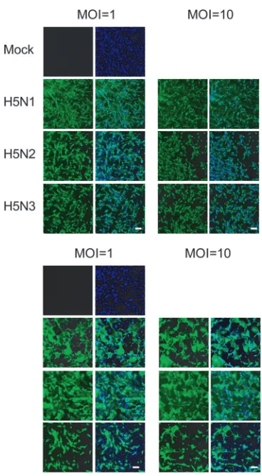
FIG. 2. Expression of virus antigen in CEF (top) and pAEpC
(bot-tom) at 24 h p.i. with avian influenza viruses. Virus antigen was
de-tected by immunostaining using a monoclonal antibody (3C11) that
recognizes H5 HA (green). Counterstaining with Hoechst 33342 was
used for detection of cellular nuclei (blue). Scale bars, 100
m.
on November 8, 2019 by guest
http://jvi.asm.org/
can cause severe outbreaks of disease in poultry (35), and the
virulence of avian influenza viruses toward poultry in vivo does
not depend on the HA0 cleavage site, although the extent of
mortality does (1, 19, 23, 35, 41, 43, 46, 47). Our present results
in primary cultured CEF might correspond to the pathogenesis
of avian influenza virus in vivo. Supporting this relationship
between cytopathicity in vitro and biological phenotype in vivo,
a 2-amino-acid change in the hepatitis B virus X protein
in-duces cytopathicity in primary hepatocytes of the tree shrew,
which is susceptible to hepatitis B virus, and this mutation is
directly linked to the pathological disease, fulminant hepatitis
(4). The present results in CEF and a report by Baumer et al.
(4) suggest that primary cultured cells can reflect pathogenesis
in vivo.
Despite the similar susceptibilities of pAEpC to H5N1,
H5N2, and H5N3 infection, only H5N1 was intensely
cyto-pathic, consistent with the fact that among avian H5 influenza
viruses, only H5N1 is virulent to humans in vivo (5, 59, 63).
Therefore, considering that results in primary cultured cell
systems can be closely correlated with pathogenesis in vivo, the
H5N1 avian virus may be as virulent as human-derived
influ-enza virus toward mammalian airway epithelial cells in vitro.
When pAEpC were independently infected with H5N1,
H5N2, and H5N3 and TUNEL staining was performed, the key
feature of apoptosis, intranucleosomal DNA fragmentation
(25), was observed only in H5N1-infected cells (Fig. 5). Thus,
the intense cytopathicity observed in pAEpC infected with
H5N1 is due to apoptosis. These results led us to examine
whether the cytopathicity in H5 virus-infected pAEpC is
quan-titatively or qualitatively regulated by a viral factor(s). The
growth kinetics of H5N1, H5N2, and H5N3 were comparable
(Fig. 2 to 4), although in one batch of pAEpC (pAEpC-8) at
8 h p.i., intracellular viral-protein synthesis was slightly higher
in H5N1-infected pAEpC than in H5N2- or H5N3-infected
pAEpC (Fig. 4). Thus, the intense cytopathicity and apoptosis
observed in H5N1-infected pAEpC could depend on one or
more H5N1-specific viral factors.
Several viral factors, including M1, NS1, and NA proteins,
have been reported to be related to apoptosis induction (24,
30, 40, 66). Recently, PB1-F2, an 87-amino-acid protein
syn-FIG. 3. Avian influenza virus production from pAEpC. pAEpC-7, -8, and -9 were infected with H5N1, H5N2, or H5N3 virus at MOIs of 1 and
10. The numbers of progeny viruses at 8, 16, and 24 h p.i. were measured as TCID
50(A) and by antigen-capture ELISA (B).
V
OL. 82, 2008
H5N1 INDUCES APOPTOTIC CELL DEATH
11299
on November 8, 2019 by guest
http://jvi.asm.org/
thesized from an alternate reading frame of the influenza A
PB1 gene, was shown to induce cell death (9). Zamarin et al.
(64) have also found that the PB1-F2 protein sensitizes cells to
apoptotic stimuli, as demonstrated by increased cleavage of
caspase 3 substrates in PB1-F2-expressing cells. In this study,
we demonstrated that rH5N3KYHA and rH5N3ThaiHA, but
not rH5N3HAvir, induced significant levels of
caspase-depen-dent apoptosis (Fig. 7), suggesting that apoptotic cell death was
caused by the H5N1 HA protein and not by differences in HA0
cleavage. Therefore, H5N1 HA could be one of the major
factors involved in the induction of apoptosis. The importance
of HA in apoptotic cell death in vitro is further supported by
recent findings showing that the HA of the 1918 pandemic
influenza virus enhances mortality in mice (21, 31, 51). Further
investigations will be required to determine the minimal HA
domain(s) required for apoptosis induction and if there is a
correlation between high pathogenicity and the ability of
vi-ruses to induce apoptosis in vivo.
Wurzer et al. recently reported that caspase 3 enhances virus
production from MDCK cells infected with avian H7N7
influ-enza virus and that the pancaspase inhibitor z-VAD-FMK
in-hibits virus production from infected cells (61). However, in
our present study, the production levels of progeny virus from
pAEpC and CEF infected with avian influenza virus were
comparable in the presence and absence of z-VAD-FMK (Fig.
6). This incongruity in the regulation of virus production by
caspase 3 could be due to differences in intracellular signal
transduction between different cell types. Apoptosis induced
by influenza viruses has been shown in a variety of cell lines
(17, 24, 30, 33, 40, 48, 64, 66); however, in the last few years, a
viral protein, NS1, has been shown to block apoptosis (14, 65).
These opposite effects make the precise mechanism of
virus-FIG. 4. Western blot analysis of the growth kinetics of viral proteins in pAEpC at 8, 16, and 24 h p.i. with avian influenza virus. pAEpC-7, -8,
and -9 were infected with H5N1, H5N2, or H5N3 virus at MOIs of 1 and 10. (A) Viral NP and M1 were detected by anti-NP and anti-M polyclonal
antibodies, respectively.
␣
-Tubulin was used as a control. (B) Relative band intensities of the viral proteins (NP and M1) in panel A. The intensities
of H5N1 viral proteins at each time point were set to 1.
on November 8, 2019 by guest
http://jvi.asm.org/
[image:7.585.138.454.67.484.2]induced apoptosis unclear. The discrepancy might be based on
the different cell lines and virus strains used in each
experi-ment. In this study, H5N1-infected MDCK and A549 cells,
derived from human lung carcinoma, showed little
cytopathic-ity at 24 h p.i., although large amounts of viral proteins were
made in the two cell lines (data not shown). In addition,
in-fection of A549 with H5N1 did not change the activity of
caspase 3, despite the large amounts of viral proteins
synthe-FIG. 5. TUNEL assay for detection of apoptosis in pAEpC at 16 h p.i. with avian influenza virus. pAEpC-7, -8, and -9 were infected with H5N1,
H5N2, or H5N3 virus at an MOI of 1 (in the absence of caspase inhibitor) or 10 (in the presence of the caspase inhibitor z-VAD-FMK). Parallel
samples were cultured with the caspase inducer staurosporine (STS) as a control or cultured with the pancaspase inhibitor z-VAD-FMK in
H5N1-infected cells. The arrows indicate apoptotic cells (green). Viral antigen was detected simultaneously with a polyclonal antibody against the
H5 virus (red). Cellular nuclei were counterstained with Hoechst 33342 (blue). Scale bars, 80
m. The results are shown as low-magnification
(bottom) and high-magnification (top) micrographs.
V
OL. 82, 2008
H5N1 INDUCES APOPTOTIC CELL DEATH
11301
on November 8, 2019 by guest
http://jvi.asm.org/
[image:8.585.75.514.72.606.2]11302
on November 8, 2019 by guest
sized (see Fig. S2 in the supplemental material). This lack of
H5N1-induced cytopathicity might suggest that immortalized
cell lines are not suitable as in vitro models for analyzing
virus-induced CPE. We therefore investigated whether
pri-mary cells derived from mammalian airway epithelium could
be a better system to evaluate the pathogenesis of influenza
virus in humans.
The use of primary human airway epithelial cells is the most
suitable for analyzing the pathogenesis of influenza virus in
humans. Although primary human cells have been used for this
purpose (7, 18, 22, 27, 28, 49, 56), infection experiments with
these cells are restricted in practice because of the limited
number of donors, the limited number of cells per person,
and/or ethical issues. As shown in this study, the similar H5
virus growth and cytopathicity in pAEpC and human SAEC
suggest that pAEpC may be a good experimental system to
model the human airway epithelium. pAEpC can be prepared
on a large scale, and large numbers of cells allow us to perform
many kinds of in vitro infection experiments, such as
evalua-tion of viral growth kinetics, virus-induced cytopathicity, and
expression profiles of host genes, including those in
apop-totic pathways. Furthermore, this system has the advantage
of being able to compare viral growth and cytopathicity in
different types of airway epithelial cells, from trachea to
alveoli, in a single animal. We did not use ciliated cells in
this study; however, the pAEpC were monolayered and were
confirmed to be epithelial cells by staining them with an
antibody against pancytokeratin, an epithelial cell marker
(see Fig. S1 in the supplemental material). Further
investi-gations using well-differentiated primary epithelial cells
could generate a lot of useful information, and comparative
experiments using ciliated and nonciliated primary cells
might be necessary in future studies.
Generally, the major cause of death in patients infected with
H5N1 has been shown to be ARDS concomitant with DAD,
including the loss of alveolar epithelial cells and widespread
FIG. 6. Caspase activities in pAEpC and CEF at 16 h p.i. with avian influenza virus. pAEpC-7, -8, and -9 and CEF were infected with H5N1,
H5N2, or H5N3 virus at an MOI of 1. Parallel samples were cultured with the caspase inducer staurosporine (STS) as a control or with the
pancaspase inhibitor z-VAD-FMK in the H5N1-infected cells. Caspase activity was determined with substrates appropriate for each caspase
(caspase 3, Ac-DEVD-
p
NA; caspase 8, Ac-IETD-
p
NA; caspase 9, Ac-LEHD-
p
NA). (A) Caspase 3, 8, and 9 activities in pAEpC at 16 h p.i. with
avian influenza virus. Each bar is an average of three pAEpC. The cell morphology of infected cells is shown. (B) Caspase 3 activity in CEF at 16 h
p.i. with avian influenza virus. The cell morphology of infected cells is shown. (C) Virus production from CEF and pAEpC in the presence (
⫹
)
or absence (
⫺
) of z-VAD-FMK. The viral titer was determined by the TCID
50(top) or by ELISA (bottom). The results (except those in panel B)
[image:10.585.115.473.70.329.2]were obtained individually from three different pAEpC and are presented as means
⫾
standard errors.
*
,
P
⬍
0.05, and
**
,
P
⬍
0.01 versus H5N1.
Scale bars, 100
m.
FIG. 7. Caspase 3 activity in pAEpC at 16 h p.i. with recombinant H5N3 viruses. pAEpC-7, -8, and -9 were infected with recombinant H5N3
viruses (rH5N3HAvir, rH5N3KYHA, and rH5N3ThaiHA) at an MOI of 1. Parallel samples were cultured with the caspase inducer staurosporine
(STS) as a control or with the pancaspase inhibitor z-VAD-FMK in the cases of rH5N3KYHA- and rH5N3ThaiHA-infected cells. Caspase 3
activity was determined by using the appropriate substrate, Ac-DEVD-pNA. (A) Caspase 3 activity in pAEpC at 16 h p.i. with recombinant H5N3
viruses. The cell morphology of the infected cells is shown. (B) Sequence identities between H5N1 HAs and H5N3 HAs. The results were obtained
individually from three different pAEpC and are presented as means plus standard errors.
**
,
P
⬍
0.01 versus H5N3HAvir. Scale bar, 100
m.
V
OL. 82, 2008
H5N1 INDUCES APOPTOTIC CELL DEATH
11303
on November 8, 2019 by guest
http://jvi.asm.org/
hemorrhage in the lung (10, 11, 32, 50, 54, 55). Uiprasertkul et
al. (54) reported that TUNEL-positive cells, together with a
loss of alveolar epithelial cells and widespread hemorrhage,
were found in autopsy lung alveolar sections from
H5N1-infected humans. This report indicates that the pathogenesis
of H5N1, including DAD, is possibly caused by apoptosis. In
addition, several reports suggested that apoptosis is a
pre-requisite for DAD followed by ARDS (16, 29). Our
estab-lished in vitro system to evaluate apoptotic cell death in
mammalian airway epithelial cells caused by H5N1 virus
could be useful for revealing the molecular mechanisms by
which the recently emerged H5N1 virus induces severe
ARDS in humans.
[image:11.585.111.471.70.569.2]For the past several years, the pathogenesis of H5N1 has
FIG. 8. Cytopathicity and virus propagation in human SAEC at 24 h p.i. with avian influenza virus. Human SAEC were infected with H5N1,
H5N2, or H5N3 at an MOI of 1 or 10. (A and B) Morphological changes (A) and virus antigen expression (B) in SAEC at 24 h p.i. Virus antigen
was detected by immunostaining, as for Fig. 2. (C) Progeny virus production from infected SAEC at 24 h p.i. The viral titer was determined by
TCID
50(top) or ELISA (bottom). Scale bars, 100
m.
on November 8, 2019 by guest
http://jvi.asm.org/
been of major concern worldwide. While knowledge of the
mechanisms governing viral entry, propagation, and budding
has advanced considerably, we still know relatively little about
other aspects of H5N1 virulence, such as cytopathicity, in part
because of a lack of suitable in vitro infection systems that
properly mimic the human airway epithelium. In this study, we
used primary airway epithelial cells prepared from porcine
lung and presented evidence that the pathogenicity of H5N1
in humans is based on virus-induced apoptosis, which could
lead to DAD. Given that H5N1-induced cytopathicity is due
to caspase-dependent apoptosis, decreasing the activity of
caspase 3, 8, or 9 by pharmaceutical, neutraceutical, or other
techniques may provide patient relief from the DAD
in-duced by H5N1. Further studies are also required to
deter-mine the host factors that interact with H5N1 HA protein in
the apoptotic pathway that elicits apoptosis in mammalian
alveolar epithelium.
ACKNOWLEDGMENTS
We thank Ritsuko Koketsu, Masanobu Yamate, Yohei Watanabe,
Takuhiro Matsumura, Yo Sugawara, Yohei Hayashi, and Yuki
Takegahara (RIMD, Osaka University); Hiroshi Yokota, Hidetomo
Iwano, and Katsuro Hagiwara (Rakuno Gakuen University); and
Masahiko Nakamura (Kyoto Gakuen University) for helpful
discus-sions. We also thank Yoshinobu Okuno, Osaka Prefectural Institute of
Public Health, for providing viruses and helpful discussions.
This work was supported in part by a Grant-in-Aid for Scientific
Research from the Ministry of Education, Science, Sports, Culture,
and Technology (MEXT) to T.N.; a Grant-in-Aid for Young Scientists
from the Japan Society for the Promotion of Science (JSPS) to T.D.;
the Sasakawa Scientific Research Grant from the Japan Science
Soci-ety to T.D.; a grant from Kato Memorial Bioscience Foundation to
T.D.; and a project for the International Research Center for
Infec-tious Diseases, RIMD, Osaka University from the MEXT to T.D. and
T.N. M.U. is a research fellow of JSPS Research Fellowships for
Young Scientists.
REFERENCES
1.Alexander, D. J.2007. An overview of the epidemiology of avian influenza.
Vaccine25:5637–5644.
2.Ashkenazi, A.2002. Targeting death and decoy receptors of the
tumour-necrosis factor superfamily. Nat. Rev. Cancer2:420–430.
3.Basler, C. F., A. H. Reid, J. K. Dybing, T. A. Janczewski, T. G. Fanning, H. Zheng, M. Salvatore, M. L. Perdue, D. E. Swayne, A. Garcia-Sastre, P. Palese, and J. K. Taubenberger. 2001. Sequence of the 1918 pandemic influenza virus nonstructural gene (NS) segment and characterization of recombinant viruses bearing the 1918 NS genes. Proc. Natl. Acad. Sci. USA
98:2746–2751.
4.Baumert, T. F., C. Yang, P. Schurmann, J. Kock, C. Ziegler, C. Grullich, M. Nassal, T. J. Liang, H. E. Blum, and F. von Weizsacker.2005. Hepatitis B virus mutations associated with fulminant hepatitis induce apoptosis in
pri-mary Tupaia hepatocytes. Hepatology41:247–256.
[image:12.585.80.504.73.386.2]5.Beigel, J. H., J. Farrar, A. M. Han, F. G. Hayden, R. Hyer, M. D. de Jong, S. Lochindarat, T. K. Nguyen, T. H. Nguyen, T. H. Tran, A. Nicoll, S. Touch,
FIG. 9. TUNEL assay for detection of apoptosis in human SAEC at 16 h p.i. with avian influenza virus. Human SAEC were infected with H5N1,
H5N2, or H5N3 at an MOI of 1 (in the absence of caspase inhibitor) or 10 (in the presence of the caspase inhibitor z-VAD-FMK). Parallel samples
were cultured with the caspase inducer staurosporine (STS) as a positive control or with the pancaspase inhibitor z-VAD-FMK in H5N1-infected
cells. The arrows indicate apoptotic cells (green). Virus antigen was detected simultaneously with a polyclonal antibody that recognizes all avian
influenza viruses (red). Cellular nuclei were counterstained with Hoechst 33342 (blue). Scale bars, 80
m. The results are shown as
low-magnification (bottom) and high-low-magnification (top) micrographs.
V
OL. 82, 2008
H5N1 INDUCES APOPTOTIC CELL DEATH
11305
on November 8, 2019 by guest
http://jvi.asm.org/
and K. Y. Yuen.2005. Avian influenza A (H5N1) infection in humans.
N. Engl. J. Med.353:1374–1385.
6.Castrucci, M. R., L. Campitelli, A. Ruggieri, G. Barigazzi, L. Sidoli, R. Daniels, J. S. Oxford, and I. Donatelli.1994. Antigenic and sequence anal-ysis of H3 influenza virus haemagglutinins from pigs in Italy. J. Gen. Virol.
75:371–379.
7.Chan, M. C., C. Y. Cheung, W. H. Chui, S. W. Tsao, J. M. Nicholls, Y. O. Chan, R. W. Chan, H. T. Long, L. L. Poon, Y. Guan, and J. S. Peiris.2005. Proinflammatory cytokine responses induced by influenza A (H5N1) viruses
in primary human alveolar and bronchial epithelial cells. Respir. Res.6:135.
8.Chan, P. K.2002. Outbreak of avian influenza A(H5N1) virus infection in
Hong Kong in 1997. Clin. Infect. Dis.34(Suppl. 2):S58–S64.
9.Chen, W., P. A. Calvo, D. Malide, J. Gibbs, U. Schubert, I. Bacik, S. Basta, R. O’Neill, J. Schickli, P. Palese, P. Henklein, J. R. Bennink, and J. W. Yewdell.2001. A novel influenza A virus mitochondrial protein that induces
cell death. Nat. Med.7:1306–1312.
10.Chokephaibulkit, K., M. Uiprasertkul, P. Puthavathana, P. Chearskul, P. Auewarakul, S. F. Dowell, and N. Vanprapar.2005. A child with avian
influenza A (H5N1) infection. Pediatr. Infect. Dis. J.24:162–166.
11.Chotpitayasunondh, T., K. Ungchusak, W. Hanshaoworakul, S. Chunsuthi-wat, P. Sawanpanyalert, R. Kijphati, S. Lochindarat, P. Srisan, P. Suwan, Y. Osotthanakorn, T. Anantasetagoon, S. Kanjanawasri, S. Tanupattarachai, J. Weerakul, R. Chaiwirattana, M. Maneerattanaporn, R. Poolsavathitikool, K. Chokephaibulkit, A. Apisarnthanarak, and S. F. Dowell.2005. Human
disease from influenza A (H5N1), Thailand, 2004. Emerg. Infect. Dis.11:
201–209.
12.Cummings, B. S., J. McHowat, and R. G. Schnellmann.2000. Phospholipase
A(2)s in cell injury and death. J. Pharmacol. Exp. Ther.294:793–799.
13.Degterev, A., M. Boyce, and J. Yuan.2003. A decade of caspases. Oncogene
22:8543–8567.
14.Ehrhardt, C., T. Wolff, S. Pleschka, O. Planz, W. Beermann, J. G. Bode, M. Schmolke, and S. Ludwig.2007. Influenza A virus NS1 protein activates the PI3K/Akt pathway to mediate antiapoptotic signaling responses. J. Virol.
81:3058–3067.
15.Fodor, E., L. Devenish, O. G. Engelhardt, P. Palese, G. G. Brownlee, and A. Garcia-Sastre.1999. Rescue of influenza A virus from recombinant DNA.
J. Virol.73:9679–9682.
16.Guinee, D., Jr., E. Brambilla, M. Fleming, T. Hayashi, M. Rahn, M. Koss, V. Ferrans, and W. Travis.1997. The potential role of BAX and BCL-2
ex-pression in diffuse alveolar damage. Am. J. Pathol.151:999–1007.
17.Hinshaw, V. S., C. W. Olsen, N. Dybdahl-Sissoko, and D. Evans.1994. Apoptosis: a mechanism of cell killing by influenza A and B viruses. J. Virol.
68:3667–3673.
18.Ibricevic, A., A. Pekosz, M. J. Walter, C. Newby, J. T. Battaile, E. G. Brown, M. J. Holtzman, and S. L. Brody.2006. Influenza virus receptor specificity
and cell tropism in mouse and human airway epithelial cells. J. Virol.80:
7469–7480.
19.Isoda, N., Y. Sakoda, N. Kishida, G. R. Bai, K. Matsuda, T. Umemura, and H. Kida.2006. Pathogenicity of a highly pathogenic avian influenza virus, A/chicken/Yamaguchi/7/04 (H5N1) in different species of birds and
mam-mals. Arch. Virol.151:1267–1279.
20.Ito, T., J. N. Couceiro, S. Kelm, L. G. Baum, S. Krauss, M. R. Castrucci, I. Donatelli, H. Kida, J. C. Paulson, R. G. Webster, and Y. Kawaoka.1998. Molecular basis for the generation in pigs of influenza A viruses with
pan-demic potential. J. Virol.72:7367–7373.
21.Kobasa, D., A. Takada, K. Shinya, M. Hatta, P. Halfmann, S. Theriault, H. Suzuki, H. Nishimura, K. Mitamura, N. Sugaya, T. Usui, T. Murata, Y. Maeda, S. Watanabe, M. Suresh, T. Suzuki, Y. Suzuki, H. Feldmann, and Y. Kawaoka.2004. Enhanced virulence of influenza A viruses with the
haem-agglutinin of the 1918 pandemic virus. Nature431:703–707.
22.Kogure, T., T. Suzuki, T. Takahashi, D. Miyamoto, K. I. Hidari, C. T. Guo, T. Ito, Y. Kawaoka, and Y. Suzuki.2006. Human trachea primary epithelial cells express both sialyl(alpha2-3)Gal receptor for human parainfluenza virus type 1 and avian influenza viruses, and sialyl(alpha2-6)Gal receptor for
human influenza viruses. Glycoconj. J.23:101–106.
23.Kwon, Y. K., H. W. Sung, S. J. Joh, Y. J. Lee, M. C. Kim, J. G. Choi, E. K. Lee, S. H. Wee, and J. H. Kim.2005. An outbreak of highly pathogenic avian
influenza subtype H5N1 in broiler breeders, Korea. J. Vet. Med. Sci.67:
1193–1196.
24.Lam, W. Y., J. W. Tang, A. C. Yeung, L. C. Chiu, J. J. Sung, and P. K. Chan.
2008. Avian influenza virus A/HK/483/97(H5N1) NS1 protein induces
apop-tosis in human airway epithelial cells. J. Virol.82:2741–2751.
25.Lazebnik, Y. A., S. Cole, C. A. Cooke, W. G. Nelson, and W. C. Earnshaw.
1993. Nuclear events of apoptosis in vitro in cell-free mitotic extracts: a model system for analysis of the active phase of apoptosis. J. Cell Biol.
123:7–22.
26.Lazzaro, V. A., R. J. Walker, G. G. Duggin, A. Phippard, J. S. Horvath, and D. J. Tiller.1992. Inhibition of fibroblast proliferation inL-valine reduced
selective media. Res. Commun. Chem. Pathol. Pharmacol.75:39–48.
27.Matrosovich, M. N., T. Y. Matrosovich, T. Gray, N. A. Roberts, and H. D. Klenk.2004. Human and avian influenza viruses target different cell types in
cultures of human airway epithelium. Proc. Natl. Acad. Sci. USA101:4620–
4624.
28.Matrosovich, M. N., T. Y. Matrosovich, T. Gray, N. A. Roberts, and H. D. Klenk.2004. Neuraminidase is important for the initiation of influenza virus
infection in human airway epithelium. J. Virol.78:12665–12667.
29.Matute-Bello, G., W. C. Liles, K. P. Steinberg, P. A. Kiener, S. Mongovin, E. Y. Chi, M. Jonas, and T. R. Martin.1999. Soluble Fas ligand induces epithelial cell apoptosis in humans with acute lung injury (ARDS). J.
Im-munol.163:2217–2225.
30.Morris, S. J., G. E. Price, J. M. Barnett, S. A. Hiscox, H. Smith, and C. Sweet.1999. Role of neuraminidase in influenza virus-induced apoptosis.
J. Gen. Virol.80:137–146.
31.Pappas, C., P. V. Aguilar, C. F. Basler, A. Solorzano, H. Zeng, L. A. Perrone, P. Palese, A. Garcia-Sastre, J. M. Katz, and T. M. Tumpey.2008. Single gene reassortants identify a critical role for PB1, HA, and NA in the high virulence
of the 1918 pandemic influenza virus. Proc. Natl. Acad. Sci. USA105:3064–
3069.
32.Peiris, J. S., W. C. Yu, C. W. Leung, C. Y. Cheung, W. F. Ng, J. M. Nicholls, T. K. Ng, K. H. Chan, S. T. Lai, W. L. Lim, K. Y. Yuen, and Y. Guan.2004. Re-emergence of fatal human influenza A subtype H5N1 disease. Lancet
363:617–619.
33.Price, G. E., H. Smith, and C. Sweet.1997. Differential induction of cyto-toxicity and apoptosis by influenza virus strains of differing virulence. J. Gen.
Virol.78:2821–2829.
34.Reed, L. J., and H. Muench.1938. A simple method of estimating fifty per
cent endpoints. Am. J. Hyg.27:493–497.
35.Rohm, C., T. Horimoto, Y. Kawaoka, J. Suss, and R. G. Webster.1995. Do hemagglutinin genes of highly pathogenic avian influenza viruses constitute
unique phylogenetic lineages? Virology209:664–670.
36.Rowe, R. K., S. L. Brody, and A. Pekosz.2004. Differentiated cultures of primary hamster tracheal airway epithelial cells. In Vitro Cell Dev. Biol.
Anim.40:303–311.
37.Saelens, X., N. Festjens, L. Vande Walle, M. van Gurp, G. van Loo, and P. Vandenabeele. 2004. Toxic proteins released from mitochondria in cell
death. Oncogene23:2861–2874.
38.Salvesen, G. S., and V. M. Dixit.1997. Caspases: intracellular signaling by
proteolysis. Cell91:443–446.
39.Saraste, A. 1999. Morphologic criteria and detection of apoptosis. Herz
24:189–195.
40.Schultz-Cherry, S., N. Dybdahl-Sissoko, G. Neumann, Y. Kawaoka, and V. S. Hinshaw.2001. Influenza virus ns1 protein induces apoptosis in cultured
cells. J. Virol.75:7875–7881.
41.Shalaby, A. A., R. D. Slemons, and D. E. Swayne.1994. Pathological studies of A/chicken/Alabama/7395/75 (H4N8) influenza virus in
specific-pathogen-free laying hens. Avian Dis.38:22–32.
42.Shinya, K., M. Ebina, S. Yamada, M. Ono, N. Kasai, and Y. Kawaoka.2006.
Avian flu: influenza virus receptors in the human airway. Nature440:435–
436.
43.Slemons, R. D., and D. E. Swayne.1992. Nephrotropic properties demon-strated by A/chicken/Alabama/75 (H4N8) following intravenous challenge of
chickens. Avian Dis.36:926–931.
44.Smith, M. A., J. Swann, and D. Acosta.1988. Isolation and primary culture
of rat renal cortical epithelial cells. J. Tissue Cult. Methods11:207–210.
45.Steimer, A., M. Laue, H. Franke, E. Haltner-Ukomado, and C. M. Lehr.
2006. Porcine alveolar epithelial cells in primary culture: morphological,
bioelectrical and immunocytochemical characterization. Pharm. Res.23:
2078–2093.
46.Swayne, D. E.1997. Pathobiology of H5N2 Mexican avian influenza virus
infections of chickens. Vet. Pathol.34:557–567.
47.Swayne, D. E., M. J. Radin, T. M. Hoepf, and R. D. Slemons.1994. Acute renal failure as the cause of death in chickens following intravenous inocu-lation with avian influenza virus A/chicken/Alabama/7395/75 (H4N8). Avian
Dis.38:151–157.
48.Takizawa, T., S. Matsukawa, Y. Higuchi, S. Nakamura, Y. Nakanishi, and R. Fukuda.1993. Induction of programmed cell death (apoptosis) by influenza
virus infection in tissue culture cells. J. Gen. Virol.74:2347–2355.
49.Thompson, C. I., W. S. Barclay, M. C. Zambon, and R. J. Pickles.2006. Infection of human airway epithelium by human and avian strains of
influ-enza A virus. J. Virol.80:8060–8068.
50.To, K. F., P. K. Chan, K. F. Chan, W. K. Lee, W. Y. Lam, K. F. Wong, N. L. Tang, D. N. Tsang, R. Y. Sung, T. A. Buckley, J. S. Tam, and A. F. Cheng.
2001. Pathology of fatal human infection associated with avian influenza A
H5N1 virus. J. Med. Virol.63:242–246.
51.Tumpey, T. M., C. F. Basler, P. V. Aguilar, H. Zeng, A. Solorzano, D. E. Swayne, N. J. Cox, J. M. Katz, J. K. Taubenberger, P. Palese, and A. Garcia-Sastre.2005. Characterization of the reconstructed 1918 Spanish
influenza pandemic virus. Science310:77–80.
52.Tumpey, T. M., A. Garcia-Sastre, A. Mikulasova, J. K. Taubenberger, D. E. Swayne, P. Palese, and C. F. Basler.2002. Existing antivirals are effective against influenza viruses with genes from the 1918 pandemic virus. Proc.
Natl. Acad. Sci. USA99:13849–13854.
53.Ueda, M., M. Yamate, A. Du, T. Daidoji, Y. Okuno, K. Ikuta, and T. Nakaya.
on November 8, 2019 by guest
http://jvi.asm.org/
2008. Maturation efficiency of viral glycoproteins in the ER impacts the
production of influenza A virus. Virus Res.136:91–97.
54.Uiprasertkul, M., R. Kitphati, P. Puthavathana, R. Kriwong, A. Kongchan-agul, K. Ungchusak, S. Angkasekwinai, K. Chokephaibulkit, K. Srisook, N. Vanprapar, and P. Auewarakul.2007. Apoptosis and pathogenesis of avian
influenza A (H5N1) virus in humans. Emerg. Infect. Dis.13:708–712.
55.Uiprasertkul, M., P. Puthavathana, K. Sangsiriwut, P. Pooruk, K. Srisook, M. Peiris, J. M. Nicholls, K. Chokephaibulkit, N. Vanprapar, and P. Auewarakul.2005. Influenza A H5N1 replication sites in humans. Emerg.
Infect. Dis.11:1036–1041.
56.Wan, H., and D. R. Perez.2007. Amino acid 226 in the hemagglutinin of H9N2 influenza viruses determines cell tropism and replication in human
airway epithelial cells. J. Virol.81:5181–5191.
57.Webster, R. G., G. B. Sharp, and E. C. Claas.1995. Interspecies transmission
of influenza viruses. Am. J. Respir. Crit. Care Med.152:S25–S30.
58.Webster, R. G., S. M. Wright, M. R. Castrucci, W. J. Bean, and Y. Kawaoka.1993. Influenza—a model of an emerging virus disease.
Inter-virology35:16–25.
59.Wong, S. S., and K. Y. Yuen. 2006. Avian influenza virus infections in
humans. Chest129:156–168.
60.World Health Organization.2008. Cumulative number of confirmed hu-man cases of avian influenza A/(H5N1) reported to WHO. World Health
Organization, Geneva, Switzerland. http://www.who.int/csr/disease/avian _influenza/country/cases_table_2008_04_17/en/index.html. Accessed 20 April 2008.
61.Wurzer, W. J., O. Planz, C. Ehrhardt, M. Giner, T. Silberzahn, S. Pleschka, and S. Ludwig.2003. Caspase 3 activation is essential for efficient influenza
virus propagation. EMBO J.22:2717–2728.
62.You, Y., E. J. Richer, T. Huang, and S. L. Brody.2002. Growth and differ-entiation of mouse tracheal epithelial cells: selection of a proliferative
pop-ulation. Am. J. Physiol. Lung Cell Mol. Physiol.283:L1315–L1321.
63.Yu, H., Y. Shu, S. Hu, H. Zhang, Z. Gao, H. Chen, J. Dong, C. Xu, Y. Zhang, N. Xiang, M. Wang, Y. Guo, N. Cox, W. Lim, D. Li, Y. Wang, and W. Yang.
2006. The first confirmed human case of avian influenza A (H5N1) in
Mainland China. Lancet367:84.
64.Zamarin, D., A. Garcia-Sastre, X. Xiao, R. Wang, and P. Palese.2005. Influenza virus PB1-F2 protein induces cell death through mitochondrial
ANT3 and VDAC1. PLoS Pathog.1:e4.
65.Zhirnov, O. P., T. E. Konakova, T. Wolff, and H. D. Klenk.2002. NS1 protein
of influenza A virus down-regulates apoptosis. J. Virol.76:1617–1625.
66.Zhirnov, O. P., A. L. Ksenofontov, S. G. Kuzmina, and H. D. Klenk.2002. Interaction of influenza A virus M1 matrix protein with caspases.
Biochem-istry67:534–539.
V
OL. 82, 2008
H5N1 INDUCES APOPTOTIC CELL DEATH
11307
on November 8, 2019 by guest
http://jvi.asm.org/