0022-538X/08/$08.00⫹0 doi:10.1128/JVI.00865-08
Copyright © 2008, American Society for Microbiology. All Rights Reserved.
Establishment of Murine Cytomegalovirus Latency In Vivo Is Associated
with Changes in Histone Modifications and Recruitment of Transcriptional
Repressors to the Major Immediate-Early Promoter
䌤
Xue-feng Liu,
1Shixian Yan,
1Michael Abecassis,
1,2*† and Mary Hummel
1,2,3†
Transplant Division, Department of Surgery,1Department of Microbiology and Immunology,2and Robert H. Lurie Cancer Center,3
Northwestern University Feinberg School of Medicine, Chicago, Illinois
Received 23 April 2008/Accepted 19 August 2008
Human cytomegalovirus (CMV) is a ubiquitous herpesvirus with the ability to establish a lifelong latent infection. The mechanism by which this occurs is not well understood. Regulation of, for example, immediate-early (IE) gene expression is thought to be a critical control point in transcriptional control of the switch between latency and reactivation. Here, we present evidence that supports previous studies showing that the majority of genomes are quiescent with respect to gene expression. To study the possible role of epigenetic factors that may be involved in repression ofiegene expression in latency, we have analyzed changes in the patterns of modifications of histones bound to the major IE promoter (MIEP) in the kidneys of acutely and latently infected mice. Our studies show that, like herpes simplex virus, murine CMV genomes become relatively enriched in histones in latent infection. There are dramatic changes in modifications of histones associated with the MIEP when latency is established: H3 and H4 become hypoacetylated and H3 is hypo-methylated at lysine 4, while H3 lysine 9 is hyperhypo-methylated in latently infected mice. These changes are accompanied by a relative loss of RNA polymerase and gain of heterochromatin protein 1␥and Yin-Yang 1 bound to the MIEP. Our studies suggest that, in the majority of cells, CMV establishes a true latent infection, defined as the lack of expression of genes associated with productive infection, and that this occurs through changes in histone modifications and recruitment of transcriptional silencing factors to the MIEP.
Human cytomegalovirus (HCMV) is a ubiquitous herpesvi-rus with the ability to establish a lifelong latent infection. Reactivation of latent virus is associated with increased risk of morbidity and mortality in immunocompromised hosts, includ-ing AIDS patients, cancer patients, and recipients of solid organ and bone marrow transplants (41). Reactivation of virus in immunocompetent individuals is generally asymptomatic but contributes to the pool of infectious virus, which can infect susceptible hosts. Infection of developing fetuses during preg-nancy, resulting from either primary infection or reactivation in the mother, can result in death or long-term neurological deficits, including deafness. Currently available antiviral ther-apies are effective in blocking viral replication but are associ-ated with toxicity and resistance. Furthermore, these therapies do not eliminate reservoirs of latently infected cells, and thus do not prevent future episodes of reactivation. An understand-ing of the molecular mechanism by which CMV establishes latency is necessary for the development of new therapeutic strategies targeting reservoirs of latent infection.
Because HCMV does not infect other species, we and others have used murine CMV (MCMV), which is similar to HCMV with respect to genome organization, pathogenesis, and ability to establish latent infection and to reactivate, as a convenient
model system for the study of CMV latency in vivo. Viral gene expression during MCMV and HCMV productive infection in vitro follows the regulatory cascade observed in other herpes-viruses (39). Viral replication is initiated by expression of the immediate-early (ie) genes. The RNAs encoded by the HCMV major immediate-early region, IE-1 and IE-2, are differentially spliced transcripts which are initiated at the major immediate-early promoter (MIEP). Theie-1andie-2genes encode tran-scriptional regulatory proteins which are required for induc-tion of early and late gene expression, viral DNA synthesis, and production of infectious virus. Thus, expression of theie-1and
ie-2genes is required for all subsequent phases of viral repli-cation. MCMV encodes similar genes, which are calledie-1
andie-3(25, 26, 37).
Latency has been defined operationally as the presence of viral DNA in the absence of detectable virus. However, the molecular definition of latency has been more controversial. It has not been clear whether there is a true state of latency, in which the viral genome is transcriptionally silent with respect to expression of genes involved in lytic replication, or whether latency is maintained solely as a result of elimination of pro-ductively infected cells by immunosurveillance. In the case of the former, reactivation would be due to transcriptional reac-tivation of viral gene expression; in the latter case, reacreac-tivation would be due to loss of immune control (32). Because theie
genes are the master regulators of viral replication, transcrip-tional control of latency is likely to be mediated by regulation of these genes.
Expression of both the HCMV and MCMViegenes is con-trolled by the enhancer region of the MIEP (8, 11). This region contains putative binding sites for many cellular transcription
* Corresponding author. Mailing address: Division of Organ Trans-plantation, Northwestern Memorial Hospital, 675 N. St. Clair St., Galter Pavilion, Suite 17-200, Chicago, IL 60611. Phone: (312) 695-0359. Fax: (312) 695-1817. E-mail: mabecass@nmh.org.
† Michael Abecassis and Mary Hummel were co-senior authors of this report.
䌤Published ahead of print on 27 August 2008.
10922
on November 8, 2019 by guest
http://jvi.asm.org/
factors, including NF-B, AP-1, and ATF (20, 36). These fac-tors are not active in resting cells (6, 23). Thus, transcriptional inactivity of theiegenes could be due in part to the absence of positive-acting factors required for transcription. However, previous studies have suggested thatie gene expression may also be subject to negative regulatory factors which control access of the transcription apparatus to the DNA (3).
Cellular DNA is complexed with histones and other nuclear proteins to form chromatin. The structural subunit of chroma-tin is the nucleosome, which consists of 146 bp of DNA wrapped around a histone core octamer composed of two copies each of H2A, H2B, H3, and H4. The amino-terminal tails of these histones and some residues located at exposed sites within the globular domain of histones are subject to a wide variety of posttranslational modifications, including acet-ylation, methacet-ylation, phosphoracet-ylation, and ubiquitination (30). These modifications can have a direct impact on transcription by altering local chromatin architecture or work indirectly through the recruitment oftrans-acting factors that recognize specific histone modifications (the “histone code” hypothesis) (22, 52, 54). Because these patterns of histone modifications, and thus patterns of gene expression, can be transmitted to daughter cells, they have been called epigenetic modifications. Recent studies suggest that epigenetic factors may also reg-ulate latency and reactivation of herpesviruses (1–3, 9, 14, 21, 27, 31, 35, 43, 55). Analysis of patterns of chromatin marks associated with the CMV genome in different phases of infec-tion has been very limited and has been restricted to cell culture and ex vivo model systems (21, 40, 43). In order to understand possible mechanisms involved in establishment of latency in vivo, we have investigated changes in association of the MCMV MIEP with enzymes and effector proteins that control transcriptional activity and in patterns of histone mod-ifications in the kidneys of mice during acute and latent stages of infection. Our studies support a model of CMV latency in which the MIEP becomes transcriptionally inactive and suggest that this occurs as a result of changes in histone modifications and recruitment of transcriptional silencing factors to the MIEP.
MATERIALS AND METHODS
Mice and virus.Three-week-old, female, specific-pathogen-free BALB/c mice were purchased from Jackson Laboratory, Bar Harbor, ME. Mice were main-tained in isolation cages and fed and watered ad libitum. This study protocol was reviewed and approved by the Northwestern University Institutional Animal Care and Use Committee. MCMV (Smith strain; ATCC 194-VR) was purchased from the American Type Culture Collection (Rockville, MD). Virus stocks were generated by routine propagation in mouse salivary glands as previously de-scribed (28). BALB/c mice were infected by intraperitoneal injection with 105
PFU of purified MCMV. Mice were sacrificed at 4 days and⬎90 days postin-fection for acute and latent studies, respectively.
Antibodies.Antibodies against histone H4 (pan), acetyl-histone H4 (Lys5, -8, -12, and 16), acetyl-histone H3 (Lys9 and -14), trimethyl-histone H3 (Lys4), monomethyl-histone H3 (Lys9), dimethyl-histone H3 (Lys9), trimethyl-histone H3 (Lys9), and heterochromatin protein 1␥(HP1␥; clone 42s2) were obtained from Millipore Corporation (Billerica, MA). Antibodies for histone H3 (pan), histone deacetylase 2 (HDAC2), and HDAC3 were purchased from Sigma-Aldrich, Inc. (St. Louis, MO). RNA polymerase II (Pol II) antibody raised against the N terminus of murine Pol II was obtained from Santa Cruz Biotech-nology (Santa Cruz, CA).
Nucleic acid extraction.Kidneys from anesthetized, infected mice were re-moved and immediately frozen in liquid nitrogen. DNA was purified from mouse kidneys with a Puregene tissue kit (Gentra, Minneapolis, MN) according to the
manufacturer’s instruction. Total RNA was isolated and purified with the TRIzol Plus RNA purification kit (Invitrogen, Carlsbad, CA). On-column DNase I digestion was performed during RNA purification. DNA and RNA were quan-tified by absorbance at 260 nm on a DU640B spectrophotometer (Beckman Coulter, Fullerton, CA).
Reverse transcription.Oligo(dT)-primed cDNA was generated from 5g of total RNA using an AffinityScript multiple temperature cDNA synthesis kit (Stratagene, La Jolla, CA). Reverse transcriptase was inactivated at 70°C for 15 min, and samples were cooled to 37°C and treated for 30 min with 0.15 U/l RNase H (Invitrogen) and 1l RNase cocktail (Ambion, Austin, TX). Subse-quently, the cDNA was purified with a QIAquick PCR purification kit (Qiagen, Valencia, CA) and quantified using a Quant-iT Oligreen single-stranded DNA assay kit (Invitrogen).
Real-time PCR analysis.Real-Time PCRs were performed with an ABI 7500 Fast system using the standard mode and standard curve (absolute quantitation) assay. Primers and Taqman MGB probes (Table 1) were designed by ABI or by using Primer Express 3.0 software. Reaction mixtures contained 1⫻Taqman gene expression master mix, 900 nM primers, and 250 nM Taqman MGB probe. Each sample was analyzed in triplicate. The thermal cycling conditions were 50°C for 2 min and 95°C for 10 min, followed by 50 cycles of 95°C for 15 s and 60°C for 1 min.
Absolute quantification of MCMV DNA and RNA by real-time PCR.Plasmid pIE111 (37), harboring the MIEP,ie-1, andie-3 genes, PCR-TOPO-M82, con-taining part of M82, and PCR-TOPO-M102, concon-taining a segment of M102, were used to generate standards for absolute quantification of IE-1, M82, and M102 RNAs and DNAs. Serial dilutions of the plasmids containing 3⫻106, 3⫻105,
3⫻104
, 3⫻103
, 3⫻102
, 3⫻101
, or 1⫻101
copies were added to 800 ng of genomic DNA from uninfected mouse kidneys for quantification of viral DNA. Standards were diluted into 8 ng of cDNA from uninfected mice to quantify IE-1, M82, and M102 RNAs. A total of 800 ng of genomic DNA or 8 ng of cDNA from uninfected mouse kidneys was used as a no-template control for analysis of DNA or RNA, respectively. Primers and probe fromie-1exon 4 and the M82 and M102 open reading frames (Table 1) were used to detect IE-1, M82, and M102 DNAs and RNAs, respectively. The DNA or RNA copy number in each sample was calculated from a standard curve generated in parallel with each individual assay. The sensitivity of this assay was less than 10 copies (r ⫽ 0.99); the efficiencies of amplification were 97.4% and 95.6% for analysis of the DNA and RNA, respectively.
In order to determine the ratio of RNA to DNA, kidneys from three acutely or latently infected mice were divided in half and weighed; one half of each kidney was used to purify RNA and the other half was used to purify DNA. The DNA copy number in each sample was determined from the standard curve, and viral DNA copy number per mg of tissue (D) was calculated according to the formulaD⫽ND⫻YD, whereNDis the viral DNA copy number perg of
cellular DNA andYDis the yield of cellular DNA (g DNA/mg of tissue).
To quantify IE-1, M82, and M102 RNA copy number, cDNAs from each sample generated by reverse transcription were purified to remove unincorpo-rated nucleotides and residual RNA and quantified to determine the efficiency of conversion of the RNA to cDNA (ng cDNA/g RNA). The RNA copy number per ng of cDNA was then determined from a standard curve generated by diluting plasmids harboring a given DNA into cDNA from uninfected mice, and the viral RNA copy number per mg of tissue (R) was calculated according to the formulaR⫽NR⫻E⫻Y, whereNRis the RNA copy number per ng of cDNA,
Eis the efficiency of reverse transcription (ng cDNA/g RNA), andYis the yield (g RNA/mg tissue). This analysis corrects for variability in conversion of RNA to cDNA.
ChIP.The chromatin immunoprecipitation (ChIP) protocol (12) was modified for isolation of chromatin from tissue (38). Mouse kidneys were minced and fixed with 1% formaldehyde for 15 min at room temperature, followed by termination of the reaction with 125 mM glycine (final concentration). The samples were then washed twice in phosphate-buffered saline, resuspended with hypotonic solution (10 mM HEPES pH 7.9, 10 mM KCl, 2 mM EDTA, 5% sucrose, 0.2% NP-40, 0.15 mM spermine, 0.5 mM spermidine, 2 mM dithiothreitol, 1g/ml pepstatin, 1 mM phenylmethylsulfonyl fluoride, and 2⫻protease inhibitor cocktail [Roche Diagnostics, Indianapolis, IN]), and homogenized with 11 strokes of a Dounce homogenizer. Cell homogenates were gently layered on 5 ml of cushion buffer (10 mM Tris-Cl, 15 mM NaCl, 60 mM KCl, 2 mM EDTA, 10% sucrose, 0.15 mM spermine, 0.5 mM spermidine, and protease inhibitor in hypotonic solution), and centrifuged at 4°C, 5,000⫻gfor 1 min. The pellets were washed twice with cold phosphate-buffered saline and resuspended in sonication buffer (1 mM EDTA, 0.5 mM EGTA, 10 mM Tris-Cl pH 8.0, 0.05% sodium dodecyl sulfate [SDS], 1 mM phenylmethylsulfonyl fluoride), and sonicated with a 4710 series ultrasonic homogenizer (Cole-Parmer Instrument Co., Chicago, IL) to obtain chromatin
on November 8, 2019 by guest
http://jvi.asm.org/
fragments ranging in size from 500 to 1,000 bp. After centrifugation (12,000⫻g, 10 min) the supernatants were collected and diluted with a one-ninth volume of 10⫻RIPA buffer (10% Triton X-100, 1% sodium deoxycholate, 1.5 M NaCl). Soluble chromatin was precleared with normal mouse/rabbit immunoglobulin G (IgG) and GammaBind G-Sepharose (GE Healthcare, Piscataway, NJ). The chromatin was incubated overnight at 4°C with target-specific antibodies. Immu-noprecipitates were harvested with 30l GammaBind G-Sepharose slurry per ml of sample and washed sequentially with low-salt washing buffer (0.1% SDS, 1% Triton X-100, 2 mM EDTA, 20 mM Tris-Cl pH 8.1, 150 mM NaCl), high-salt washing buffer (0.1% SDS, 1% Triton X-100, 2 mM EDTA, 20 mM Tris-Cl pH 8.1, 500 mM NaCl), LiCl washing buffer (0.25 M LiCl, 1% NP-40, 1% Na deoxycholate, 1 mM EDTA, 10 mM Tris-Cl pH 8.1), and Tris-EDTA buffer (1 mM EDTA, 10 mM Tris-HCl pH 8.1). Immune complexes were eluted with 500 l elution buffer (0.1 M NaHCO3, 1% SDS) by rotating at room temperature for
30 min. The cross-links were reversed by incubation overnight at 65°C in the presence of 200 mM NaCl followed by incubation with 0.2g/l protease K at 45°C for 1.5 h. The DNA was extracted with phenol-chloroform and precipitated with a 1/10 volume of 3 M sodium acetate, pH 5.2, and 2.5 volumes of ethanol. Prior to the addition of antibody to the sheared chromatin, an aliquot (subse-quently referred to as the input) was removed and purified in a manner similar to that for the bound ChIP fraction. Generally, one-third of the immunoprecipi-tated DNA was used for semiquantitative and/or real-time PCR amplification.
Semiquantitative PCR amplification of the MCMV MIEP segment was per-formed in the presence of 400 nM of each primer (Table 1), 0.2 mM each of dATP, dCTP, dGTP, and dTTP, 1⫻PCR buffer, 1.5 mM MgCl2, 1 unit of
AmpliTaq Gold DNA polymerase (Applied Biosystems, Foster City, CA), and 2 units of Perfect Match PCR enhancer (Stratagene). The thermal cycling param-eters for amplification were 95°C for 5 min and 45 to 50 cycles at 95°C for 15 s, 60°C for 1 min, and 72°C for 30 s.
Amplification of the-actin and Ant4 promoter regions by semiquantitative PCR was performed using 200 nM of each primer (Table 1), 1⫻PCR master mix (Promega, Madison, WI) at 95°C for 30 s, 60°C for 1 min, and 72°C for 30 s for 28 cycles. The amplified DNAs were analyzed by 1.2% agarose gel electrophore-sis; images were acquired using a Lambda Gel photo system. As a negative
control, the chromatin was immunoprecipitated with normal mouse/rabbit IgG and processed as above.
Quantitative analysis of ChIP DNA samples by real-time PCR.Primers and probes specific for the MIEP region were designed using the Primer Express software. To validate the assay for real-time PCR analysis of MIEP DNA, we first analyzed serial dilutions of plasmid pIE111 (ranging from 1⫻101to 3⫻106
copies) diluted into genomic DNA. The sensitivity of this assay was less than 10 copies (efficiency, 98.3%;r⫽0.99). The relative amount of MCMV MIEP DNA in each ChIP sample was determined using the relative standard curve method as described in the Applied Biosystems support material, Chemistry guide: Applied Biosystems real-time PCR systems (part no. 4348358; http://www .appliedbiosystems.com/support/apptech/). Serial dilutions of sheared genomic DNA purified from acutely infected mouse kidneys were used to generate the standard curve. The same set of standards was used for all analyses. For analysis of total H3 and H4, HDACs, RNA Pol II, and YY1, the results are expressed as the percentage of immunoprecipitated chromatin relative to the total input chromatin in the immunoprecipitation mixture. In addition, the analyses of histone modifications are expressed as the ratio of the modified histone to the corresponding total histone after normalization to input DNA. ChIP assays were analyzed in triplicate, and the results presented are the averages of three inde-pendent assays plus standard errors. Attest was used to determine statistical significance.Pvalues of⬍0.05 were considered statistically significant.
RESULTS
[image:3.585.45.541.81.377.2]MCMV iegene expression is turned off in latent infection. Previous studies of MCMV latency in the lungs have shown that the number of detectable IE-1 RNA molecules is much lower than the number of detectable DNA molecules, suggest-ing that most genomes are transcriptionally inactive (42, 56). The estimates of the ratio of RNA to DNA have varied widely. One study, using semiquantitative PCR, found ratios of 0.02 to
TABLE 1. List of primers and probes
PCR type and primer or
probe target region Sequence Accession no. (nucleotide nos.)
Real-time PCR
Mouse-actin promoter 5⬘-CGTTCCGAAAGTTGCCTTTTA-3⬘(forward) DD173001 (429–489) 5⬘-GCCGCCGGGTTTTATAGG-3⬘(reverse)
5⬘-CTCGAGTGGCCGCTG-3⬘(probe)
Mouse Ant4 promoter 5⬘-CAGGCTAGTGTCTGCACCTG-3⬘(forward) AC146980 (102991–103117) 5⬘-ACCAACCCGGTGATTAACTG-3⬘(reverse)
5⬘-ATTACAGGCGTGCAGCATCT-3⬘(probe)
Mouse rpL30 gene YY1 5⬘-GGCTGGTGTTGGTGAGTGA3⬘(forward) K02928 (489–599) Binding region 5⬘-ACACAGAGGACAGAAGAGAGGATT-3⬘(reverse)
5⬘-CCAGAGCGTCAAACAC-3⬘(probe)
MCMV MIEP 5⬘-GGTGGTCAGACCGAAGACT-3⬘(forward) MCU68299 (182873–182944)
5⬘-GCTGAGCTGCGTTCTACGT-3 (reverse) 5⬘-CTGGTCGCGCCTCTTA-3⬘(probe)
MCMVie-1mRNA 5⬘-CAACAGCGGCAGCTTCTTC-3⬘(forward) L06816 (7694–7754)
5⬘-CATGGCGTACTGCCTCTTGA-3⬘(reverse) 5⬘-ACTGCTCCTCGCCC-3⬘(probe)
MCMV M82 mRNA 5⬘-GTCAGGGTGGCCAGTCT-3⬘(forward) U68299 (115883–115962)
5⬘-CGGCCGCTCAGAGACA-3⬘(reverse) 5⬘-CAGCCCCACGATCTGA-3⬘
MCMV M102 mRNA 5⬘-ACGACCGCCGCTAACG-3⬘(forward) U68299 (145836–145912)
5⬘-CGATCGGCGCACTCATCA-3⬘(reverse) 5⬘-CCGCTCCCGCCTCC-3⬘(reverse) Semiquantitative PCR
Mouse-actin promoter 5⬘-TCCGAAAGTTGCCTTTTATGGCTCGAG-3⬘(forward) DD173001 (432–556) 5⬘-CAACGAAGGAGCTGCAAAGAAGCTG-3⬘(reverse)
Mouse Ant4 promoter 5⬘-CAGAGTGTGCCTTCTGGTAAGTAAAG-3⬘(forward) AC146980 (102794–103068) 5⬘-GAGGATGCTGGGAGAACAGCGCATCC-3⬘(reverse)
MCMV MIEP 5⬘-GCAACGTGACCTTTAAACGGTACTTTCCC-3⬘(forward) MCU68299 (182877–183100) 5⬘-TCAGACCGAAGACTGCGACGGTAC-3⬘(reverse)
on November 8, 2019 by guest
http://jvi.asm.org/
0.2 (56), while another study, based on a statistical analysis of the frequency of detection, estimated that there is 1 IE-1 tran-scriptional event per 25,000 viral genomes (42). We have pre-viously developed a kidney transplant model for inducing tran-scriptional reactivation ofiegene expression (20). Because we were interested in studying latency in the kidney, we developed a real-time PCR absolute quantification assay in order to an-alyze the ratio of IE-1 RNA to DNA in different phases of infection in the kidney, despite the fact that the viral load in the kidneys is approximately 10-fold lower than in the lungs (4).
Using this assay, MCMV DNA was consistently detected in both acutely and latently infected mice, although the DNA copy number was approximately 13-fold lower in latently in-fected mice than in acutely inin-fected mice at 4 days postinfec-tion (Table 2). No-template controls were consistently nega-tive. In other studies, we have shown that viral DNA replication in the kidney peaks at 7 days postinfection. The ratio of acute versus latent DNA taken at the peak of infection is much higher (X.-F. Liu, unpublished observations). IE-1 RNA was also consistently detected in acutely infected mice at 4 days postinfection, with approximately two copies of IE-1 RNA per genome. In contrast to acute infection, IE-1 RNA was only occasionally detectable in kidneys of latently infected mice. Of three latently infected mice analyzed in triplicate, IE-1 RNA was detected in one out of three replicates in two mice and was undetectable in the third. The ratio of RNA to DNA dropped 10-fold in latently infected mice, to 0.22. The difference in the ratio of RNA to DNA in acute and latent infection was statistically significant (P⬍0.01). Similar ratios were observed for RNAs encoding genes expressed at later stages of infection, M82 and M102. These data demonstrate that in latently infected mice the majority of MCMV genomes are transcriptionally silent with respect to expression of genes involved in productive infection.
Chromatin immunoprecipitation assays. Previous studies with cells latently infected with HCMV indicated that the con-formation of the chromatin associated with the MIEP, which is determined by histone modifications, may controlie gene ex-pression in latent infection (35, 40, 43). We therefore used ChIP assays to analyze changes in interactions of the MIEP during the acute and latent stages of infection in vivo with various factors that control transcriptional activity. To validate our ChIP procedure for chromatin isolated from mouse kidney tissue, we selected two cellular genes, -actin and Ant4, as controls for transcriptionally active and inactive genes, respec-tively. -Actin is widely expressed in all organs; Ant4 is a developmentally regulated gene which is silenced through
DNA methylation in adult mouse tissues (46). After isolation, shearing, and cross-linking, chromatin was immunoprecipi-tated with antibodies specific for histone H3 (all forms), RNA polymerase II, HP-1␥, or IgG. Immunoprecipitated DNA was then amplified with primers specific for the-actin or Ant4 promoter regions. As expected, histone H3 was associated with both the-actin and Ant4 promoters (Fig. 1, lanes 3 and 4). RNA polymerase II was associated with the transcriptionally active -actin promoter, but binding of RNA Pol II to the transcriptionally silent Ant4 promoter was not above back-ground (Fig. 1, lanes 5 and 6). Conversely, HP-1␥, a marker for silenced chromatin (5, 45, 51), preferentially bound to the Ant4 promoter (Fig. 1, lanes 7 and 8). There was no detectable difference between acute infection and latent infection with any of the antibodies used (Fig. 1, compare lanes 3 and 4, lanes 5 and 6, and lanes 7 and 8). These results indicate that the -actin and Ant4 genes are valid controls for our experimental system and that our ChIP assay procedure works appropriately with chromatin isolated from mouse kidney tissue.
Because the MCMV DNA copy number is lower in latently infected mice, we titrated serial dilutions of acute chromatin to determine the ratio of acute versus latent chromatin required for equal detection of MCMV DNA after amplification. Our results showed that 10- to 20-fold less chromatin from acutely infected mice gives a signal comparable to that observed with chromatin from latently infected mice (data not shown). These results are consistent with our observation that the DNA copy number is approximately 13-fold lower in latently infected mice than in acutely infected mice at 4 days postinfection (Table 2).
Interaction of RNA polymerase II with the MIEP is lost in latency.Using an acute/latent chromatin ratio of 1:20, we then examined the interaction of RNA polymerase with the MIEP in chromatin from acutely and latently infected mice. Chroma-tin from uninfected mice was used as a negative control. There was no detectable amplification of the MIEP region with
chro-TABLE 2. Ratio of RNA to DNA in acute and latent infection
Gene
No. of RNA or DNA copies/mg of tissue (avg⫾SEM)
Pvaluea
Acute infection Latent infection
DNA RNA RNA/DNA DNA RNA RNA/DNA
ie-1 1,076⫾104 2,135⫾262 2.04⫾0.37 82⫾7 16⫾9 0.22⫾0.13 0.01
M102 958⫾53 1,643⫾369 1.74⫾0.43 74⫾7 11⫾6 0.17⫾0.09 0.001
M82 1,254⫾84 2,227⫾192 1.77⫾0.11 96⫾9 16⫾2 0.17⫾0.06 0.03
[image:4.585.43.548.82.158.2]aPvalue for comparison of acute versus latent infection in mice.
FIG. 1. Validation of ChIP assays. Chromatin from kidneys of acutely (A) and latently (L) infected mice was subject to ChIP analysis with antibodies against histone H3, RNA Pol II, HP-1, or normal IgG as described in Materials and Methods. PCR was performed with primers specific to the-actin gene promoter region (-actinP) or the Ant4 gene promoter region (Ant4P).
on November 8, 2019 by guest
http://jvi.asm.org/
[image:4.585.333.513.620.669.2]matin from uninfected mice (Fig. 2A, lanes 1, 4, and 7) or after immunoprecipitation of chromatin from infected mice with nonspecific IgG antibody control (Fig. 2A, lanes 8 and 9), demonstrating the specificities of our PCR amplification and immunoprecipitation, respectively. As expected, RNA poly-merase II was associated with the MIEP in chromatin from acutely infected mice (Fig. 2A, lane 5). Very little RNA poly-merase II was bound to the MIEP in chromatin from latently infected mice (Fig. 2A, lane 6). It is unlikely that this difference is due to differences in copy number, since the DNAs are equally detectable in the input DNA (Fig. 2A, lanes 2 and 3). To confirm this result, we used real-time PCR to analyze DNA copy number. Relative MIEP DNA copy number in ChIP and input DNA was determined by the relative standard curve method as described in Materials and Methods, and the amount bound to RNA Pol II was normalized to input DNA to control for variability in DNA copy number between acutely and latently infected mice. The percentage of IE-1 DNA mol-ecules bound to RNA polymerase II in acute infection was greater than that of-actin. However, in latent infection, this percentage fell 24-fold, such that the percentage of MIEP molecules bound to RNA polymerase II was similar to that of the transcriptionally inactive Ant4 promoter (Fig. 2B). The difference between acutely and latently infected mice in bind-ing of RNA polymerase II to the MIEP was statistically signif-icant (P⬍0.001). These data are consistent with our previous results (Table 2) showing that the majority of genomes in latently infected mice are transcriptionally inactive.
The MCMV MIEP is associated with histones in infected cells. Previous studies have indicated that the genomes of herpesviruses are not associated with histones in the virion but become chromatinized during infection (27, 55). We therefore
investigated the association of the MIEP with histones H3 and H4 during acute and latent stages of infection, using antibodies that recognize both modified and unmodified forms of the histones. Associations of the-actin and Ant4 promoters with histones H3 and H4 were analyzed as controls. ChIP products were amplified by real-time PCR, and values were normalized to input DNA analyzed in parallel. Our results (Fig. 3) show that both the-actin and Ant4 promoters are associated with histones H3 and H4 in latently and acutely infected mice, with little difference between these groups. The MIEP is also asso-ciated with histones in both acutely and latently infected mice. At 4 days postinfection, the percentage of IE-1 molecules as-sociated with histones was similar to that of cellular genes. However, in contrast to cellular genes, the percentage of IE-1 molecules associated with histones significantly increased in latent infection (P⬍0.05), such that it was greater than that of cellular genes. These results are consistent with previous stud-ies of herpes simplex virus (HSV) (27, 55).
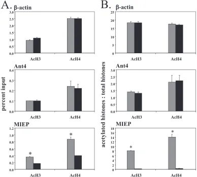
Histones bound to the MIEP are hypoacetylated in latent infection. It is well-established that modifications of specific histone residues play an important role in transcriptional reg-ulation of eukaryotic genes (30). Acetylation of lysines 9 and 14 of histone 3 (H3K9 and H3K14) is associated with transcrip-tionally active genes, while deacetylation is associated with transcriptional inactivity. We therefore examined interactions of the MIEP with acetylated histones H3 and H4 during acute and latent infection by ChIP followed by real-time PCR anal-ysis (Fig. 4). Associations of acetylated histones with the -ac-tin and Ant4 promoters were analyzed in parallel as controls for transcriptionally active and inactive genes, respectively. Our results showed that binding of acetylated histones to the -actin promoter was approximately 10-fold higher than
[image:5.585.72.253.67.239.2]bind-FIG. 2. ChIP analysis of RNA polymerase II binding to the MCMV MIEP. (A) Anti-Pol II- or IgG-precipitated DNA samples subjected to semiquantitative PCR amplification with primers specific to the MIEP region. N, uninfected mice; A, acutely infected mice; L, latently in-fected mice. (B) ChIP/real-time PCR analysis of the association of RNA polymerase II with the MIEP,-actin, and Ant4 promoters in chromatin from acutely infected and latently infected mice. The results (means of three experiments ⫹standard errors of the means) are shown as percentages of input chromatin immunoprecipitated with anti-RNA polymerase II antibody. ⴱ, P ⬍ 0.001. Gray bars, acute infection; black bars, latent infection.
FIG. 3. ChIP analysis of the interaction of the MIEP with histones in acutely and latently infected mice. Chromatin was immunoprecipi-tated with antibodies recognizing all forms of histone H3 or histone H4. ChIP products were analyzed by real-time PCR using primers and probes specific for the cellular genes-actin or Ant4 and for the MCMV MIEP. The results (means of three experiments⫹standard errors of the means) are shown as percentages of input chromatin immunoprecipitated with anti-histone H3 (top) or anti-histone H4 (bottom).ⴱ,P⬍0.05. Gray bars, acute infection; black bars, latent infection.
on November 8, 2019 by guest
http://jvi.asm.org/
[image:5.585.350.494.68.250.2]ing to the Ant4 promoter (⬃1% of input versus 0.1% for H3 and 2.5% versus 0.23% for H4) (Fig. 4A, top and middle panels). There is no difference between acute and latent infec-tion in binding of acetylated histones to cellular promoters. In contrast, a greater proportion of MIEP molecules are bound to acetylated histones in acute infection than in latent infection (Fig. 4A, bottom panel). However, a greater proportion of viral genomes are associated with histones in latent infection than acute infection (Fig. 3). When this is taken into account by calculating the ratio of acetylated histones to total histones bound to the promoter, the proportion of MIEP molecules associated with acetylated H3 in latently infected mice is re-duced 40-fold, from 8 in acutely infected mice to 0.2 in latently infected mice (Fig. 4B, bottom panel). Similarly, the ratio of acetylated H4 bound to the MIEP falls 35-fold from 14 to 0.4 in acutely and latently infected mice, respectively. The ratios of acetylated histones to total histones are greater than 1 because of differences in the efficiencies of the antibodies for modified histones, which recognize epitopes in the freely accessible his-tone tails, and unmodified hishis-tones, which recognize epitopes
buried within the molecule. In contrast, the ratio of acetylated versus total histones bound to the promoters of cellular genes in acutely and latently infected mice remained unchanged (Fig. 4B, top and middle panels). These results demonstrate that histones bound to the MIEP are hypoacetylated in latently infected mice.
HDACs are recruited to the MIEP in latent infection. Deacetylation of histones is catalyzed by HDACs. Among 10 classical HDACs, 5 are expressed in kidney tissue (10). We selected HDAC2 and HDAC3, which have been well-charac-terized and have relatively high expression in the kidney, as targets to investigate. Binding levels of HDACs to the-actin and Ant4 promoters were analyzed as controls. There was little binding of HDACs to the MIEP in acute infection. In contrast, binding of HDAC2 to the MIEP was 30-fold higher in latently infected mice compared to acutely infected mice, while binding of HDAC3 was enriched 4.6-fold in latently infected mice (Fig. 5). Thus, the percentage of MIEP molecules bound to HDACs was much greater than that bound to the transcriptionally inactive Ant4 gene. Unlike the MIEP, CpGs in the Ant4
pro-FIG. 4. Association of acetylated histones with the MIEP region. Chromatin from acutely and latently infected mice was subjected to ChIP with antibodies against histone H3, histone H4, acetylated histone H3 (Ac-H3), or acetylated histone H4 (Ac-H4), followed by quantitative real-time PCR with primers/probe specific to-actin (top panels), Ant4 (middle panels), or the MCMV MIEP region (bottom panels). The data are presented as the percent input (A) or as the ratio of DNA bound to acetylated histones versus that bound to the corresponding total histone after normalization to input DNA (B). Results shown are the means⫹standard errors of the means of three independent experiments.ⴱ,P⬍0.01. Gray bars, acute infection; black bars, latent infection.
on November 8, 2019 by guest
http://jvi.asm.org/
[image:6.585.98.486.69.418.2]moter are methylated. These observations suggest that tran-scriptional silencing of Ant4 may be regulated primarily by factors other than HDAC2 and HDAC3. However, our results suggest that HDACs do play a role in transcriptional silencing of the MIEP in latent infection.
Patterns of histone methylation change upon establishment of latency.Methylation of histones has been correlated with both positive and negative effects on transcription. Trimeth-ylation of histone H3 lysine 4 (H3K4me3) is strongly indicative of transcriptional activation, whereas methylation of other ly-sine residues of H3 (e.g., lyly-sine 9 or 27) is associated with transcriptional repression (5, 7, 30, 44, 47). Histone H3 lysine 9 can be mono-, di-, or trimethylated, denoted H3K9me1, H3K9me2, and H3K9me3, respectively. To determine the methylation status of histone H3 associated with the MIEP, we performed ChIP analysis of chromatin from acutely and la-tently infected mice with antibodies specific for trimethylated H3K4 and for the mono-, di-, or trimethylated forms of H3K9 as well as antibodies that recognize total H3. The promoter regions of the -actin and Ant4 genes were amplified from ChIP products in parallel with the MIEP as controls. Our results showed that, as expected, the-actin promoter is en-riched in H3 methylated at lysine 4 and is deficient in H3 methylated at lysine 9 (Fig. 6A and B, top panel). The reverse is true for the transcriptionally silent Ant4 gene (Fig. 6A and B, middle panel). There is little difference between latently and acutely infected mice in the methylation patterns of histones bound to cellular promoters. In contrast to cellular genes, the patterns of methylation of histones bound to the MIEP are dramatically different in acutely and latently infected mice
(Fig. 6A, bottom panel). In latently infected mice, the MIEP becomes markedly enriched in H3 methylation of lysine 9. Methylation of lysine 4, which is associated with active tran-scription, appears to be the same in acutely and latently in-fected mice. However, the proportion of MIEP molecules as-sociated with histones is much greater in latently infected mice (Fig. 3). When this is taken into account by calculating the ratio of methylated H3 to total H3, it is apparent that meth-ylation of H3 lysine 4 is markedly reduced in latently infected mice, while mono- and dimethylation of H3 lysine 9 is dramat-ically increased (Fig. 6B, bottom panel). There is little associ-ation of the trimethylated form of H3K9, which is generally found in permanently silenced pericentric heterochromatin (44), with the MIEP. As with our analysis of the state of histone acetylation (Fig. 4), the results of our analysis of meth-ylation of histones associated with the MIEP in latently in-fected mice are consistent with those observed for transcrip-tionally silent genes.
HP-1␥is bound to the MIEP in latent infection.It is thought that methylation of H3K9 facilitates formation of heterochro-matin and represses transcription by recruiting HP-1, which interacts with itself and other factors to create a compact chromatin structure that is not permissive to transcription (5, 45, 51). We therefore assessed recruitment of HP-1␥ to the MIEP by quantitative PCR analysis of ChIP products. The results (Fig. 7) showed that, relative to acute infection, the level of HP-1␥ associated with the MIEP was enhanced 14-fold in latent infection, a difference which was statistically significant (P⫽
0.02).
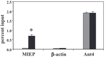
YY1 is recruited to the MCMV MIEP in latent infection. YY1 is a highly conserved zinc-finger transcription factor that can act as either an activator or repressor of transcription (13). Previous studies have shown that YY1 binds to negative reg-ulatory elements in the HCMV enhancer and mediates repres-sion of the HCMV enhancer (29, 34). The MCMV MIEP contains the sequence CCATATT, which is an imperfect match for the YY1 consensus binding site (49). We therefore investigated binding of YY1 to the MIEP by ChIP analysis. We analyzed YY1 binding to ribosomal protein L30 (rpL30) and -actin as positive and negative controls, respectively. YY1 is a positive regulator of rpL30 (15, 16). Our studies showed that there is a dramatic increase in the association of YY1 with the MIEP in latently infected mice (P⬍0.01) (Fig. 8).
DISCUSSION
The relative roles of the immune response and transcrip-tional control of viral gene expression in controlling CMV latency and reactivation have not been clear. CMV disease is generally observed only in immunocompromised individuals, and thus, there is no question that immunosurveillance plays a critical role in controlling reactivation once it has begun. How-ever, the role of transcriptional regulation of viral gene expres-sion, and particularlyie gene expression, has been more ob-scure. The high frequency of CMV-specific CD4⫹and CD8⫹T cells in healthy, seropositive individuals, which suggests re-peated stimulation of the immune response (18, 24, 48), the observation that viral shedding is frequently observed in healthy individuals (53), and the high frequency of detection of
[image:7.585.79.248.67.283.2]iegene expression in mice with latent MCMV infection (17, 33,
FIG. 5. Recruitment of histone deacetylases to the MIEP in latent infection. Chromatin from acutely or latently infected mice was immu-noprecipitated with antibodies specific to histone deacetylase 2 (top) or histone deacetylase 3 (bottom) followed by real-time PCR analysis with primers/probe specific to the MCMV MIEP,-actin, or Ant4. The results (means of three experiments⫹standard errors of the means) are shown as percentages of input chromatin immunoprecipitated with anti-HDAC antibody.ⴱ,P⬍0.01. Gray bars, acute infection; black bars, latent infection.
on November 8, 2019 by guest
http://jvi.asm.org/
56), has cast doubt on the idea there is a true state of latency, as defined by the absence of expression of genes associated with productive infection.
However, other studies support the hypothesis that there is a true state of latency and that transcriptional reactivation ofie
gene expression is a key event in reactivation of CMV. Studies
[image:8.585.117.467.68.377.2]in our lab have shown that MCMViegene expression can be induced by allogeneic transplantation of latently infected kid-neys, where it is not detectable in the contralateral nontrans-planted control kidney from the same mouse (20). Similarly, activation of the HCMV enhancer can be induced by alloge-neic transplantation of kidneys from transgenic mice carrying a
FIG. 6. Analysis of methylation of histone H3 bound to the MIEP in acute and latent infection. Chromatin from kidneys of acutely or latently infected mice was analyzed by ChIP with antibodies against histone H3, H3K4me3, H3K9me1, H3K9me2, or H3K9me3. Real-time PCR was performed on the immunoprecipitates with primers/probe specific to-actin (top panels), Ant4 (middle panels), or the MIEP (bottom panels). The data are presented as the percent input (A) or as the ratio of DNA bound to methylated histones versus that bound to the corresponding total H3 after normalization to input DNA (B). Results shown are the means⫹standard errors of the means of three independent experiments.ⴱ,P⬍
0.02. Gray bars, acute infection; black bars, latent infection.
[image:8.585.78.249.549.641.2]FIG. 7. Heterochromatin protein 1␥binds to the MIEP in latent infection. ChIP/quantitative real-time PCR was performed on chro-matin from acutely and latently infected mice with antibody specific to HP-1␥. Immunoprecipitated DNA was analyzed by real-time PCR with primers/probe specific to the MIEP,-actin, or Ant4 promoters. The results (means of three experiments ⫹ standard errors of the means) are shown as percentages of input chromatin immunoprecipi-tated with anti-HP-1␥antibody.ⴱ,P⬍0.001. Gray bars, acute infec-tion; black bars, latent infection.
FIG. 8. Recruitment of YY1 to the MIEP in latent infection. ChIP/ quantitative real-time PCR was performed on chromatin from acutely and latently infected mice with antibody specific to YY1. Immunopre-cipitated DNA was analyzed by real-time PCR with primers/probe specific to the MIEP,-actin, or rpL30 promoters. The results (means of three experiments⫹standard errors of the means) are shown as percentages of input chromatin immunoprecipitated with anti-YY1.ⴱ,
P⬍0.001. Gray bars, acute infection; black bars, latent infection.
on November 8, 2019 by guest
http://jvi.asm.org/
[image:8.585.335.506.558.650.2]-galactosidase reporter gene compared to their contralateral controls (20). In these uninfected mice, increased expression of the reporter can only be due to activation of the enhancer. Additional studies in our lab and others have shown thatie
gene expression is much higher in mice treated with tumor necrosis factor than in control mice (20, 50). Studies with HCMV naturally infected monocytes have shown thatiegene expression can be induced ex vivo in cells which were negative prior to treatment (43). Furthermore, analysis of the ratio of RNA to DNA in mice latently infected with MCMV has shown that the number of viral DNA molecules vastly exceeds the number of IE-1 RNA transcripts, and thus, that most viral genomes are silent with respect toiegene expression (42, 56). Here, we have used direct quantification to examine the ratio of MCMV DNA to RNA in acutely and latently infected mice. Our studies show that, although the DNA is consistently detectable in latently infected mice, IE-1 RNA is often below the level of detection. Of three replicates of samples from three mice, IE-1 RNA was detectable two of nine times. Over-all, the ratio of RNA to DNA was approximately 0.22 IE-1 transcripts per genome. While this is substantially higher than previous estimates of RNA expression derived from statistical analysis of the frequency of detection in the lungs (42), it is similar to previous estimates based on semiquantitative PCR analysis of latently infected lungs (56). Because detection of IE-1 RNA is likely due to sporadic reactivation, the absolute ratio of RNA and DNA may be quite variable. In any case, the results of our analysis of RNA expression in the kidneys of latently infected mice support previous analyses of the lung, which indicate that the majority of genomes are transcription-ally inactive in latently infected mice.
In order to investigate the mechanisms by which ie gene expression is repressed in latent infection, we have analyzed epigenetic factors associated with the MIEP. Methylation of cytosine residues in CpG dinucleotides and specific modifica-tions of histones bound to promoter regions have been shown to be associated with repression of transcription (45). Our previous studies have shown that the MIEP is not methylated in latently infected mice (19). We therefore investigated mod-ifications of histones bound to the MIEP in both acutely in-fected mice, in which theiegenes are actively transcribed, and in latently infected mice, in whichiegene expression is turned off. Our studies showed that, like HSV (27, 55), association of the genome with histones is much greater in latently infected mice than in acute infection. In addition, our studies showed that the patterns of histone modifications change when latency is established. In acute infection, histones H3 and H4 bound to the MIEP are acetylated at lysine residues associated with active transcription and H3 is methylated at lysine 4. These chromatin modifications are lost when latency is established and replaced by methylation of lysine 9, deacetylation of H3 and H4, and recruitment of HDACs. Concomitant with changes in histone modifications, association of RNA polymer-ase with the MIEP is lost when latency is established. Thus, changes in histone modifications correlate with loss of tran-scription. In addition, HP-1␥, which is thought to facilitate formation of a tightly condensed heterochromatin structure that is inaccessible to the transcription apparatus (5, 45, 51), becomes associated with the MIEP in latent infection.
Our results are consistent with a previous study of human
monocytes naturally infected with HCMV (43). This study showed that the HCMV MIEP is bound to HP-1, but not acetylated H4 in latently infected cells, and that after reacti-vation induced ex vivo the converse was true. Similarly, a study using untreated and tetradecanoyl phorbol acetate-treated HCMV-infected THP-1 monocytic cells as in vitro models for latency and reactivation, respectively, found that tetradecanoyl phorbol acetate-induced reactivation results in deposition of acetylated histone H3 to the MIEP (21). This study found little association of dimethylated H3K9 with the MIEP in latently infected THP-1 cells. Our study shows that, while there is some binding of dimethylated H3K9 to the MCMV MIEP in latent infection, the MIEP is more highly enriched in the mono-methylated form of H3K9. Thus, our studies provide further evidence of the similarity between MCMV and HCMV in regulation of ie gene expression and support the validity of using MCMV as a model to study CMV latency and reactiva-tion in vivo. Using this model, we have been able to substan-tially expand our knowledge of chromatin modifications and enzymes bound to the MIEP during latent infection. This knowledge will provide an important foundation for future studies investigating changes in chromatin bound to the MIEP that occur during transcriptional reactivation ofiegene expres-sion induced by transplantation.
An understanding of the mechanism by which transcrip-tional repressors are recruited to the MIEP is essential to designing future therapies to prevent establishment of latency or to eliminate existing reservoirs of latent infection. It is likely that this process is mediated by factors that bind to specific DNA sequences in the MIEP (3). Our studies show that YY1, which has been proposed to mediate repression of the HCMV enhancer (3), binds to the MCMV MIEP in latently infected mice. This observation suggests that YY1 may play such a role in the establishment of MCMV latency. It has been postulated that viral factors mediate chromatinization of the HSV ge-nome as a mechanism for the establishment of latency (27). Whether recruitment of transcriptional silencing factors to the MIEP is mediated by viral or host factors during the establish-ment of latency in CMV infection and determining the iden-tities of those factors are important questions that remain to be answered.
ACKNOWLEDGMENTS
This study was supported by Public Health Service grants R01 AI42898 (M.A.) and R21 AI076771 (M.H.) from the NIH.
REFERENCES
1.Alberter, B., and A. Ensser.2007. Histone modification pattern of the T-cellular herpesvirus saimiri genome in latency. J. Virol.81:2524–2530. 2.Amelio, A. L., N. V. Giordani, N. J. Kubat, E. J. O’Neil, and D. C. Bloom.
2006. Deacetylation of the herpes simplex virus type 1 latency-associated transcript (LAT) enhancer and a decrease in LAT abundance precede an increase in ICP0 transcriptional permissiveness at early times postexplant. J. Virol.80:2063–2068.
3.Bain, M., M. Reeves, and J. Sinclair.2006. Regulation of human cytomeg-alovirus gene expression by chromatin remodeling, p. 167–183.InM. Red-dehase (ed.), Cytomegaloviruses: molecular biology and immunology. Caister Academic Press, Norfolk, United Kingdom.
4.Balthesen, M., M. Messerle, and M. J. Reddehase.1993. Lungs are a major organ site of cytomegalovirus latency and recurrence. J. Virol.67:5360–5366. 5.Bannister, A. J., P. Zegerman, J. F. Partridge, E. A. Miska, J. O. Thomas, R. C. Allshire, and T. Kouzarides.2001. Selective recognition of methylated lysine 9 on histone H3 by the HP1 chromo domain. Nature410:120–124. 6.Barnes, P. J., and M. Karin.1997. Nuclear factor-B: a pivotal transcription
factor in chronic inflammatory diseases. N. Engl. J. Med.336:1066–1071.
on November 8, 2019 by guest
http://jvi.asm.org/
7.Bernstein, B. E., E. L. Humphrey, R. L. Erlich, R. Schneider, P. Bouman, J. S. Liu, T. Kouzarides, and S. L. Schreiber.2002. Methylation of histone H3 Lys 4 in coding regions of active genes. Proc. Natl. Acad. Sci. USA
99:8695–8700.
8.Boshart, M., F. Weber, G. Jahn, K. Dorsch-Hasler, B. Fleckenstein, and W. Schaffner.1985. A very strong enhancer is located upstream of an immediate early gene of human cytomegalovirus. Cell41:521–530.
9.Day, L., C. M. Chau, M. Nebozhyn, A. J. Rennekamp, M. Showe, and P. M. Lieberman.2007. Chromatin profiling of Epstein-Barr virus latency control region. J. Virol.81:6389–6401.
10.de Ruijter, A. J., A. H. van Gennip, H. N. Caron, S. Kemp, and A. B. van Kuilenburg.2003. Histone deacetylases (HDACs): characterization of the classical HDAC family. Biochem. J.370:737–749.
11.Dorsch-Hasler, K., G. M. Keil, F. Weber, M. Jasin, W. Schaffner, and U. H. Koszinowski.1985. A long and complex enhancer activates transcription of the gene coding for the highly abundant immediate early mRNA in murine cytomegalovirus. Proc. Natl. Acad. Sci. USA82:8325–8329.
12.El-Osta, A., and A. P. Wolffe.2001. Analysis of chromatin-immunopurified MeCP2-associated fragments. Biochem. Biophys. Res. Commun.289:733– 737.
13.Gordon, S., G. Akopyan, H. Garban, and B. Bonavida.2006. Transcription factor YY1: structure, function, and therapeutic implications in cancer biol-ogy. Oncogene25:1125–1142.
14.Gruffat, H., E. Manet, and A. Sergeant.2002. MEF2-mediated recruitment of class II HDAC at the EBV immediate early gene BZLF1 links latency and chromatin remodeling. EMBO Rep.3:141–146.
15.Hariharan, N., D. E. Kelley, and R. P. Perry.1991. Delta, a transcription factor that binds to downstream elements in several polymerase II promot-ers, is a functionally versatile zinc finger protein. Proc. Natl. Acad. Sci. USA
88:9799–9803.
16.Hariharan, N., D. E. Kelley, and R. P. Perry.1989. Equipotent mouse ribosomal protein promoters have a similar architecture that includes inter-nal sequence elements. Genes Dev.3:1789–1800.
17.Henry, S. C., and J. D. Hamilton.1993. Detection of murine cytomegalovirus immediate early 1 transcripts in the spleens of latently infected mice. J. In-fect. Dis.167:950–954.
18.Holtappels, R., M. F. Pahl-Seibert, D. Thomas, and M. J. Reddehase.2000. Enrichment of immediate-early 1 (m123/pp89) peptide-specific CD8 T cells in a pulmonary CD62Llomemory-effector cell pool during latent murine
cytomegalovirus infection of the lungs. J. Virol.74:11495–11503. 19.Hummel, M., S. Yan, Z. Li, T. K. Varghese, and M. Abecassis.2007.
Tran-scriptional reactivation of murine cytomegalovirusie gene expression by 5-aza-2⬘-deoxycytidine and trichostatin A in latently infected cells despite lack of methylation of the major immediate-early promoter. J. Gen. Virol.
88:1097–1102.
20.Hummel, M., Z. Zhang, S. Yan, I. DePlaen, P. Golia, T. Varghese, G. Thomas, and M. I. Abecassis.2001. Allogeneic transplantation induces ex-pression of cytomegalovirus immediate-early genes in vivo: a model for reactivation from latency. J. Virol.75:4814–4822.
21.Ioudinkova, E., M. C. Arcangeletti, A. Rynditch, F. De Conto, F. Motta, S. Covan, F. Pinardi, S. V. Razin, and C. Chezzi.2006. Control of human cytomegalovirus gene expression by differential histone modifications during lytic and latent infection of a monocytic cell line. Gene384:120–128. 22.Jenuwein, T., and C. D. Allis.2001. Translating the histone code. Science
293:1074–1080.
23.Karin, M., and E. Gallagher.2005. From JNK to pay dirt: jun kinases, their biochemistry, physiology and clinical importance. IUBMB Life57:283–295. 24.Karrer, U., S. Sierro, M. Wagner, A. Oxenius, H. Hengel, U. H. Koszinowski, R. E. Phillips, and P. Klenerman.2003. Memory inflation: continuous ac-cumulation of antiviral CD8⫹T cells over time. J. Immunol.170:2022–2029. 25.Keil, G. M., A. Ebeling-Keil, and U. H. Koszinowski.1987. Immediate-early genes of murine cytomegalovirus: location, transcripts, and translation prod-ucts. J. Virol.61:526–533.
26.Keil, G. M., A. Ebeling-Keil, and U. H. Koszinowski.1987. Sequence and structural organization of murine cytomegalovirus immediate-early gene 1. J. Virol.61:1901–1908.
27.Knipe, D. M., and A. Cliffe.2008. Chromatin control of herpes simplex virus lytic and latent infection. Nat. Rev. Microbiol.6:211–221.
28.Koffron, A. J., M. Hummel, B. K. Patterson, S. Yan, D. B. Kaufman, J. P. Fryer, F. P. Stuart, and M. I. Abecassis.1998. Cellular localization of latent murine cytomegalovirus. J. Virol.72:95–103.
29.Kothari, S., J. Baillie, J. G. Sissons, and J. H. Sinclair.1991. The 21bp repeat element of the human cytomegalovirus major immediate early en-hancer is a negative regulator of gene expression in undifferentiated cells. Nucleic Acids Res.19:1767–1771.
30.Kouzarides, T.2007. Chromatin modifications and their function. Cell128:
693–705.
31.Kubat, N. J., R. K. Tran, P. McAnany, and D. C. Bloom.2004. Specific histone tail modification and not DNA methylation is a determinant of herpes simplex virus type 1 latent gene expression. J. Virol.78:1139–1149.
32.Kurz, S., H. P. Steffens, A. Mayer, J. R. Harris, and M. J. Reddehase.1997. Latency versus persistence or intermittent recurrences: evidence for a latent state of murine cytomegalovirus in the lungs. J. Virol.71:2980–2987. 33.Kurz, S. K., M. Rapp, H. P. Steffens, N. K. Grzimek, S. Schmalz, and M. J.
Reddehase.1999. Focal transcriptional activity of murine cytomegalovirus during latency in the lungs. J. Virol.73:482–494.
34.Liu, R., J. Baillie, J. G. Sissons, and J. H. Sinclair.1994. The transcription factor YY1 binds to negative regulatory elements in the human cytomega-lovirus major immediate early enhancer/promoter and mediates repression in non-permissive cells. Nucleic Acids Res.22:2453–2459.
35.Meier, J. L.2001. Reactivation of the human cytomegalovirus major imme-diate-early regulatory region and viral replication in embryonal NTera2 cells: role of trichostatin A, retinoic acid, and deletion of the 21-base-pair repeats and modulator. J. Virol.75:1581–1593.
36.Meier, J. L., and M. F. Stinski.2006. Major immediate-early enhancer and its gene products, p. 151–166.InM. J. Reddehase (ed.), Cytomegaloviruses: molecular biology and immunology. Caister Academic Press, Norfolk, United Kingdom.
37.Messerle, M., B. Buhler, G. M. Keil, and U. H. Koszinowski.1992. Structural organization, expression, and functional characterization of the murine cy-tomegalovirus immediate-early gene 3. J. Virol.66:27–36.
38.Miao, J., S. Fang, Y. Bae, and J. K. Kemper.2006. Functional inhibitory cross-talk between constitutive androstane receptor and hepatic nuclear factor-4 in hepatic lipid/glucose metabolism is mediated by competition for binding to the DR1 motif and to the common coactivators, GRIP-1 and PGC-1␣. J. Biol. Chem.281:14537–14546.
39.Mocarski, E. S., and C. T. Courcell.2001. Cytomegaloviruses and their replication, p. 2629–2673.InD. M. Knipe and P. M. Howley (ed.), Fields virology, 4th ed., vol. 2. Lippincott Williams & Wilkins, Philadelphia, PA. 40.Murphy, J. C., W. Fischle, E. Verdin, and J. H. Sinclair.2002. Control of
cytomegalovirus lytic gene expression by histone acetylation. EMBO J.21:
1112–1120.
41.Pass, R. F.2001. Cytomegalovirus, p. 2675–2705.InD. M. Knipe and P. M. Howley (ed.), Fields virology, 4th ed., vol. 2. Lippincott Williams & Wilkins, Philadelphia, PA.
42.Reddehase, M. J., J. Podlech, and N. Grzimek.2002. Mouse models of cytomegalovirus latency: overview. J. Clin. Virol.25:S23–S36.
43.Reeves, M. B., P. A. MacAry, P. J. Lehner, J. G. Sissons, and J. H. Sinclair.
2005. Latency, chromatin remodeling, and reactivation of human cytomeg-alovirus in the dendritic cells of healthy carriers. Proc. Natl. Acad. Sci. USA
102:4140–4145.
44.Rice, J. C., S. D. Briggs, B. Ueberheide, C. M. Barber, J. Shabanowitz, D. F. Hunt, Y. Shinkai, and C. D. Allis.2003. Histone methyltransferases direct different degrees of methylation to define distinct chromatin domains. Mol. Cell12:1591–1598.
45.Richards, E. J., and S. C. Elgin.2002. Epigenetic codes for heterochromatin formation and silencing: rounding up the usual suspects. Cell108:489–500. 46.Rodic, N., M. Oka, T. Hamazaki, M. R. Murawski, M. Jorgensen, D. M. Maatouk, J. L. Resnick, E. Li, and N. Terada.2005. DNA methylation is required for silencing of ant4, an adenine nucleotide translocase selectively expressed in mouse embryonic stem cells and germ cells. Stem Cells23:1314– 1323.
47.Santos-Rosa, H., R. Schneider, A. J. Bannister, J. Sherriff, B. E. Bernstein, N. C. Emre, S. L. Schreiber, J. Mellor, and T. Kouzarides.2002. Active genes are tri-methylated at K4 of histone H3. Nature419:407–411.
48.Sester, M., U. Sester, B. Gartner, B. Kubuschok, M. Girndt, A. Meyerhans, and H. Kohler.2002. Sustained high frequencies of specific CD4 T cells restricted to a single persistent virus. J. Virol.76:3748–3755.
49.Shi, Y., J.-S. Lee, and K. Galvin.1997. Everything you ever wanted to know about Yin Yang 1. Biochim. Biophys. Acta1332:F49–F66.
50.Simon, C. O., C. K. Seckert, D. Dreis, M. J. Reddehase, and N. K. Grzimek.
2005. Role for tumor necrosis factor alpha in murine cytomegalovirus tran-scriptional reactivation in latently infected lungs. J. Virol.79:326–340. 51.Stewart, M. D., J. Li, and J. Wong.2005. Relationship between histone H3
lysine 9 methylation, transcription repression, and heterochromatin protein 1 recruitment. Mol. Cell. Biol.25:2525–2538.
52.Strahl, B. D., and C. D. Allis.2000. The language of covalent histone modifications. Nature403:41–45.
53.Toro, A. I., and J. Ossa.1996. PCR activity of CMV in healthy CMV-seropositive individuals: does latency need redefinition? Res. Virol.147:233– 238.
54.Turner, B. M.2002. Cellular memory and the histone code. Cell111:285– 291.
55.Wang, Q. Y., C. Zhou, K. E. Johnson, R. C. Colgrove, D. M. Coen, and D. M. Knipe.2005. Herpesviral latency-associated transcript gene promotes assem-bly of heterochromatin on viral lytic-gene promoters in latent infection. Proc. Natl. Acad. Sci. USA102:16055–16059.
56.Yuhasz, S. A., V. B. Dissette, M. L. Cook, and J. G. Stevens.1994. Murine cytomegalovirus is present in both chronic active and latent states in persis-tently infected mice. Virology202:272–280.