Virus Capsid and E2 Proteins with Reverse Genetics
Jonathan E. Snyder,aChristian J. Berrios,a* Thomas J. Edwards,aJoyce Jose,aRushika Perera,aand Richard J. Kuhna,b
Markey Center for Structural Biology, Department of Biological Sciences, Purdue University, West Lafayette, Indiana, USA,aand Bindley Bioscience Center, Purdue University, West Lafayette, Indiana, USAb
A 7-Å cryoelectron microscopy-based reconstruction of Sindbis virus (SINV) was recently generated. Fitting the crystal structure of the SINV capsid protein (Cp) into the density map revealed that the F2-G2 loop of the Cp was shifted away from cytoplasmic domain of E2 (cdE2) in the 7-Å reconstruction relative to its position in the Cp crystal structure. Furthermore, the reconstruc-tion demonstrated that residue E395 in region I of the cytoplasmic domain of the E2 envelope protein (cdE2-RI) and K252 of Cp, part of the Cp F2-G2 loop, formed a putative salt bridge in the virion. We generated amino acid substitutions at residues K250 and K252 of the SINV Cp and explored the resulting phenotypes. In the context of cells infected with wild-type or mutant virus, reversing the charge of these two residues resulted in the appearance of Cp aggregates around cytopathic vacuole type I (CPV-I) structures, the absence of nucleocapsid (NC) formation, and a lack of virus particle release in the infected mammalian cell. How-ever, expressing the same Cp mutants in the cell without the envelope proteins or expressing and purifying the mutants from an
Escherichia coliexpression system and assemblingin vitroyielded NC assembly in all cases. In addition, second-site mutations within cdE2 restored NC assembly but not release of infectious particles. Our data suggest an early temporal and spatial interac-tion between cdE2-RI and the Cp F2-G2 loop that, when ablated, leads to the absence of NC assembly. This interacinterac-tion also ap-pears to be important for budding of virus particles.
T
he alphaviruses are a group of enveloped, positive-sense, sin-gle-stranded RNA viruses in the Togaviridae family. Well-studied members include the Eastern, Western, and Venezuelan equine encephalitis viruses, Semliki Forest virus, Ross River virus, Chikungunya virus, and the type member, Sindbis virus (SINV) (42). The emergence of Chikungunya virus as an epidemic threat has reemphasized the medical importance of the alphaviruses (47).The⬃11.5-kb genome encodes four nonstructural proteins that primarily facilitate genome replication and four structural proteins, capsid, pE2, 6K, and E1, which are initially translated from a subgenomic message as a polyprotein (42). Co- and post-translational processing by viral and host proteases yields the ma-ture structural proteins. The capsid protein (Cp) is an autopro-tease that acts to cotranslationally cleave itself from the growing polypeptide (30) and later associates with viral genomic RNA to form a nucleocapsid (NC) core. The remaining polyprotein in-serts into the endoplasmic reticulum (ER) in a signal sequence-mediated manner (2). Signalase processing yields pE2 and E1, the envelope proteins, and 6K, a small membrane-resident protein whose function in the virus life cycle remains unclear (26). pE2 is later cleaved by furin to yield the small protein E3 and the larger protein E2 (52). E1 and E2 are type I integral membrane proteins, with E2 primarily responsible for host receptor engagement (3,
24), while E1 contains pH-dependent fusogenic properties re-quired for virus entry (14,50). The ectodomains of both E1 and E2 comprise the bulk of each protein, although both E1 and E2 con-tain some residues, termed cytoplasmic domains, which are found on the inner face of the envelope and, at least in the case of E2, interact with the NC (15,17,28).
The icosahedrally arranged virus particle, released by budding from the host plasma membrane (PM), is comprised of two pro-tein shells (4). In the outer shell, 240 copies each of E1 and E2, arranged as 80 trimers of E1-E2 heterodimers, are embedded in a
plasma membrane-derived lipid bilayer (33). The membrane layer also contains substoichiometric amounts of 6K and sur-rounds the NC core. The NC is comprised of 240 copies of Cp and one copy of genomic RNA and also demonstrates icosahedral symmetry (4). The icosahedral symmetry of the NC may derive from the 1:1 protein-protein interaction between each molecule of Cp in the NC and a molecule of E2 in the envelope, as the icosahedral arrangement of prebudded, cytoplasmic NCs remains loosely defined (4,12,32).
The alphaviruses have long proven a model system for struc-tural studies of enveloped mammalian viruses owing, in part, to the T⫽4 icosahedral arrangement of both the envelope and the NC core of budded virus particles. Genetic and structural evidence suggests that a hydrophobic pocket on the face of the Cp interacts with the cytoplasmic domain of E2 (cdE2) in the context of a budded virion (36,38,44). Specifically, a conserved YxL motif in cdE2 is involved in side-chain interactions with residues resident in the Cp hydrophobic pocket, and other evidence suggests inter-actions of C-terminal cdE2 residues, relative to the YxL domain, with Cp (23,25, 31, 36,51). Thus, Cp and E2 interact during budding, and this interaction is required for budding to occur (43,
53). However, details regarding the arrangements of, and the in-teractions between, the Cp and the E1 and E2 envelope proteins prior to budding events remain unclear.
Received18 May 2012Accepted30 August 2012
Published ahead of print5 September 2012
Address correspondence to Richard J. Kuhn, [email protected]. * Present address: Christian J. Berrios, Harvard Medical School, Boston, Massachusetts, USA.
Copyright © 2012, American Society for Microbiology. All Rights Reserved.
doi:10.1128/JVI.01220-12
on November 7, 2019 by guest
http://jvi.asm.org/
(34). Likewise, a detailed understanding of the NC assembly path-way is not available. While Cp homodimers are thought to be key assembly intermediates, and charge neutralization between genomic RNA and the basic Cp likely provides the source for the initial assembly nucleation, no NC assembly intermediates have been identified by either biophysical or microscopy-based tech-niques (27,46). Additionally, the site of NC assembly is unclear, although NC may assemble at or near the site of viral genome replication (11, 40). Interestingly, Cp mutants have been de-scribed in which NC formation in the cytoplasm is disrupted but in which wild-type (wt)-like virus particles are nevertheless bud-ded from the cell (9).
While it has long been established that NCs and envelope pro-tein spikes interact during budding at the plasma membrane, emerging data suggest that the interaction of NC and envelope proteins may occur before either arrives at the plasma membrane, in the form of membranous virus-induced type II cytopathic vac-uoles (CPV-II) (16). These CPV-II molecules have been impli-cated in the traffic of envelope proteins to the plasma membrane and appear to be decorated with NCs (19,41). The precise origin of CPV-II vacuoles and the temporal and spatial aspects of the initial interaction between NC and envelope proteins on these CPV-II structures have yet to be detailed. However, the cotraffick-ing of NC and E2 to the site of buddcotraffick-ing hints at interactions be-tween Cp and the envelope proteins well before virus budding at the plasma membrane.
A 7-Å cryoelectron microscopy (cryo-EM)-based reconstruc-tion of SINV virus demonstrated several novel molecular links between the E2 envelope protein and the NC in the context of a virus particle (44). Among these data, it was shown that a flexible loop on the edge of the hydrophobic pocket of Cp, the F2-G2 loop composed of amino acids 248-NSKGK-252, was shifted⬃3.5 Å away from cdE2 relative to the loop position in the SINV Cp crys-tal structure. Furthermore, a salt bridge between cdE2 E395 and Cp K252, part of the F2-G2 loop, was proposed. On the basis of those results and the observation that Cp residue K250 was impli-cated in cross-capsomere contacts (45), we performed a muta-tional analysis of Cp residues K250 and K252.
Simultaneous mutation of SINV Cp residues K250 and K252 led to production of Cp aggregates around cytopathic type I rep-lication vacuoles (CPV-I) and the complete abrogation of NC for-mation in the cell concomitant with a lack of virus particle release. Surprisingly, though, when the mutant proteins were expressed and assembledin vitro(45), expressed in the absence of the enve-lope proteins, or expressed via a replicon or by a mammalian expression plasmid, formation of core-like particles (CLPs) was detected in all cases. Double SINV mutants were generated in which both cdE2-RI (cdE2 391-KARRE-395) and the Cp residues K250 and K252 were simultaneously mutated. These double mu-tants restored NC formation for the Cp K250D/K252D and K250E/K252E mutant backgrounds but did not restore the release of virions.
Taken together, our data suggest (i) that an early temporal and
MATERIALS AND METHODS
Cell culture.BHK-15 cells obtained from the American Type Culture Collection were maintained in minimal essential medium (MEM) (Life Technologies) supplemented with 10% fetal bovine serum (FBS), except for enzyme-linked immunosorbent assay (ELISA) studies in which the FBS was omitted.
Generation of mutants in virus cDNA.All mutants were generated in the pToto64 SINV cDNA plasmid using standard overlap PCR mutagen-esis with Platinum Pfx DNA polymerase (Invitrogen). Infectious full-length RNA was produced by linearizing the plasmids with SacI followed by SP6-mediatedin vitrotranscription (Ambion). The Cp-expressing rep-licon was created in the pToto64 background by deleting the nucleotides corresponding to E3, E2, 6K, and E1 via overlap PCR mutagenesis. The first nine nucleotides of E3 were retained in addition to the stop codon utilized by E1. Plaque diameter analysis was performed by incubating BHK cells seeded in 6-well culture dishes with 1.5 ml of 0.2 mg/ml DEAE dextran–50 mM Tris (pH 7.4) at 37°C for 1 h, followed by incubation with
in vitro-transcribed RNA at 25°C for 30 min and subsequent overlay with 1% agarose–MEM.
NC accumulation assays.BHK cells were electroporated (1.5 kV, 25 F, 200⍀, 0.2-cm-gap cuvette) with 10g ofin vitro-transcribed RNAs derived from the Toto64 cDNA clones. Infection was allowed to proceed for 8 h, at which time media were removed and cells washed 3 times with phosphate-buffered saline (PBS) and then scraped off the cell culture dish using 1 ml PBS. Cells were subjected to pellet formation for 10 min at 750⫻gand resuspended in hypotonic buffer (10 mM NaCl, 10 mM Tris [pH 7.4], 20 mM EDTA) to swell for 20 min on ice. Triton X-100 (20%) was added to achieve a final concentration of 4% (vol/vol). Lysed cell debris and nuclei were subjected to pellet formation at 2,000⫻gfor 10 min. The solution was then loaded onto a continuous iodixanol gradient (0% to 30%) in TNE-Triton X-100 (50 mM Tris [pH 7.4], 100 mM NaCl, 1 mM EDTA, 0.1% Triton X-100) and centrifuged at 32,000 rpm in a Beckman SW-41 rotor for 2.5 h. All steps were carried out at 4°C. The gradient was fractionated, and each fraction was analyzed by SDS-PAGE on a 12% polyacrylamide gel. Separated proteins were electroeluted and adsorbed to 0.4-m-pore-size nitrocellulose membranes before being probed with a rabbit polyclonal anti-Cp primary antibody and an IRDye 680 goat anti-rabbit secondary antibody conjugate (Li-COR). Stained membranes were scanned with a Li-COR Odyssey infrared imager. Cp intensities were measured using the Odyssey software and relative inten-sity values plotted as a function of fraction number.
In vitroNC assembly.The wild-type or mutant Cp genes were sub-cloned into pET-11a prokaryotic expression vector (Novagen). Protein expression and purification andin vitroassembly of NCs were performed as described previously (45), using an arbitrary single-stranded DNA (ssDNA) 109-mer (5=-GATCCGAAAGCGCGCCGTGAGTGCCTGACG CCATACGCCCTGGCCCCAAACGCCGTAATCCCAGCGGCCGCT GCCGCCGCGTGCTGCGTTAGGTCGGCCAATGCTTAGC-3=).
Transient transfection and expression of SINV Cp or Cp plus enve-lope proteins.The SINV sequences encoding the structural proteins (Cp, pE2, 6K, E1), or the sequences encoding just the Cp, were cloned into pcDNA4/TO/myc/his/A mammalian expression vector (Invitrogen). BHK cells were seeded into 60-mm-diameter culture dishes at⬃70% confluence and then transiently transfected with 10g of plasmid with Lipofectamine (Invitrogen) 18 h prior to lysis and gradient analysis as described above.
on November 7, 2019 by guest
http://jvi.asm.org/
Multistep growth curve analysis.BHK cells were infected with stock virus at a multiplicity of infection (MOI) of 0.5. Virus was allowed to adsorb to cells at 25°C for 1 h followed by incubation at 37°C. Media were removed and replaced with fresh media at 30-min intervals for the first 2 h and then at 1-h intervals thereafter. The amount of infectious virus release was quantified by titration on BHK cells.
Plaque assay.Serial dilutions (10-fold) of culture media or virus were made in PBS containing 1% FBS plus 10 mM CaCl2and 10 mM MgCl2.
BHK cells at⬃90% confluence were inoculated with 250l of the dilu-tions for 1 h at room temperature and then overlaid with MEM containing 5% FBS and 1% agarose. Cells were incubated at 37°C for 48 h and stained with neutral red.
ELISA.Posttransfection media (2 ml) were concentrated to 100l using Amicon 100-kDa centrifugal filters and applied to a 96-well Poly-sorp ELISA plate (Sigma) overnight at 4°C. The samples were blocked with 5% BSA–PBS for 2 h followed by three washes with PBS-T (PBS, 0.1% Tween 20). A 1:500 dilution of a monoclonal anti-E2 antibody (202) was applied to the sample for 2 h, followed by three washes with PBS-T. A 1:1,500 dilution of a horseradish peroxidase (HRP)-conjugated goat anti-mouse secondary antibody was applied to the sample for 1 h, followed by three PBS-T washes. A TMB peroxidase detection system (KPL) was used to develop the ELISA according to the manufacturer’s instructions. The optical density (OD) was determined at 450 nm.
Electron microscopy analyses.BHK cells were electroporated within vitro-transcribed RNAs or transfected (Lipofectamine 2000) with plas-mids expressing the viral Cp or structural polyprotein. Cells were fixed and processed at 8 h postelectroporation of RNA or 18 h posttransfection of plasmid. Samples were fixed in 2% glutaraldehyde– cacodylate buffer (0.1 M sodium cacodylate, 2 mM MgCl2, 2 mM CaCl2, 0.5% NaCl, pH
7.4) and then washed in cacodylate buffer followed by water. Samples were then postfixed with 2% OsO4and 1.5% K4Fe(CN)6and then rinsed
with water. Processing of cells for immunostaining omitted the osmium tetroxide and potassium ferrocyanide. Cells were subjected to pellet for-mation at 4,000⫻gin 1% low-melt agarose. The pellet was dehydrated in sequential concentrations of ethanol from 10% to 100%. Pellets were then embedded in Durcupan resin and hardened at 60°C for 72 h. Sections (50 to 70 nm thick) were collected on carbon-coated copper mesh grids (car-bon-coated gold mesh grids for immunostaining) using a diamond knife. Sections received a postembedding stain of 2% uranyl acetate and Sato’s lead. Images were collected on a Philips CM-10 Biotwin transmission electron microscope (TEM) with film at 80 kV, a FEI CM200F cryoelec-tron microscope with a 1-k charge-coupled-device (CCD) camera at 200 kV (FEI Company, Hillsboro, OR), or an FEI Titan Krios TEM at 300 kV with a 4-k CCD camera. For immunostaining, sections were etched with 200 mM NaOH in ethanol and rinsed in ethanol and water followed by incubation in 0.005% Triton X–PBS for 10 min. Grids were then blocked with 2% bovine serum albumin–1% fish gelatin–PBS for 30 m, followed by exposure to anti-Cp antibody (1:150 in 10% blocking solution–PBS) for 1.5 h. Samples were then rinsed in PBS and blocked with 3% normal goat serum in PBS for 10 min followed by exposure of rabbit anti-body conjugated to 6-nm-diameter gold (EMS) (1:40 in PBS) for 1.5 h. Samples were rinsed in PBS and cross-linked with 2.5% glutaraldehyde for 5 min and then poststained with 2% uranyl acetate and Sato’s lead.
RESULTS
Phenotype of the SINV Cp F2-G2 loop mutants.It has previously
been suggested that the SINV Cp F2-G2 loop is involved in a putative interaction with cdE2 (44). Furthermore, Cp K250 was proposed to mediate cross-capsomere contacts within the NC (46) and Cp K252 was postulated to participate in salt bridge con-tacts with cdE2. However, analysis of virus assembly phenotypes after mutation of these residues has not yet been described. Thus, we generated SINV mutants in which K250 and K252 were simul-taneously mutated to alanine (K250/2A), aspartic acid (K250/2D), or glutamic acid (K250/2E) and a mutant in which the sequence
encoding the tripeptide sequence 250-KGK-252 was deleted (⌬KGK).
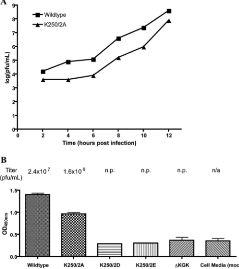
After BHK cells were electroporated within vitro-transcribed viral RNA, infectious virus was recovered from only the wild type and the K250/2A mutant; no infectious virus was recovered from the K250/2D, K250/2E, or⌬KGK mutant. Growth curve analysis of the wild type and the K250/2A mutant indicated that the K250/2A mutant released⬃1.5-log-fewer infectious units than the wild type throughout the 12-h time course (Fig. 1A).
To ascertain if noninfectious particles were being released in the K250/2D, K250/2E, or⌬KGK backgrounds, we collected me-dia 8 h after electroporation ofin vitro-transcribed viral RNAs and assayed the amount of infectious particles in the media and the amount of total virus particles in parallel by plaque assay and ELISA, respectively (Fig. 1B). The K250/2A mutant showed a de-crease in signal in the ELISA relative to the growth curve, indicat-ing no modulation in specific infectivity in that mutant. The K250/2D, K250/2E, and⌬KGK mutants, however, failed to release any virus particles as gauged by the ELISA, indicating that, in these backgrounds, virus particle release is completely abrogated. Elec-troporating BHK cells within vitro-transcribed RNA generated from wild-type or mutant cDNAs and growing the electroporated cells at 30°C, rather than 37°C, did not rescue infectious virus
FIG 1Analysis of infectious particle release. (A) Growth curve of the wild type and the K250/2A Sindbis capsid mutant. Infectious stocks of the wild type or the K250/2A mutant were used to inoculate a monolayer of BHK cells at a multiplicity of infection of 0.5. Media was collected at various time intervals postinoculation, and the amount of infectious particles released from the cells was determined via titration on BHK cells. (B) ELISA to detect total Sindbis particles released after electroporation ofin vitro-transcribed viral RNA. BHK cells were electorporated with RNA, and the media were collected at 8 h post-transfection. In parallel, the titer was determined by plaque assay on BHK cells and the total amount of virions determined by ELISA. The mock sample was derived from BHK cells electroporated without RNA and processed as de-scribed above. n.p., no plaques; n/a, not applicable.
on November 7, 2019 by guest
http://jvi.asm.org/
[image:3.585.301.536.70.335.2]particle release for the K250/2D, K250/2E, and⌬KGK mutants (data not shown). Thus, when the lysines present at Cp positions 250 and 252 were replaced with a small, neutral, and nonpolar residue, release of infectious virus was attenuated. However, re-versing the charge of these two lysines, as in the K250/2D and K250/2E mutants, or deleting the residues led to a complete dis-ruption of virus particle release from cells.
Reversing the charge of Cp residues K250 and K252 leads to
incomplete NC assembly in the mammalian cell.Tang et al. (44)
suggested that the Cp F2-G2 loop, which contains the K250 and K252 residues, might shift position when cdE2 inserts into the Cp hydrophobic pocket; thus, these Cp residues may be important for budding. Tellinghuisen and Kuhn suggested that Cp K250 may be involved in cross-capsomere contacts in the context of an assem-bled NC core (46). Thus, disruption of the lysine residues in the Cp F2-G2 loop could be expected to affect NC assembly or bud-ding or both.
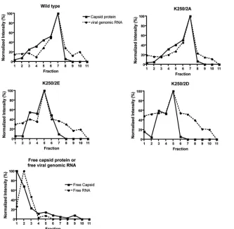
To determine if NC assembly was affected in the F2-G2 loop mutants, we performed a NC accumulation assay in BHK cells after electroporation ofin vitro-transcribed viral RNAs in a fash-ion similar to that described previously (29). Briefly, 8 h postelec-troporation, lysates of BHK cells were applied to a continuous rate-zonal gradient and centrifuged and the migration of Cp-con-taining complexes was determined by fractionation and
densi-tometry after immunoblot analysis. With the exception of the ⌬KGK mutant, equivalent amounts of total Cp were detected in the cell (data not shown). The migration of viral genomic RNA-containing complexes was also determined by fractionation and quantitative reverse transcriptase PCR (qRT-PCR).
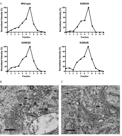
As shown inFig. 2, free Cp (45) and freein vitro-transcribed viral genomic RNA (assayed independently of each other) showed only slight sedimentation into the gradient. Lysates from cells transfected with the wild type and the K250/2A mutant demon-strated cosedimentation of Cp and viral genomic RNA to fraction 7, the position typically occupied by NC (data not shown). The K250/2D and K250/2E mutants, however, demonstrated sedi-mentation of the bulk of Cp- and genomic RNA-containing com-plexes to fractions 4 and 5, unlike the wild type, suggesting that, in these mutants, Cp-genomic RNA complexes are formed but are conformationally different from that of authentic NC. In addition to mock-electroporated cells, the⌬KGK mutant failed to generate full-length Cp in the cell (data not shown) and was not considered further. We unsuccessfully attempted to visualize the putative ag-gregates isolated from the K2502/2D or K250/2E gradient frac-tions by negative-stain electron microscopy.
To validate the lack of NC formation in the K250/2D and K250/2E mutant backgrounds, we examined transfected BHK cells by electron microscopy. In thin-section EM, we were unable
FIG 2Rate-zonal gradient analysis of infected cell lysates. BHK cells were electroporated within vitro-transcribed full-length RNA and hypotonically lysed at 8 h posttransfection. Clarified cell lysates were loaded onto an iodoxinol gradient and fractionated from the top of the gradient after 2.5 h. Fraction 1 refers to the top of the gradient, whereas fraction 11 was collected from the bottom. The amount of capsid protein and genomic RNA in each fraction was determined by immunoblotting and densitometry and by qRT-PCR, respectively. Theyaxis represents the normalized intensity in each fraction relative to the fraction containing the most abundant signal.
on November 7, 2019 by guest
http://jvi.asm.org/
[image:4.585.134.467.62.388.2]to identify NC structures in the cells transfected with the K250/2D and K250/2E mutants (data not shown), whereas we were able to identify such structures in the cells transfected with the wild type
(Fig. 3A). Using immuno-EM with an anti-Cp antibody, however,
we identified what appeared to be aggregates of Cp surrounding CPV-I replication vacuoles (Fig. 3B). These aggregates did not resemble the canonical NC structure. Taken together, the gradient and EM analyses suggested that, whereas the K250/2A mutant was restricted at a point after NC assembly, but before completion of the budding process, the K250/2D and K250/2E mutants were deficient in NC assembly.
The SINV Cp F2-G2 loop mutants assemble into core-like
particlesin vitro.Given the lack of knowledge of the NC assembly
process, we sought to interrogate NC assemblyin vitrousing the K250/2D and K250/2E mutants, as the gradient analysis suggested that the Cp aggregates found after expression of mutant Cp might represent a NC assembly intermediate. The wild-type and mutant Cp was expressed inEscherichia coliand purified using chroma-tography as previously described (45).
Like the wild type, the K250/2A mutant readily assembled into core-like particles (CLPs)in vitrousing a single-stranded DNA
template but, surprisingly, the K250/2D and K250/2E mutants also assembled into CLPs under identical standard assembly reac-tion condireac-tions as gauged by native agarose gel electrophoresis
(Fig. 4A) and negative-stain electron microscopy of assembly
re-actions (data not shown). Interestingly, though, when subjected to a temperature gradient, the K250/2D (data not shown) and K250/2E NC mutants appeared less thermostable than the wild type and the K250/2A mutants (Fig. 4B), suggesting that these charged residues may indeed be involved in intercapsomere con-tacts in the context of an assembled NC as previously suggested (46). Nevertheless, the mutant Cp is capable of assembling into NC-like structuresin vitroand, thus, some other factor is respon-sible for the lack of NC formationin vivofor the K250/2D and K250/2E Cp mutants.
When expressed ectopically, the SINV Cp F2-G2 loop
mu-tants assemble into NCs in the cell.The disparate results with
respect to NC assembly for the K250/2D and K250/2E mutants between infected cells andin vitroresults led us to assay the ability of the mutant Cp to assemble into NC in the cell when expressed alone using a mammalian expression vector. BHK cells were transfected with a plasmid expressing wild-type or mutant Cp and were lysed 18 h after transfection. The cell lysate was subjected to the same ultracentrifugation and densitometric analyses as before. As seen inFig. 5, all mutants produced Cp-containing structures that migrated in a manner similar to that seen with the wild type. Previously published work has shown that expression of the al-phavirus structural proteins ectopically leads to the formation and
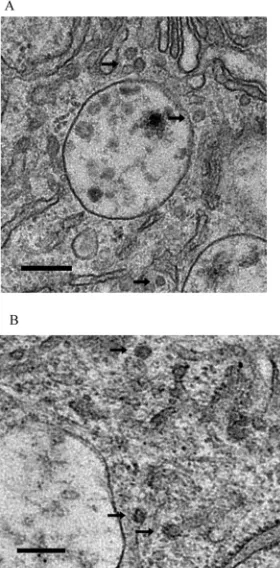
FIG 3Electron microscopic analysis of infected BHK cells. BHK cells were electroporated within vitro-transcribed infectious RNA and processed for EM at 8 h posttransfection, followed by staining with uranyl acetate. (A) Thin-section EM analysis of a cell infected with the wild type. Nucleocapsid cores in close proximity to CPV-II replication structures are readily apparent (black arrows). Cores were not detected via thin-section EM of cells infected with the K250/2D or K250/2E mutant (data not shown). Bar, 200 nm. (B) Immunogold EM analysis of a cell infected with the K250/2E mutant. Cells were stained with an anti-capsid primary antibody and a gold-conjugated secondary antibody in addition to uranyl acetate. Aggregates of protein assuming no regular shape were stained with the gold label (black arrows), and they lie on or near the surface of CPV-I replication vacuoles (white arrow). Bar, 200 nm.
FIG 4Agarose gel electrophoretic analysis ofin vitroassembly reactions. Cap-sid protein was expressed and purifiedin vitro. (A) Capsid and mutant proteins all assemble into core-like particles in a nucleic acid-dependent fashion. As-sembly reactions were carried out as described in the text. Aliquots of each reaction were electrophoresed on 0.8% agarose and stained with 0.05% Coo-massie blue. The small shift in mobility of the assembled CLPs was due to the difference in the pI of the proteins. The top of the image represents the cathode and the bottom the anode. The basic unassembled capsid protein can be seen migrating toward the cathode when nucleic acid is absent. (B) Assembled CLPs demonstrate a difference in levels of thermostability. Assembled CLPs were subjected to an increasing temperature gradient at the temperatures indicated for 15 min. Treated CLPs were analyzed by agarose gel electrophoresis as
described above.
on November 7, 2019 by guest
http://jvi.asm.org/
[image:5.585.93.235.68.350.2] [image:5.585.337.506.71.282.2]release of virus-like particles, and alphavirus NC assembly in cells that lack nucleic acid has not been described; thus, NCs presum-ably assemble here with the aid of cellular RNAs (1). Therefore, the complexes found in these cell lysates likely represent NCs and, as such, the K250/2D and K250/2E mutant Cp are also capable of NC assembly in cells. Taken further, there is apparently some fac-tor or environment in the infected cell that prevents authentic NC assembly using K250/2D and K250/2E mutant Cp.
When expressed via a SINV replicon, the SINV Cp F2-G2
loop mutants assemble into NCs in the cell.In the infected cell,
NCs are found in close proximity to CPV-I structures which com-prise both the site of viral RNA synthesis and the site of NC assem-bly (11). NCs also interact with the envelope proteins (23,25,31,
36,51) frequently during the viral life cycle. Thus, in an effort to determine whether it is the viral replicase, the viral envelope pro-teins, or the environment in an infected cell that is responsible for the conflicting results seen with infected cells versus the other experimental conditions with regard to NC assembly for the K250/2D and K250/2E mutants, we generated a SINV replicon expressing only the wild-type or mutant Cp. This construct, which lacked the sequence encoding the viral glycoproteins, was ex-pected to better mimic the environment of an infected cell, given the expression of the viral enzymatic proteins and the concurrent establishment of viral replicases on CPV-I replication vacuoles. As before, BHK cells were electroporated and the cell lysates were subjected to the same ultracentrifugation and densitometric anal-ysis. Again (Fig. 6A), the mutants all produced Cp-containing structures that migrated in a manner similar to that seen with the wild type and apparently represented NCs. Indeed, electron mi-croscopy studies validated the presence of NC structures for both the wild type and the mutants (Fig. 6B). To exclude the possibility
that the shorter length of the replicon nucleic acid, relative to that of the full-length viral genomic RNA molecule, played a role in rescuing NC assembly, we created a separate construct in which we replaced the fourth and fifth codons of E3 with stop codons. In this system, only Cp is produced via the subgenomic message, as in the replicon, but the replicated nucleic acid is the same length as in the full-length genome. Both the wt and the K250/2D Cp mu-tant were tested in this background, and we detected NC assembly in cell lysates after sedimentation analysis in the ultracentrifuge
(Fig. 7). Therefore, it appeared that it was the envelope proteins
that were responsible for the lack of NC assembly in infected cells expressing the K250/2D and K250/2E mutant Cp.
Double mutants containing mutations in both the Cp F2-G2 loop and cdE2 region I rescue NC assembly but not virus particle
release.Given the 7-Å SINV reconstruction, as well as previously
published biophysical analyses of residues involved in Cp:E2 in-teractions (22,44), we speculated that region I of the cytoplasmic domain of E2 (cdE2-RI), when present, may be responsible for the lack of NC assembly in the K250/2D and K250/2E mutant Cp backgrounds. We therefore created a set of double mutants in which cdE2-RI (cdE2 391-KARRE-395) and Cp residues K250 and K252 were simultaneously changed. Interestingly, gradient analysis of the double mutants demonstrated restoration of NC assembly in the K250/2D and K250/2E Cp backgrounds (Table 1). Lysates were scored as containing NCs (⫹) if the most intense Cp signal was found in fraction 6 or 7 or scored as lacking NCs if the most intense Cp signal was found in fraction 1, 2, 3, 4, or 5. Neg-ative-stain electron microscopy analyses of a subset of the double mutants validated the gradient findings (Fig. 8). Specifically, we identified electron-dense NC structures adjacent to CPV-I struc-tures for both the wt and K250/2E Cp mutant backgrounds
con-FIG 5Rate-zonal gradient analysis of cells transfected with a mammalian expression vector producing capsid protein. BHK cells were transfected with an expression plasmid producing wild-type or mutant capsid proteins and hypotonically lysed at 16 h posttransfection. Clarified cell lysates were loaded onto an iodoxinol gradient and fractionated after 2.5 h. The amount of capsid protein in each fraction was determined by immunoblotting and densitometry. Theyaxis represents the normalized intensity in each fraction relative to the fraction containing the most abundant signal.
on November 7, 2019 by guest
http://jvi.asm.org/
[image:6.585.137.455.65.318.2]currently with a lack of aggregates of Cp around these CPV-I structures using immuno-EM (data not shown). However, recov-ery of infectious particles as determined by plaque titration of posttransfected BHK cell media, or immediate agarose overlay after transfection of infectious RNA, was not restored for the K250/2D or K250/2E Cp background despite the formation of pinpoint-sized syncytium-derived plaques (Table 2). Further-more, modulation of the sequence of cdE2 R-I residues 393-RRE-395 also impaired the ability of virus with a wild-type or K250/2A Cp background to release infectious particles. Taken together,
these data suggest that cdE2-RI is important for efficient budding but also that cdE2-RI may interact with Cp at a prebudding stage, perhaps before or during NC assembly.
Ectopic expression of the entire structural polyprotein with Cp F2-G2 loop mutants yields NC assembly in the mammalian
cell.Next, we generated a set of mammalian expression plasmids
in which only the structural polyprotein, containing the wild-type envelope proteins, with either the wild-type Cp or the F2-G2 loop mutant Cp, was produced from a single message. Given the pres-ence of the wild-type cdE2 R-I sequpres-ence, and its implication in NC
FIG 6Analysis of cells transfected with Sindbis replicon expressing the capsid. BHK cells were electroporated within vitro-transcribed replicon RNA. (A) Cells were hypotonically lysed at 8 h posttransfection. Clarified cell lysates were loaded onto an iodoxinol gradient and fractionated after 2.5 h. The amount of capsid protein in each fraction was determined by immunoblotting and densitometry. Theyaxis represents the normalized intensity in each fraction relative to the fraction containing the most abundant signal. (B) Cells transfected with a SINV replicon expressing wt capsid protein were processed for negative-stain electron microscopy at 8 h posttransfection. Clusters of cores are indicated by black arrows. Bar, 200 nm. (C) Cells transfected with a SINV replicon expressing the K250E/K252E capsid protein were processed for negative stain and EM at 8 h posttransfection. Clusters of cores are indicated by black arrows. Bar, 200 nm.
on November 7, 2019 by guest
http://jvi.asm.org/
[image:7.585.83.509.60.535.2]assembly defects as described above, we expected to find a lack of NC structures in cells transfected with plasmids expressing the wild-type envelope proteins but the K250/2D and K250/2E mu-tant Cp. Cell lysates were subjected to the same gradient analysis described above, and, surprisingly, we detected molecules that sedimented like wild-type NC structures in all cases (Fig. 9). Given the absence of a viral replicase in this study, and the transfected cells devoid of CPV-I replication vacuoles, the data suggest that NC assembly may occur in a noncanonical fashion when the structural proteins are expressed ectopically or perhaps due to a lack of viral RNA synthesis. Therefore, the proposed cdE2:Cp in-teractions may occur only on virus-induced structures, which were absent in the transfected cells in the experiment described here. Indeed, although cdE2 R-I has been shown to interact with NCin vitro(22), we were unable to inhibitin vitroNC assembly of the wild type or the F2-G2 loop Cp mutants when GFP:cdE2 pro-tein (22) was spiked into assembly reaction mixtures at up to a 10-mole excess (data not shown).
DISCUSSION
Genetic and other evidence has convincingly implicated an inter-action of Cp with a conserved YxL motif on cdE2 as necessary for budding to occur (36,43,53). However, whether there are addi-tional Cp:cdE2 interactions, as well as the pathway by which the
assembled NCs as well as the envelope proteins traffic to the site of budding, remains unclear.
Recent evidence has suggested that NCs and envelope proteins cotraffic from cytoplasmic domains to the plasma membrane on virus-induced membrane structures termed cytopathic vacuoles type II (CPV-II), and NC and the envelope proteins likely interact on these CPV-II structures (19,41). Furthermore, a morphologi-cally distinct virus-induced membrane structure, termed cyto-pathic vacuole type I (CPV-I), likely contains the viral replicase and has been implicated as the site of initial NC assembly (11). Thus, NCs likely assemble on or near CPV-I structures prior to
[image:8.585.79.249.65.155.2]FIG 7Rate-zonal gradient analysis of cells transfected with viral RNA con-taining premature stop codons in E3. BHK cells were transfected within vitro -transcribed viral RNA producing either wild-type or K250/2D capsid protein in the absence of viral envelope protein expression. Cells were hypotonically lysed at 8 h posttransfection. Clarified cell lysates were loaded onto an iodoxi-nol gradient and fractionated after 2.5 h. The amount of capsid protein in each fraction was determined by immunoblotting and densitometry. Theyaxis represents the normalized intensity in each fraction relative to the fraction containing the most abundant signal.
TABLE 1Rate zonal gradient analysis of cell infected with Sindbis virus containing mutations in both capsid and cdE2a
Sequence
Presence or absence of NC
250KGK252 (wt) 250AGA252 250DGD252 250EGE252
391KARRE395(wt) ⫹ ⫹ ⫺ ⫺
391KAERE395 ⫹ ⫹ ⫹ ⫹
391KARRK395 ⫹ ⫹ ⫹ ⫹
391KARAA395 ⫹ ⫹ ⫹ ⫹
391KAARE395 ⫹ ⫹ ⫹ ⫹
391KAAAA395 ⫹ ⫹ ⫹ ⫹
aColumn headings represent the capsid protein primary sequences, and rows represent
the cdE2 primary sequences. Lysates were scored as containing NCs (⫹) if the most intense Cp signal was found in fraction 6 or 7 or scored as lacking NCs if the most intense Cp signal was found in fractionⱕ5. Boldface represents the amino acids mutated in this study.
FIG 8EM analysis of cells infected with double cdE2/capsid mutants. (A) Cells transfected with a SINV full-length infectious RNA expressing wt capsid protein in the cdE2 R393E background were processed for negative-stain elec-tron microscopy at 8 h posttransfection. Clusters of cores are indicated by black arrows. Bar, 200 nm. (B) Cells transfected with a SINV full-length infec-tious RNA expressing the K250E/K252E capsid protein in the cdE2 R393E background were processed for negative-stain electron microscopy at 8 h post-transfection. Clusters of cores are indicated by black arrows. Bar, 200 nm.
TABLE 2Plaque diameter analysis of cells infected with Sindbis virus containing mutations in both capsid and cdE2a
Sequence
Plaque diam (mm)
250KGK252 (wt) 250AGA252 250DGD252 250EGE252
391KARRE395 (wt) 3 2 ⬍1 ⬍1
391KAERE395 ⬍1 ⬍1 ⬍1 ⬍1
391KARRK395 2 1.5 ⬍1 ⬍1
391KARAA395 ⬍1 1.5 ⬍1 ⬍1
391KAARE395 ⬍1 ⬍1 ⬍1 ⬍1
391KAAAA395 ⬍1 ⬍1 ⬍1 ⬍1
a
Column headings represent the capsid protein primary sequences, and rows represent the cdE2 primary sequences.
on November 7, 2019 by guest
http://jvi.asm.org/
[image:8.585.350.490.68.352.2] [image:8.585.39.286.588.678.2] [image:8.585.301.543.612.706.2]their association with envelope proteins on CPV-II structures. It is unknown, however, whether any interactions exist between Cp/ NCs and the envelope proteins prior to their association on CPV-II vacuoles.
Here, we provide evidence that Cp and the E2 envelope protein may interact earlier than previously thought in the virion assem-bly pathway, perhaps in an event that precedes cotrafficking of NC and envelope proteins to the PM via CPV-II structures.
Modeling of cdE2 into and out of the hydrophobic pocket of Cp in a 7-Å cryo-EM reconstruction of SINV virus provided the opportunity for structure-guided mutagenesis (44). Specifically, Tang and colleagues suggested that a flexible loop on the Cp, the F2-G2 loop, was shifted⬃3.5 Å from its position observed in the crystal structure of SINV Cp (5,48). One residue in this loop, lysine 250, had been previously implicated in intercapsomere con-tacts within the NC (46), and Tang et al. suggested that the F2-G2 loop might move when the NC encounters the cdE2 domain of E2 (44). Furthermore, a second lysine in the F2-G2 loop, K252, was implicated as being a part of a salt bridge pair with D395 of cdE2 (44). Given these data, we performed structure-guided mutagen-esis, simultaneously mutating both K250 and K252 in the Cp F2-G2 loop to alanine, aspartic acid, or glutamic acid, and ex-pressed these proteins in a variety of ways.
Disruption of the lysines present in the F2-G2 loop led to a lack of NC assembly in the infected cell, with Cp found in aggregates around CPV-I vacuoles.In vitroassembly of the same mutant Cp, however, led to formation of core-like particles (CLPs). Interest-ingly, though, the NCs assembled from K250/2D and K250/2E Cp were not as thermostable as the wild type and the K250/2A mu-tant, in line with the proposal that at least one residue, K250, in the
F2-G2 loop might be involved in cross-capsomere contacts in the context of an assembled NC (46). A crystal structure of the SINV Cp revealed that K252 is involved in a salt bridge with Cp E111 (25). Residue E111 is part of the N-terminal arm of Cp shown to interact with the hydrophobic pocket of an adjacent Cp molecule in the crystal lattice and, thus, perhaps in the context of a NC prior to binding of cdE2 into the hydrophobic pocket. Taken together, these observations could explain the difference in thermostability. Despite these differences in thermostability, CLPs assembled with both wild-type and mutant Cp remained intact during gradient analysis ofin vitroassembly reactions (data not shown), and thus the differences in thermostability are unlikely to be related to the phenotype observed in the cell. Therefore, importantly, and un-expectedly, thein vitroassembly data demonstrated that the mu-tant Cp contains the information necessary to assemble into NC-like structures.
Expression of Cp in the cell by a SINV replicon or by a mam-malian expression plasmid with or without the viral envelope pro-teins also readily led to NC assembly in the cell, as gauged by gradient and negative-stain electron microscopy analyses. In the former system, not only were the viral nonstructural and the viral envelope proteins not produced but the reorganization of internal cellular membranes that are a hallmark of alphavirus infection (37) was also absent. Previously, overexpression of the alphavirus structural proteins in a similar fashion was shown to yield the formation and release of virus-like particles (1). In the latter sys-tem, the replicon that we created contained a SINV RNA in which the envelope protein gene segment was missing. In this replicon, Cp is still produced via translation of a subgenomic message; the viral replicase, the genomic RNA packaging signal, and enzymatic
FIG 9Rate-zonal gradient analysis of cells transfected with a mammalian expression vector producing the Sindbis structural polyprotein. BHK cells were transfected with an expression plasmid producing wild-type or mutant capsid protein, in addition to the wild-type envelope proteins, and hypotonically lysed at 16 h posttransfection. Clarified cell lysates were loaded onto an iodoxinol gradient and fractionated after 2.5 h. The amount of capsid protein in each fraction was determined by immunoblotting and densitometry. Theyaxis represents the normalized intensity in each fraction relative to the fraction containing the most abundant signal.
on November 7, 2019 by guest
http://jvi.asm.org/
[image:9.585.136.454.64.315.2]fected cell.
Previously published genetic and structural data strongly sug-gested that cdE2 interacts closely with a hydrophobic pocket on the surface of Cp (36,38,44). Jose and colleagues recently dem-onstrated that region I of cdE2 (cdE2-RI), comprised of E2 391-KARRE-395, specifically interacts with both NCs isolated from cells infected with SINV, as well asin vitro-assembled CLPs (22). cdE2 E395 has been specifically implicated in virus assembly (13), and Tang et al. proposed that cdE2 residue E395, part of region I, formed a salt bridge with Cp K252, a residue in the F2-G2 loop under investigation here (44). Thus, we speculated that cdE2-RI, when present, was responsible for the lack of NC assembly in the K250/2D and K250/2E mutant Cp backgrounds.
Double mutants in which the primary sequences of both the Cp F2-G2 loop and cdE2-RI were mutated showed that multiple different alterations of the cdE2 sequence 393-RRE-395 were ca-pable of restoring the ability of the K250/2D and K250/2E mutant Cp to incorporate into NCs in the cell. The fact that multiple different substitutions in this region of cdE2 rescued the NC as-sembly defect suggests that it is not necessarily one particular Cp: cdE2 side-chain interaction that is responsible for the K250/2D and K250/2E mutant phenotypes but perhaps more likely the shape or conformation of cdE2. Indeed, it has been suggested that, given the conservation of the positions of prolines and cysteines in cdE2 across the alphavirus genus, cdE2 might adopt a conserved conformation important for Cp binding (42).
While mutation of cdE2-RI restored the capacity for NC for-mation in the K250/2D and K250/2E backgrounds, it did not re-store the release of infectious particles. Except for the E2 E395K mutant, all other cdE2-RI mutants prevented release of infectious particles by virus containing wild-type Cp. That cdE2 region I might be important in virus assembly was also recently proposed by Jose et al. (22). Thus, cdE2-RI may have a multifaceted role in the virus assembly process through interaction with the Cp. Inter-estingly, as previously noted, some degree of covariance exists between cdE2-RI and the F2-G2 loop of Cp (22). Specifically, in most alphaviruses sequenced to date, when the Cp residue corre-sponding to SINV K250 is basic, the cdE2 residue correcorre-sponding to E395 is acidic, and when the Cp residue corresponding to K250 is acidic, the cdE2 residue is basic. Note that it is Cp residue K252, not K250, that is reported to form a salt bridge with cdE2 residue E395 (44); thus, modeling based on static images of virus particles and isolated NC may underscore the true fluidity of the cdE2 and Cp domains that apparently interact with one another.
When we assayed the ability of the K250/2D and K250/2E Cp mutants to assemble when they were ectopically expressed as part of the complete structural polyprotein, we expected that the K250/2D and K250/2E Cp mutants would not assemble into NCs. However, gradient analysis demonstrated the presence of Cp-con-taining structures that sedimented just like the wild type. These data suggest that, while cdE2 does play a role in the NC assembly defect phenotypes, the ultrastructure of the infected cell does as well. Indeed, in this system, CPV-I vacuoles are absent, and the
arrangement of viral proteins in the uninfected versus infected cell. Indeed, when GFP:cdE2 protein was added at up to a 10-mole excess intoin vitroassembly reactions using wild-type or mutant F2-G2 loop Cp, assembly of CLPs was unaffected, again demon-strating that the presence of cdE2 alone is not sufficient to recon-stitute the Cp:cdE2 interactions that occur in the infected cell.
The results described here provide additional insights into the temporal and spatial interactions between SINV Cp and cdE2. We demonstrated that Cp containing mutations in the F2-G2 loop still retained the information necessary to assemble into NCs. CPV-I vacuoles are proposed to be the site of normal NC assembly (11), but in cells transfected within vitrotranscripts containing the Cp mutation K250/2D or K250/2E, an accumulation of aggre-gates of Cp and viral genomic RNA complex at CPV-I vacuoles was detected. Given that this phenotype can be rescued by modu-lation of the primary sequence of cdE2-RI, a region already impli-cated in Cp:E2 interactions (44), and that, in the absence of these second-site mutations, K250/2D and K250/2E Cp form aggregates on CPV-I vacuoles, our data suggest that Cp and E2 may interact on or near CPV-I vacuoles, the site of NC assembly (11), soon after Cp synthesis.
It is tempting to speculate that cdE2 might act as a chaperone, aiding the assembly of NCs. Other viruses employ such a mecha-nism (7); however, additional data are required to interrogate this hypothesis. Another mechanistic possibility, and perhaps one that is more likely, is that changing the local charge of the Cp F2-G2 loop in the K250/2D and K2502/E mutants increases the affinity of Cp for cdE2. This could lead to an altered rate of nucleation be-tween Cp and cdE2 and the formation of aggregates. In the in-fected cell, the K250/2D Cp and the K2502/E Cp may become kinetically trapped by this interaction, unable to continue down the NC assembly pathway. Computational studies with other RNA viruses have demonstrated, counterintuitively, that increas-ing the affinity of a viral Cp for its cognate RNA leads to an aber-rant and incorrect NC assembly pathway by altering the rate of nucleation (8,21).
Nevertheless, the data presented here have provided additional knowledge concerning the interactions of Cp and E2 in the in-fected cell and, coupled with recent additions to the literature, have provided a new level of insight into the interactions and trafficking of the alphavirus NC and envelope proteins and allow us to propose a more detailed model regarding the NC assembly pathway. Wilkinson et al. proposed that Cp and RNA may initially associate on the cytoplasmic faces of intracellular membranes (51). Here, we show that Cp and cdE2 may interact on CPV-I vacuoles, although direct evidence of the presence of E2 in CPV-I structures has yet to be published. However, the analysis of alpha-virus chimeras with respect to genome replication kinetics as well as work with an anti-idiotypic antibody in which the original an-tigen was cdE2 have led to speculation that molecules of E2 might indeed be present in the replicase complex (42,43). Given that CPV-I vacuoles are derived from the plasma membrane (10), the
on November 7, 2019 by guest
http://jvi.asm.org/
ultimate destination for E2, the presence of the envelope proteins in or near the viral replicase may not actually be that surprising.
Soonsawad and colleagues showed that CPV-II vacuoles con-tain envelope proteins spikes, are decorated with NCs, and traffic from sites internal to the cell to the plasma membrane (41). Un-published electron tomography data (T. J. Edwards and R. J. Kuhn) have demonstrated the movement of assembled NCs from CPV-I vacuoles to CPV-II vacuoles, and recent studies using mi-croinjection of viral RNA into envelope protein-expressing cells demonstrated a preference for newly synthesized envelope pro-teins during envelopment of NCs (39).
Taking these data together, we can propose a more detailed model regarding the fate of Cp and envelope proteins in the cell. Specifically, newly translated Cp may associate with CPV-I mem-branes, interact with viral genomic RNA, and assemble into NC structures. Molecules of cdE2 resident in the CPV-I structures may temporarily interact with Cp before or during NC assembly. Assembled NCs may then traffic from CPV-I vacuoles to CPV-II vacuoles, where they interact with additional molecules of cdE2. Presumably, NC and cdE2 interact strongly here, perhaps via the conserved cdE2 YxL motif and the hydrophobic pocket on Cp. The C-terminal domain of cdE2 (cdE2-CTD) is reported to ini-tially transverse the membrane, flipping back and exposing cdE2-CTD to the cytosol well after translation in a phosphorylation-dependent manner (28). Thus, cdE2 may assume different conformations within the cell, providing scaffolds of different lev-els of affinity for Cp when embedded in CPV-I versus CPV-II membranous structures. A strong interaction between NC and cdE2 at CPV-II may tether NCs to CPV-II, and NCs and envelope proteins could cotraffic to the plasma membrane (41). The struc-ture, or perhaps composition, of CPV-II is such that premature budding is inefficient (41). Only when CPV-II reaches and fuses with the PM do the envelope proteins form a 2D lattice that is planar enough to envelop the NC, providing the free energy to bud a virus particle through the PM. This model, while attractive, is almost certainly overly simplistic and requires further studies be performed to completely elucidate the alphavirus virion assembly pathway.
The work presented here correlates with previously published genetic and structural analyses and expands our knowledge of alphavirus assembly. Taken together, our data (i) suggest that an early temporal and spatial interaction occurs between Cp and E2 at or near the CPV-I replication structures and (ii) provide a de-scription of a new set of cdE2:Cp amino acid interactions, involv-ing the N-terminal region of cdE2, that are important for alpha-virus NC assembly as well as budding.
ACKNOWLEDGMENTS
We thank Anita Robinson for clerical assistance as well as the Life Science Microscopy Facility and Biological Electron Microscopy Facility at Pur-due University.
We acknowledge support from the NIH through NIGMS award GM56279 to R.J.K. and NIH Biophysics Training Grants 5T32GM008296-21 and 5T32GM008296-22 to J.E.S.
REFERENCES
1.Akahata W, et al.2010. A virus-like particle vaccine for epidemic Chi-kungunya virus protects nonhuman primates against infection. Nat. Med.
16:334 –338.
2.Bonatti S, Migliaccio G, Blobel G, Walter P.1984. Role of signal recog-nition particle in the membrane assembly of Sindbis viral glycoproteins. Eur. J. Biochem.140:499 –502.
3.Byrnes AP, Griffin DE.1998. Binding of Sindbis virus to cell surface heparan sulfate. J. Virol.72:7349 –7356.
4.Cheng RH, et al.1995. Nucleocapsid and glycoprotein organization in an enveloped virus. Cell80:621– 630.
5.Choi HK, et al.1991. Structure of Sindbis virus core protein reveals a chymotrypsin-like serine proteinase and the organization of the virion. Nature354:37– 43.
6.Cristea IM, et al.2010. Host factors associated with the Sindbis virus RNA-dependent RNA polymerase: role for G3BP1 and G3BP2 in virus replication. J. Virol.84:6720 – 6732.
7.Dokland T.1999. Scaffolding proteins and their role in viral assembly. Cell. Mol. Life Sci.56:580 – 603.
8.Elrad OM, Hagan MF.2010. Encapsulation of a polymer by an icosa-hedral virus. Phys Biol.7:045003. doi:10.1088/1478-3975/7/4/045003. 9.Forsell K, Griffiths G, Garoff H.1996. Preformed cytoplasmic
nucleo-capsids are not necessary for alphavirus budding. EMBO J.15:6495– 6505. 10. Frolova EI, Gorchakov R, Pereboeva L, Atasheva S, Frolov I. 2010. Functional Sindbis virus replicative complexes are formed at the plasma membrane. J. Virol.84:11679 –11695.
11. Froshauer S, Kartenbeck J, Helenius A.1988. Alphavirus RNA replicase is located on the cytoplasmic surface of endosomes and lysosomes. J. Cell Biol.107:2075–2086.
12. Fuller SD, Berriman JA, Butcher SJ, Gowen BE.1995. Low pH induces swiveling of the glycoprotein heterodimers in the Semliki Forest virus spike complex. Cell81:715–725.
13. Gaedigk-Nitschko K, Schlesinger MJ.1991. Site-directed mutations in Sindbis virus E2 glycoprotein’s cytoplasmic domain and the 6K protein lead to similar defects in virus assembly and budding. Virology183:206 – 214.
14. Garoff H, Frischauf AM, Simons K, Lehrach H, Delius H.1980. Nucle-otide sequence of cdna coding for Semliki Forest virus membrane glyco-proteins. Nature288:236 –241.
15. Garoff H, Simons K.1974. Location of the spike glycoproteins in the Semliki Forest virus membrane. Proc. Natl. Acad. Sci. U. S. A.71:3988 – 3992.
16. Garoff H, Sjoberg M, Cheng RH.2004. Budding of alphaviruses. Virus Res.106:103–116.
17. Garoff H, Soderlund H.1978. The amphiphilic membrane glycoproteins of Semliki Forest virus are attached to the lipid bilayer by their COOH-terminal ends. J. Mol. Biol.124:535–549.
18. Gorchakov R, Garmashova N, Frolova E, Frolov I.2008. Different types of nsP3-containing protein complexes in Sindbis virus-infected cells. J. Virol.82:10088 –10101.
19. Griffiths G, et al.1989. The dynamic nature of the Golgi complex. J. Cell Biol.108:277–297.
20. Gualtieri EJ, et al. 2011. Detection of membrane protein two-dimensional crystals in living cells. Biophys. J.100:207–214.
21. Hagan MF, Elrad OM.2010. Understanding the concentration depen-dence of viral capsid assembly kinetics—the origin of the lag time and identifying the critical nucleus size. Biophys. J.98:1065–1074.
22. Jose J, et al.2012. Interactions of the cytoplasmic domain of Sindbis virus E2 with nucleocapsid cores promote alphavirus budding. J. Virol.86: 2585–2599.
23. Kail M, et al.1991. The cytoplasmic domain of alphavirus E2 glycopro-tein contains a short linear recognition signal required for viral budding. EMBO J.10:2343–2351.
24. Klimstra WB, Ryman KD, Johnston RE.1998. Adaptation of Sindbis virus to BHK cells selects for use of heparan sulfate as an attachment receptor. J. Virol.72:7357–7366.
25. Lee S, et al.1996. Identification of a protein binding site on the surface of the alphavirus nucleocapsid and its implication in virus assembly. Struc-ture4:531–541.
26. Liljeström P, Garoff H. 1991. Internally located cleavable signal se-quences direct the formation of Semliki Forest virus membrane proteins from a polyprotein precursor. J. Virol.65:147–154.
27. Linger BR, Kunovska L, Kuhn RJ, Golden BL. 2004. Sindbis virus nucleocapsid assembly: RNA folding promotes capsid protein dimeriza-tion. RNA10:128 –138.
28. Liu N, Brown DT. 1993. Transient translocation of the cytoplasmic (endo) domain of a type I membrane glycoprotein into cellular mem-branes. J. Cell Biol.120:877– 883.
29. Lopez S, Yao JS, Kuhn RJ, Strauss EG, Strauss JH.1994.
on November 7, 2019 by guest
http://jvi.asm.org/
ture similar to that of nucleocapsid cores in mature virus. J. Virol.76: 11128 –11132.
33. Mukhopadhyay S, et al.2006. Mapping the structure and function of the E1 and E2 glycoproteins in alphaviruses. Structure14:63–73.
34. Mulvey M, Brown DT.1996. Assembly of the Sindbis virus spike protein complex. Virology219:125–132.
35. Neuvonen M, et al.2011. SH3 domain-mediated recruitment of host cell amphiphysins by alphavirus nsP3 promotes viral RNA replication. PLoS Pathog.7:e1002383. doi:10.1371/journal.ppat.1002383.
36. Owen KE, Kuhn RJ. 1997. Alphavirus budding is dependent on the interaction between the nucleocapsid and hydrophobic amino acids on the cytoplasmic domain of the E2 envelope glycoprotein. Virology230: 187–196.
37. Schwartz M, Chen J, Lee WM, Janda M, Ahlquist P.2004. Alternate, virus-induced membrane rearrangements support positive-strand RNA virus genome replication. Proc. Natl. Acad. Sci. U. S. A.101:11263–11268. 38. Skoging U, Vihinen M, Nilsson L, Liljestrom P.1996. Aromatic inter-actions define the binding of the alphavirus spike to its nucleocapsid. Structure4:519 –529.
39. Snyder JE, et al.2011. Rescue of infectious particles from preassembled alphavirus nucleocapsid cores. J. Virol.85:5773–5781.
40. Söderlund H.1973. Kinetics of formation of the Semliki Forest virus nucleocapsid. Intervirology1:354 –361.
41. Soonsawad P, et al.2010. Structural evidence of glycoprotein assembly in
45. Tellinghuisen TL, Hamburger AE, Fisher BR, Ostendorp R, Kuhn RJ.
1999. In vitro assembly of alphavirus cores by using nucleocapsid protein expressed in Escherichia coli. J. Virol.73:5309 –5319.
46. Tellinghuisen TL, Kuhn RJ.2000. Nucleic acid-dependent cross-linking of the nucleocapsid protein of Sindbis virus. J. Virol.74:4302– 4309. 47. Thiboutot MM, et al.2010. Chikungunya: a potentially emerging
epi-demic? PLoS Negl. Trop. Dis.4:e623. doi:10.1371/journal.pntd.0000623. 48. Tong L, Choi HK, Minor W, Rossmann MG. 1992. The structure determination of Sindbis virus core protein using isomorphous replace-ment and molecular replacereplace-ment averaging between two crystal forms. Acta Crystallogr. A48(Pt 4):430 – 442.
49. von Bonsdorff CH, Harrison SC.1978. Hexagonal glycoprotein arrays from Sindbis virus membranes. J. Virol.28:578 –583.
50. Wahlberg JM, Bron R, Wilschut J, Garoff H.1992. Membrane fusion of Semliki Forest virus involves homotrimers of the fusion protein. J. Virol.
66:7309 –7318.
51. Wilkinson TA, Tellinghuisen TL, Kuhn RJ, Post CB.2005. Association of Sindbis virus capsid protein with phospholipid membranes and the E2 glycoprotein: implications for alphavirus assembly. Biochemistry44: 2800 –2810.
52. Zhang X, Fugere M, Day R, Kielian M.2003. Furin processing and proteolytic activation of Semliki Forest virus. J. Virol.77:2981–2989. 53. Zhao H, Lindqvist B, Garoff H, von Bonsdorff CH, Liljestrom P.1994.
A tyrosine-based motif in the cytoplasmic domain of the alphavirus enve-lope protein is essential for budding. EMBO J.13:4204 – 4211.