JOURNAL OFVIROLOGY, Sept. 2007, p. 9718–9726 Vol. 81, No. 18 0022-538X/07/$08.00⫹0 doi:10.1128/JVI.00746-07
Preferential Integration of Adeno-Associated Virus Type 2 into a
Polypyrimidine/Polypurine-Rich Region within
AAVS1
䌤
Victor J. McAlister and Roland A. Owens*
Laboratory of Molecular and Cellular Biology, National Institute of Diabetes and Digestive and Kidney Diseases, National Institutes of Health, Department of Health and Human Services, Bethesda, Maryland 20892
Received 5 April 2007/Accepted 5 July 2007
Adeno-associated virus type 2 (AAV2) preferentially integrates its genome into theAAVS1locus on human chromosome 19. Preferential integration requires the AAV2 Rep68 or Rep78 protein (Rep68/78), a Rep68/78 binding site (RBS), and a nicking site withinAAVS1and may also require an RBS within the virus genome. To obtain further information that might help to elucidate the mechanism and preferred substrate configurations of preferential integration, we amplified junctions between AAV2 DNA and AAVS1 from AAV2-infected HeLaJW cells and cells with defective Artemis or xeroderma pigmentosum group A genes. We sequenced 61 distinct junctions. The integration junction sequences show the three classical types of nonhomologous-end-joining joints: microhomology at junctions (57%), insertion of sequences that are not normally contiguous with either the AAV2 or theAAVS1sequences at the junction (31%), and direct joining (11%). These junctions were spread over 750 bases and were all downstream of the Rep68/78 nicking site withinAAVS1.Two-thirds of the junctions map to 350 bases ofAAVS1that are rich in polypyrimidine tracts on the nicked strand. The majority of AAV2 breakpoints were within the inverted terminal repeat (ITR) sequences, which contain RBSs. We never detected a complete ITR at a junction. Residual ITRs at junctions never contained more than one RBS, suggesting that the hairpin form, rather than the linear ITR, is the more frequent integration substrate. Our data are consistent with a model in which a cellular protein other than Artemis cleaves AAV2 hairpins to produce free ends for integration.
Adeno-associated virus type 2 (AAV2) is a naturally defec-tive human parvovirus that usually requires a helper virus, such as an adenovirus or a herpesvirus, for productive infection (41). In the absence of helper virus, AAV2 can establish a latent infection by episomal persistence (54) or by integrating its DNA into the host chromosomes, preferentially within a 4-kb region of human chromosome 19 designatedAAVS1(11). This is not site-specific integration in the classic sense, but approximately 70% of integration events occur somewhere within this locus (Fig. 1A) (20, 26, 51). The mechanism is unknown, but it does require the Rep68 or Rep78 (Rep68/78) protein encoded by AAV2 and a 33-bp region ofAAVS1(Fig. 1A) that includes a Rep68/78 binding site (RBS) and a nicking site for Rep68/78 that resembles the terminal resolution site (trs) within the 145-base inverted terminal repeats (ITRs) of the AAV2 genome (Fig. 2) (29, 57, 64, 67). Thetrsgot its name because of its role in AAV2 replication. AAV2 has a linear, single-stranded DNA genome (4,679 bases; GenBank acces-sion no. AF043303), and the ITRs are essential for replication. The ITRs are palindromic and fold into T-shaped hairpin structures, one of which provides a 3⬘end that primes second-strand synthesis by a cellular DNA polymerase (Fig. 2) (10, 65). The hairpin is then nicked by Rep68/78 attrs(Fig. 2) and unwound, and the end is replicated. As a result of this mode of
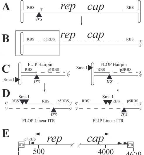
replication, the AAV2 ITRs can exist in two configurations, called flip and flop (Fig. 2) (10, 65).
Most published AAV2 breakpoints at integration junctions are near RBSs (11, 12, 46). Figure 2 shows that AAV2 has RBSs within its ITRs (bases 18 to 33, 93 to 108, 4572 to 4587, and 4647 to 4662) and near the promoter at map position 5 (p5) (bases 262 to 277) (33, 34, 42, 67). Resolution of the hairpin into a linear extended form results in a duplication of the hairpin sequence. The duplicated region includes the RBS within the ITR (Fig. 2D).
One curious feature is that all AAV2 junctions within the
4-kbAAVS1locus are to one side of the nicking site (11). This
side represents the direction in which unidirectional DNA syn-thesis (using the intact strand as a template) can be initiated by Rep68/78 nicking and a crude cell lysate in vitro (Fig. 1C) (64). Preferential integration has been detected in human and nonhuman primate tissue culture cell lines (2, 12, 13, 24, 27, 43, 47, 49, 53, 55, 62), cells and tissues of transgenic rodents con-taining the humanAAVS1locus (3, 48, 70), and one patient sample (35). However, each publication described only a small number of junctions. The goal of the work presented here was to generate numbers of junctions sufficient for subtle patterns in the junction sites to be detected, which might shed light on the mechanism of integration. Since AAV2-AAVS1junctions resemble those found with cellular nonhomologous DNA end joining, we also examined integration in two cell lines with known defects in DNA repair.
MATERIALS AND METHODS
Human cell lines and virus.The HeLaJW cell line, a derivative of a HeLa cervical carcinoma cell line that has previously been selected for attachment to plastic dishes (36), was grown in Dulbecco’s modified Eagle medium
supple-* Corresponding author. Mailing address: Laboratory of Molecular and Cellular Biology, National Institute of Diabetes and Digestive and Kidney Diseases, Bldg. 8, Rm. 307, National Institutes of Health, Department of Health and Human Services, 8 Center Drive MSC 0840, Bethesda, MD 20892-0840. Phone: (301) 496-3359. Fax: (301) 402-0053. E-mail: address: [email protected].
䌤Published ahead of print on 11 July 2007.
9718
on November 8, 2019 by guest
http://jvi.asm.org/
mented with 10% fetal bovine serum (FBS), penicillin, streptomycin, and neo-mycin (Gibco, Carlsbad, CA). A fibroblast line with a defective xeroderma pigmentosum group A (XPA) gene (repository identification no. GM04429) was obtained from Coriell Cell Repositories (Camden, NJ) and grown in minimal essential medium (Gibco) and 10% FBS. SC-3 cells, telomerase-immortalized skin fibroblasts from a patient that produces a defective Artemis protein (40), were a kind gift from Katheryn Meek of Michigan State University and were grown in Ham F-12 medium (Gibco), with antibiotics and 20% FBS.
Wild-type AAV2 was prepared as described previously (6), and titers were determined by DNA slot blot analysis. Cells were routinely infected at a multi-plicity of infection of approximately 9⫻103
viral genomes per cell.
Revisions to theAAVS1sequence.There are multiple differences between the initially reportedAAVS1sequence (GenBank accession no. S51329.1) (25) and the newer sequence generated as part of the sequencing of human chromosome 19 (GenBank accession no. AC010327.8). In the numbering system we use here, the complement base 11429 of the newer sequence (AC010327.8), which corre-sponds to base 1 of the old sequence (S51329.1), is designated base 1. The complement of base 7319 of the newer sequence (AC010327.8), which corre-sponds to base 4067 in the initially reported sequence (S51329.1) (25), is desig-nated base 4111 in the numbering system used in this work. By our new num-bering system, the Rep68/78 primary nicking site (64) is between T396 and T397. The RBS (67) runs from base 408 through base 423 (Fig. 1).
Integration assay.Cells were harvested 48 h after infection, and genomic DNA was isolated using a DNeasy tissue kit (QIAGEN, Valencia, CA). Primers 6-13-6-1 (5⬘-CAG GAC AGA GAT GTG TAC CTT CAG GG-3⬘[AAV2 bases 4021 to 4046]) and 6-13-6-4 (5⬘-AGG CAG ATA GAC CAG ACT GAG CTA TG G-3⬘[AAVS1bases 1239 to 1213]) were used in the first round of PCR ampli-fication, with 0.5 ng of genomic DNA as the substrate in a 50-l reaction volume. After an initial incubation for 4 min at 94°C, the reaction mixture was subjected to 28 cycles of PCR amplification for 1 min at 94°C, 1 min of annealing at 63°C using FastStart DNA polymerase (Roche) or 60°C using Herculase Faststart polymerase (Stratagene), and 3 min at 72°C. One percent of the amplification product was diluted into a new reaction mixture containing a set of nested primers, 6-13-6-2 (5⬘-TGG ACA CTA ATG GCG TGT ATT CAG AGC-3⬘
[AAV2 bases 4343 to 4369]) and 6-13-6-3 (5⬘-CAG GGA AGG AGA CAA AGT CCA GGA-3⬘[AAVS1bases 1182 to 1159]). The PCR parameters were the same as those for the first amplification. For a subset of integration assays, nested AAV2 primers 6-13-6-5 (5⬘-CTC AAT CAG ATT CAG ATC CAT GTC AGA
ATC TGG-3⬘[AAV2 bases 467 to 435]) and 6-13-6-6 (5⬘-TGG GGA CCT TAA TCA CAA TCT CGT AAA ACC-3⬘[AAV2 bases 357 to 328]) were used for the first and second PCRs, respectively. An additional subset of assays was per-formed using primer pairs 10-25-5-4 (5⬘-AGG ACA GAG ATG TGT ACC TTC AGG-3⬘[AAV2 bases 4022 to 4045]) and 8-15-5-13 (5⬘-CCC TGG AAG ATG CCA TGA C-3⬘[AAVS1bases 2034 to 2016]) for the first PCR and 8-25-5-3 (5⬘-ACG TAG ATA AGT AGC ATG GCG GGT-3⬘ [AAV2 bases 4496 to 4519]) and 8-15-5-7 (5⬘-CAG GGC AGG GAA GGA GAC [AAVS1bases 1187 to 1170]) for the second PCR, using Herculase Hotstart polymerase and an annealing temperature of 59°C for 30 cycles of amplification for each round of PCR.
Sequence analysis.Topoisomerase-mediated ligation was used to insert PCR products into plasmid pCR4TOPO (Invitrogen). The ligation products were then transformed intoEscherichia coliOne Shot TOP10 competent cells (Invitrogen). Plasmids for sequencing were purified using a QIAGEN plasmid mini kit or a QIAprep spin miniprep kit. Sequencing was performed by Asthagen, Inc. (Gaith-ersburg, MD), or MWG Biotech, Inc. (High Point, NC), using either M13 universal primers or the same AAV2 primers used for the second round of the nested PCR.
Candidate cloned junction sequences were first compared directly toAAVS1
[image:2.585.88.238.69.273.2]by use of the BLAST 2 sequence program (63), available at the website http: //www.ncbi.nlm.nih.gov/BLAST/bl2seq/wblast2.cgi. The filters (for low-complex-ity DNA and human repeats) had to be turned off, since there are multiple
FIG. 1.AAVS1locus. (A) Schematic of theAAVS1locus. Positions oftrsand the RBS and the range of reported AAV2 integration sites (integration zone) are indicated. (B) Expanded view of bases 1 to 2100, with both strands indicated. The two upper arrows represent the po-sitions of the first-round PCR primers, and the lower arrow represents the positions of the two second-round primers (arrows not drawn to scale). (C) Nicking attrscan result in covalent attachment of Rep68/78 (R) to the 5⬘side of the nick and induce unidirectional DNA synthesis
(dashed line). stranded AAV2 DNA (not drawn to scale). The ITRs are shown asFIG. 2. AAV2 replication. (A) Schematic of packaged single-hairpins, and the RBSs within the ITRs and the positions of the replication (rep) and capsid (cap) genes are indicated. For simplicity, only the minus strand is shown, even though the plus strand is pack-aged with the same efficiency. (B) Second-strand synthesis (dashed line) initiated from an ITR creates an additional RBS in the promoter for therepgene (p5RBS). (C) Expanded views of the region boxed in panel B, shown in the flip or flop configuration. (D) Rep68/78 nicking attrsand replication of the end of the genome (lower dashed line) result in conversion from flip hairpin to flop linear ITR and vice versa (as indicated by the change in the distance from the SmaI site[s] to the end of the genome). RBS⬘, second RBS created by replication of the ITR. (E) Schematic of the AAV2 genome, showing the positions of the PCR primers. The two upper arrows represent the positions of the first-round PCR primers, and the three lower arrows represent the posi-tions of the second-round primers (arrows not drawn to scale).
on November 8, 2019 by guest
http://jvi.asm.org/
[image:2.585.298.544.70.334.2]sequences betweenAAVS1bases 325 and 717 that are masked by these filters. The sequence was then compared to the GenBank nonredundant database by use of the BLASTn program (1), available at http://www.ncbi.nlm.nih.gov /BLAST/, to identify the AAV2 sequences. By convention, AAV2 sequences in databases have the left ITR in the flip configuration and the right ITR in the flop configuration, even though either conformation may exist at either end of the viral genome. The junction sequences were therefore checked manually against a flop left ITR and a flip right ITR sequence. Rare junction sequences with more than two ambiguously called bases within 15 bases of the junction were dis-carded, since this led to difficulty in identifying the precise junction point and determining whether or not the junction was distinct.
Putative binding motifs for cellular proteins were identified using the CONS ENSUS algorithm (17), applying the web-based interface offered at http://rsat .ccb.sickkids.ca. The relative nucleotide frequencies in all selected upstream regions were used to estimate the prior frequencies, which enter into the algo-rithm estimation of information content andPvalues. The expected matrix length was left at the default value of 10.
RESULTS
Previously reported data are consistent with the hypothesis that Rep68/78-mediated preferential integration intoAAVS1
employs elements of the cellular nonhomologous-end-joining DNA repair pathways. Since AAV2 integration is atypically specific, other cellular components may be involved. We set out to establish a baseline for junction characteristics by using HeLaJW cells (a derivative of a very commonly used human cell line) and then to look for qualitative differences in cell lines with known DNA repair defects, namely in the genes encoding the Artemis and XPA proteins. The Artemis protein is involved in locus-specific, nonhomologous end joining by cleaving hairpin DNA intermediates during immunoglobulin gene rearrangement (28, 30, 31, 40). Since fibroblasts from patients with Artemis gene defects are hypersensitive to ion-izing radiation (40), the protein is suspected to have a broader role in DNA repair. The XPA protein is believed to be in-volved primarily in the recognition of bulky DNA adducts, as part of nucleotide excision repair (52). We hypothesized that the covalent attachment of Rep68/78 to the 5⬘side of the nick (Fig. 1C) might be perceived as a bulky adduct. In addition, a recent report shows that XPA can bind tightly to transition points between single-stranded and double-stranded DNA (69) similar to those found in hairpin ITRs (Fig. 2).
We also make use of the latest sequence data for AAVS1. Most reports of AAV2-AAVS1 integration junctions use an older and less accurate sequence (GenBank accession no. S51329.1) (25; see reference 11 for a review). By mapping published junctions onto a newer, more correct AAVS1 se-quence (GenBank accession no. AC010327.8), we have deter-mined that several of the sequences at junctions that were reported as being of unknown origin are actually fromAAVS1
(data not shown). As a result, the frequency of junctions con-taining microhomologies has been underestimated and the fre-quency of junctions containing inserts has been overestimated.
Junction amplification. We used a nested-PCR system to amplify AAV2-AAVS1junctions (see Fig. 1 and 2 for primer locations). One set of primers was withinAAVS1and was used in conjunction with a primer set within the capsid gene that was designed to detect junctions at the right end of the AAV2 genome or a primer set within the AAV2rep gene that was designed to detect junctions at the left end of AAV2.
AAV2-AAVS1junction amplification products were ligated into a T/A
cloning vector and sequenced. Although junctions could be
defined with as little as 21 bases each of AAV2 andAAVS1
sequence, we typically obtained over 80 bases of sequence on the AAV2 side of the junction and over 100 bases on the
AAVS1side (data not shown). We found no differences in the
AAVS1sequences (other than those attributable to PCR
rep-lication errors) among the three cell lines.
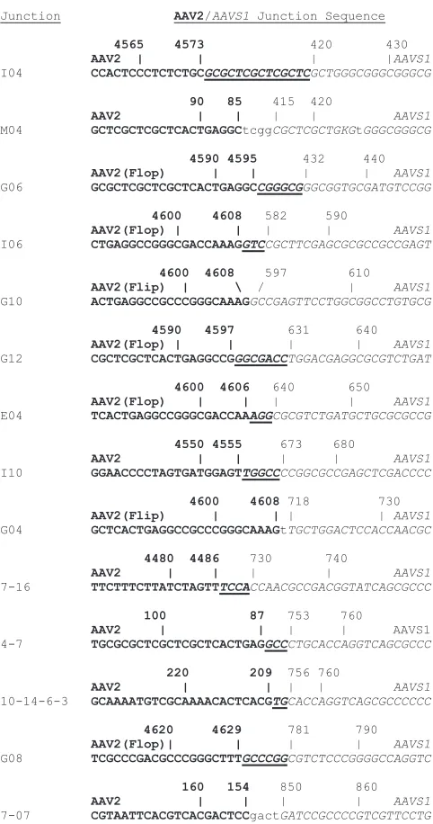
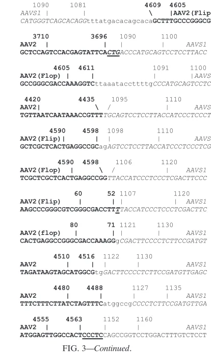
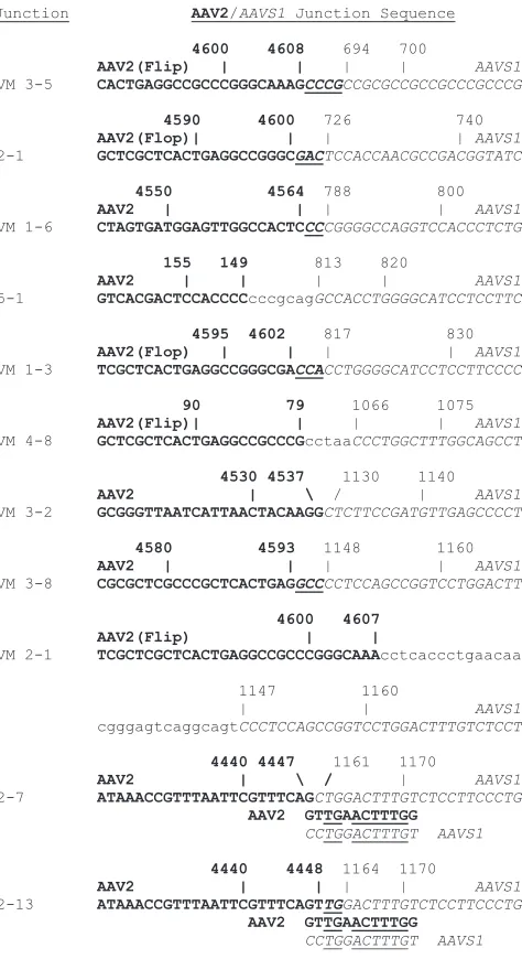
Classes of AAV2-AAVS1junctions. The junction sequences isolated from HeLaJW and XPA- and Artemis-defective cells are shown in Fig. 3, 4, and 5, respectively. Over 100 cloned PCR products were sequenced, yielding 61 distinct
AAV2-AAVS1junctions. Most of the junctions (57%) contained
[image:3.585.301.542.64.522.2]mi-crohomologies ranging from 1 to 14 bases that could have come from either AAV2 orAAVS1(Table 1). The second most
FIG. 3. AAV2-AAVS1 junctions from HeLaJW cells. AAV2 se-quences are shown in bold type.AAVS1sequences are in italics. Other sequences are in lowercase. Regions of microhomology are under-lined.
9720 MCALISTER AND OWENS J. VIROL.
on November 8, 2019 by guest
http://jvi.asm.org/
prevalent class overall (31%) was that of junctions with DNA inserted that was not normally contiguous with either AAV2 or
AAVS1 DNA. With the XPA-defective cells, these were as
frequent as microhomology junctions (Table 1), but given the small number of junctions, the significance of this observation is not clear. Several of these inserts contained 1 to 6 bases that represented a repeat of AAV2 orAAVS1DNA at or near the junction. For example, junction K08 (Fig. 3) contains a con-tiguous direct repeat of the last 2AAVS1bases (TT) within the insert. The insert of junction 8-9 (Fig. 3) contains a 3-base (TTT), noncontiguous direct repeat of AAV2 sequence near the junction and a 7-base (CAGCACA), noncontiguous direct repeat ofAAVS1sequence near the junction. Direct joining of AAV2 toAAVS1DNA represented the smallest class of junc-tions (11% overall) in all three cell types. Although our PCR assay is not sufficiently quantitative to detect even significant drops in integration efficiency, we can say that there does not
appear to be an absolute requirement for a fully functional Artemis or XPA protein in AAV2 preferential integration.
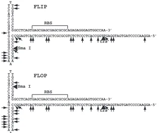
[image:4.585.40.308.63.521.2] [image:4.585.330.537.68.414.2]AAV2-AAVS1junctions do not contain more than one ITR RBS.Since preferential integration can be seen with input of either packaged, single-stranded AAV2 DNA or a plasmid containing AAV2 DNA, there is a question as to whether the preferred substrate is single-stranded or double-stranded DNA. Since many AAV2 junction breakpoints are within the ITRs, there is also a question of whether the linear or hairpin form of the ITR (Fig. 2) is preferred. In addition, several reports show that significant numbers of AAV2 breakpoints occur in or near the p5 promoter (11). Our PCR primers within the AAV2repand capgenes were designed to amplify junc-tions up to about 180 bases away from the left ITR (which includes the p5 RBS) and up to about 160 bases away from the right ITR, respectively (Fig. 2). However, many of the reported breakpoints in p5 region-AAVS1junctions result in a deletion of most of therepgene (12), where our left-end PCR primers anneal. It was therefore not surprising that the majority of AAV2 breakpoints we identified at either end are within the 145-base ITRs (Table 1). Figure 6 shows the distribution of AAV2 breakpoints within the ITRs presented in their hairpin conformations. In their extended linear conformation, the complementary sequences that make up the RBS separate to form two RBSs (Fig. 2) (60). Although there are multiple breakpoints within what would be the RBS furthest from the
FIG. 3—Continued.
FIG. 3—Continued.
on November 8, 2019 by guest
http://jvi.asm.org/
end of the genome in the linear ITR and within the upper and lower stem loops, none were detected in the RBS that would be closest to the end of the genome in the linear ITR (Fig. 2 and 6). As seen in Table 1, the breakpoints are distributed approximately evenly between the flip and flop configurations in those junctions for which the configuration could be deter-mined.
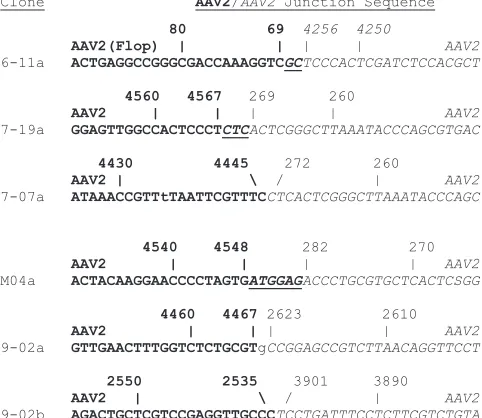
AAV2-AAV2 junctions. Several of the junctions we se-quenced contained AAV2-AAV2 junctions in addition to AAV2-AAVS1junctions (Fig. 7). For example, AAV2-AAV2 junctions 9-02a and 9-02b (Fig. 7) were both upstream of junction 9-02c (Fig. 3), all within the same PCR product. The AAV2 junctions shared several features with
AAV2-AAVS1junctions. Half of them (three of six) involved an ITR,
although none involved two ITRs. They also showed the three classical nonhomologous-end-joining joints, with three
micro-homology junctions, one with an insertion at the junction, and two direct joinings.
AAVS1 breakpoints cluster in a region rich in pPy tracts.
OurAAVS1breakpoints at junctions ranged from base 413,
[image:5.585.304.541.67.502.2]within theAAVS1RBS (67), through base 1163 (Fig. 8). Due to the positions of ourAAVS1primers, we could not detect junctions further downstream. We detected a small cluster of integration junctions immediately downstream of the nicking site withinAAVS1, near the region of homology with AAV2 that contains the RBS (Fig. 8), as had been seen in one pre-vious study (12). Over two-thirds (69%) of our distinct se-quenced junctions mapped toAAVS1bases 813 to 1163. One obvious feature of this region is a high frequency of polypyrim-idine (pPy) tracts on the strand that is nicked by Rep68/78.
FIG. 4. AAV2-AAVS1junctions from XPA-defective cells. AAV2 sequences are shown in bold type.AAVS1 sequences are in italics. Other sequences are in lowercase. Regions of microhomology are underlined.
FIG. 5. AAV2-AAVS1 junctions from Artemis-defective cells. AAV2 sequences are shown in bold type.AAVS1sequences are in italics. Other sequences are in lowercase. Regions of microhomology are underlined. Additional homologous sequences are shown below the sequences for junctions 2-7 and 2-13 and are underlined.
9722 MCALISTER AND OWENS J. VIROL.
on November 8, 2019 by guest
http://jvi.asm.org/
[image:5.585.47.286.69.480.2]Figure 8 shows that there are eight pPy tracts of 9 bases or more betweenAAVS1bases 800 and 1200. In contrast,AAVS1
bases 1 to 800 contain no pPy tracts of 9 bases or more. Further evidence for a correlation between pPy tracts and integration junctions is seen in the region betweenAAVS1bases 430 and 550, which contains no integration junctions and no pPy tracts of greater than 3 bases on the nicked strand (Fig. 8).
DISCUSSION
Several patterns have emerged from our analysis of
AAV2-AAVS1junctions. All integration junctions are on the same
side of the Rep68/78 nicking site ofAAVS1, consistent with the hypothesis that unidirectional DNA replication, initiated at the nick (Fig. 1), is somehow involved (64). Our data also suggest that the majority of junctions are the result of
microhomology-mediated end joining, which implies a strand invasion or sin-gle-strand annealing event as part of the mechanism.
In addition, about two-thirds of junctions were found in a 350-base region enriched for pPy tracts on the nicked strand of
AAVS1. One trivial explanation for this clustering of junctions
could be that the amplification reaction is more efficient over shorter distances (18). However, it should also be noted that there is not a perfect correlation between the distance from the
AAVS1primer and the frequency of integration junctions. For
[image:6.585.44.542.84.152.2]example, we detected six junctions betweenAAVS1bases 851 and 900 but only two junctions betweenAAVS1bases 951 and 1000 (Fig. 8). In addition, at least six different studies using proximalAAVS1primers ranging from bases 1209 to 1621 have reported two or more junctions within the region from base 800 to base 1200 (18, 27, 43, 48, 49, 51, 53). This is one of the few regions in which all of the above-cited works reported
FIG. 6. Mapping of AAV2-AAVS1and AAV2-AAV2 junction breakpoints on the AAV2 ITR. For simplicity, only hairpin ITRs at the 3⬘end of the genome are shown. Narrow arrowheads indicate the breakpoints (estimated for microhomology junctions). Broad arrowheads indicate the positions of thetrsand SmaI sites. Both the flip and flop hairpins are shown. Breakpoints that could not be unambiguously mapped to flip or flop are indicated on both hairpins.
TABLE 1. Comparison of AAV2-AAVS1junction type frequencies among cell lines
Cell line Defective geneb
No. (%) of distinct junctionsa
Microhomology Insertion at
junction Direct joining Total ITR Flip
c Flopc
HeLa NA 24 (62) 11 (28) 4 (10) 39 31 (79) 8 (20) 12 (31)
GM04429 XPA 5 (45) 5 (45) 1 (9) 11 9 (82) 3 (27) 3 (27)
SC-3 Artemis 6 (55) 3 (27) 2 (18)d 11 8 (73) 3 (27) 2 (18)
aOnly one instance of a particular junction sequence was counted.
bOther defects that have not been characterized may also exist. NA, not applicable. cCounting only junctions in the hairpin region that are unambiguously flip or flop. dPossible microhomeology in one junction.
on November 8, 2019 by guest
http://jvi.asm.org/
[image:6.585.133.458.415.684.2]AAV2-AAVS1 junctions. Due to the small number of se-quences analyzed in each of these studies, the pattern was not detected.
Polypurine (pPu) tracts and their complementary pPy tracts are believed to stimulate recombination (4, 21–23). They can form triplex DNA, which can stabilize strand invasion events (21–23). In addition, pPy tracts as short as 18 bp have been demonstrated to promote oligonucleotide-directed gene cor-rection at least 23 bp away in vivo and strand invasion (D-loop formation) at sites up to 4 kb away in vitro (4). As most of our junctions were outside the actual pPy tracts, we hypothesize that smaller pPy tracts may be able to stimulate recombination over shorter distances. A minimum of 9 bp was arbitrarily set for identifying a pPy tract because the longest pPu tract on the nicked strand from bases 1 through 1200 is 8 bases. It should also be noted that there is a 21-base pPy tract comprised of
AAVS1bases 1717 to 1737 (data not shown), which is in theory
large enough to stimulate strand invasion throughout the
en-tireAAVS1locus. There are also 10-base pPy/pPu tracts within
the AAV2 ITRs (bases 8 to 17, 109 to 118, 4562 to 4571, and 4663 to 4672), which may contribute to the ITRs’ role in recombination (50, 60).
In addition to the intrinsic properties of pPu/pPy tracts, there are cellular proteins that bind specifically to these se-quences. For example, recent work shows that a complex of pPy tract binding protein-associated splicing factor with a 54-kDa nuclear RNA-binding protein may be involved in DNA double-strand-break rejoining (5). Also, the human purine-rich element binding protein alpha (PUR␣) is a single-stranded DNA-binding protein that binds preferentially to the purine-rich element termed PUR, which is present at origins of rep-lication and in gene-flanking regions in a variety of eukaryotes (14, 15, 32, 56, 61, 68). PUR␣is believed to be involved in the control of both DNA replication and transcription and can
promote the unwinding of double-stranded DNA containing pPu/pPy tracts (68). Computer-assisted analysis (17) of the junction sites revealed that several were near potential binding sites for the human PUR␣protein (data not shown).
We also identified potential binding sites for transcription factors SP1 and MZF1 (38) near several of our AAV2-AAVS1
junctions (data not shown). SP1 has been reported to bind Rep68/78, and this interaction may allow the creation of DNA loops which might help to direct or restrict integration (16, 44, 45). SP1 is also a substrate for DNA-dependent protein kinase, a key protein in nonhomologous end joining (9, 19).
[image:7.585.302.541.71.339.2]The pattern of AAV2 breakpoints at junctions is explained most easily by the ITRs being in the hairpin, as opposed to the linear conformation, prior to junction formation (Fig. 2 and 6). If the breakpoints were random or strictly dependent on the primary base sequence, we would expect to see at least a few breakpoints within the RBS nearest to the end of the genome if the linear ITR were the more common substrate. As with our data in Fig. 6, a recent large-scale study ofrep⫺AAV2 vector “random” integration shows a similar lack of cellular junction breakpoints within the terminal 40 bases of the AAV2 ITR (37). This pattern is therefore Rep independent, indicating that it is determined by cellular proteins. Another recent study of the concatemerization and circularization of rep⫺ AAV2 vectors also concluded that a forced hairpinned ITR is pre-ferred, as a recombination substrate, to an ITR that could exist in either a hairpinned or an extended linear conformation (8). We detected several complex junctions which involved one
FIG. 7. AAV2-AAV2 junctions. AAV2 sequences upstream of the junction are shown in bold type. AAV2 sequences downstream of the junction are in italics. Other sequences are in lowercase. Regions of microhomology are underlined.
FIG. 8. Mapping of AAV2-AAVS1 junction breakpoints onto
AAVS1. pPy tracts of greater than 8 bases are in bold type. The 2 bases flankingtrs(T396 and T397) are in lowercase, and the RBS (bases 408 to 423) is in lowercase italics. Breakpoints found at one or more junctions are underlined. For microhomology junctions, the center of the microhomology is underlined.
9724 MCALISTER AND OWENS J. VIROL.
on November 8, 2019 by guest
http://jvi.asm.org/
[image:7.585.43.284.72.281.2]or two AAV2-AAV2 junctions in addition to an AAV2-AAVS1
junction. We hypothesize that such junctions result from cir-cularization of AAV2 genomes by cellular proteins, followed by a Rep68/78-induced opening of the circles, prior to forming the junctions withAAVS1. Recent work has demonstrated that in the absence of Rep68/78, circularization efficiency can be greater than 90% within 24 h postinfection (7, 8). Work from other groups suggests that circularization is a dead end for integration (39, 58, 59). Since Rep68/78 is required for the efficient rescue of AAV2 ITR-containing DNA from plasmids (66), it is reasonable to speculate that Rep68/78 can resolve AAV2 circles, producing ends that can recombine with AAV2 or chromosomal DNA.
In theory, our assay should have detected so-called head-to-head and tail-to-tail fusions of AAV2 genomes. Such fusions are common replicative intermediates during productive infec-tion (65) and have been detected previously in patient samples (54). Our results indicate that these are rare with a wild-type AAV2 infection in the absence of helper virus. This observa-tion is also consistent with circularizaobserva-tion being an efficient reaction compared to dimerization of the AAV2 genome. Cir-cularization of AAV2 monomer DNA would result in a head-to-tail junction that would not be amplified by our system.
We originally hypothesized that Artemis might stimulate recombination by cleaving hairpins within the ITRs, but our data suggest that a protein other than Artemis can cleave the AAV2 hairpins. We cannot, however, rule out the possibility that Artemis is one of several proteins that cleaves the ITR.
We have also identified two junctions (Fig. 5) consistent with homeologous recombination (recombination between slightly mismatched sequences). Junctions 2-7 and 2-13 are at approx-imately the same sites, just upstream of an 11-base pPy tract in
AAVS1.Although technically defined as a direct joining and a
2-base microhomology junction, respectively, each had 8 of 10 bases in common between AAVS1 and AAV2 (Fig. 5). We hypothesize that the recombination events were initiated by a strand invasion event, followed by the use of the partially mismatched sequence as a primer for DNA synthesis. This would have been followed by DNA mismatch repair that in some cases corrected the mismatch in favor of AAV2 and in other cases corrected in favor of AAVS1. Microhomeology junctions were detected only with the Artemis-defective cells. Since the Artemis protein is not known to be involved in homeologous recombination, we suspect that this is the result of a secondary defect.
Based on our observations, we propose a novel predominant mechanism for Rep68/78-mediated integration intoAAVS1in which a hairpinned ITR is first processed by a cellular endo-nuclease to produce a 3⬘overhang. This overhang then anneals to its complementary sequence within the intact strand of
AAVS1, which has been exposed by DNA unwinding ahead of
an active or stalled replication complex assembled at the Rep68/78 nicking site. This annealed 3⬘end is then extended by a DNA polymerase to create the first de facto joining of AAV2 toAAVS1DNA sequences. Further analysis of AAV2 integration in cells with known defects in DNA damage repair/ signaling should help to further constrain models of preferen-tial integration.
ACKNOWLEDGMENTS
We thank Peggy Hsieh, Anthony Furano, and Kevin Gardner for their critical reading of the manuscript as well as for other useful advice. We thank Sven Bilke for assistance with the sequence analysis. We thank Irving Miller for supplying much of the purified AAV2 used in this study and J. Rodney Brister and Dik van Gent for useful technical advice. We also thank Katheryn Meek for supplying the Artemis cell line.
R.A.O. is a coinventor on several patents related to recombinant AAV technology. To the extent that the work in the manuscript in-creases the value of this patent, he has a conflict of interest.
This research was supported by the Intramural Research Program of the NIH, National Institute of Diabetes and Digestive and Kidney Diseases.
REFERENCES
1.Altschul, S. F., T. L. Madden, A. A. Scha¨ffer, J. Zhang, Z. Zhang, W. Miller, and D. J. Lipman.1997. Gapped BLAST and PSI-BLAST: a new generation of protein database search programs. Nucleic Acids Res.25:3389–3402. 2.Amiss, T. J., D. M. McCarty, A. Skulimowski, and R. J. Samulski.2003.
Identification and characterization of an adeno-associated virus integration site in CV-1 cells from the African green monkey. J. Virol.77:1904–1915. 3.Bakowska, J. C., M. V. Di Maria, S. M. Camp, Y. Wang, P. D. Allen, and
X. O. Breakefield. 2003. Targeted transgene integration into transgenic mouse fibroblasts carrying the full-length humanAAVS1locus mediated by HSV/AAV rep⫹hybrid amplicon vector. Gene Ther.10:1691–1702. 4.Biet, E., J. S. Sun, and M. Dutreix.2003. Stimulation of D-loop formation by
polypurine/polypyrimidine sequences. Nucleic Acids Res.31:1006–1012. 5.Bladen, C. L., D. Udayakumar, Y. Takeda, and W. S. Dynan.2005.
Identi-fication of the polypyrimidine tract binding protein-associated splicing factor●p54(nrb) complex as a candidate DNA double-strand break rejoining factor. J. Biol. Chem.280:5205–5210.
6.Carter, B. J., C. A. Laughlin, L. M. de la Maza, and M. Myers.1979. Adeno-associated virus autointerference. Virology92:449–462.
7.Choi, V. W., D. M. McCarty, and R. J. Samulski.2006. Host cell DNA repair pathways in adeno-associated viral genome processing. J. Virol.80:10346– 10356.
8.Choi, V. W., R. J. Samulski, and D. M. McCarty.2005. Effects of adeno-associated virus DNA hairpin structure on recombination. J. Virol.79:6801– 6807.
9.Collis, S. J., T. L. DeWeese, P. A. Jeggo, and A. R. Parker.2005. The life and death of DNA-PK. Oncogene24:949–961.
10.Cotmore, S. F., and P. Tattersall.2006. A rolling-hairpin strategy: basic mechanisms of DNA replication in the parvoviruses, p. 171–188.InJ. R. Kerr, S. F. Cotmore, M. E. Bloom, R. M. Linden, and C. R. Parrish (ed.), Parvoviruses. Edward Arnold Limited, London, United Kingdom. 11.Dutheil, N., and R. M. Linden.2006. Site-specific integration by
adeno-associated virus, p. 213–236.InJ. R. Kerr, S. F. Cotmore, M. E. Bloom, R. M. Linden, and C. R. Parrish (ed.), Parvoviruses. Edward Arnold Lim-ited, London, United Kingdom.
12.Giraud, C., E. Winocour, and K. I. Berns.1995. Recombinant junctions formed by site-specific integration of adeno-associated virus into an episome. J. Virol.69:6917–6924.
13.Giraud, C., E. Winocour, and K. I. Berns.1994. Site-specific integration by adeno-associated virus is directed by a cellular DNA sequence. Proc. Natl. Acad. Sci. USA91:10039–10043.
14.Gupta, M., V. Sueblinvong, J. Raman, V. Jeevanandam, and M. P. Gupta.
2003. Single-stranded DNA-binding proteins PUR␣and PURbind to a purine-rich negative regulatory element of the␣-myosin heavy chain gene and control transcriptional and translational regulation of the gene expres-sion. Implications in the repression of␣-myosin heavy chain during heart failure. J. Biol. Chem.278:44935–44948.
15.Herault, Y., G. Chatelain, G. Brun, and D. Michel.1993. The PUR element stimulates transcription and is a target for single strand-specific binding factors conserved among vertebrate classes. Cell. Mol. Biol. Res.39:717–725. 16.Hermonat, P. L., A. D. Santin, and R. B. Batchu.1996. The adeno-associated virus Rep78 major regulatory/transformation suppressor protein binds cel-lular Sp1 in vitro and evidence of a biological effect. Cancer Res.56:5299– 5304.
17.Hertz, G. Z., and G. D. Stormo.1999. Identifying DNA and protein patterns with statistically significant alignments of multiple sequences. Bioinformatics
15:563–577.
18.Hu¨ser, D., S. Weger, and R. Heilbronn.2002. Kinetics and frequency of adeno-associated virus site-specific integration into human chromosome 19 monitored by quantitative real-time PCR. J. Virol.76:7554–7559. 19.Jackson, S. P., J. J. MacDonald, S. Lees-Miller, and R. Tjian.1990. GC box
binding induces phosphorylation of Sp1 by a DNA-dependent protein ki-nase. Cell63:155–165.
20.Kearns, W. G., S. A. Afione, S. B. Fulmer, M. G. Pang, D. Erikson, M. Egan,
on November 8, 2019 by guest
http://jvi.asm.org/
M. J. Landrum, T. R. Flotte, and G. R. Cutting.1996. Recombinant adeno-associated virus (AAV-CFTR) vectors do not integrate in a site-specific fashion in an immortalized epithelial cell line. Gene Ther.3:748–755. 21.Knauert, M. P., and P. M. Glazer.2001. Triplex forming oligonucleotides:
sequence-specific tools for gene targeting. Hum. Mol. Genet.10:2243–2251. 22.Knauert, M. P., J. M. Kalish, D. C. Hegan, and P. M. Glazer.2006. Triplex-stimulated intermolecular recombination at a single-copy genomic target. Mol. Ther.14:392–400.
23.Knauert, M. P., J. A. Lloyd, F. A. Rogers, H. J. Datta, M. L. Bennett, D. L. Weeks, and P. M. Glazer.2005. Distance and affinity dependence of triplex-induced recombination. Biochemistry44:3856–3864.
24.Kotin, R. M., and K. I. Berns.1989. Organization of adeno-associated virus DNA in latently infected Detroit 6 cells. Virology170:460–467.
25.Kotin, R. M., R. M. Linden, and K. I. Berns.1992. Characterization of a preferred site on human chromosome 19q for integration of adeno-associ-ated virus DNA by non-homologous recombination. EMBO J.11:5071–5078. 26.Kotin, R. M., M. Siniscalco, R. J. Samulski, X. Zhu, L. Hunter, C. A. Laughlin, S. McLaughlin, N. Muzyczka, M. Rocchi, and K. I. Berns.1990. Site-specific integration by adeno-associated virus. Proc. Natl. Acad. Sci. USA87:2211–2215.
27.Lamartina, S., G. Roscilli, D. Rinaudo, P. Delmastro, and C. Toniatti.1998. Lipofection of purified adeno-associated virus Rep68 protein: toward a chro-mosome-targeting nonviral particle. J. Virol.72:7653–7658.
28.Lees-Miller, S. P., and K. Meek.2003. Repair of DNA double strand breaks by non-homologous end joining. Biochimie85:1161–1173.
29.Linden, R. M., E. Winocour, and K. I. Berns.1996. The recombination signals for adeno-associated virus site-specific integration. Proc. Natl. Acad. Sci. USA93:7966–7972.
30.Ma, Y., U. Pannicke, K. Schwarz, and M. R. Lieber.2002. Hairpin opening and overhang processing by an Artemis/DNA-dependent protein kinase complex in nonhomologous end joining and V(D)J recombination. Cell
108:781–794.
31.Ma, Y., K. Schwarz, and M. R. Lieber.2005. The Artemis:DNA-PKcs en-donuclease cleaves DNA loops, flaps, and gaps. DNA Repair4:845–851. 32.Ma, Z. W., A. D. Bergemann, and E. M. Johnson.1994. Conservation in
human and mouse Pur alpha of a motif common to several proteins involved in initiation of DNA replication. Gene149:311–314.
33.McCarty, D. M., D. J. Pereira, I. Zolotukhin, X. Zhou, J. H. Ryan, and N. Muzyczka.1994. Identification of linear DNA sequences that specifically bind the adeno-associated virus Rep protein. J. Virol.68:4988–4997. 34.McCarty, D. M., J. H. Ryan, S. Zolotukhin, X. Zhou, and N. Muzyczka.1994.
Interaction of the adeno-associated virus Rep protein with a sequence within the A palindrome of the viral terminal repeat. J. Virol.68:4998–5006. 35.Mehrle, S., V. Rohde, and J. R. Schlehofer.2004. Evidence of chromosomal
integration of AAV DNA in human testis tissue. Virus Genes28:61–69. 36.Miller, B. W., and J. Williams.1987. Cellular transformation by adenovirus
type 5 is influenced by the viral DNA polymerase. J. Virol.61:3630–3634. 37.Miller, D. G., G. D. Trobridge, L. M. Petek, M. A. Jacobs, R. Kaul, and D. W.
Russell.2005. Large-scale analysis of adeno-associated virus vector integra-tion sites in normal human cells. J. Virol.79:11434–11442.
38.Morris, J. F., R. Hromas, and F. J. Rauscher III.1994. Characterization of the DNA-binding properties of the myeloid zinc finger protein MZF1: two independent DNA-binding domains recognize two DNA consensus se-quences with a common G-rich core. Mol. Cell. Biol.14:1786–1795. 39.Nakai, H., S. Fuess, T. A. Storm, L. A. Meuse, and M. A. Kay.2003. Free
DNA ends are essential for concatemerization of synthetic double-stranded adeno-associated virus vector genomes transfected into mouse hepatocytes in vivo. Mol. Ther.7:112–121.
40.Noordzij, J. G., N. S. Verkaik, M. van der Burg, L. R. van Veelen, S. de Bruin-Versteeg, W. Wiegant, J. M. Vossen, C. M. Weemaes, R. de Groot, M. Z. Zdzienicka, D. C. van Gent, and J. J. van Dongen.2003. Radiosensitive SCID patients with Artemis gene mutations show a complete B-cell differ-entiation arrest at the pre-B-cell receptor checkpoint in bone marrow. Blood
101:1446–1452.
41.Owens, R. A.2006. Latent infection of the host cell by AAV and its disrup-tion by helper viruses, p. 237–252.InJ. R. Kerr, S. F. Cotmore, M. E. Bloom, R. M. Linden, and C. R. Parrish (ed.), Parvoviruses. Edward Arnold Lim-ited, London, United Kingdom.
42.Owens, R. A., M. D. Weitzman, S. R. M. Kyo¨stio¨, and B. J. Carter.1993. Identification of a DNA-binding domain in the amino terminus of adeno-associated virus Rep proteins. J. Virol.67:997–1005.
43.Palombo, F., A. Monciotti, A. Recchia, R. Cortese, G. Ciliberto, and N. La Monica.1998. Site-specific integration in mammalian cells mediated by a new hybrid baculovirus–adeno-associated virus vector. J. Virol.72:5025– 5034.
44.Pereira, D. J., and N. Muzyczka.1997. The adeno-associated virus type 2 p40 promoter requires a proximal Sp1 interaction and a p19 CArG-like element to facilitate Rep transactivation. J. Virol.71:4300–4309.
45.Pereira, D. J., and N. Muzyczka.1997. The cellular transcription factor SP1 and an unknown cellular protein are required to mediate Rep protein acti-vation of the adeno-associated virus p19 promoter. J. Virol.71:1747–1756. 46.Philpott, N. J., C. Giraud-Wali, C. Dupuis, J. Gomos, H. Hamilton, K. I.
Berns, and E. Falck-Pedersen.2002. Efficient integration of recombinant adeno-associated virus DNA vectors requires a p5-repsequence incis. J. Vi-rol.76:5411–5421.
47.Pieroni, L., C. Fipaldini, A. Monciotti, D. Cimini, A. Sgura, E. Fattori, O. Epifano, R. Cortese, F. Palombo, and N. La Monica.1998. Targeted inte-gration of adeno-associated virus-derived plasmids in transfected human cells. Virology249:249–259.
48.Recchia, A., L. Perani, D. Sartori, C. Olgiati, and F. Mavilio.2004. Site-specific integration of functional transgenes into the human genome by adeno/AAV hybrid vectors. Mol. Ther.10:660–670.
49.Rinaudo, D., S. Lamartina, G. Roscilli, G. Ciliberto, and C. Toniatti.2000. Conditional site-specific integration into human chromosome 19 by using a ligand-dependent chimeric adeno-associated virus/Rep protein. J. Virol.74:
281–294.
50.Ruffing, M., H. Heid, and J. A. Kleinschmidt.1994. Mutations in the carboxy terminus of adeno-associated virus 2 capsid proteins affect viral infectivity: lack of an RGD integrin-binding motif. J. Gen. Virol.75:3385–3392. 51.Samulski, R. J., X. Zhu, X. Xiao, J. D. Brook, D. E. Housman, N. Epstein,
and L. A. Hunter.1991. Targeted integration of adeno-associated virus (AAV) into human chromosome 19. EMBO J.10:3941–3950.
52.Sancar, A., L. A. Lindsey-Boltz, K. U¨ nsal-Kac¸maz, and S. Linn.2004. Mo-lecular mechanisms of mammalian DNA repair and the DNA damage check-points. Annu. Rev. Biochem.73:39–85.
53.Satoh, W., Y. Hirai, K. Tamayose, and T. Shimada.2000. Site-specific inte-gration of an adeno-associated virus vector plasmid mediated by regulated expression of Rep based on Cre-loxPrecombination. J. Virol.74:10631– 10638.
54.Schnepp, B. C., R. L. Jensen, C. L. Chen, P. R. Johnson, and K. R. Clark.
2005. Characterization of adeno-associated virus genomes isolated from human tissues. J. Virol.79:14793–14803.
55.Shelling, A. N., and M. G. Smith.1994. Targeted integration of transfected and infected adeno-associated virus vectors containing the neomycin resis-tance gene. Gene Ther.1:165–169.
56.Shimotai, Y., H. Minami, Y. Saitoh, Y. Onodera, Y. Mishima, R. J. Kelm, Jr., and K. Tsutsumi.2006. A binding site for Pur␣and Puris structurally unstable and is required for replication in vivo from the rat aldolase B origin. Biochem. Biophys. Res. Commun.340:517–525.
57.Snyder, R. O., R. J. Samulski, and N. Muzyczka.1990.In vitroresolution of covalently joined AAV chromosome ends. Cell60:105–113.
58.Song, S., P. J. Laipis, K. I. Berns, and T. R. Flotte.2001. Effect of DNA-dependent protein kinase on the molecular fate of the rAAV2 genome in skeletal muscle. Proc. Natl. Acad. Sci. USA98:4084–4088.
59.Song, S., Y. Lu, Y. K. Choi, Y. Han, Q. Tang, G. Zhao, K. I. Berns, and T. R. Flotte.2004. DNA-dependent PK inhibits adeno-associated virus DNA in-tegration. Proc. Natl. Acad. Sci. USA101:2112–2116.
60.Srivastava, A., E. W. Lusby, and K. I. Berns.1983. Nucleotide sequence and organization of the adeno-associated virus 2 genome. J. Virol.45:555–564. 61.Stacey, D. W., M. Hitomi, M. Kanovsky, L. Gan, and E. M. Johnson.1999.
Cell cycle arrest and morphological alterations following microinjection of NIH3T3 cells with Pur␣. Oncogene18:4254–4261.
62.Surosky, R. T., M. Urabe, S. G. Godwin, S. A. McQuiston, G. J. Kurtzman, K. Ozawa, and G. Natsoulis.1997. Adeno-associated virus Rep proteins target DNA sequences to a unique locus in the human genome. J. Virol.
71:7951–7959.
63.Tatusova, T. A., and T. L. Madden.1999. BLAST 2 sequences, a new tool for comparing protein and nucleotide sequences. FEMS Microbiol. Lett.174:
247–250.
64.Urcelay, E., P. Ward, S. M. Wiener, B. Safer, and R. M. Kotin.1995. Asymmetric replication in vitro from a human sequence element is depen-dent on adeno-associated virus Rep protein. J. Virol.69:2038–2046. 65.Ward, P.2006. Replication of adeno-associated virus DNA, p. 189–211.In
J. R. Kerr, S. F. Cotmore, M. E. Bloom, R. M. Linden, and C. R. Parrish (ed.), Parvoviruses. Edward Arnold Limited, London, United Kingdom. 66.Ward, P., E. Urcelay, R. Kotin, B. Safer, and K. I. Berns.1994.
Adeno-associated virus DNA replication in vitro: activation by a maltose binding protein/Rep 68 fusion protein. J. Virol.68:6029–6037.
67.Weitzman, M. D., S. R. M. Kyo¨stio¨, R. M. Kotin, and R. A. Owens.1994. Adeno-associated virus (AAV) Rep proteins mediate complex formation between AAV DNA and its integration site in human DNA. Proc. Natl. Acad. Sci. USA91:5808–5812.
68.Wortman, M. J., E. M. Johnson, and A. D. Bergemann.2005. Mechanism of DNA binding and localized strand separation by Pur␣and comparison with Pur family member, Pur. Biochim. Biophys. Acta1743:64–78.
69.Yang, Z., M. Roginskaya, L. C. Colis, A. K. Basu, S. M. Shell, Y. Liu, P. R. Musich, C. M. Harris, T. M. Harris, and Y. Zou.2006. Specific and efficient binding of xeroderma pigmentosum complementation group A to double-strand/single-strand DNA junctions with 3⬘- and/or 5⬘-ssDNA branches. Bio-chemistry45:15921–15930.
70.Young, S. M., D. M. McCarty, N. Degtyareva, and R. J. Samulski.2000. Roles of adeno-associated virus Rep protein and human chromosome 19 in site-specific recombination. J. Virol.74:3953–3966.
9726 MCALISTER AND OWENS J. VIROL.