Copyright ( 1972 American Society forMicrobiology Printed inU.S.A.
Temperature-Dependent
Transformation
of
Cells
Infected with
aMutant of
Bryan
Rous
Sarcoma
Virus
JOHN P. BADER
ChemistryBranch, NationtalCancer Institute, NationalInstitutesofHealth, Bethesda, Maryland20014
Received forpublication 18 April 1972
Chick embryocells infected with a mutant (Ta) of the Bryan high-titer strain of Rous sarcoma virus (RSV-BH) are morphologically transformed at 36 C but
appearsimilartouninfected cellsat41C. Whencellsinfected with RSV-BH-Taare
switchedfrom 41 to 36 C, morphological changes characteristic oftransformation
areobservable within 10min. The transformation is
reversible;
cells shifted from 36 to41 C havebeenobservedtolosetheir transformedmorphology within1 hr. The transformation afterashift intemperatureis unaffected by inhibitionof deoxyribo-nucleic acid (DNA), ribonucleic acid (RNA), or protein synthesis, demonstratingthat theproteins involvedinthemorphological change arealreadypresent. Trans-formedcells infected with RSV-BH orRSV-BH-Tatake up hexose and synthesize
hyaluronic acidat higherrates than uninfected cells orRSV-BH-Ta-infected cells grown at 41 C. However, inhibition of either protein or RNA synthesis, but not
DNAsynthesis, prevented the inductionof increased hexose uptake andhyaluronic acidsynthesis aftera shift ofRSV-BH-Ta-infected cellsfrom 41 to36 C.Therefore, these biochemical changes are secondary to a more basic change responsible for
morphologicaltransformation.
Several investigators have described the
iso-lation of avian sarcoma virusmutantswith
tem-perature-sensitive properties. Some mutants can go through full cycles of reproduction at both
high (41 C) and low(36 C)temperatures, but the
infected cells become transformed only at the low temperature (6, 11, 13, 19). Other mutants aretemperaturedependentforvirusreproduction
as well as transformation (21). All of these mutants were derived from either the
Schmidt-Ruppin or B77 strains ofavian sarcoma virus. Transformation by these viruses is recognized by theincreased refractility or roundingofcells,
or both, morphological changes which often accompany malignant transformation in other systems.
Transformation ofchickembryo fibroblastsby
the Bryanhigh-titerstrain ofRous sarcomavirus (RSV-BH) ismorphologically unique, consisting
of a change from elongated to polygonal or
rounded shape, and the appearance of
large
numbers of vacuoles. Single transformed cells can be identified against a background of
non-transformed cells (15). Both lethal and
tempera-ture-dependent mutations in RSV-BH have been found after exposure of newly infected cells to 5-bromodeoxyuridine (4, 5). One mutant
isolate,
RSV-BH-Ta, was selected for its ability to re-produce at 41 and 36 C and to transform cells
only at the lower temperature. Several featuresof cells infected with RSV-BH-Ta were examined
andaredescribedhere.
MATERIALS AND METHODS
Cell cultures. Chick embryo cells were prepared
from
10-day-old embryos and were replated at in-tervals asdescribed previously (15). Growth medium consisted ofEagleminimalessential medium(MEM) supplemented with dextrose (2 g/liter finalconcen-tration), sodium pyruvate (5 mM),
10%7c
tryptosephosphate broth (Difco), 5% fetal bovine serum,
penicillin (50
,.gg/ml),
streptomycin (50,ug/ml), and tylosine (50 ,Ag/ml). Cultures were maintained in humidified, C02-atmosphere incubators. Cells in50-mm plastic petridishes wereused beforereaching confluency.
Virus. RSV-BH is the Bryan high-titer strain of
RSVo mixed withanontransformingRous-associated virus
(RAVy).
Virus mutants were obtained aftertreatmentofnewly infected cellswith 5-bromodeoxy-uridine (5). Mutants were selected for ability to
re-produce atboth41 and 36 C, andall containRAV1. The mutants ofthe transformingparticles may have
mutations in other regions ofthe virusgenome, but if the products ofthese mutant regionsare required
for virus reproduction they are complemented by
267
on November 10, 2019 by guest
http://jvi.asm.org/
BADER
RAV1 (5). Themutant RSV-BH-Ta described herein
was propagated in chick embryo cells by infecting
cells atahigh multiplicity of virus tocell, maintain-ing the growmaintain-ing cells at 39 C through two passages at 3-day intervals, and collecting the cell culture fluids. New cells were infected monthly and were
used in transformation experiments within 1 to 6
weeks after infection. During this time, practically allcells assumed the morphology of transformed cells
uponincubationat36C.
The focus formation method (20) was used for
assaysof Rous sarcomavirus, and infectiousvirus is
counted in focus-forming units (FFU). Focus agar medium consisted of the above growth mediumwith
3j'- calf serum substituting for fetal bovine serum, and with the addition of1% beefembryo extractand
0.8%o agar. When RSV-BH-Ta was being assayed,
cultures were incubated for 2 days at 41 C before being shiftedto 36 C for an additional 5 days.
Neu-tralred (0.01%inEagle MEM) waslayeredontothe
nutrientagarthe day before microscope scanning for foci.
The assay method for thegrowth ofinfected cells
incolonies suspended in softagarhasbeen described
(2).
Antimetabolites. Cytosine arabinoside
(NIH-CCNSC) at 10-4 M reduced incorporation of
deoxy-thymidine-3H into deoxyribonucleic acid (DNA) to
less than 10% within 30 min. Actinomycin D (2
mg/ml; Merck, Inc.) reduced incorporation of uri-dine-:1H into ribonucleic acid (RNA) to less than
5% within 30 min. Cycloheximide (10,g/ml; Cal-biochem) and puromycin (50 jig/ml; Calbiochem) reduced incorporation of leucine-14C into protein to
less than 2% within 30min. When cytosine arabino-side was used, tryptose phosphate broth was elimi-nated from the medium.
Hyaluronic acid synthesis. Hyaluronic acidcontent
was determined by quantitative electrophoresis in
polyacrylamide-agarose mixed gels (J. P. Bader, D. A. Ray, and T. L. Steck, Biochim. Biophys. Acta,
inpress). Cell culture fluids were applied directly to
thegels aftertreatmentfor 15 min with 0.1 NNaOH. Cellular cytoplasms were separated from nuclei
aftertreatment with0.5%cl Nonidet P-40(J. P. Bader
et al., in press). These cytoplasmic extracts were
treated with Pronase (50jg/ml; 37 C; 30min), then 0.1 N NaOH (15 min; room temperature). The
samples were dialyzed, lyophilized to dryness, then resuspended in theelectrophoresis buffer atone-fifth the original volume. Nonconcentrated cytoplasmic samples taken directly after treatment with Pronase and NaOH gave comparable results, as determined
by viewing of stained gels, but amounts of
hyalu-ronateweretoolow forquantitation.
Hexose uptake. The rate of hexose uptake was
determined by the incorporation ofdeoxyglucose-3H (9). Culture fluids were removed, and the cells were
rinsed once with Dulbecco's phosphate-buffered
sa-line (PBS). Deoxyglucose-3H (2,Ci in 2 ml of PBS)
was added, and all cultures were incubated at 39 C
for15min. (Under theseconditions deoxyglucose-3H
isincorporated at alinear rateforat least 30 min.) ColdPBSwasaddedtothecultures, and the cellswere
rinsed fourtimes withcoldPBS. NonidetP-40 (0.5c%0 in PBS) was added, and after 15 min at room tem-perature the cytoplasmic fluids were analyzed for radioactivity and protein (12).
RESULTS
Reproduction of RSV-BH-Ta. The mutant, RSV-BH-Ta, was selected for its ability to re-produce withoutrestriction over the temperature range 36 to 41 C. Chick embryo cells were ex-posed to a high multiplicity ofRSV-BH-Ta, the cells were incubated, and the amount of progeny virus appearing in the cell culture fluidsthe next day was determined (Table 1). Reproduction occurred at all temperatures. Inhibition of the appearanceof progeny virusby cytosine arabino-side demonstrated that the usual reproductive cycle wasresponsible for the appearance of virus, rather than merely the persistence and release of superficially adsorbed virions.
Focus formation and growth of colonies in agar. WVhen cells were infected with RSV-BH-Ta at either high (>2 FFU/cell) or low (<1 FFU/ cell) multiplicity and cultures were maintained in liquid growth medium, no morphological trans-formation was observed at 41 C (Fig. 1). Similar cultures incubated at 36 C became transformed to about the same extent as wild-type RSV-BH, athigh andlow multiplicities of infection.
The development of foci of transformed cells under agar was also examined. Cells were in-fected with dilutions of RSV-BH or RSV-BH-Ta, then were overlaid with focus agar medium and incubated at 41 or 36 C (after 2 initial days at 41 C). Seven days later, the number of recog-nizable foci appearing at 36 C exceeded by 100-foldthe number at 41 C (Table 2).
[image:2.499.269.462.494.599.2]The cells comprising the foci at 36 C were deeply stained with neutral red, a characteristic ofRSV-BH-transformed cells. Foci induced by
TABLE 1. Reproductionz of RSV-BH-Ta at various
temperatures"
FFU X 10-'perml Cells infected with
36C 37C 39C 41C RSV-BH ... 140b 150 180 260
RSV-BH-Ta... 240 340 360 420
RSV-BH-Ta +
ara-C ... 2.3 2.1 0.7 1.6
Chick embryo cells were exposed to about 3
focus-forming units (FFU) per BH or RSV-BH-Ta cell. Growth medium or medium
contain-ing cytosine arabinoside (ara-C, 10-4 M) was
added, and24hrlater culture fluidswere removed for subsequent assay.
268 J. VIROL.
on November 10, 2019 by guest
http://jvi.asm.org/
TEMPERATURE-DEPENDENT TRANSFORMATION BY RSV
269
/,,5
P.J
{t
'
'j4;,/
,;-' j
A,
Jlvon
[image:3.499.47.433.67.576.2]tj1;~~~~~~~~~~~~~~~
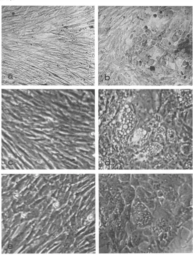
FIG. 1. Effects oftemperattare onl morphology of cells in2fected wit/i RSV-BH-Ta. Chlick embryo cells were
infected wit/i RSV-BHorRSV-BH-Ta anidtranisferredseveraltimes. Light microscopy (X160) ofRSV-BH-Ta infectedcellsat41 C (a)a,idat36 C(b). Plhase-conitrast micrography (X256) ofnoninrJctedchickembryo cells at41 (c), RSV-BH-infectedcellsat41 C(d), RSV-BH-Ta-inifectedcells at 41C(e),anldat36 C(f).
RSV-BH-Ta at 41 C usually could be identified
only as bare areas or "plaques" in the cellular
monolayer,aphenomenonoftenfoundwhenfoci
of RSV-BH-transformed cells develop under
nutrientagar. Such plaque formation isprobably due to the loss of adherence of transformed cells
to the substratum, since transformed cells are
found piled up at the periphery of the plaque.
VOL. 10, 1972
on November 10, 2019 by guest
http://jvi.asm.org/
TABLE2. Focusformation and suspeiided colony formation of infected cellsathigh aid low temperatures
-No. of focia No.ofcoloniesb
Cellsnfecte with Dilutionof_______________________ ___________________
Cellsinfected wvith virus
36C 41C 36C 41C
RSV-BH 10-2 Confluent Confluent 171 390
10-3 504 726 . 6 19
104 42 68 0 0
RSV-BH-Ta 10iO Confluent (66)c Confluent 700
10-12 Confluent (16) 370 57
10-° 540
(3)
24 71O-4 48 0 0 0
a Sparse monolayers were exposed to various dilutions of RSV-BH or RSV-BH-Ta, then overlaid with focus agarmedium. Six days later, neutral red (0.01%') wasadded, andfoci were observed witha
microscope thenext day.
IColonies of cellsgrowing suspended in nutrient agar. Cellsweredispersed withtrypsin,exposedto
virus, then mixed with soft nutrient agar andlayeredinto 50-mmpetri dishes. Ten (41 C) and12 (36 C) days later colonies werecountedwith the aid ofamicroscope.
cNumbersin parentheses representatypical foci composed of lightly stained cells, incontrast to the
darkly stained cells of RSV-BH foci.
However, deeply stained cells were rarely found in thefoci of RSV-BH-Ta-infected cells develop-ing at 41 C.
In the same experiment, infected cells were
suspendedin softnutrient agar, and theabilityof these cells toformcolonies (2) wasexamined. A temperature restriction on the growth of RSV-BH-Ta-infected cells in suspension was found, compared with cells infected with the wild type (Table 2). However, some colonies were formed
at 41 C even after infection at low multiplicity;
thenumber at 41 C was about one-fifth the num-ber at 36 C. The generally lower efficiency of
colony formationat36C,asshownby the num-ber of colonies developingafterinfection ofcells
with RSV-BH, indicates that the number of colonies developing from RSV-BH-Ta-infected cells was about 10-fold greaterat 36 than 41 C. The growth at 41 C of RSV-BH-Ta-infected cells into colonies suspended in agar, or into plaques in a monolayer, may be due to either a
partial reversion for the transformation
pheno-typein somevirions, or to ageneral "leakiness" of RSV-BH-Ta.
Cells infected with RSV-BH-Ta athigh
multi-plicity could be grown and subcultured at 41 C for several weekswhile maintaining essentially a normal appearance. At any time, these cultures could be shifted to 36 C, and morphological changes soon became apparent. All of the fol-lowingstudies were done with cultures infected at highmultiplicity and in which practically all cells were carrying transforming virus.
Time course of transformation and reversal. Cells infected with RSV-BH-Ta were grown at 41 C, then shifted to 36 C andexamined with a
microscope at periodic intervals for morphologi-cal changes. By using standard light microscopy we detected some rounding of the cells by 1 hr after changing temperature. Within 3 to 4 hr, cell shapes and intercellular associations were clearly different from cel-ls kept at 41 C, and by12 hr the cultures were indistinguishable from cul-tures infected with wild-type RSV-BH.
Cytoplasmic vacuolization is a characteristic
feature of cells transformed by RSV-BH or RSV-BH-Ta, and is rarely found in uninfected chick embryo cells or RSV-BH-Ta-infected cells kept at 41 C. This vacuolization is more readily
detectable by using phase-contrast microscopy than standard light microscopy. Examination of RSV-BH-Ta-infected cultures by phase-contrast microscopy revealed newly formed vacuoles in 1 to 5%O of the cells within 10 min after shifting from 41 to 36 C (Table 3, Fig. 2). By 20 to 30 min, the vacuolization was extensive; vacuoles were found throughout the cytoplasm of many
cells, and as many as half the cells contained vacuoles. Vacuolization often was found to precede achange in cell shape.
The transformation was reversible.
Trans-formedcells maintained at 36 C lost their vacuoles andbecameelongated after being shiftedto41C. This reversal of vacuolization was less readily detectable than the appearance of vacuoles after a shift from 41 to 36 C (Table 3). Both the
generalcellmorphologyof thetransformedcells,
as seenin thelight microscope, andthedegreeof vacuolization, as assessed by phase-contrast mi-croscopy, took considerably longer to revert to normal with increased temperature than to be transformed afteratemperatureshiftdown.Also,
270 BADER J. VIROL.
on November 10, 2019 by guest
http://jvi.asm.org/
TEMPERATURE-DEPENDENT TRANSFORMATION BY RSV
t
A
A
9
.
.
l
i
|
i.g
,,:e,
-
jkaEZ
!hi;
11"|
115i
Xffif
*_.s w | x
111, Eiii f.
j6,.-gu.ts
<F- 4| | S |Es k _.
'
SIIF3--B-w
;.;>--w " F!w z.
:'s
S So
|
....v
.
.
3s_s3t--aiW*...
.,
g
i.s
§
ll
5K
1
u3
tq'
^|^'
-^e
..^'
-
t_<
3...1..S;
[image:5.499.50.443.82.442.2],S.,t;,.;'..tSa o l.
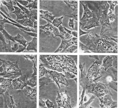
FIG. 2. Effectsoftemperatureshiftotnmorphology ofRSV-BH-Ta iiifected cells. Cellsgrowntat 41C(a) were
shiftedto36 Candobserved byphase-coiitrastmicrographly after 10 miii (b), 15miii (c), 20 miii (d), 25miin(e), anid30 min (I). Note the inierease inperinuclear vacuiolizationz withl time at36 C.
whencellswere grownfor2ormoredays at 36 C, the reversal to the nontransformed state at 41 C took several hours longer than if the cells had
originally been maintained for less than 12 hr at 36C.
Effects ofantimetabolites. The rapidity ofthe
morphological changes occurring in shifted cul-tures indicated that the degree ofcomplexity of metabolic reactions was small. The specific metabolic requirements for transformation to occur in "shifted-down" cultures was examined by using cytosine arabinoside to inhibit DNA synthesis, actinomycin D to inhibit RNA
syn-thesis, and cycloheximide or puromycin to in-hibitproteinsynthesis.Noneofthesecompounds
prevented the appearance of vacuoles or gross transformation in RSV-BH-Ta-infected cultures
when-added simultaneously with a shift of the cultures from 41 to 36 C (Table 4; Fig. 3). No differences were observed either in the rate of morphologicalchanges or in the number ofcells exhibiting these changes during thefirst 6 hrafter treatment and temperature change. Also, pre-incubation of cells with antimetabolites at the higher temperature for upto6 hrbefore shifting to thelower temperature had little effect onthe morphological change occurring duringthe 2 hr after temperature shift.
Theseresults demonstrated that transcriptional and translationalrequirementsformorphological transformation had been fulfilledat 41C.Aslight decrease in the extent of vacuolization was no-ticed in cultures treated with cycloheximide or puromycinforlongerthan 8 hr (Fig. 3), although
VOL. 10, 1972 271
on November 10, 2019 by guest
http://jvi.asm.org/
BADER
TABLE3. Timecourseofappearanceofand reversal
of traisformationa
Transformation Time after shift
41 -+36C 36 +41C
0 0
++++
10 min + ++++
20min ++ ++++
30 min ++ ++++
I hr ++ +++
3 hr +++ +
6 hr +++ +
12 hr ++++ 0
aCultures wereplaced at41 or36C 1 dayafter
a cell transfer. The following day, cultures were shifted to the opposite temperature. At periodic intervals, cultures were removed from the incu-bators along with cultures which had not been shifted, and each was observed by both light and phase-contrast microscopy. The degree of trans-formationwasrelated to that of RSV-BH infected cultures. Symbols: 0, no detectable vacuolization; +,detectable vacuolization in less than10%of the cells; ++, vacuoles easily recognizable in5-20%o of the cells; +++, vacuolization of 20 to 50% of the cells; ++++, vacuolization of greater
than 50%c of the cells.
the cells never completely reverted to normal. This phenomenon was observed in cells infected with either mutant orwild-type virus, suggesting that newproteins eventually were needed for the
maintenance ofthe transformed state.
Theantimetabolites, likewise, had little effect on
reversal, i.e., the loss of transformation upon
shifting cultures from 36 to 41 C (Table 4). In
fact, culturescould be shifted up and down at the two temperatures several times at 2-hr intervals, resulting in the appearance or loss of transfor-mation, and the antimetabolites had no effect on these morphological changes.
Uptakeofhexose.The rate of uptake of hexose is increased in cells after transformation by RNA tumor viruses (8, 17). The uptake of deoxyglu-cose-3H into chick embryo cells and into cells infected with RSV-BH or RSV-BH-Ta was ex-amined after these cells had been cultured at 36 or 41 C. The rate of deoxyglucose uptake was greater in cells transformed by RSV-BH than noninfected cells (Table 5), and the growth temperature had little influence on the uptake. Cells infected with RSV-BH-Ta responded as transformed cells after growth at 36 C, and as normal cells after growth at 41 C (Table 5), consistent with the morphological observations reported above.
It was possible that the uptake of hexose was
itself temperature sensitive.
Temperature-sensi-tive cells grown at the two temperatures were
incubated with deoxyglucose-3H at 36 and 41 C (Table 6). Uptake was dependent only on the temperature of growth of cultures and not on
thetemperature during the short incubation with deoxyglucose. This result indicated that the al-teration in deoxyglucose uptake accompanying transformation was not due to a temperature sensitivity of molecules interacting directly with deoxyglucosetoaffectitsuptake.
Reduction of thetemperature of RSV-BH-Ta-infected cells grown at 41 C resulted in an ex-pected increase in deoxyglucose uptake after a
short delay (Fig. 4). However, both actinomycin D and the protein inhibitors, puromycin and cycloheximide, prevented this increase in
deoxy-glucose uptake, indicating that both new RNA and newprotein were requiredfor the increased uptake to occur. In the same cultures, morpho-logical changes were unaffected by these anti-metabolites. Cytosine arabinoside had no effect on the increase in hexose uptake found after the cells wereshiftedto 36 C.
Cultures grown at36C showedrelatively high levels of hexose uptake which remained high during the 6 hours following a shift to 41 C. ActinomycinDhad little influenceontheratesof hexose uptakeoverthisinterval. However, cyclo-heximide, whether added to shifted cultures or those heldat36 C, reduced hexose uptake levels to those commensurate with cultures grown at 41 C.These results demonstrate that the reversi-bility of the rate of hexose uptake is a more gradual process than the reversal of transforma-tion,andfurthersuggestthat theproteins involved
TABLE 4. Effects of a,itimetabolites oni chalnges inl cellular morphologya
Transformation
Treatment - 36
41C 41- 36C 36C 41C
None ... 0 ++++4+++-+ +
Cytosine arabinosidel 0 ++++ ++++' +
ActinomycinD... 0 ++±A-±+++ +
Cycloheximide... 0 +++ +++ 0
Puromycin . 0 +++ ++++ 0
a, Cultures infected with RSV-BH-Ta were
grown at41 or 36 C. Antimetaboliteswereadded;
some cultureswere shifted tothe higher orlower temperature; and thedegreeof transformation (0 to ++++; see footnote a in Table 3) was
re-corded 12 hr later. No delayin theappearanceor
reversal oftransformation was detected in
drug-treatedcultures observedatearliertimes.
272 J. VIROL.
on November 10, 2019 by guest
http://jvi.asm.org/
[image:6.499.269.460.459.579.2]FIG. 3. Effects of cycloheximide antdpuromycini onz transformation of RS V-BH-Ta-inifected cells. Cyclohexi-inide (10 jig/ml) orpuromycin2 (50
Ag/ml)
wasadded toculturesbeforeshifting from 41 to 36 C. At initervals, thesecultures werecompared with otherculturesheld at 41 C. Phase-co,,trastmicrographs were takeii 12hr afterthetemperatutre chanige: (a)41C, (b)41Cpluscycloheximide, (c) 41 Cpluispuromycini; (d)shiftedto 36C, (e)
36Cplus cycloheximide, (f) 36 Cpluspuromycini. Vacuolizationi isseeni onily in those clultulresshiftedto 36 C.
Theisolatedrefractile bodies seeninallcultureswereextracelllular.
in maintaining high levels of hexose uptake in transformed cellsarerelatively unstable.
Increased hyaluronic acid synthesis. Another
characteristic of cells transformed by avian sarcoma viruses is the increased synthesis of
hyaluronic acid (7, 10, 18). Cells transformed by RSV-BH not only produce more hyaluronate thannormal cells butmore hyaluronateis found associated with cells (Table 7). As anticipated,
RSV-BH-Ta cells contained morehyaluronateat 36than41 C.
The effect oftemperature shift on synthesis of
hyaluronate was examined. When the
tempera-ture-sensitive cellswere treated withactinomycin D or cycloheximide at the time of the tempera-ture change, the induction of hyaluronate syn-thesis found in untreated cultures was inhibited (Table 8). Increases of hyaluronate, both intra-cellularly and incellculturefluids, wasprevented by actinomycin Dand cycloheximide, but notby cytosine arabinoside, demonstrating that RNA and protein synthesis but not DNA synthesis, were required. The morphological changes ob-servedat 36C,therefore, were notdependent on increased hyaluronate synthesis.
273
on November 10, 2019 by guest
http://jvi.asm.org/
BADER
TABLE 5. Effects ofgrowth temperature on deoxyglucose uptakea
Deoxyglucoseuptakeb Cells infected with
36C 41C
Noninfected... 62 60
RSV-BH 183 176
RSV-BH-Ta 156 68
aCells wereplaced at 36 or41 C the day after transfer. Thefollowing day, the growth medium was removed, and deoxyglucose-3H (1.0,uCi/ml) in hexose-free medium was added. After incuba-tionfor15minat39C, intracellularradioactivity was measured.
b3H counts per minute per microgram of
pro-tein.
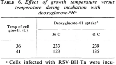
TABLE 6. Effect of growth temperature versus temperature during incubation with
deoxyglucose-3Ha
Deoxyglucose-311uptake'
Tempofcell growth(C)
36C 41C
36 233 239
41 123 135
a
Cells
infected with RSV-BH-Ta wereincu-bated overnight at 36 or 41 C. Deoxyglucose-3H uptake during a 15-min incubation period at 36 or 41 C wasmeasured.
bH counts per minute per microgram of
pro-tein.
DISCUSSION
The mutant of Rous sarcoma virus described in this paper provides hope for a biochemical definition of malignancy and for this purpose may have advantages over those mutants previ-ously described (6, 11, 13, 19, 21). The morpho-logical transformation induced by the Bryan high-titer strain of RSV is characteristic ofthe virus,israrely seeninuninfected cultures, andis unambiguous with respect toother physiological changeswhich may affectthemorphology of the cell. The mutant, RSV-BH-Ta, is capable of full cycles of reproduction at temperatures in which transformation of infected cells is inapparent. This observation demonstrates that, as in the case of some other avian sarcoma viruses (6, 11, 13, 19), the functional product of the gene re-sponsible for transformation is not required for virus reproduction; i.e., the gene responsible for transformation is gratuitous.
Theproductionoffullyinfectious virus at 41 C shows that the synthesis and packaging of viral
L'
I
120
100 80
604 40
a *41°_36°
4M°36°+Cycloheximide
20 _ _---O-- 41_36°6ActAnomycin
°41° Cyclohexim,de
0-*36--41°
_ _36°-4l'Actinomycin 0---436°-346e4Cycloheximide
- 036° Cyclohexrnide
0 1 _.Il_ L L_lJIIl
2 3 4 5 6 2 3 4 5 6
HOURSAFTER TEMPERATURESHIFT
FIG. 4. Effects oftemperature shifts on hexose
up-take. Cultures grown at 41 or 36 C for I day were
shiftedtothe opposite temperature athourly intervals before the removalof the cell culture medium andthe addition ofdeoxyglucose-3H (2.0 ,Ci/plate). All
cul-tures were incubatedat39 Cfor15 miii before
deter-mination of intracellular radioactivity. Left, Cultures
grownat41C. Note theincrease in rateofhexose
up-take in cultures shifted to 36 C beginning at 3 hr.
Cycloheximide (10 ,ug/ml) and actinomycini D (2 ,ug/ml) preventedthisincrease. Right, Culturesgrowit at36C. Norapid effect ofanincrease intemperature
wasseeii;however, abilityto takeuphexose decreased
ili cultures containingcycloheximide.
TABLE 7. Effect of growth temperature onz
cell-associated hyaluronic acida
Hyaluronic acid contentb Cellsinfected with
36C 41C
Noninfected .1 0.16 0.14
RSV-BH. 0.86 0.78
RSV-BH-Ta 0.90 0.22
aCells were placed at 36 or 41 C overnight.
Culture fluidswere removed, and cellsrinsed and suspended with trypsin. Cells were counted, and cytoplasmic fractions were analyzed for hya-luronic acidcontentbyquantitative electrophore-sis inpolyacrylamide-agarosemixed gels.
IPicograms of hyaluronate per cell.
RNAintovirions continuesatthis high tempera-ture. The temperature-sensitive molecule, there-fore, is a gene product rather than something interfering with transcription of the viralgenome. This was demonstrated in another way through theuseof metabolicinhibitors. Inhibitionof DNA synthesis, RNA synthesis, or protein synthesis had noeffect on the changein cellular morphol-ogy after a shift of RSV-BH-Ta-infected cells from41 to 36C. Onthe other hand, both actino-mycin D and cycloheximide interfere rapidly with production ofoncornaviruses (1, 3); virus production virtually ceases withinafew hours of the addition of either antimetabolite.
Nonethe-J. VIROL.
274
on November 10, 2019 by guest
http://jvi.asm.org/
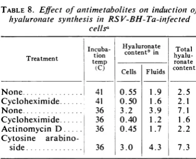
[image:8.499.66.262.74.175.2] [image:8.499.67.262.265.374.2]TABLE 8. Effect of anitimetabolites oninductioni of hyaluroniate synthesis in RSV-BH-Ta-infected
cellsa
Incuba- Hyaluronate Total Treatment tion content5 in
hyalu-temp ronate
(C) Cells Fluids content
None... 41 0.55 1.9 2.5
Cycloheximide... 41 0.50 1.6 22.1 None... 36 3.2 3.9 7.1 Cycloheximide... 36 0.40 1.2 1.6 Actinomycin D ... 36 0.45 1.7 2.2
Cytosine
arabino-side... 36 3.0 4.3 7.3
Cells infected with RSV-BH-Ta were grown at 41 C. Fluids were replaced with fresh growth medium, antimetabolites were added, and
cul-tures wereincubated for6 hr at 36 or 41 C. Culture fluids were analyzed directly by electrophoresis inacrylamide-agarose gels. Cytoplasmic fractions wereprocessedfor hyaluronate determinationsas
described inMaterials and Methods. bMicrograms per culture.
less, pretreatment of cells with antimetabolite foraslongas6hrbeforeshifting from41 to 36 C did not prevent transformation by RSV-BH-Ta, demonstrating that virus production is not cor-related with transformation even at the
trans-forming temperature.
Thus, it seems likely that the temperature-sensitive molecule is a protein, perhaps an en-zyme involved in synthesis of other cellular components or participating directly in the structural organization of the cell, or both. The partial loss of the transformed morphology upon
continued exposure to cycloheximide or
puro-mycinfurther suggests thatproteinsare involved
in the maintenance of the transformed state. It is possible that more than onevirus gene is responsible for the changesobservedduring
trans-formation with RSV-BH, and that the products
of these genes are both temperature sensitive in
cells infectedwith RSV-BH-Ta.Complementation
of at least two mutant classes of the
Schmidt-Ruppin strain of RSV has been observed (H. Hanafusa, personal communication). Possible
complementation among mutants of RSV-BH
(5) remainstobeanalyzed.
Inhibitors ofnucleic acid or protein synthesis
failedtopreventreversiontothenontransformed stateuponashift from 36 to 41 C.Thisindicates
that the "transformation molecule" does not produce its effectbyinterferingwith thesynthesis
ofa specific nucleic acid or
protein. Otherwise,
in the absence of this nucleic acid or protein,
reversal to nontransformed morphology could not occur.
Other investigators have found that inhibition ofprotein synthesis is sufficient to prevent mor-phological transformation in their temperature-dependent avian sarcoma systems (6, 11). Clearly, the transformation they observe requires the induction of other proteins. Since actinomycinD did not preventtransformation in these systems, an interesting, but complex, mechanism involv-ingtranslational control is suggested.
A multitude of biochemical changes has been reported to occur in malignant cells, and each laboratory has its favorite potential cancer mole-cule. We have examined the increase in hyalu-ronic acid synthesis and the increase in hexose uptake which accompany the transformation of cells by RSV-BH. These capacities are both induced upon a shift of RSV-BH-Ta-infected cells from 41 to 36 C. The induction was shown tobeatthe level of transcription for both hyalu-ronic acid synthesis and hexose uptake, as the inhibition of induction by both cycloheximide and actinomycin demonstrate. Therefore, the change in morphology cannot be attributed to increased hyaluronate synthesis or molecules responsible for increased hexose uptake. The outcome of these experiments suggests that we should look elsewhere for the primary tempera-ture-sensitive molecule.
In another study, levels of cyclic adenosine monophosphate (AMP) were shown to be cor-related with transformation of chick embryo cells by RSV-BH and RSV-BH-Ta (16). Also, transformation of RSV-BH-Ta-infected cells shifted from 41 to 36 C could be partially pre-vented by the addition of exogenous dibutyryl cyclic AMP and theophylline, an inhibitor of phosphodiesterase. Experiments to define more clearly the role of cyclicAMPin transformation by RSV-BH are in progress.
The induction of increased hexose uptake and hyaluronate synthesis as described for this
tem-perature-dependent system provides an excellent system for the analysis of such induction in
eukaryotic cells. It is possible that analyses of temperature-dependent regulation ofhexose up-take or hyaluronate synthesis will lead to a moleculardefinitionof someregulatory processes invertebrate cells.
ACKNOWLEDGMENTS
Theexperiments describedhere werelargelydependent upon theexcellenttechnical assistance of Nancy R. Brown and David A.Ray.
LITERATURE CITED
1. Bader,J. P. 1964. The role ofdeoxyribonucleicacidin the synthesisof Roussarcomavirus.Virology22:462-468. 2. Bader, J. P. 1967.Achange in growthpotentialof cells after
conversionbyRoussarcomavirus. J. Cell.Physiol. 70:301-307.
on November 10, 2019 by guest
http://jvi.asm.org/
[image:9.499.40.231.77.232.2]276 BADER
3. Bader, J.P. 1970. Synthesis of the RNA of RNA-containing tumorviruses. I. The interval between synthesis and en-velopment.Virology 40:494-504.
4. Bader, J. P., and A.V. Bader. 1970. Evidencefor a DNA replicative genome for RNA-containing tumor viruses. Proc. Nat.Acad. Sci. U.S.A.67:843-850.
5. Bader, J. P., and N. R. Brown. 1971.Inductionofmutations in an RNA tumor virus by an analogue of a DNA precur-sor.Nature N.Biol.234:11-12.
6. Biquard, J., andP.Vigier. 1972.Characteristicsof a condi-tional mutant of Rous sarcoma virus defectiveinability to transformcells athightemperature. Virology 47:444-455. 7. Ericksen,S., J. Eng, and H. R. Morgan.1961.Comparative studiesin Roussarcomuawithvirus,tumorcells, and chick embryo cells transformed in vitrobyvirus. I. Production ofmucopolysaccharides.0.J. Expt. Med.114:435-440. 8. Hatanaka, M., R. J. Huebner, and R. V. Gilden. 1969.
Altera-tionsin thecharacteristicsof sugar uptake bymouse cells transformed by murine sarcoma viruses. J. Nat. Cancer Inst.43:1091-1096.
9. Hattanaka, M.,and H. Hanafusa. 1970. Analysis of a func-tional change in membrane in the process of cell transforma-tionby Roussarcoma virus; alteration in the characteris-ticsof sugartransport.Virology 41:647-652.
10. Kabat, E. A.1939. Apolysaccharidein tumors due to a virus ofleucosisand sarcomaof fowls. J.Biol. Chem.
130:143-147.
I l. Kawai, S., andH.Hanafusa.1971. The effects ofreciprocal changes intemperatture on thetransformed state of cells infected with a Rous sarcomamutant. Virology46:470-479. 12. Lowry,0.H., N.Rosebrough,A.L. Farr,and R. J. Randall.
J. VIROL.
1951.Protein measurement with the Folin phenol reagent. J.Biol. Chem. 193:265-275.
13. Martin, G. S. 1970. Rous sarcoma virus: afunction required for the maintenance of the transformed state. Nature
(London) 227:1021-1023.
14. Martin, G. S., S. Venuta, M. Weber, and H. Rubin. 1971. Temperature-dependent alterations in sugar transport in cells infected by a temperature-sensitive mutant of Rous sarcomavirus. Proc. Nat. Acad. Sci. U.S.A. 68:2739-2741. 15. Nakata,Y., and J. P. Bader. 1968. Studies on the fixation and development of cellular transformation by Rous sarcoma virus. Virology 36:401-410.
16. Otten, J., J. P. Bader, G. J.Johnison,andI. Pastan.1972. A mutation in a Rous sarcoma virus gene that controls adeno-sine-3'5'-monophosphate levels and transformation. J. Biol. Chem. 247:1632-1633.
17. Steck, T. L., S. Kaufman, and J. P. Bader. 1968.Glycclysis inchick embryo cell cultures transformed by Rous sarcoma virus.Cancer Res. 28:1611-1619.
18. Temin, H. R. 1965. The mechanism of carcinogenesis by avian sarcoma viruses. I. Cell multiplication and differentia-tion. J. Nat. Cancer Inst. 35:679-993.
19. Temin, H. M. 1971. The role of the DNA provirus in carcino-genesis by RNA tumor viruses, p. 176-187. Int I. Silvestri (ed.),Thebiology ofoncogenicviruses;Proceedingsof the Second Lepetit Colloquium. North Holland Publ., Amster-dam.
20. Temin, H. M., and H. Rubin.1958. Characteristics of an assay forRous sarcoma virus and Rous sarcoma cellsin tissue culture. Virology 6:669-688.
21. Toyoshima, K., and P. K. Vogt. 1969.Temperaturesensitive mutantsof anavian sarcomavirus.Virology39:930-931.