Quantifying
bacterial
transfer
from
patients
to
staff
during
burns
dressing
and
bed
changes:
Implications
for
infection
control
Sarah
E.
Bache
a,b,*
,
Michelle
Maclean
b,
George
Gettinby
c,
John
G.
Anderson
b,
Scott
J.
MacGregor
b,
Ian
Taggart
aaBurnsUnit,CanniesburnPlasticSurgeryUnit,GlasgowRoyalInfirmary,Glasgow,UnitedKingdom b
TheRobertsonTrustLaboratoryforElectronicSterilisation Technologies(ROLEST),Department ofElectronicand Electrical Engineering, UniversityofStrathclyde,Glasgow,UnitedKingdom
cDepartmentofMathematicsandStatistics,UniversityofStrathclyde,Glasgow,UnitedKingdom
1.
Introduction
Advances in fluid resuscitation, organ support, and early excision and grafting have all improved survival rates followingasevereburn[1]. However,thishasalsohadthe effectofshiftingthecauseofmorbidityandmortalityaway
fromhypovolemiaandtowardssepsis.Sepsisisaprimaryrisk factorofmortalityfollowingaburn[2,3].Itisnowestimated thatinpatientswithburnsover40%totalbodysurfacearea (TBSA), 75% of all deaths are related to infection and/or inhalationinjury[1].Followingasevereburn,physical, non-specificandspecificimmunedefencesareallaffected,leading toastateofimmunosuppression.Coupledwithlarge
bacteria-*Correspondingauthor.
E-mailaddress:sarahbache@doctors.org.uk(S.E.Bache).
a
r
t
i
c
l
e
i
n
f
o
Articlehistory:
Accepted4December2012
Keywords: Infectioncontrol Nosocomialinfection Healthcareworkers Contamination Dressingchange Bedchange
a
b
s
t
r
a
c
t
Routinenursingactivitiessuchasdressing/bedchangesincreasebacterialdispersalfrom burnspatients,potentiallycontaminatinghealthcareworkers(HCW) carryingoutthese tasks.HCWthusbecomevectorsfortransmissionofnosocomialinfectionbetweenpatients. Thesuspectedrelationshipbetween%totalbodysurfacearea(%TBSA)ofburnandlevelsof bacterialreleasehasneverbeenfullyestablished.
BacterialcontaminationofHCWwasassessedbycontactplatesamples(n=20)from initially sterile gowns worn by the HCW during burns patient dressing/bed changes. Analysisof24 gownswasundertakenandexaminedforrelationshipsbetween%TBSA, timetakenforactivity,andcontaminationreceivedbytheHCW.
RelationshipsbetweensizeofburnandlevelsofHCWcontamination,andtimetakenfor thedressing/bedchangeandlevelsofHCWcontaminationwerebestdescribedby expo-nentialmodels.Burnsizecorrelatedmorestrongly(R2=0.82,p<0.001)thantimetaken (R2=0.52,p<0.001),withlevelsofcontaminationreceivedbytheHCW.Contamination doubledwithevery6–9%TBSAincreaseinburnsize.
Burnsizewasusedtocreateamodeltopredictbacterialcontaminationreceivedbya HCWcarryingoutbed/dressingchanges.Thismayhelpwiththecreationofburn-specific guidelinesonprotectiveclothingwornbyHCWcaringforburnspatients.
#2012ElsevierLtdandISBI.Allrightsreserved.
Available
online
at
www.sciencedirect.com
journalhomepage:www.elsevier.com/locate/burns
0305-4179/$36.00#2012ElsevierLtdandISBI.Allrightsreserved.
harbouringwounds,thisrendersburnspatientsboth suscep-tibletoinfection and potent dispersersof bacteria[4]. The consequencesofnosocomialpropagationcanbefelt through-out the entire hospital, increasing costs and the risk of outbreaksofmultidrug-resistantbacteriaonthe burnsunit andbeyond[5].
Transmissionofinfectionbetweenburnspatientsmainly occursthroughairbornetransmissionordirectandindirect contact[1,6].Routinenursingactivitymaycreateperiodsof increasedbacterialdispersalintotheairandontosurfacesand otherindividuals presentinthevicinity.Thepresentstudy examines the contamination of healthcare workers (HCW) resultingfromburnwounddressingchanges,whichareoften coupledwithbedsheetchanges.
Dressingchangesonevensmallnon-burnwoundscreate airbornedispersalofbacteria[7].Bedsheetchangeshavealso beenshowntoliberatebacteriaintotheair[8].Inthe1970s, attemptsweremadetolinkthesizeofaburnandtheairborne dispersalofStaphylococcus aureus duringadressing change, whichimpliedthatthesizeoftheburnwasrelatedtolevelsof bacteriafoundonsettleplatesoveraperiodofdays[9].More recently, it was shown that 31% of dressing changes on methicillinresistantS.aureus(MRSA)positiveburnspatients liberatedtheorganismintotheair[10].
HCWuniformsareapotentialreservoirofinfection[11–13], andtheircontaminationcanbedirectlyattributedtopatients [14,15]. Not only can bacteria be transferred from burns patients to uniforms during dressing changes, but also laboratorysimulationshavedemonstratedthatthesebacteria can be transferred from the uniform to patients [17,18]. Despite this, there is little consensus for the appropriate protectiveattire to beworn byHCW carrying out dressing changesonburnspatients.InasurveyofUSburnsunits,only 24%ofunitsrequiredfullprotectivecoverageonenteringa patient’sroomandchangingadressing[19].UKguidelinesare similarly vagueandnotburns-specific [20–22]. Quantitative dataon keyissues mayhelpintheir development. Inthis context, the current study was set up to address the hypothesis that the level of contamination received by a HCWwouldberelatedtothesizeoftheburnandthetime takenforthedressingchange.
2.
Materials
and
methods
2.1. Setting
QuantificationofHCWcontaminationwascarriedoutduring burn dressing changes.For patientswith largerburns, the dressingchangewouldusuallyalsoincorporateabedsheet changewhilerollingthepatienttoapplybandages(hereafter termed ‘dressing/bed change’).Data including age ofburn, recent routine wound swab results, time taken for the dressing/bedchangetotakeplaceandthe%TBSAburnwere recordedforeachpatient.Patientsweretreatedaccordingto standardpracticeonourburnsunit.Weaimforearlyexcision andsplitthicknessskinautograftorcoveragewithadermal substituteinalldeepdermalandfullthicknessburns.Patients withsuperficialburns,orthosedeemedtoosickforsurgical interventionaremanagedconservativelywithdressingsand
topicalagents.Patientswithburnwoundsover10daysold wereexcludedfromthestudy.
2.2. Samplestandardisation
To ensure that samples were taken from a standardised baseline, HCW were asked to don sterile, impermeable, disposable full-body gowns over their uniforms prior to performingdressing/bedchanges.Thiswasdonetoeliminate naturalvariationsinbacterialcontaminationbetween differ-entHCWsbeforethebeginningofthedressing/bedchange.It also provided a consistent sampling material, which was preferabletosamplingfromavarietyoftexturesandsurfaces includingcottonandskin.GownswerethuswornbytheHCW onlytofacilitate thestudydesignand samplingobjectives. Usually, disposable plastic aprons would be worn over uniforms as routine bed/dressing changes are carried out. All HCWmaintainedstandard handhygieneby decontami-natinghandsandputtingonfreshdisposableglovesbefore enteringthepatient’sroomtocarryoutthenursingactivity. Thereafter,withtheexceptionofwearingdisposablegowns ratherthandisposableplasticapronsoveruniforms,theHCW carried out the dressing/bed change in the usual manner. Gloves were removed and hands washed following the dressingchangeandgownsampling,beforeleavingtheroom. Samplesweretaken fromthetwo most‘involved’HCW carryingoutthedressingchange,eachofwhomwouldusually stand eitherside ofthe bedand carry out undressing and redressing of wounds alongside one another. For smaller burns,oneHCWoftencarriedoutthedressingchangealone, andonlyonesetofsampleswasobtained.Samplingduring dressing/bedchangesonanyonepatientwasonlycarriedout once.
2.3. Samplingsites
Followingthedressing/bedchange,andwhiletheHCWwas still wearing the disposable gown, and remained in the patient’s room, the gown was sampled. To estimate the contaminationthatwouldbereceivedduringadressing/bed change by a HCW who had not been wearing an apron, samplesweretakenfrom20sitesacrossthefrontofthegown. The20‘noapron’sitesareillustratedinFig.1.Ofnote,thesites areallacrossthefrontofthegown,asitwastheaimofthe studytocollectsamplesfromareasthatwerelikelytobecome mostcontaminatedduringdressing/bedchanges.Inorderto estimate the protection afforded had a disposable plastic apron been worn, a subset of 15 ‘with apron’ sites were analysedseparately.Theseexcludedfivesamplingsitesonthe chest and abdomen that would normally be covered by a disposableapron.ThesearealsodemonstratedinFig.1.
2.4. Bacteriologicalmethods
Samples were taken from the 20 sites using 25cm2 Baird Parker Agar (BPA) contact plates that were pressed firmly againstthesamplingsiteforapproximately2s,bythesame investigator (SEB). BPA allows for selective isolation of staphylococcal-typeorganisms,whichareanacceptedmarker ofbacteriaoriginatingfromahumansource[23].Aselective burnsxxx (2013) xxx–xxx
2
JBUR-3935;No.ofPages9
agar was chosen over a non-selective agar as preliminary studiesindicated thatnon-selective agar yielded toomany bacterialcolony-formingunits (cfu)per agar plateto accu-rately enumerate. Contact agar plates allow direct sample collectionfromthecontaminatedgowns,andenableaccurate reproductionofsamplingduetothedefinedsurfaceareaofthe agar plates.Sampleplates wereincubatedat 378Cfor48h beforeenumeration.
Thetimetakenforthedressing/bedchangetotakeplace wasmeasuredfromwhentheHCWenteredthepatient’sroom tocommence the dressing/bed change(the pointat which theywouldusuallydonaplasticapron).Itfinishedatthepoint whenthe dressing and bed change (if thatwas also being carriedout)wascompleted,whentheywouldusuallyremove theirapronandglovespriortoleavingtheroom.Atthispoint the gown was sampled. Any further activities, including tidying the room, assisting with feeding, or brushing the patient’shairorteethwerenotincludedinthetimetakenfor dressing/bed change.The gown was sampled before these extraactivitiestookplace.Thismeantthatthecontamination measured was that received only during the dressing/bed change.Itwasnotpossibletoseparatethedressingandbed changecomponentsoftheactivity,asthebedsheetchange
was often integrated into the dressing change when the patientwasrolledforapplicationofbandages.Weintendedto mimicreal-life situationsas muchaspossibleand didnot want toinconvenience thepatientor HCW,or prolongthe activity bycarrying out separatedressing changesand bed changes,duringwhatcanbeadistressinganduncomfortable time.
2.5. Statisticalanalysis
Inundertakingthestudyconsiderationwasgiventopower andsamplesizerequiredforthepurposesoftheregression andcorrelationanalysis.Itwasestimatedthatmeasurements wouldberequiredonbacterialcfuandassociated%TBSAfora minimum of10patientsin ordertohave inexcess of90% statistical power to detect a correlation of 0.9 with 95% confidence. A random sample size of between 10 and 15 patientswasplannedwithreplicatecfumeasurementsbeing observedonuptotwoHCWcarryingoutdressing/bedchanges perpatient.
HCW bacterial contamination was expressed as mean numberofbacterialcfuper25cm2agarplate,ormeancfu/ plate.Foreachsamplingsessionthiswascalculatedforall20
Fig.1–DiagramtodemonstratesamplingsitesonthefrontofHCWgowns.Theimageontheleftshowsthepositionsofall
20samplingsites(termed‘noapron’sites).Theimageontherighthighlightsthe15samplingsitesleftexposediftheHCW
[image:3.595.88.498.62.417.2]‘no apron’ sites, and also for the 15 ‘with apron sites’, excludingthose 5sites thatwouldhave beencovered bya disposable plastic apron, had one been worn. Statistical analysis was carried out using NCSS Windows Version 7 software. Relationships were examined for between three variables:%TBSAandHCWcontamination;timetakenforthe dressing/bed change and HCW contamination; %TBSA and timetakenforthedressing/bedchange.Separateanalysiswas carriedoutonall20‘noapron’sites,andonthe15‘withapron’ samplingsites.Mathematicalmodellingwasusedtoidentify equationswhichbestdescribedthethreerelationships.These wereusedtopredictthecontaminationaHCWwouldreceive duringdressing/bedchangeofaburnpatientby%TSBA.The coefficientofdetermination,R2wasusedtomeasurehowwell the model fitted to the observed data and p<0.05 was consideredsignificant.
3.
Results
3.1. Patientdemographicsandwoundinformation
Sampleswerecollectedfromthegownsof24HCWcarrying outdressing changeson15differentpatients,withamean burnsizeof19%TBSA(range1–51%TBSA).Meanageofpatient was39years(range19–85years).Samplesweretakenamean of6.4daysaftertheburn(range2–10days).Meantimetaken forthe dressingchange was45min(range10–90min).The mostcommonorganismidentifiedonroutinewoundswabs wasS.aureus.Bacillussp.,coliforms,andStreptococcussp.were alsocommonlyisolated.ResultsaresummarisedinTable1.
3.2. Relationshipbetweentimetakenfordressing/bed
changeand%TBSA
Asignificantrelationshipwasdemonstratedbetweenthetime takenforthedressing/bedchangetotakeplaceandthesizeof theburn(%TBSA).Thiswasexplainedbyalinearcorrelation (coefficient of determination, R2=0.76; p<0.001). This is demonstratedinFig.2.
3.3. Analysisof20‘noapron’sites
ThevariationincontaminationreceivedbyaHCWduringa dressing/bedchangewhen20‘noapron’samplingsiteswere analysedwasexaminedinrelationto%TBSAoftheburnand time takenfor thedressing/bed change.Bothrelationships wereexplainedbyexponentialmodels.Thesewereasfollows: Relationship between HCW contamination and %TBSA (coefficientofdetermination,R2=0.82;p<0.001):
Meancfu=plate¼8:59 Exp0:080%TBSA
Relationshipbetweentimetakeninminfordressing/bed changeandHCWcontamination(coefficientofdetermination, R2=0.52;p<0.002):
Meancfu=plate¼17:44 Exp0:034timetakeninmin
ThesecurvesareillustratedinFig.3.Bothcharts demon-strate an exponential relationship between the variable (%TBSA or time taken forthe dressing/bed change totake place)andthecontaminationreceivedbytheHCW.However, although theyarebothsignificantrelationships,timetaken correlateslessstronglythan%TBSAasshownbythelowerR2. %TBSAisamoreaccuratepredictorofHCWcontamination thantimetakenforthedressing/bedchangetotakeplace.
3.4. Analysisof15‘withapron’sites
ThevariationincontaminationreceivedbyaHCWduringa dressing/bedchangewhen15‘withapron’samplingsiteswas examinedinrelationto%TBSAoftheburnandtimetakenfor thedressing/bedchange.Bothrelationshipswereexplainedby exponentialmodels.Thesewereasfollows:
Relationship between HCW contamination and %TBSA (coefficientofdetermination,R2=0.86;p<0.001):
Meancfu=plate¼2:05 Exp0:110%TBSA
RelationshipbetweenHCWcontaminationandtimetaken inminfordressing/bedchange(coefficientofdetermination, R2=0.44;p=0.007):
Meancfu=plate¼15:98 Exp0:034timetakeninmin
0 25 50 75 100
0 15 30 45 60
Size of burn (%TBSA)
Time taken for dressing change (min)
Fig.2–Chartdemonstratinglinearrelationshipbetween%TBSAoftheburn,andtimetakeninmintocompletethe
dressing/bedchange.
burnsxxx (2013) xxx–xxx
4
JBUR-3935;No.ofPages9
[image:4.595.173.412.541.719.2]thickness); age of burn in days; the %TBSA that has been harvested as a split thickness skin graft; the %TBSA that has been covered by autograft or dermal substitute; recent wound swabs; whether a dressing change and bed change took place; time taken for the dressing/bed change; and the mean cfu per plate for all 20 ‘no apron’ sites, and the 15 ‘with apron’ sites.
Study no.
Patient Pt age
(years) %TBSA burn Site of burn Depth of burn Age of burn (days) %TBSA donor site harvested %TBSA covered in skin or substitute
Wound swab results Dressing
change Bed sheet change Time taken (min) Mean cfu/plate 20 sites Mean cfu/plate 15 sites
1 A 19 1 UL DPT 6 0 0 Not taken Yes No 10 23 18
2 B 24 2 AT SPT 6 0 0 Not taken Yes No 25 12 9
3 C 26 2 AT SPT 6 0 0 Not taken Yes No 10 14 5
4 D 44 2 UL SPT 10 0 0 Not taken Yes No 20 13 4
5 E 34 6 AT DPT/FT 8 6 6 Staphylococcus aureus,
Bacil-lussp.
Yes No 40 40 27
6 E 34 6 AT DPT/FT 8 6 6 S. aureus,Bacillussp. Yes No 40 13 5
7 F 33 6 LL DPT 9 6 6 coliforms,S. aureus, Gp G
Streptococcus,Bacillussp.
Yes No 50 1 1
8 G 22 7 UL SPT 8 0 0 coliforms,S. aureus, Gp A
Streptococcus,Bacillussp.
Yes No 20 50 22
9 H 45 15 UL, AT, HN FT 6 9 15 S. aureus,,Bacillussp., Clos-tridium perfringens
Yes Yes 55 54 41
10 H 45 15 UL, AT, HN FT 6 9 15 S. aureus,,Bacillussp.,C. perfringens
Yes Yes 55 50 21
11 I 85 16 AT DPT/FT 120 0 0 S. aureus,Bacillussp. Yes Yes 25 101 90
12 I 85 16 AT DPT/FT 120 0 0 S. aureus,,Bacillussp. Yes Yes 25 20 20
13 J 39 30 UL, LL, AT, PT DPT/FT 7 9 15 S. aureus,Streptococcus pneu-monia
Yes Yes 50 108 118
14 J 39 30 UL, LL, AT, PT DPT/FT 7 9 15 S. aureus,S. pneumoniae Yes Yes 50 97 52 15 K 46 30 UL, LL, PT DPT/FT 6 0 0 S. aureus,Streptococcussp.,
Bacillussp.
Yes Yes 55 28 7
16 K 46 30 UL, LL, PT DPT/FT 6 0 0 S. aureus,Streptococcussp.,
Bacillussp.
Yes Yes 55 25 26
17 L 55 35 UL, LL, AT, DPT/FT 4 0 0 Methicillin resistantS. aur-eus(MRSA)
Yes Yes 60 177 126
18 L 55 35 UL, LL, AT, DPT/FT 4 0 0 MRSA Yes Yes 60 66 71
19 M 29 41 UL, PT, HN FT 8 18 18 coliforms,S. aureus,S.
pneumonii,bacillussp.
Yes Yes 90 142 96
20 M 29 41 UL, PT, HN FT 8 18 18 coliforms,S. aureus,S.
pneumonii,bacillussp.
Yes Yes 90 294 233
21 N 45 43 UL, LL, AT, HN FT 2 1 18 No growth Yes Yes 85 287 259
22 N 45 43 UL, LL, AT, HN FT 2 1 18 No growth Yes Yes 85 420 341
23 O 40 51 UL, AT, PT, HN FT 6 4 32 Enterococcus cloacae Yes Yes 78 662 569
24 O 40 51 UL, AT, PT, HN FT 6 4 32 E. cloacae Yes Yes 78 333 569
[image:5.595.49.726.109.525.2]Thesecurves areillustratedinFig. 4. Again,bothcharts demonstrateanexponentialrelationshipbetweenthevariable (%TBSA or timetaken for thedressing/bed change totake place)andthecontaminationreceivedbytheHCW.However, althoughtheyarebothsignificantrelationships,timetaken correlateslessstronglythan%TBSAasshownbythelowerR2. %TBSAisamoreaccuratepredictorofHCWcontamination thantimetakenforthedressing/bedchangetotakeplace.
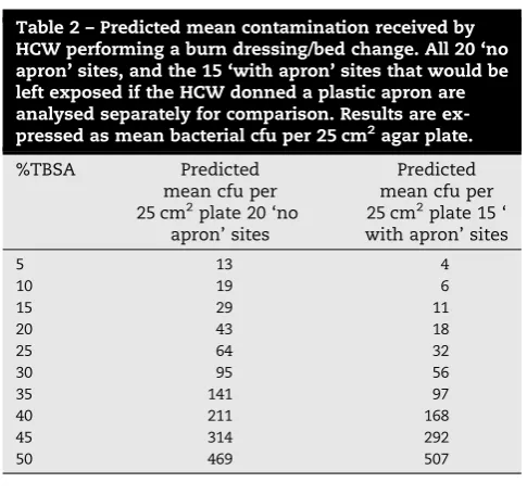
3.5. PredictedcontaminationofHCW
Usingtheabovestatisticalmodels,theexpectedmeannumber ofbacterialcfuper25cm2platefromaHCW performinga burns dressing/bed change can be predicted. This was producedfromdatasetsforall 20‘no apron’sitesandthe 15‘withapron’sites.ThesevaluesaresummarisedinTable2. Itwasfoundthatforevery9%TBSAincreaseinburnsize,the mean number of cfu/platedoubled when all 20 sites were analysed.Thiswastrueforevery6%TBSAincreaseinburnsize when15‘withapron’siteswereanalysed.
0 200 400 600 800
0 15 30 45 60
Burn size (%TBSA)
Mean cfu/plate
(
n
=20)
0 200 400 600 800
0 25 50 75 100
Time taken (min)
Mean cfu
/plate (
n
=20)
[image:6.595.69.518.61.216.2] [image:6.595.77.516.290.450.2]Fig.3–Chartsdemonstratingexponentialrelationshipsbetween%TBSAandmeancfuperplate(left)andtimetakenin
minutesfordressingchangeandmeancfuperplate(right)whenall20‘noapron’samplingsitesonaHCWgownare
analysed.
0 150 300 450 600
0 15 30 45 60
Burn size (%TBSA)
Mean cfu
/plate
(
n
=15)
0 150 300 450 600
0 25 50 75 100
Time taken (min)
Mean cfu
/plate
(
n
=1
5)
[image:6.595.303.546.530.758.2]Fig.4–Chartdemonstratingexponentialrelationshipsbetween%TBSAandmeancfuperplate(left)andtimetakenin
minutesfordressingchangeandmeancfuperplate(right)when15‘withapron’samplingsitesonaHCWgownare
analysed.
Table2–Predictedmeancontaminationreceivedby HCWperformingaburndressing/bedchange.All20‘no apron’sites,andthe15‘withapron’sitesthatwouldbe leftexposediftheHCWdonnedaplasticapronare analysedseparatelyforcomparison.Resultsare ex-pressedasmeanbacterialcfuper25cm2agarplate.
%TBSA Predicted
meancfuper 25cm2plate20‘no
apron’sites
Predicted meancfuper 25cm2plate15‘ withapron’sites
5 13 4
10 19 6
15 29 11
20 43 18
25 64 32
30 95 56
35 141 97
40 211 168
45 314 292
50 469 507
burnsxxx (2013) xxx–xxx
6
JBUR-3935;No.ofPages9
4.
Discussion
The consequencesof nosocomial infectionsfrom a burns patientcross-contaminating otherpatientsare potentially devastating [1,24]. Prevention of cross-contamination is thusbecominganincreasinglyimportantareaofburncare research. The potential for HCW to act as vectors of transmissionbetweenpatients,andtheincreasedbacterial dispersalduringdressingandbed sheetchangesonburns patientshaslongbeenknown[6–9,11–18].Thecurrentstudy highlights high levels of HCW contamination following a dressing/bed change and quantifies levels of bacterial contaminationforthefirsttime.
Duringadressing/bedchangetheHCWcanbeexpected tocomeintocontact withthepatient, theirdressingsand thesurroundingenvironment,allofwhicharelikelytobe heavily contaminatedon the burns unit.AHCWwhohas become contaminated by carrying out a dressing change will proceed to make contact with other patients or environmentalsurfaces,dispersingorganisms,wherethey cansurviveforseveralweeks andform anenvironmental reservoir[25–27]. Theenvironmentmaythencontaminate another patient directly or indirectly via the hands or uniform of a HCW acting as a carrier for nosocomial infection[28,4,29].
Guidelines on the use of protective clothing for HCW during burns dressing/bedchangesarenot burns-specific. Basedonthe resultsofthisstudy,theymayrequire tobe revisedwithconsiderationoftheamountofcontamination received by HCW during performance of these routine nursingactivities.Theuseofgloves andmeticuloushand hygiene for alldressing changes is acceptedpractise and wasnotexaminedhere[15,30].Ofnote,WHOrecommenda ‘5 moments for hand hygiene’ approach whereby hands should be cleaned before and after all procedures and contact with patient surroundings [31]. It may be argued thattheHCWinthisstudyshouldhavebeenencouragedto washtheirhandsseveraltimes during theactivity, rather thanjustatthebeginningandend.Howeverastheywerein constantcontact withthe environment,patient,andopen woundsthroughoutthedurationoftheactivity,dividingthe dressing/bed change into distinct ‘moments for hand hygiene’ was difficult. One compromise that may be employedin the future is to encouragea pause for hand hygiene and change of gloves only, between removing dressings and applying fresh dressings. The compliance withtheserecommendationsishoweverunlikely toaffect thelevelsofbacteriafoundonthegowns,astheyconcern onlyhandhygiene.
Disposablefull-bodygownswereonlywornforthisstudy toenablesamplingfromasurfacethatwasknowntobesterile priortothenursingactivities.Standardpracticeonourunitis for plastic aprons to be worn for most dressing and bed changes,excludingthose takingplace inICU oron known heavilycontaminatedpatients.Theresultsofthisstudyhave ledtoareviewofourclinicalpractice,andrevisedguidelines onprotectiveattirewornbyHCW.
ThemathematicalmodelsproducedindicatethataHCW performingadressingchangeonapatientwitha15%TBSA
burn could be expected to become contaminated with a mean of29bacterialcfu/25cm2if they woreno protective clothing and11bacterialcfu/25cm2if a plastic apron was worn, supposing absolute protection is afforded by the apron.Forlargeburns,predictionoflevelsofcontamination whenaHCWwearsordoesnotwearanapronhighlightsthe limitation of relying only on the apron as a means of preventionofHCWcontamination.Forexample,50%TBSA burnisestimatedtoproduce469cfu/platewhenwearing‘no apron’,comparedto507cfu/plate‘withapron’.Themajority of samples were collected from the forearms, arms, shouldersandchest:areasthatofskinanduniformwhich wouldnotbeprotectedorcleanedduringhandwashingand may comeinto contactwith otherpatients orequipment. Beforethestudywasinitiated,HCWwereencouragedtoact exactlyastheywouldwerethey wearinganapron. Whilst this was the agreed intention, it is nevertheless possible that theymayhavebeenlesscarefulthanusual knowing theywerecoveredbyagown,ormorecarefulastheywere conscious they were part of a study. Regardless of this possibleeffect,theresultshighlighttheneedforareviewof protectiveguidelinesforHCW.
Burns between 2 and 10 days old were examined, although numerous factors such as the site of the burn, whether debridement had taken place, donor site size, comorbidities andbacteria isolated from the wound were unabletobecontrolled.Despitetheinclusioncriteriabeing fairly broad, %TBSA was still shown to be an important predictor ofHCWcontamination.Future studieswouldbe useful tomonitor the changeinHCWcontamination asa burnprogressestowardshealing,orasthepatientbecomes colonised with increasingly resistant organisms. Further-more,BPAwasusedthroughouttomonitor staphylococcal-typebacteria,butotherselectivemediamaybeusedinthe future to identify other organisms that colonise burns wounds,suchasGram-negatives,whichmayshowdifferent transfer characteristics betweenpatients andHCW. Were the studies to be repeated on a larger sample size, quantitative analysis of wound contamination may be attempted, although this would only be an estimate. However thiswouldnotbe helpfulin predicting contami-nationandthusguidingHCWonwhichprotectiveattireto wear;resultsnotbeingknownuntilafterthe dressing/bed changehadtakenplace.
transferratefromtheHCWtoanothersurfaceorpatient.In theabsenceofthis,anarbitraryfiguremaybeassignedasa pre-determinedcutoffpointabovewhichfull-body protec-tionshouldbeworn.Thecostoffullbodyprotectionmust also be considered andweighed up against the perceived riskoftransferfromaHCW.
Itislogicaltoassumethatingeneralalargerburnwilltake longer to dress, and indeed this was shown by a linear relationship between%TBSAandtotal time taken(Fig. 2). Although time taken was related to the level of HCW contamination,itexplainedlessofthevariationthanburn size,withalowercoefficientofdetermination,R2. Further-more,asthetimetakenforthedressingchangewillnotbe known until after the event, and may depend on HCW experience,%TBSAwaspreferentiallyconsideredtopredict HCW contamination. A rough guide is that for every 6–9%TBSA increase in burn size, bacterial contamination doubles.
Thisstudyincreasesknowledgeofthetransferofbacteria from burns patients to HCW. It highlights the need for guidelines on protective clothing worn by HCW to be developed, asburnspatients have beenshown to disperse high levels of bacteria onto HCW. For the first time, a quantitativeanalysisofbacterialcontaminationreceivedby HCWperformingburnsdressingandbedchangeshavebeen performed.TherisksofHCWcontaminationmustbebalanced against the cost of protective measures and resources availabletoburnsunitsworldwide.
Conflict
of
interest
Allauthorsdeclarenofinancialorpersonalassociationsthat couldinappropriatelyinfluencethiswork.
Funding
SEB would like to thank the Royal College of Surgeons of Edinburghfortheawardofasmallresearchgrantforcosts towardsconsumablesforthisresearch.Norolewasplayedby thefundingsourcebeyondthis.
Ethical
approval
EthicalapprovalwasgrantedbythelocalRECforthestudy oflevelsofenvironmentalbacterialcontaminationaround burnspatients,aspartofalargerbodyofwork.Allsamples were taken from disused gowns and no direct patient involvementwasrequiredtocarryoutthisresearch.Verbal consentwasobtainedfromthepatientandHCWpriortothe study.
Acknowledgements
SEBwouldliketothankthestaffandpatientsonWard45at GRIfortheirhelpwiththisstudy,andMr.StuartWatsonfor hissupportandadvice.
r
e
f
e
r
e
n
c
e
s
[1] RaflaK,TredgetEE.Infectioncontrolintheburnunit.Burns 2011;37:5–15.
[2] KastenKR,MakleyAT,KaganRJ.Updateonthecriticalcare managementofsevereburns.JIntensiveCareMed 2011;26:223–36.
[3] WangY,TangHT,XiaZF,ZhuSH,MaB,WeiW,etal. Factorsaffectingsurvivalinadultpatientswithmassive burns.Burns2010;36:57–64.
[4] WeberJ,McManusA.Infectioncontrolinburnpatients. Burns2004;30:A16–24.
[5] TalonD.Theroleofthehospitalenvironmentinthe epidemiologyofmulti-resistantbacteria.JHospInfect 1999;43:13–7.
[6] GouldD.Isolationprecautionstopreventthespreadof contagiousdiseases.NursStand2009;23:47–55. [7] ThomBT,WhiteRG.Thedispersaloforganismsfrom
minorsepticlesions.JClinPathol1962;15:559–62. [8] ShiomoriT,MiyamotoH,MakishimaK,YoshidaM,
FujiyoshiT,UdakaT,etal.Evaluationofbedmaking-related airborneandsurfacemethicillin-resistantStaphylococcus aureuscontamination.JHospInfect2002;50:30–5.
[9] HambreusA.DispersalandtransferofStaohylococcusaureus inanisolationwardforburnedpatients.JHygCamb 1973;71:787–97.
[10] DansbyW,PurdueG,HuntJ,ArnoldoB,PhillipsD,MoodyB, etal.Aerolizationofmethicillin-resistantStaphylococcus aureusduringanepidemicinaburnintensivecareunit.J BurnCareRes2008;29:331–7.
[11] BabbJR,DaviesJG,AyliffeGAJ.Contaminationofprotective clothingandnurses’uniformsinanisolationward.JHosp Infect1983;4:149–57.
[12] PerryC,MarshallR,JonesE.Bacterialcontaminationof uniforms.JHospInfect2001;48:238–41.
[13] CallaghanI.Bacterialcontaminationofnurses’uniforms:a study.NursStand1998;13:37–42.
[14] SpeersR,ShooterRA,GayaH,PatelN.Contaminationof nurses’uniformswithStaphylococcusaureus.Lancet 1969;2:233–5.
[15] SnyderGM,ThomKA,FurunoJP,PerencevichEN, RoghmannMC,StraussSM,etal.Detectionof methicillin-resistantStaphylococcusaureusandvancomycin-resistant enterococcionthegownsandglovesofhealthcare workers.InfectControlHospEpidemiol2008;29:583–9. [16] MorganDJ,LiangSY,SmithCL,JohnsonJK,HarrisAD,
FurunoJP,etal.Frequentmultidrug-resistantAcinetobacter baumanniicontaminationofgloves,gowns,andhandsof healthcareworkers.InfectControlHospEpidemiol 2010;31:716–21.
[17] HambreusA.TransferofStaphylococcusaureusvianurses’ uniforms.JHygCamb1973;71:799.
[18] HambreusA,RansjoU.Attemptstocontrolclothes-borne infectioninaburnunit.JHygCamb1977;79:193–203. [19] HodleAE,RichterKP,ThompsonRM.Infectioncontrol
practisesinUSburnsunits.JBurnCareRes2006;27: 142–51.
[20] PrattRJ.Epic2:nationalevidence-basedguidelinesfor preventinghealthcareassociatedinfectionsinNHS hospitalsinEngland.JHospInfect2007;65(S):51–64. [21] Goodpracticeininfectionpreventionandcontrol–
guidelinesfornursingstaff.www.wales.nhs.uk/sites3/ Documents/739/RCN%20infection%20control.doc.pdf [accessed03.11.11].
[22] NICEinfectioncontrolguidelines:preventionofhealthcare associatedinfectioninprimaryandcommunitycareJune 2003.http://www.nice.org.uk[accessed03.11.11].
burnsxxx (2013) xxx–xxx
8
JBUR-3935;No.ofPages9
[23] DancerSJ,WhiteL,RobertsonC.Monitoringenvironmental cleanlinessontwosurgicalwards.IntJEnvironHealthRes 2008;18:357–64.
[24] ChurchD,ElsayedS,ReidO,WinstonB,LindsayR.Burn woundinfections.ClinMicrobiolRev2006;19:403–34. [25] BoyceJM.Environmentalcontaminationmakesan
importantcontributiontohospitalinfection.JHospInfect 2007;65(S2):50–4.
[26] NeeleyAN.Asurveyofgram-negativebacteriasurvivalon hospitalfabricsandplastics.JBurnCareRehabil
2000;21:523–7.
[27] CurtisL.Environmentalcontrolofmethicillinresistant Staphylococcusaureusandotherhospitalacquired infections.JBurnCareRes2008;29:1015.
[28] ArnowPM,AllynPA,NicholsEM,HillDL,PezzloM,Bartlett RH.Controlofmethicillin-resistantStaphylococcusaureusin aburnunit:roleofnursestaffing.JTrauma1982;22:954–9. [29] BoyceJM,Potter-BynoeG,ChenevertC,KingT.
Environmentalcontaminationduetomethicillin-resistant Staphylococcusaureus:possibleinfectioncontrol
implications.InfectControlHospEpidemiol1997;18:622–7. [30] BhallaA,PultzNJ,GriesDM,RayAJ,EcksteinEC,AronDC, etal.Acquisitionofnosocomialpathogensonhandsafter contactwithenvironmentalsurfacesnearhospitalized patients.InfectControlHospEpidemiol2004;25:164–7. [31] WHOguidelinesonhandhygieneinhealthcare.WHO