International Journal of Innovative Technology and Exploring Engineering (IJITEE) ISSN: 2278-3075, Volume-8 Issue-8, June, 2019
Abstract: Breast cancer disease history goes back to around 1,500 years B.C. Old Egyptians were the first to report the infection over 3,500 years ago. Breast cancer is the main cause of death among technologically advanced and underdeveloped countries. After lung cancer, more than 10% of women are diagnosed with Breast cancer. Breast Cancer can be determined by features like as redness of the skin, change in size, abdominal pain and texture change. Though the main cause of breast cancer is ambiguous in some cases, stoppage of the disease becomes difficult. However, detection of Tumour in the breast at an early stage is the only method to cure this disease. Radiologist recognizes breast cancer using various technologies like mammography, thermography etc. Optical coherence tomography is new invention found in medical services before five years; it is microscopic needle method for detection of Breast Cancer. Computer-aided diagnosis is the most appropriate method for diagnosis of disease. Quantitative Assessment of lower half of breast can be done using contour outlining with related measures such as breast symmetrical approach, the mass of breast can be recognized. In Existing research, two classifiers were used namely Naïve Bayes and k nearest neighbor (KNN) for the classification of breast cancer. The experimental result improves the performance with high accuracy and less error rate with k nearest neighbor. In research work, the data sets are gathered which is downloaded from the UCI (University of California Irvine)
machine Knowledge Depository Site. Machine learning methods are used for the prediction and classification of features of breast cancer. Various stages used for extraction of the feature of breast cancer. Firstly, the pre-processing phase is used for searching for the missed attributes and then feature extraction method is used for finding feature vectors in BCD (Breast Cancer Dataset). Experimental result use classification method (gene-BPNN) for the prediction of benign and malign in MATLAB (Matrix Laboratory) 2016a Simulation metrics are evaluated using accuracy, specificity and sensitivity.
Index Terms: Breast Cancer, Mammography, Quantitative Assessment, Contour outlining, Machine learning.
Revised Manuscript Received on June 15, 2019
Amandeep kaur,Research Scholar,Dept. of CSE, Sachdeva Engineering College for Girls, Gharuan, Mohali,India.
Prabhjeet Kaur,Assistant Professor, Dept. of CSE, Sachdeva Engineering College for Girls,Gharuan, Mohali,India.
I. INTRODUCTION
Breast Cancer is mainly due to the gathering of the one cell that causes obstruction to the whole system and forms the division of the cell. If the disease is not detected at an early stage then that mass forms a large quantity of cells after the passage of time. In This way, the Tumour is expanded to other parts of the body through a cell that may result in extra size and where benign mass is expanded to another part of the tissue [1]. Breast Cancer is difficult to detect at the beginning because symptoms may not be easily detected, but actual discrimination between the benign and malignant tumors is done after medical examination [2]. In recent research, a large quantity of women has breast cancer dies every year according to the world health organization because the disease is not detected at an early stage [3]. Prognostic factor plays a main role in the recognizing the size of the cancer cells that include the chemotherapy [4][5]. Breast Cancer is a disease that is found in the most developed countries and about 70 to 80% of the population diagnosed with this disease. However, the disease may cause death and early detection of the disease increase the rate of survival of the patient. The main cause of breast cancer may be heredity, hormonal change and unhealthy diet environmental factors. Signs and symptoms of Breast cancer are required to be seen at an early stage and symptoms are:
A. Pain in the abdomen B. Breasts lump
C. Changed colour of breast D. Change in size.
E. Discharge in the nipple. F. Swollen breast
G. Loss of weight
H. Weakness and body ache.
Detection of breast cancer disease can be done through mammography [5][6]. Mammography is the best technique because it is cost effective and helps in the diagnosis of the disease at an early stage. Radiologists aim in detecting the abnormal stage and complexity using mammography with low x-ray projection [7]. Breast Cancer generally increases day by day by the enlargement of the cells up to 3mm in every 3 to 4 months because multiplication of the cells may spread to other parts of the body[8].
Breast Cancer Detection and Classification using
Analysis and Gene-Back Proportional Neural
Network Algorithm
Breast cancer is the most common source of the death rate among women [9]. Detection and diagnosis of the breast cancer are used through various methods which are mammography, biopsy and maybe through needle [10]. Unusual disorder in mammograms like as asymmetrical, multi-mass. A radiologist with their naked eyes requires a large quantity of the mammograms for the detection of breast cancer [11]. This may result in causing stress to the eyes during pattern recognition and image processing of breast cancer. Mainly, screening of breast cancer is done through mammography, magnetic resonance imaging (MRI), clinical results and also breast images are rebuilt through X-ray [12].
Fig.1 Screening method of Breast Cancer [13]
In the last few decades, the lifespan of the American Society of Medical Oncology determines that management of breast cancer replaced by the biology of therapeutic approaches [20]. Such approaches transform the disease from local therapy to minimal therapy [13][14]. In Existing research, two classifiers were used namely Naïve Bayes and k nearest neighbor for the classification of breast cancer. The experimental result improves the performance with high accuracy and less error rate with k nearest neighbor. In research algorithm works on these symptoms are lose weight, change of Size and change color of breast. In the proposed research, detection and classification of breast cancer are done using gene-back propagation neural network algorithm and Principal Component Analysis algorithm.
Section 1 described an overview of Breast cancer. Section 2 explained about literature survey of breast cancer. Section 3 determined the proposed methodology for the detection and classification of breast cancer. Section 4 illuminated about experimental results using various performance parameters. Section 5 concluded the proposed study of detection and prediction of breast cancer.
II. LITERATURESURVEY
Zhao L., Cheong A., et al., (2016) [15] author presented research on the automatic detection of the internal contour of the breast in three-dimensional pictures. The method works on investigation of the area curve and detection of breast contour with maximum exactness, less error 1.65dice coefficients ranging 0.75 to 0.88 when ground truth with annotated contour. Moreover, the detection was done with the less available features of the breast. Krawczyk B and Schaefer G, (2014) [16] author proposed research on extraction features of breast explaining dual symmetry from pictures and using classification approach for gathering judgment building. Significantly, the classification method discovers the issue of the unnecessary objects distributing in the same in clinical information analyser. They construct subspace features from balancing information subgroup and training various classifiers on various subspaces. The comparison of the single classifier was done by investigating
various methods. Firstly, dynamic classifier mass based on evolution criteria, other was a neural network for fusion classification. The experimental results’ using both approaches helps to enhance the comparison of canonical arrangement structures. Bhowmik M. K, et al., (2018) [17] Author described the establishment of the Organisation of Biotechnology-Tripura University-Jabalpur University (DBT-TU-JU) with breast thermo gram data. The main goal of the building of Organisation of Biotechnology-Tripura University-Jabalpur University was presenting breast thermo gram data with actual images of the desired regions. Thermo gram protocol consists of the thermo gram protocol based on various factors contains 1000 breast thermo grams with several subjects. In this research, an actual observation was done by comparison of the state of art images for evaluation metrics. Segmentation methods include k mean clustering and threshold is a high proficient method as compared to particle swarm optimisation, fuzzy c means clustering. Shirmohammadli V and Manavizadeh N, (2018)[18], the
author researched on mathematical modelling of the particle projection based methods in the fluid device on the basis of dielectric phoresis( DEP). The device was used for the discrimination of cancer-based cells from normal blood cells in the specified approach. The desired method worked in channel contains friction for the gathering of the cells. The predicted equation recognise the location of cells with x and y-axis equation. In this research, the software approach was developed in MATLAB for the prediction of the cells. Experimental result describes the software for the prediction of the error rate. Amrane M and Oukid S et al., (2018)[21]
International Journal of Innovative Technology and Exploring Engineering (IJITEE) ISSN: 2278-3075, Volume-8 Issue-8, June, 2019
There was a comparison was performed between these methods and the accuracy of KNN was higher as compared to Naïve Bayes with the low error rate. The accuracy was recorded at 97.51%. Jafarpisheh, N., et al., 2018 [23]
performed the relapse prognosis on the breast cancer disease through the combination of basic and modern structure of MLA (Machine Learning Algorithms). It was assumed that, the current most harmful disease was cancer disease and breast cancer was a sub category of this. The majority of women deaths were recorded to be due to the breast cancer (BC). Therefore, MLA was assigned to detect and diagnose BC. The purpose of these machine learning methods to be sure about the breast cancer was relapsed or not. The present work was linked to the predictions of BC through MLPN (Multi-Layer Perceptron Network) along with its two different outputs. First one was DNN (Deep Neural Network) which worked as a feature extraction in which MLPN was a classifier. On the other side, the second one was RNN (Rough Neural Network), finally SVM (Support Vector Machine) was trained. The comparison of performance of each method demonstrated that the RNN worked well on the generation of two outputs and gained the higher percentage of accuracy in the shortest variance of structures. The obtained accuracy of proposed method by RNN was 92.77% with variance at 12.2333. Hela, B., et al., 2013 [24] proposed a work on the detection of breast cancer and the approach was based on the mammograms. Generally, the mammogram was a form of x ray specifically utilized for detection of nodules that represents the occurrence of abnormal cells in the breast and shows the existence of breast cancer. It was considered by researchers and the doctors that, the early detection of cancer disease was easier to detect and for diagnose rather than the detection of higher stages of disease. It would decline the long survival of the patients. The research was described the initial stages of breast disease in tiny lesion boost prognosis and conveyed it to expel the mortality. Hence, the mammography become best from other techniques for the better diagnose of the disease by screening [19]. Although the procedure of mammograms was not a simple task because of the presence of different tissues in the image and it was specifically accessed the dense breast. The work was all about the study of the early detection methods of BC through the analysis and the analysis give access to the radiologists to understand the type of images to detect the disease. The early detection was useful to almost decline the mortality rate by 25%. Gayathri, B. K., et al., 2016 [25] utilized the image segmentation based approaches for the early detection of breast cancer (BC). Basically, digital image processing become an interesting area of research almost in every field and had a lot of attention in the medical field for its vast range of application of detection diseases. Image segmentation process was an approach to detect and screening of images of diseases such as cancer disease. The image segmentation was briefly described in this work and it included various kinds of segmentation methods as thresholding, edge based that further categorized as gray and gradient based, region based segmentation that was a collection of various methods as sobel, prewitt, Laplacian and canny approach. The advantages and disadvantages of each of method were represented in this work. The procedure of segmentation was to partition the images according to
their relevant data which obtained via a region growing segmentation and give access for the quick seed and to choose the early seeds.
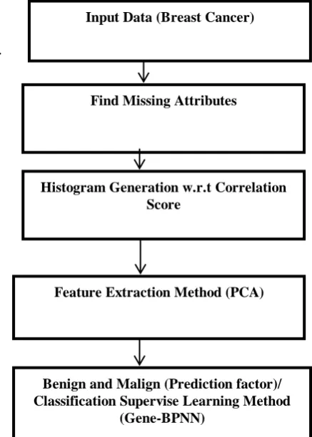
III. RESEARCHPROPOSEDWORK
In this research methodology section, collection of the Breast Cancer Dataset downloads from the UCI Machine Learning Repository Site.
[image:3.595.306.525.158.465.2].
Fig 2. Proposal Flow Chart
Study the various prediction and classification Machine Learning Algorithms to design a data pre-processing and feature extraction phase is to find the missing or irrelevant attributes and search unique feature vector in the BC (Breast Cancer Dataset). To implement a novel prediction and classification approach to find breast cancer such as Benign and Malign. Compute and compare the performance metrics like as accuracy rate, error rate, specificity and sensitivity.
A. Kernel PCA algorithm used for Feature Extraction for BCD[26]
1. Select a kernel mapping .
2. Get KP to depend on training data , (n=1,….. NN)}. 3. Resolve E (Eigen Value) issue of KP to obtain and . 4. Each data point given xx, get its PCs (Principal
Components) the feature vector : ) =
Below method steps described is based on the assumption that the data-point has 0 mean and therefore, the covariance of two data-points is similar to their correlation.
Input Data (Breast Cancer)
Find Missing Attributes
Histogram Generation w.r.t Correlation Score
Feature Extraction Method (PCA)
Benign and Malign (Prediction factor)/ Classification Supervise Learning Method
Table 1. Different Types of Kernels
Kernel Name Equation
Gaussian Kernel
Sigmoid Kernel +
Polynomial Kernel
(
Feature Vector or Space:
….. (i)
Equation (i) mapping is not elucidate and F(xxkp) is not available. Moreover, it can still get the kernel matrix K for the 0 mean data points F(xx) in terms of KP for F(xx):
= )T1 F( ) = [F( - T1
[F( ) - )]
= )T1 ) – T1
F( T F
= - - +
………….. (ii)
In detail description, the mapping in the kernel Principle Component Analysis is not obviously detailed is the dimensionality of feature vector F. Similarity, the Covariance Matrix and its E are only described in the above derivative, but they don't require to be evaluated in design. The High-Dimensional of FF doesn't extra evaluation as only
the KERNEL is required in the designed.
B. Gene-BPNN Algorithm for Breast Cancer Classification
1. Initialization 2. Fitness Evaluate
3. New Feature Set (Population Size) Operators (i) Selection
(ii) Crossover (iii) Mutation 4. Replace
5. Test (BPNN). 6. Exit.
Back Proportional Neural Network 1. Input Layer
2. Hidden Layer 3. Output Layer
= = a ( ) ………..(i)
It will positively require understanding how to train it. BPNN is a normally used method for training NN. NN is structured as a series of LAYERS, individually composed of one or more neurons. Individual neurons produce an output and activation function, based on the O/Ps of the existing layer and a set of weights.
= act (W1xx1 + ……. +WnnXnn)………..(ii)
Pseudo Code in Gene-BPNN algorithm
1. Start all weights with rand numbers, normally -1 to 1. Repeat
2. For every design in the Training_Set
3. For each_layer in the network 4. For every node in the Layer
(i) C
alculate the wts, sum of inputs to feature.
(ii) A
dd the threshold_val to the sum.
(iii) E
valuate the Activation Function for the Feature. End
End
5. For every_feature in the output layer. 6. Evaluate the error_signal.
End
7. For all Hidden_Layers. For every feature in the layer.
(i) E
valuate the feature’s signal_err.
(ii) T
he update features weight in the NNs. End
End
8. Compute the Error Methods End
9. While (Max_iterations) < than specified and (Err_method is > than specified).
IV . EXPERIMENTAL RESULT A. Dataset Description
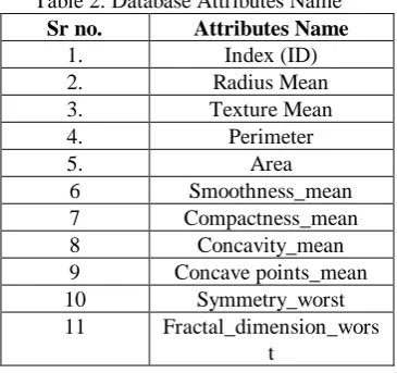
[image:4.595.340.523.514.685.2]The Dataset (BCD) that used is taken from UCI (University of California, Irvine). There are eleven attributes and the initial one is an identity that it will delete. 9th criterions are as explained earlier in BC classification section, they are intended to determine if a cancer is benign and malign, the last property contains a binary value (0,1). The attribute set consists of 699 medical cases. Breast Cancer Detection contains missing 16 values, which partial proposed work dataset to 683 sample values.
Table 2. Database Attributes Name
Sr no. Attributes Name
1. Index (ID)
2. Radius Mean
3. Texture Mean
4. Perimeter
5. Area
6 Smoothness_mean
7 Compactness_mean
8 Concavity_mean
9 Concave points_mean
10 Symmetry_worst
11 Fractal_dimension_wors
t
International Journal of Innovative Technology and Exploring Engineering (IJITEE) ISSN: 2278-3075, Volume-8 Issue-8, June, 2019
Fig 3. Breast Cancer Datasets (Wisconsin)
Fig 3 defines the count of benign and malignant cancer percentage calculated which 65% non-cancerous and 212 (35 %) cancerous type.
B. Result and Discussions
Overall research, two models were nearly studied and computed for datasets using the above mention in methodology phase described like as accuracy, specificity and sensitivity rate. In this research work, the evaluation of different models using WISCONSIN BREAST CANCER DIAGNOSTIC DATASET and the consequences were as follows:
Table 3: Performance Metrics
Parameters Values
Accuracy 99
Error Rate 0.0034
Specificity 0.98
Sensitivity 0.0078
Table 3 define performance of the classifier was calculated using metrics like as an accuracy rate, error rate, specificity and sensitivity etc. The sensitivity which is also known as TPR (True Positive Rate) is the % age of benign cancer data classified as benign by classification. It can correctly benign cancers will have a higher consequence insensitivity.
C. Formula:[26]
*100 ……… (i)
Specificity is the percentage of malignant cancers data classification. It classification that can appropriately classification Malignant Cancer will have a better result.
*100 ………..(ii) The accuracy rate is the metric for model calculation. It combines sensitivity and specificity for complete data combined.
[image:5.595.60.272.53.220.2]*100 ………(iii)
Fig 4. Comparison – Accuracy rate (%)
Table 4. Comparison between Gene-BPNN, KNN and NB
Techniques Accuracy Rate (%) Gene-BPN
N
99%
KNN 97.5%
NB 96.19%
Table 4 and Fig 4, it can recognize that three classification methods are efficient in the diagnosis, all of which define a High-Level of accuracy rate despite the small and large datasets. We analyze the various methods in machine learning methods to analyses the diagnosis of the breast cancer disease in data mining.
Illustration, Gene-BPNN classification method are high accuracy rate achieved at 99% value. As a consequence, Gene-BPNN is the most efficient classifier for BREAST CANCER classification difficulty. Though, if the huge amount of dataset, the Gene-BPNN will resolve the time complexity issue.
V. CONCLUSIONANDFUTURESCOPE
hor-1 Photo
Though cancer is predicted and classified through various approaches, still automatic detection of disease is difficult to approach due to different categories of cancer. In Future Scope, various new deep learning algorithms are required to be implemented for the detection of different stages and categories of breast cancer simultaneously.
REFERENCES
1. Davis, P. L., Staiger, M. J., Harris, K. B., Ganott, M. A., Klementaviciene, J., McCarty, K. S., & Tobon, H. (1996). Breast cancer measurements with magnetic resonance imaging, ultrasonography, and mammography. Breast cancer research and treatment, 37(1), 1-9.
2. Leitch, A. M., Dodd, G. D., Costanza, M., Linver, M., Pressman, P., McGinnis, L., & Smith, R. A. (1997). American Cancer Society guidelines for the early detection of breast cancer: update 1997. CA: A cancer Journal for Clinicians, 47(3), 150-153.
3. Alarabeyyat, A., & Alhanahnah, M. (2016). Breast Cancer Detection Using K-Nearest Neighbor Machine Learning Algorithm. In 2016 9th International Conference on Developments in eSystems Engineering
(DeSE) (pp. 35-39). IEEE.
4. Prabhakar, S. K., & Rajaguru, H. (2018). Performance analysis of breast cancer classification with softmax discriminant classifier and linear discriminant analysis. In Precision Medicine Powered by pHealth and
Connected Health (pp. 197-201). Springer, Singapore.
5. Pisani, P., Parkin, D. M., Ngelangel, C., Esteban, D., Gibson, L., Munson, M., ... & Laudico, A. (2006). Outcome of screening by clinical examination of the breast in a trial in the Philippines. International journal of cancer, 118(1), 149-154.
6. Dheeba, V., Singh, N. A., & Singh, J. A. P. (2014). Breast Cancer Diagnosis: An Intelligent Detection System Using Wavelet Neural Network. In Proceedings of the International Conference on Frontiers of
Intelligent Computing: Theory and Applications (FICTA) 2013 (pp.
111-118). Springer, Cham.
7. Janghel, R. R., Shukla, A., Tiwari, R., & Kala, R. (2010). Intelligent decision support system for breast cancer. In International Conference in
Swarm Intelligence (pp. 351-358). Springer, Berlin, Heidelberg.
8. Kattepura, V., & Kurian, M. Z. (2017). Effectiveness of Existing CAD-Based Research Work towards Screening Breast Cancer. International Journal of Advanced Computer Science And Applications, 8(9), 121-128.
9. Francis, S. V., & Sasikala, M. (2013). Automatic detection of abnormal breast thermograms using asymmetry analysis of texture features. Journal of medical engineering & technology, 37(1), 17-21.
10. Müller, A., Homey, B., Soto, H., Ge, N., Catron, D., Buchanan, M. E., ... & Barrera, J. L. (2001). Involvement of chemokine receptors in breast cancer metastasis. nature, 410(6824), 50.
11. Miki, Y., Swensen, J., Shattuck-Eidens, D., Futreal, P. A., Harshman, K., Tavtigian, S., ... & Ding, W. (1994). A strong candidate for the breast and ovarian cancer susceptibility gene BRCA1. Science, 266(5182), 66-71. 12. Babu, G. R., Lakshmi, S. B., & Thiyagarajan, J. A. (2013).
Epidemiological correlates of breast cancer in South India. Asian Pacific
Journal of Cancer Prevention, 14(9), 5077-5083.
13. Singletary, S. E. (2001). New approaches to surgery for breast cancer. Endocrine-related cancer, 8(4), 265-286.
14. Blackburn, H. L., Ellsworth, D. L., Shriver, C. D., & Ellsworth, R. E. (2015). Role of cytochrome P450 genes in breast cancer etiology and treatment: effects on estrogen biosynthesis, metabolism, and response to endocrine therapy. Cancer Causes & Control, 26(3), 319-332. 15. Zhao, L., Cheong, A., Reece, G. P., Fingeret, M. C., Shah, S. K., &
Merchant, F. A. (2016). Inferior breast-chest contour detection in 3-d images of the female torso. IEEE Journal of Translational Engineering in Health and Medicine, 4, 1-10.
16. Krawczyk, B., & Schaefer, G. (2014). Breast thermogram analysis using classifier ensembles and image symmetry features. IEEE Systems Journal, 8(3), 921-928.
17. Bhowmik, M. K., Gogoi, U. R., Majumdar, G., Bhattacharjee, D., Datta, D., & Ghosh, A. K. (2018). Designing of ground-truth-annotated DBT-TU-JU breast thermogram database toward early abnormality prediction. IEEE journal of biomedical and health informatics, 22(4), 1238-1249.
18. Shirmohammadli, V., & Manavizadeh, N. (2018). Numerical Modeling of Cell Trajectory Inside a Dielectrophoresis Microdevice Designed for Breast Cancer Cell Screening. IEEE Sensors Journal, 18(20), 8215-8222.
19. Amrane, M., Oukid, S., Gagaoua, I., & Ensarİ, T. (2018). Breast cancer classification using machine learning. In 2018 Electric Electronics,
Computer Science, Biomedical Engineerings' Meeting (EBBT) (pp. 1-4).
IEEE.
20. Sotiriou, C., Neo, S. Y., McShane, L. M., Korn, E. L., Long, P. M., Jazaeri, A., ... & Liu, E. T. (2003). Breast cancer classification and prognosis based on gene expression profiles from a population-based study. Proceedings of
the National Academy of Sciences, 100(18), 10393-10398.
21. Amrane, M., Oukid, S., Gagaoua, I and Ensarİ, T. (2018). Breast cancer classification using machine learning. In 2018 Electric Electronics, Computer Science, Biomedical Engineerings' Meeting (EBBT) (pp. 1-4). IEEE.
22. Jafarpisheh, N., Nafisi, N. and Teshnehlab, M. (2018). Breast cancer relapse prognosis by classic and modern structures of machine learning algorithms. In Fuzzy and Intelligent Systems (CFIS), 2018 6th Iranian Joint Congress on (pp. 120-122). IEEE.
23. Hela, B., Hela, M., Kamel, H., Sana, B and Najla, M. (2013). Breast cancer detection: A review on mammograms analysis techniques. In Systems, Signals & Devices (SSD), 2013 10th International Multi-Conference on (pp. 1-6). IEEE.
24. Gayathri, B. K and Raajan, P. (2016). A survey of breast cancer detection based on image segmentation techniques. In Computing Technologies and Intelligent Data Engineering (ICCTIDE), International Conference on (pp. 1-5). IEEE.
25. Schölkopf, B., Smola, A., & Müller, K. R. (1998). Nonlinear component analysis as a kernel eigenvalue problem. Neural computation, 10(5), 1299-1319.
26. Habib, A., Alalyani, M., Musa, I. H., & Almutheibi, M. S. (2015). Brief review on sensitivity, specificity and predictivities. IOSR J Dent Med Sci, 14(4), 64-8.
AUTHORSPROFILE
Amandeep kaur is an M.Tech Student at Department of CSE in Sachdeva Engineering College For Girls,Gharuan,Mohali.She completed her B.Tech from Rayat And Bahra College Of Engineering And Biotechnology for Women, Kharar in year 2017. Now she is pursing M.Tech from SECG,Gharuan,Mohali.She is doing her research in field of data mining and machine learning,titled as “Breast Cancer Detection and Classification Using Analysis and Gene-Back Proportional Neural Network Algorithm”.Beside this her other research interest includes cloud computing ,information security.