Abstract: This paper discusses various plant diseases and how to improve precision agriculture (PA) using Image processing. The aspects considered are the higher yielding and result in good quality of crop production. Precision agriculture (PA) is necessary to improve agricultural productivity of specific crop. Image processing is an important tool for identification of plant diseases, whereas manual detection of crop plant disease is a difficult task as it takes serious observation (need implementation expert of automated system) and consumes much time. Another outcome from the study is that automatic detection can be very good aspect for identification of crop disease.
Index Terms: Plant pathology, nutrients, leaf sheath, image processing
I. INTRODUCTION
India’s economy is one of the fastest growing economies of the world, and livelihood of 58% of rural household depends on agriculture. Now a day’s Growing potential of Indian processing sector poised India’s contribution to world food trade. Indian retail market of food industry contributing 70% of the sales and grocery market becomes sixth largest is the world. Recent trends of technical advancement in food processing industry establish ranked fifth in terms of production, consumption, export and expected growth. it accounts 32% of the country’s total food market. Behavioral practice of soil, climate and cultivation method admire to grow variety of food crops in different parts of the country. A large number of the crops grown in our country are rice, wheat, sugarcane, pulses etc. Instead of huge production we are still lagging behind, because existing literature does not derive any comprehensive technique which can deal with the complete identification of the plant disease. We should develop a technique or framework for soil testing and disease identification via single platform. Some basic steps required for image processing are discussed in later parts of this study. Image processing deals with image acquisition, image preprocessing, disease segmentation, feature extraction and disease classification, which are explained in the Fig.1.
Revised Manuscript Received on May 06, 2019
Shashank Chaudhary, Computer Science & Engineering, AKTU Lucknow, India
Dr Upendra kumar, Computer Science & Engineering IET Lucknow India.
Fig.1 Steps of Image Processing
A. Image Acquisition
The real time images are fed directly from the camera. For further analysis, proper visibility and easy analysis of images, white background is created because most of leaves colour varies from red to green for exact segmentation.
B. Image Preprocessing
Image preprocessing is required to resize captured image from high resolution to low resolution. The image resizing can be done through the process of interpolation. Captured input image is being converted into a grayscale image using colour conversion by the equation
Image = 0.3R + 0.59G + 0.11B
The captured image placed in white background results in large differences between grey values of object and background. References [1] have discussed the application of computer vision technique to enhance the plant leave in order to detect diseases. Computer vision image enhancement (Colour conversion and Histogram equalization) can be detecting highly enhanced images with higher clarity than captured images. Captured infected plant leaves images can be diagnose using Grayscale translation and histogram equalization.
C. Disease Segmentation
Disease Segmentation is an important step to make something that is more meaningful and easier to analyze. The goal of segmentation is to simplify or change the representation of an image into multiple segments for further analysis.
A Review: Crop Plant Disease Detection Using
Image Processing
We can identify objects or other relevant information in digital images. Through image statistics, maximum and minimum grey levels are obtained and threshold values are calculated by averaging the obtained values. These threshold values transfers the grey scale image to binary image. Reference [2] proposes a classification method where, image segmentation and SVM techniques named as Otsu’s thresholding is used for plant disease classification involves all the possible threshold values through iteration and calculate threshold for each side of pixel level. Separate the pixels into two clusters, then find the mean of each cluster and finally squaring the difference of the means. Reference [3] findsan image segmentation method using support vector machine and Otsu’s method for apple sorting and grading. Results derive by this application of above mentioned technique, shows segmentation error of 3% to 25% for rigid SVM and 2% for flexible SVM.
D. Feature Extraction
Feature Extraction is one of the most interesting steps of image processing to reduce the efficient part of an image or dimensionally reduction of interesting parts of an image as a compact feature vector. Feature reduction representation is useful when the image size is large and required to rapidly complete the tasks such as image matching and retrieval. Other common feature extraction techniques include:
Histogram of oriented gradients (HOG)
Speeded-up robust features (SURF)
Local binary patterns (LBP)
Haar wavelets
Colour histograms
Once the features have been extracted, they may be used to build machine learning models for accurate object recognition or object detection. Reference [4] applies feature extraction to recognise leaf for plant classification using GLCM and PCA methods. Different features are needed to describe the different properties of the leaves. For feature extraction of leaves recognition, gray-level co-occurrence matrix method is introduced. Gray-level co-occurrence matrix (GLSM matrices) is designed to measure the spatial relationships between pixels.
E. Classification of Image
Classification of image consists of database that contains pre-defined patterns that are compared with detected objects to classify them in a proper category. Classification will be executed on the basis of spectral defined feature such as density, texture etc. [5] suggests image classification using Convolution Neural Networks and Deep Learning, and it has introduced the Convolution Neural Network (CNN) as a new area in machine learning and is applied to crop plant disease through classification of image. To classify an image author [6] developed mobile application for infected crop disease, classification diagnosis crop plant with symptom of signatures disease, detection of signature disease that is expressed as a number of rules that concern the colour, the shape of the spots, historical weather data. It is based on mobile phone detection. Above developed application allows an agriculturist acting as an end user, extend or to customize the supported set of plant diseases.
II. LITERATURE REVIEW
Literature review technique used in this paper is the structured literature review and the basic steps or structures are discussed as below –
A. Soil Testing
Crops need nutrients in the same way as humans. Healthy soil contains the three nutrients for plant such as N (nitrogen), P (Phosphorus), K (Potassium) which are the three essential nutrition of soil, along with this as other nutrients are also required in smaller quantities such as calcium, magnesium, sulphur, iron, zinc, copper, boron, molybdenum and nickel which are also to be measured. Other reasons for reduction in Soil nutrient value are continuous use of soil along with the overuse of insecticides. Fertility of soils depends adequate levels required nutrients in the soil.
Nutrients are found in abundance in the form of fertilizers for plant growth and the soil is re-enriched by the use of proper fertilizer in analyzed quantity, as per the requirement. For healthy production of crop Soil testing is done which evaluates the deficient nutrient from the soil. Some nutrients are not readily available to plants; perform the soil test to avail the required nutrient. Reference [7] proposed to utilize fly ash material as a soil nutrient to improve the quality of soil which results in the increase of the crop production.
Fly ash used as a required nutrient substitute for increase crop production. it is useful to improve the quality of soil; it is good source of required nutrient and micronutrients. It can increase the production where soil yielding is poor. Fly ash changes organic values, raises water holding capacity, soil porosity, the soil texture and pH, electrical conductivity of the soil. A marginal increase found in the concentration of P, K, S, Fe, Zn, Mn, B, Ca and Mg elements, when fly ash used as supplement in the soil. Reference [8] found importance of Fly ash use as a nutrient supplement and integrated component of plant nutrient supply system, other organic waste and fly ash residue increase the crop yield and changes soil nutrient and micronutrient as per the need derived from soil characteristics.
B. Disease Identification in crop
Each crop is detected by many different types of plant pathogens, causing different diseases and some of them are significant and occur most widely around the world. Blast, Bacterial Leaf Blight, Brown spot, Sheath Blight and False smut are main diseases found in rice, whereas the important diseases of wheat are Rust, Loose smut, Karnal bunt and spot blotch. Sugarcane is a crop or we can say it's a cash crop which is affected by a number of disease or pathogens. The important ones include Red rot, Smut, Grassy shoot and Red stripe. These are the four important diseases that are considered to be a more important economically. Plant pathogens are mostly developed due to environmental factor. It affects plant leafs, roots or crop itself. Precision Agriculture is everything that makes the practice of farming more accurate and controlled. For Precision Agriculture, Image processing technique
Fig.2 Disease Identification
C. Leaf Diseases
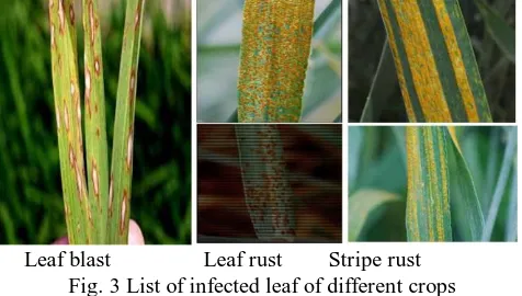
Leaf diseases are caused by pathogens brutally affecting the yield of crop. The disease of infected leaf can be identified on the basis of its symptoms such as finding the pattern of rotten leaf by bacteria. If we initially identify the disease caused by leaf based on their symptoms such as infected pattern of leaf may be rotten by any bacteria. Some of diseases we know are blast and it is a fungal disease caused by the organism magnaporte oryzae and bacterial leaf blast frequently occurs in rice. leaf disease of wheat are leaf rust or brown rust because rust pustule are brown in colour and another one is stripe rust caused due to rust pustule and its colour is yellow that is why it is called yellow rust.
Reference [9] to detect, diagnose and prescribe control options, an experts system using Rule-Based Algorithm to identify Plant Diseases in the Philippines. This application helps farmers to identify problem in rice plants and perform action to detect and diagnose rice plant disease and prescribe feasible control options. They proposed the methods to determine problems evolved due to rice plant diseases and disorders by taking interviews and surveys to farmers. Once the actual problem occurred in crop is determined, an automatic detection system can be developed using current age technologies. After determining the problems, a creative and innovative idea was created. Agile Software Development Methodology was used to come up with the expert system which emphasizes in real time communication with farmers; preferably the Rule-based algorithm was incorporated in the application for the classification of rice plants diseases and symptoms.
Leaf blast Leaf rust Stripe rust Fig. 3List of infected leaf of different crops
Identification of infected disease is very important, so [10] classify rice diseases such as leaf blast and brown spot which can be identified by using different pattern recognition techniques. They proposed that SOM (Self Organising Map) based neural network can be applied in zooming algorithm to classify diseased rice images. Boundary detection and spot detection methods were used for feature extraction of the infected parts of plant’s leaves and produced satisfactory classification result.
Manually it is difficult to identify the disease, if any automatic method is developed then it is easy to identify a disease. Reference [11] has developed an automatic grading
diagnosis via embedded image processing system for rapid and accurate identification of disease. Here wheat leaf rust detection performed in real time by adopted system of ARM9 processor with embedded Linux and program is developed in the QT Integrated Environment gives accuracy for image recognition of 96.2%.
Health card of any plant depends on the severity of disease and its occurrence. Here [12] derived diagnosis Leaf Disease Severity Measurement using Image processing
Excessive use of pesticides on fungi causes disease in sugarcane which increases cost and pollution. Simple threshold and triangle threshold method are used to segment the leaf area and infected region area. One of the leaf found disease is SEPTORIA leaf spot which is generally founds in tomatoes. This disease spreads upwards from the lower leaves to the rest of the foliage and may cause almost complete defoliation of larger plants. It is caused by the fungus Septoria lycopersici. Symptoms of the disease notified on the lower leaves as tiny grey spots with dark brown margins. These spots may remain small or may enlarge. When the spots are large in number, adjacent ones often unite as they enlarge resulting in the partial or complete collapse of a leaflet. Reference [13] detects disease in tomato leaf, by the use of automatic technique to minimize the effect of presence of vein, RGB image should be colour transformed before segmentation. After then Otsu threshold can be applied on colour component to detect disease spot accurately.
Another important leaf occurring disease is Little Leaf of Brinjal. Brinjal leaf disease caused by a virus as the name suggests that mostly new developing leaves they become little in size and they remain little in size. In exhaust conditions the entire plant may be stunted and the leaves remain little in size throughout the plant. So this is a virus disease, as [14] detect little leaf disease is a hazardous disease found in pine trees. the author found, detection of plant disease through some automatic technique is beneficial and it reduces a large work of monitoring in big farms of crops. Image segmentation and soft computing techniques is used to detect the plant disease. So, same technique can be used for brinjal little leaf disease. a we can detect the symptoms of little leaf diseases at primary stage using image segmentation technique. Some other leaf disease are Powderly mildew and Downey mildew, which is a leaf infected disease and is a fungal disease most frequently found in wheat, barley and other cereal crops as well as in apples and grapes so [15] considered these cereals crop and fruit crops for study and then proposed their view using image processing technique based on detection of fungal disease in plants and proposed statical method for quantitatively detecting and classifying fungal disease, based on disease severity fungal disease symptoms are termed as powdery mildew, downey mildey and their effects on fruit crops are being considered for study.
Bacterial leaf scorch is a leaf disease mostly active in ornamental trees and shrubs and is also found in blueberry and almond, so according to [16] has analyzed views to detect infected area of Bacterial Leaf Scorch (BLS) of Shade Trees Using Image Processing. In image segmentation, K-means is very effective
separating foreground and background images to identify infected plant from Bacterial leaf scorch infection. Segmentation technique is based on subtracting the clustered leaf images and intensity mapping for highlighting leaf area. To classify the crop species [17] develop a model for deep learning for image-based plant disease detection using deep convolution neural network with images of plant leaves with the goal to identify disease on images. Another method suggest by [18] is to classify crop disease based on image processing, developed grading method based on computer image processing technique, firstly it acquires the images of the crop disease, leaves. Vector median filtering method is used to pre-process the crop leaves. Using statistical pattern recognition method for segmentation and then it calculates the ratio percentage of the lesion area and the leaf area. Finally crop disease harm degree and classification standard were determined.
Reference [19] has conferred upon unhealthy region of plant leaves. Initially colour transformation is performed for RGB image as input, and green pixels are masked, remove specific threshold value followed by the segmentation process. Then texture statistics are computed for the useful segments, after that texture features is to classify plant leaf diseases. Finally the extracted features are passed through the classifier. Digital image processing techniques are used for bacterial infection detection as suggest by [20] on tomato and crape jasmine leaves, colour transformation method is
used to transform an RGB image into YIQ colour space, to detect bacterial disease symptoms of brown-black colour spot appear and centre becomes dry then I channel alone is taken for further analysis. Median filter is used for smoothing and filtering the image. After that brown-black colour spot of infected disease appear using Otsu’s thresholding method. Then Haralick texture features are extracted and stored for over segmented image. At last classification algorithm is applied for classification of the infected disease. Reference [21] has identified multiple plant diseases using digital image processing; suggest colour transformation and colour histograms method for disease identification, after that pair wise-based classification system is used. Its performance was tested for 82 different biotic and a biotic stresses using a large database containing images of symptoms belonging to, affecting the leaves of 12 different plant species.
I. Leaf found Disease
S.no Crop name Infected disease
1 Rice Leaf blast, brown spot, Leaf blight
2 Wheat Leaf rust, stripe rust
3 Potato Leaf blight Early blight
4 Groundnut Early and late leaf spot (Tikka) 5 Brinjal Little leaf, leaf spot
6 Tomato Leaf curl, Septoria leaf spot 7 Crucifier Alternaria leaf spot, white rust
II. Summary of Leaf found Disease and Work using Image Processing Technique
S.no Author Work Result Techniques
1 Roselia et al. (2017) Design an expert system with all possible control option to detect rice plant disease.
Real time communication with farmers
Expert system of agile software development methodology
2 Phadikar et al. (2008)
Zooming algorithm is used to detect symptoms of infected parts of plant
Classification of rice disease images.
Propose self organising map(SOM)neural network
3 Peifeng et al. (2017) Develop an automatic diagnosis method to differentiate various wheat diseases.
Diagnosis wheat leaf rust disease, with accuracy of 96.2% for image recognition.
embedded image processing system
4 Patil et al (2011) Segment the leaf area and lesion region area.
Leaf Disease Severity
Measurement by the Excessive use of pesticides on fungi caused disease in sugarcane increase cost and pollution
simple threshold and triangle threshold method are used
5 Budihal et al. (2015), Detection of Disease in Tomato Leaf
Detect disease spot accurately. Colour transformation then Otsu’s threshold
6 Singh and Misra
(2017)
Automatic technique is used for detecting little leaf disease found in pine tree.
Detects the symptoms of little leaf diseases
image segmentation and soft computing techniques
7 Pujari et al. (2015) Considered cereals crop and fruit crops are for study based on detection of fungal disease.
classifying fungal disease based on disease severity
8 Krishnan et al. (2014)
Separating foreground and background images to detect Bacterial Leaf Scorch (BLS) of shade trees.
detect infected area of Bacterial Leaf Scorch (BLS) of shade trees
K-means clustering segmentation is used
9 Sharada Prasanna
Mohanty et al. (2016)
Image-Based Plant Disease Detection
Trained a model, image of plant leaves classifying both crop species and identify disease on images.
deep convolution neural network
10 Youwen Tian et al. (2012)
Develop grading method to classify crop disease based on image processing.
Predict crop infected degree and calculate percentage of infected area.
Vector median filtering method is used for pre-process then statistical pattern recognition method for segmentation 11 Arivazhagan et al.
(2013)
Color transformation method is used for RGB images.
Lesion area of leaf detected and classifies crop disease using texture feature.
Color transformation is used followed by segmentation. 12 Revathy (2015) Colour transformation is
used to detect brown-black colour spot.
Detect the infected disease spot. Colour transformation then Otsu threshold
13 Garcia et al (2016) Created database for Identifying multiple plant diseases
Identified multiple plant diseases using digital image processing
Colour histograms and pair wise based classifier is used.
D. Disease found in root of plant
The disease infects the root of the plant are called root infected disease and it’s basically occur in plants such as chickpea. The diseases are dry root rot and ascochyta blight which infect in the root of the plant, Club root of crucifier, Root rot of pigeon pea.
Dry rot root Sclerotinia blight Root rot Fig. 4 List of infected root of different crops
III. ROOT FOUND DISEASE S.no Crop name Infected disease
1 Rice Leaf blast, brown spot, Leaf blight
2 Wheat Leaf rust, stripe rust
4 Groundnut Early and late leaf spot(Tikka) 5 Chickpea dry root rot & Ascochyta blight 6 Pigeon Pea Root rot
7 Crucifier Club root of crucifier
E. Disease found in stem of plant
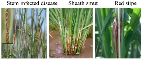
As we have already discussed the leaf infected diseases and the root infected diseases, now the stem infected crop disease is being discussed. As the crop matures the probability of occurrence of stem disease increases. So the identification of stem disease is an important factor to find out the disease. Otherwise at this condition or stage it brutally infects the crop plant. Caterpillars feed within the stem of growing canes and may cause enough damage to kill the growing point, resulting in browning holes in the stem; some species of caterpillars also infect stems of paddy, corn and sorghum.
Reference [22] detected borer diseases. The SVM classifier was chosen to recognize the diseases of sugarcane. It was unable to detect the sugarcane borer disease with the general linear methods. Author uses support vector machine, resulting in the distribution of minimum average grey value and the minimum grey value of the sugarcane as disease and disease-free.
Stem infected disease Sheath smut Red stipe
[image:5.595.311.555.411.514.2]
Fig. 5 List of infected stem of different crops
IV. Stem Found Disease
S.no Crop Name Infected disease
1 Sugar Cane sheath smut, red stripe, black stripe
2 Apple Black stripe, brown spot
3 Rice Neck blast, brown spot
4 Wheat Leaf rust, stripe rust
5 Pigeon Pea Root rot
III. CONCLUSION
In this study the main stress was on Leaf diseases since most of the diseases are found to be infecting on leaves itself, the other diseases like that of root and stem are also analyzed from different viewpoints of collective literature but they are less severe in nature and their probability of occurrence is also low so the main stress in this study was on the contemporary diseases
r-1 Photo
The paper presents a comprehensive view on the various researches done in contemporary domain of Crop diseases. The collection of analysis is done dealing with various plant diseases. Then the identification of exact disease in rice such as Blast, Bacterial Leaf Blight, Brown spot, Sheath Blight and False smut are to be achieved through the image processing. In this study the analysis was done by segmentation techniques such as Otsu’s and K-means clustering and feature extraction and classification is advocated for the usage through image processing technique.
REFRENCES
1. K.Thangadurai,K. Padmavathi(2014),computer Vision image Enhancement for Plant Leaves Disease, Computing and Communication Technologies (WCCCT), 2014 World Congress on,2014
2. K. Elangovan, S. Nalini(2017), Plant Disease Classification Using Image Segmentation and SVM Techniques 7 pp. 1821-1828,2017
3. A.Mizushima and R. Lu (2013), An image segmentation method for apple sorting and grading using support vector machine and Otsu’s method, 94,pp. 29–37, 20
4. Abdolvahab Ehsanirad,Sharath Kumar Y.H.(2010),Leaf recognition for plant classification using GLCM and PCA methods,3,pp.31-36,2010. 5. Deepika Jaswal, Sowmya.V, K.P.Soman(2014), Image Classification
Using Convolutional Neural Networks,3pp2278-7763.2014
6. N. Petrellis (2017) Mobile Application forPlant Disease Classification Based on Symptom Signatures ACM ISBN 978-1-4503-5355-7/17/09 7. Sudha jala,Dinesh goyal(2006), Fly ash as a soil ameliorant for
improving crop production—a review, ELSEVIER Volume 97, Issue 9, June 2006, Pages 1136-1147
8. B.N.Mittraa et al. (2005) Fly ash—a potential source of soil amendment and a component of integrated plant nutrient supply system, ELSEVIER Volume 84, Issue 11, August 2005, Pages 1447-1451
9. Roselia C. Morco, Fredilyn B. Calanda, Jonathan A. Bonilla, Mark Jade S. Corpuz, Junnel E. Avestro, Jean M. Angeles(2017)e-RICE: An Expert System using Rule-Based Algorithm to Detect, Diagnose, and Prescribe Control Options for Rice Plant Diseases in the Philippines 978-1-4503-5392,2017
10. Santanu Phadikar and Jaya Sil “Rice Disease identification using Pattern Recognition Techniques” IEEE Proceedings of 11th International Conference on Computer and Information Technology ( 2008) , pp. 1-4244-2136, 2008
11. Peifeng Xu ,Gangshan wu,y.gio,X.chen,H.yangi ,R.zhang(2017)Automatic Wheat leaf rust detection and grading Diagnosis via embedded image processing system,107,pp836-847,(2017)
12. S. B. Patil, S. K. Bodhe(2011), Leaf Disease Severity Measurment Using Image processing,5pp297-301,2011
13. S.Budihal, Sandhya R, Soumya D Hajawagol,(2015), Detection of Disease in Tomato Leaf,4pp.2278-5140.
14. V .Singh, A.K. Misra (2017) Detection of plant leaf diseases using image segmentation and soft computing techniques,4PP41-49,2017
15. J.D pujari,R.Yakkundimath,A.S. Byadgi(2015)image processing based on detection of fungal disease in plants,46pp.1802-1808,2015.
16. Murali Krishnan and Dr.M.G.Sumithra “A Novel Algorithm for Detecting Bacterial Leaf Scorch (BLS) of Shade Trees Using Image Processing pp. 978-1-4799-1532-3/13,2013
17. Sharada Prasanna Mohanty, David Hughes,Marcel Salathe (2016) Using Deep Learning for Image-Based Plant Disease Detection,2016. 18. Youwen Tian,Lide Wang,Qiuying Zhou(2012) Grading Method of Crop
Disease Based on Image Processing,pp.427–433, 2012.
19. S.Arivazhagan,R.Newlin Shebiah,S.Ananthi,S.Vishnu Varthini (2013) Detection of unhealthy region of plant leaves and classification of plant leaf diseases using texture features,15pp11-23,2013
20. R. Revathy, Dr. R. Roselin(2015) Digital Image Processing Techniques for Bacterial Infection Detection on Tomato and Crape Jasmine Leaves,6,pp2229-5518,2015
21. Jayme Garcia Arnal Barbedo, Luciano Vieira Koenigkan,Thiago Teixeira Santos(2016), Identifying multiple plant diseases using digital image processing,147pp104-116
22. Tisen Huang, Rui Yang,Wenshan Huang, Yiqi Huang, Xi Qiao(2018) Detecting sugarcane borer diseases using support vector machine,5pp74-82
AUTHORS PROFILE
First Author Shashank Chaudhary M-Tech IIT Dhanbad, research work on plant leaf disease using image processing and machine learning.
Second Author Dr Upendra kumar Assistant Professor in IET Lucknow. Human Cognition inspired Models, Artificial Intelligence, Fractal and Chaos based Theory, Image Processing, Biomedical Signal Processing, Soft Computing, Pattern Recognition, Robotics and Embedded in C, Machine Vision