Avian metapneumovirus
studies using
Reverse Genetics
Thesis submitted in accordance with the requirements of the
University of Liverpool
for the degree of
Doctor in Philosophy
By
MARCO FALCHIERI
1 | P a g e
2 | P a g e
T
ABLE OF CONTENTPage
Table of content....………2
Abstract………...3
Introduction.………..5
1. AVIAN METAPNEUMOVIRUS LITERATURE REVIEW………10
2. AVIAN METAPNEUMOVIRUS RT-NESTED PCR: A NOVEL FALSE POSITIVE REDUCING INACTIVATED CONTROL VIRUS WITH POTENTIAL APPLICATIONS TO OTHER RNA VIRUSES AND REAL TIME METHODS………..….……..………...….…30
3. AN INVESTIGATION INTO VECTORING PROPERTIES OF AVIAN METAPNEUMOVIRUS ………45
4. AVIAN METAPNEUMOVIRUSES EXPRESSING INFECTIOUS BRONCHITIS VIRUS GENES ARE STABLE AND INDUCE PARTIAL PROTECTION ……...………....68
5. ATTEMPT TO DEVELOP A REVERSE GENETIC SYSTEM FOR AVIAN METAPNEUMOVIRUS SUBTYPE B ……...…....90
6 GENERAL DISCUSSION AND CONCLUSION………106
References……...………...………...113
3 | P a g e
ABSTRACT
Avian metapneumovirus (AMPV) is an enveloped negative sense single stranded RNA virus which is a major endemic respiratory pathogen of global domestic poultry. Since reverse genetic (RG) techniques have been applied to this pathogen several reports have investigated the effects of single and multiple genomic mutations and gene deletions or insertions on viral biology. The aim of this study was to gain a better understanding of the viral capacity to accept and in some cases express homologous and heterologous extra sequences.
Initially an AMPV subtype A was modified to introduce a homologous 200 bp sequence within the G gene and this recombinant was suggested to be used as a positive control for validating all stages of a previously established RT-nested PCR.
4 | P a g e
The vectoring abilities of subtype A strains were further investigated to accept and express foreign genes, specifically GFP gene and both spike (S1) and nucleocapsid (N) genes of infectious bronchitis virus (IBV). After viruses had been recovered by RG, all recombinants were proven to express the inserted genes efficiently and were all found to be highly stable during passage in vitro. Subsequently IBV recombinants were tested as candidate vaccines by eye-drop inoculation of one-day-old chickens. When chicks were challenged with IBV, partial protection results were observed, as assessed by greater motility of tracheal cilia from animals receiving the recombinants.
5 | P a g e
6 | P a g e
In the late 1970 in South Africa a severe pathological condition of turkeys, characterized by an acute rhinotracheitis, hit the local poultry industry heavily (24). Although many previously known respiratory pathogens could have been responsible for such a condition, no immediate diagnosis was made. Subsequent analysis pointed to an unknown viral agent, promptly named
Turkey rhinotracheitisvirus (TRTv), as cause of that epizootic (24). The virus was later charachterized as a negative sense, non-segmented, single stranded RNA virus closely related to mammalian respiratory syncytial viruses (178). For this reason it was classified within the Paramyxoviridae family, subfamily
Pneumovirinae, genus Pneumovirus (178). More recently, following the
detection of a similar virus infecting humans, TRTv was then placed in a new genus, Avian metapneumovirus (AMPV) (224). After its first detection in Africa (24) AMPV spread rapidly to Europe (7) and then to most parts of the world (50), becoming immediately a major problem for turkey production, but also for chickens, which soon proved to be susceptible to it. Losses were mainly due to a decreased bird growth rate and sometimes to high mortalities caused by secondary bacterial infections (205). Furthermore field and experimental evidence showed the viral capacity to affect egg production in laying birds, both in terms of number of laid eggs and egg quality (69, 190). Up to now AMPV control has been possible only by using live attenuated and killed vaccines (89). However, this approach has been shown to be limited, especially regarding eradication; also live vaccines have sometimes been shown to revert to virulence in turkeys (30) and as different viral subtypes are present, protection is often limited to certain strains (51). On the other hand killed vaccines appeared to be ineffective in preventing the respiratory infection, and are mainly administered to protect against egg drops (51).
7 | P a g e
just to develop better vaccines but even to reach a better understanding of viral biology (91, 123, 158, 179). In 2004 the first AMPV reverse genetic system (RG) was developed (158), enabling the recovery of the RNA virus from a DNA copy (cDNA) of its genome. As DNA molecules are more stable and easily modifiable than RNA ones, RG has given the opportunity to scientists to introduce not only single or multiple mutations, but also deletions and insertions into the AMPV genome and, followed by viral rescue, to readily observe the effects of those modifications on virus biology.
In 2004 an RG system was specifically developed for subtype A of AMPV (158). This thesis has investigated the ability of this subtype to accept, tolerate and express inserted foreign sequences. Moreover an attempt to adapt this technique to subtype B was made.
In Chapter 2, a homologous M2 gene sequence of approximately 200 bp was introduced into the G gene using restriction endonuclease cut properties and ligation steps. The insertion was made in a such way as to increase the size of a common AMPV diagnostic RT-PCR amplicon of the G gene. Rescued virus proved to be unaffected, at least in vitro, by the insertion, and it was proposed as positive control for diagnostic PCRs.
8 | P a g e
methods. GFP expression was verified both by fluorescence observation and by specific ELISA. The latter enabled not only the detection of GFP but also quantification of the amount of protein produced.
Chapter 4 describes the modification of the AMPV genome to be able to accept and express foreign genes. Extra gene transcriptional starts and stops were included into different intergenic regions so that genes could be inserted. To assess the system, green fluorescent protein was initially added in different positions. Once expression was verified, observing the fluorescence under UV microscope, AMPV recombinants carrying infectious bronchitis virus (IBV) S1 and nucleocapside genes were constructed. After in vitro studies assessing foreign gene expression, stability, viability and cytopathogenicity, recombinant viruses were used as vaccine candidates against IBV in chicken experimental infections.
Finally, Chapter 5 describes the attempt to construct a subtype B reverse genetics system. While for A and C subtypes such a technique has been successfully applied, for subtype B, despite several efforts having been made, RG has not yet been successful. As a first step a cDNA of a full length B subtype genome had to be constructed by cloning viral PCR amplicons in a plasmid, followed by the construction of plasmids encoding for essential AMPV support proteins. This procedure has proved to be very critical in view of plasmids instability and intolerance of the cloning bacteria to certain viral sequences.
9 | P a g e
10 | P a g e
CHAPTER 1
11 | P a g e
TABLE OF CONTENT
Page
1.1. Aetiology………...……….13
1.1.1. Taxonomy………..13
1.1.2. Morphology ....………. ……….………...13
1.1.3 Genome………...14
1.1.4 Proteins………....14
1.1.5 Virus attachment and entry………..15
1.1.6 Replication and transcriptional models………..…15
1.1.7 Packaging and assembly………...16
1.1.8 Strain classification………17
1.1.9 Physical and chemical susceptibility………...17
1.2. Epidemiology………18
1.2.1. Distribution………..………..………18
1.2.2. Hosts……….………...………...18
1.2.3. Transmission………..………...19
1.3. Pathogenesis………..20
1.4.Disease in birds………..21
1.5. Post-mortem findings………..………22
1.5.1. Gross lesions…….………...22
1.5.2. Microscopic lesions……..………...23
1.6. Immunity………..………24
1.7. Diagnosis………24
12 | P a g e
1.7.1.1 Isolation ………25
1.7.1.2. Viral detection……….26
1.7.1. Indirect diagnosis………..26
1.8. Control………28
1.8.1. Vaccination………..28
13 | P a g e
1.1 AETIOLOGY
1.1.1 Taxonomy
Avian metapneumovirus (AMPV) is one of the two viral species, with human metapneumovirus (HMPV), belonging to the Metapneumovirus genus; Metapneumoviruses are part of the subfamily Pneumovirinae within the
Paramyxoviridae family, including single stranded, negative sense RNA, and enveloped viruses (178, 224, 226).
1.1.2 Morphology
Viral particles are characterized by high pleomorphism (Figure 1), both in size and length; virions can be spherical or have a filamentous form, usually of 40 – 500 nm, but sometimes even over 1000 nm. Their nucleocapsids have a helical shape and on the envelope surface projections of about 13 – 14 nm are clearly distinguishable (11, 25, 46, 51, 84, 86, 145, 240).
Figure 1: AMPV observed under electronic microscope
14 | P a g e
1.1.3 Genome
The viral genome consists of a single negative stranded RNA of about 13000 - 14000 nucleotides (181). It encodes for 8 genes (Figure 2) which lead to the synthesis of at least 9 proteins: starting from the 3’ end, we can recognize in order N, P, M, F, M2 (including two overlapping open reading frames), SH, G and L (70, 132). Every gene is flanked by a transcriptional start sequence and a transcriptional stop, and between each transcriptional units there are intergenic untranslated regions. At the leader (3’) and trailer (5’) ends, other untranslated sequences of about 40 nucleotides are known, containing promoters and regulatory sequences for transcription, replication and packaging (91, 132, 233).
Figure 2: AMPV genome
1.1.4 Proteins
15 | P a g e
inhibition properties. The small hydrophobic protein (SH) is an integral membrane polypeptide; however its function is poorly understood. The Fusion (F) and the Attachment (G) proteins are located on the external part of the envelope and are the major antigenic determinants (21, 48, 70).
1.1.5 Virus Attachment and Entry
Viral particles interact with the host cells using the surface proteins (131). The G protein enables virus attachment on the cell receptors, while F promotes the fusion of the envelope with the cell membrane; the nucleocapsid is then released in the cytoplasm, where AMPV replication occurs (70, 77).
1.1.6 Replication and Transcription models
16 | P a g e
gene. This mechanism regulates all the transcription process until the end of the viral genome, meaning a good number of polymerase molecules will dissociate at every gene junction. This results in a sequential and gradually decreasing mRNA production: the genes located proximally will be transcribed in higher quantity compared to the ones placed distally. A minor or major amount of mRNA will result in a minor or major protein production (68, 229, 233).
Replication starts with the synthesis of a complementary positive copy of the genome, called antigenome. This is used as a template for genome production. During replication the polymerase ignores the transcription start and end signals, reading fully through all the viral RNA. The mechanisms that permit the polymerase to behave differently during transcription and replication are not clear, but some studies on human respiratory syncytial virus indicate that the concentration of N may be critical in regulating this process (76, 91, 233).
1.1.7 Packaging and Assembly
17 | P a g e
1.1.8 Strain Classification
Four subtypes of AMPV have been identified: A, B, C and D. Early studies, which showed differences among AMPV strains using monoclonal antibodies (45, 57) and sequencing and amino acidic analysis (122, 157), suggested the existence of two subtypes, A and B. In 1997, following the first North American AMPV outbreak (193), subtype C was identified. Both serological (55) and genomic techniques (194, 195) have highlighted the differences with the previous subtypes. Finally retrospective analysis of French strains, isolated in 1985, resulted in the identification of subtype D (12, 213).
1.1.9 Physical and Chemical Susceptibility
18 | P a g e
1.2 EPIDEMIOLOGY
1.2.1 Distribution
AMPV has been detected worldwide, with the only exception of Oceania (14). The disease appeared for the first time in South Africa in the late ’70 (25), and then spread to Europe (6, 7, 18, 19, 73, 75, 99, 150, 177), Asia (82, 135, 168, 221, 231), South and Central America (8, 66, 115, 216) and other African countries (26, 105, 169). In North America it has been reported only since 1996 (198).
Subtypes A and B are responsible of the disease in all affected continents, with the only exception of North America, where infections are caused just by AMPV subtype C. Subtypes C related strains have been more recently detected in France (212) and Korea (130). Subtype D was isolated only once in France in 1985 (12).
1.2.2 Hosts
19 | P a g e
surveys suggest that occupational exposure to turkeys might be a risk factor for human infection (124).
1.2.3 Transmission
20 | P a g e
1.3 PATHOGENESIS
The upper respiratory tract is considered to be not just the first replicative site of the virus but also the main target tissue for viral replication. AMPV seems to have a particular tropism for the ciliated cells of the nasal cavities, conchas, infraorbital sinus and trachea. In these organs viral particles can be detected until 7-9 days post infection using immunofluorescence technique (121) and isolated until 14 days post infection (53) in turkeys. Similar findings have been demonstrated also in chickens (32). However, occasionally AMPV can reach the lungs and the air sacs (5, 32, 58, 139). Bacterial co-infections seem to facilitate viral penetration along the lower respiratory tract:
Escherichia coli (1, 219, 223), Bordetella avium (61, 113), Mycoplasma
gallisepticum (156) and imitans (80), Riemerella anatipestifer (187),
21 | P a g e
related to the virulence of the single strain than to the subtype belonging (9, 202, 222).
1.4 DISEASE IN BIRDS
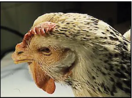
The clinical picture of AMPV infection is typically characterized by respiratory symptoms: coughing, sneezing, nasal discharge, swollen infraorbital sinus, but even conjunctivitis and submandibular oedema can be present (25, 117, 145). The disease is also called turkey rhinotracheitis (TRT) as this species appears to be more severely affected compared to chickens, where often the infection is asymptomatic (50). Morbidity is usually very high (almost 100%), while mortality can be very variable (172). The severity of the disease is highly dependent on management factors, including bird density, ventilation, temperature, hygienic conditions, and on secondary bacterial infections (89, 96, 205). Co-infections of AMPV and E.coli have been associated in chickens with swollen head syndrome (SHS). This is characterized not just by respiratory signs but also of a general head swelling (Figure 3), which leads to neurological signs, such as disorientation, torticollis and opistothonus (96, 120).
22 | P a g e
Figure 3: SHS in chicken (provided by Prof. E. Catelli)
1.5 POST-MORTEM FINDINGS
1.5.1 Gross Lesions
23 | P a g e
1.5.2 Microscopic Lesions
[image:24.595.221.446.362.574.2]No major differences have been found between turkeys and chickens. As said previously AMPV has a particular tropism for epithelial cells. The main histological lesions are located on the respiratory epithelium, characterized by deciliation, deepithelization, thickening of the mucosa, hyperaemia, mononuclear infiltration and glandular proliferation in the turbinates, infraorbital sinuses and trachea (Figure 4). Lesions are usually transient and detectable in the first 10 days after infection. After 3 weeks, birds are totally recovered (9, 32, 114, 222). Epithelial damages in the oviduct have been also observed (52).
Figure 4: microscopic lesions in trachea after AMPV infection
24 | P a g e
1.6 IMMUNITY
The immune reactions of turkeys towards AMPV infections have not been fully clarified. Some studies speculated cellular mediated immunity to be critical for protection, while humoral immunity appears to be not critical, and circulating antibody titers do not seem to be an indicator of protection (182). In experimental infections, turkeys with no detectable antibodies were protected against challenge with a virulent strain (62) and vaccinated bursectomised poults were resistant to challenge (119). Furthermore suppression of T- lymphocytes with cyclosporine A caused delayed recovery from clinical signs and more lasting microscopic lesions (186). Local immunity could be related in resistance to infection, however, as suggested for other respiratory pathogens (79), its short duration might explain recurrent infections during birds productive life in farms (182). Maternal antibodies are passed from hens to their progeny via the egg yolk, but their role does not seem to be significant, as they do not prevent infections (162) or do not interfere with early vaccination, allowing young chicks to be immunized in early stages (62) or directly in ovo
(237).
1.7 DIAGNOSIS
25 | P a g e
critical for a definitive diagnosis. This target can be reached directly by isolating or detecting the virus, or indirectly, by demonstrating specific serological responses in the host (90).
1.7.1 Direct Diagnosis
AMPV has a very short persistence in the host before clearance, both in turkeys (53) and chickens (32). For this reason virus isolation and detection are not always easy. Samples must be taken in the very early stages (at 3-5 days after infection) from birds not yet showing clinical signs (51).
1.7.1.1 Isolation
26 | P a g e
1.7.1.2 Viral Detection
Different immunochemical methods have been used to detect viral antigens in fixed and unfixed tissues and smears (89). Immunoperoxidase (32, 111, 139), immunofluorescence (116, 121) and immunogold staining (165) have been widely used during scientific studies. Specific monoclonal antibodies and immunodiffusion tests have been also used to differentiate between viral subtypes (45, 57, 90). All these methods appear to be time consuming and not particularly sensitive and they have been almost totally replaced by molecular methods, especially for diagnostic applications (89).
RT-PCRs amplify portions of viral genome, allowing a more rapid detection. Furthermore its high sensitivity enables viral presence to be revealed for a longer period compared to isolation methods or other detection techniques (51). Several PCR protocols have been described. A PCR based on the highly conserved N gene has proved to be able to detect all four AMPV subtypes (13). Subtype specific PCRs have been suggested targeting more variable genes like G (13, 154) and F (109, 143) for subtypes A and B and M for subtype C (4, 173). Real Time PCR protocols have been also developed, allowing not just a more sensitive viral detection but even viral quantification (41, 95). Multiplex RT-PCR have also been tested, able to detect AMPV and other respiratory viruses such as Influenza, Newcastle Disease and IBV (3, 83, 140).
1.7.2 Indirect Diagnosis
27 | P a g e
[image:28.595.244.423.364.544.2]immunosorbent assay (ELISA) tests have all been used. However while the first two are mainly employed for research work, ELISA is now the most common serological AMPV test, due to its sensitivity, specificity and suitability for mass serological screening. Several commercial kits have been developed (49, 89). Performances are highly dependent on the coated antigen: homologus tests have shown a higher efficiency compared to heterologous ones, especially among different subtypes (51, 148, 214). This can give rise to false negatives and the illusion that a vaccine has not ‘taken’. Subtype C antibodies are detected very poorly by subtype A and B ELISA, leading to the production of subtype C specific test (55, 138). Finally Blocking ELISAs are available in order to detect antibodies originated from sera of any avian species (33, 218).
Figure 5: IIF positivity to AMPV on tracheal section
28 | P a g e
1.8 CONTROL
As specific therapy against AMPV is not available, a preventive approach is critical both in avoiding the infection of birds and in controlling eventual losses caused by the disease. Attention to hygiene and biosecurity practices, ventilation, temperature, density, stress control, disinfection and good management procedures are all critical in reducing symptomatology and mortality (89, 205). Antibiotics can be used to prevent secondary bacterial infection (42, 97).
1.8.1 Vaccination
AMPV infections can be prevented by vaccination. Several vaccines are available and commonly used in commercial birds. Live attenuated vaccines are usually administrated by several methods (intranasal, eye-drop, drinking water or spray) to all bird categories at early stages to prevent the respiratory symptomatology (49, 89). While in broilers one administration seems to be fully protective, in growing turkeys repeated vaccination are required. Laying birds are usually vaccinated prior to the onset of lay by injection of inactivated vaccines, to avoid egg production losses (49, 63). Good cross protection has been reported between A and B subtypes (56, 74, 214). On the other hand, subtype C vaccines do not protect against A and B subgroups (55). Simultaneous vaccination with AMPV and other respiratory viruses (IBV, NDV) is not advised by pharmaceutical companies. However, experimental studies have shown no interference in protection onset (54, 79, 81). In ovo
29 | P a g e
have been recently developed and evaluated, however with poor outcomes at the time of writing, compared to conventional vaccines (107, 123, 179, 209).
1.9 REVERSE GENETICS
In 2004 Naylor et al. developed the first reverse genetic system for AMPV (158). A full length (FL) cDNA of subtype A was cloned in a plasmid vector by a series of PCR and ligation steps. The plasmid included a kanamicyn-resistant gene, essential in the cloning process, a T7 promoter and Hepatitis delta virus ribozyme. (158). N, P, L and Matrix 2 (M2) single genes, lead by a T7 promoter, were also cloned in other plasmids in order to provide the genome with the essential protein to form the RNP complex. Viral rescue was performed on Vero cells, previously infected with a recombinant Fowlpox virus
expressing the bacteriophage T7 polymerase. This polymerase is able to recognize the T7 promoter inserted in the plasmids and then to initiate transcription directly from them. Therefore the addition of the full length cDNA and the four support protein genes, in the presence of lipofectamine 2000 to allow cDNAs entrance into cells, should lead to the formation of all the RNP components (158). After the complex has been established, genome replication and gene transcription can begin as occur naturally, producing new RNA virions. A similar system, based on the same technical principles, was constructed for subtype C in 2006 in the United States (92). Up to now it has not yet been possible to apply RG system for subtype B.
30 | P a g e
CHAPTER 2
AVIAN METAPNEUMOVIRUS
RT-NESTED PCR: A NOVEL
FALSE POSITIVE REDUCING INACTIVATED CONTROL
VIRUS WITH POTENTIAL APPLICATIONS TO OTHER RNA
31 | P a g e
TABLE OF CONTENT
Page 2.1 Abstract………...……….…....32
2.2. Introduction………..33
2.3. Material and methods……….35
2.4 Results……….……….………39
32 | P a g e
2.1 ABSTRACT
33 | P a g e
2.2 INTRODUCTION
Avian metapneumovirus (AMPV) causes major disease in poultry in
most of the world and its detection and characterization has assisted (84, 98, 145, 234), and continues to assist (34, 39), in vaccine selection and disease control. RT-PCR is a powerful technique for detecting many viruses through the presence viral RNA in diagnostic samples. For Avian metapneumovirus (AMPV) tests have been reported (38, 110, 154, 167) which offer a sensitivity generally similar to, or greater than, classical virus isolation while additionally extending detection beyond loss of replicative viability (154), as well as frequently informing of virus origins through RNA sequence analysis (31, 34). Furthermore this sequence analysis avoids the need for secondary virus identification as often required in isolation techniques (17).
For PCR tests a positive control DNA sample generally accompanies sample tests to ensure the detection system is functional. However its cross contamination of test samples can lead to false positive readings (128). For RT-PCR tests, a control RNA is equally necessary and generally takes the form of an established control virus, but again its cross contamination of samples can be confused with true positive detections. This paper describes the construction and testing of a novel positive control AMPV able to identify such cross contamination events by producing amplicons of increased sizes, readily distinguishable from field virus amplicons on agarose gels, when used in conjunction with an established G gene based RT-nested-PCR method, (154).
34 | P a g e
antigenomic sense primer G1+. The resultant amplicon is further amplified using subtype specific PCRs whereby common primer G5- is paired with G8+A for subtype A specificity and G9+B for subtype B specificity. Subtype A and B viruses result in bands of 268 bp and 361 bp respectively and are readily distinguished on agarose gels.
[image:35.595.132.499.496.724.2]More recently, the presence of the AMPV G gene was demonstrated unnecessary for AMPV virus growth in Vero cells (133, 158, 160) and this made possible the generation of viruses with dysfunctional or absent G genes by reverse genetics (158, 161). This also led to the possibility of generating viable diagnostic RT-PCR control viruses containing major G gene modifications. The current study describes the construction and testing of a novel subtype A Vero-grown RT-nested-PCR positive control virus possessing two G gene modifications, with one conferring subtype B primer specificity and the other resulting in RT-nested PCR amplicons of increased size. Subsequently, the virus was absorbed onto filter paper and inactivated by microwave radiation to yield a stable, non-infectious standardized control sample.
Figure 6: Schematic diagram showing the usage of oligonucleotide primers in the established
35 | P a g e
2.3 MATERIALS AND METHODS
Establishment of an AMPV control virus. A full length (FL) cloned viral
copy of a subtype A AMPV LAH A (158) was modified in two stages; initially to add B type specificity, then a spacer was added to yield amplicons of increased size. Modifications are outlined in Figure 7 and details are given below.
AMPV full length copy: addition of G9+B sequence. A full-length DNA
36 | P a g e
37 | P a g e
Table 1: Oligonucleotide primers used in the RT-nested-PCR or construction of the modified full length cDNA
PRIMER SEQUENCES
G 1 + gggacaagtatctctatg
G 6 - ctgacaaattggtcctgatt
G 8 + A cactcactgttagcgtcata
G 9 + B tagtcctcaagcaagtcctc
G 5 - caaagagccaataagccca
M2.2 Xho + gtgcaatgctcgaggattgtgtatgg
M2-SH Xho - ccatacacaatcctcgagcattgcac
Sal Ga 235 - ggctgctcctgggtgggtcgacaccaatctctatctcctcc
Sal Ga 235 + ggaggagatagagattggtgtcgacccacccaggagcagcc
Ga-G9-b gccaatatgtacctcctcccgaggacttgcttgaggactactgcagtttgatatgc
Ga-G9+b gcatatcaaactgcagtagtcctcaagcaagtcctcgggaggaggtacatattggc
AMPV full length copy: insertion of spacer sequence. To increase
RT-nested-PCR amplicon sizes, a spacer was added within the amplified region between binding sites for the opposing primer pairs, as shown in Figure 7 (b). A section of 216 bp was amplified from the AMPV M2 gene using M2:2xho+ and M2-SHxho- primers which added Xho1 sites close to each end. A Sal1 site was introduced to the FL copy between G8+A and G5-, using Sal Ga 235 – and Sal Ga 235 + oligos. After digestion of the plasmid and 216 bp amplicon with Sal 1 and Xho1 respectively, both were ligated together (Figure 2 (c)) in the presence of Sal 1 and the ligation mixture was used to transform Invitrogen STB12 competent cells. Following colony growth and screening using a junction PCR, one was grown, from which modified plasmid was prepared. Prior to further development, PCRs using nested oligos G8+A, G9+B and G5- were performed.
Control Virus Recovery. Virus recovery was attempted as previously
38 | P a g e
were used to transfect Vero cells previously infected with a fowlpox recombinant virus expressing T7 polymerase (20). The cells were incubated for 6 days and material serially passaged twice in fresh Vero cells. From the first passage onwards, cell sheets were examined daily for cytopathic effects (CPE). Freeze thawed lysates were further passaged in fresh Vero monolayers.
Virus irradiation and storage. The Vero cell lysates containing the
modified virus were freeze-thawed twice then absorbed onto Whatman no 1 filter paper. After air-drying, this was microwave irradiated at maximum power (900 W) for two minutes using a protocol previously demonstrated to inactivate virus, but not inhibit the detection of viral RNA by RT-PCR (71). The filter papers were cut into 3 x 0.5 cm pieces and stored in 1.5 ml flip top tubes at 4°C. To assess virus inactivation, treated papers were immediately soaked in Vero cell culture medium, vortex mixed then resultant liquid was used to inoculate Vero cell monolayers.
Testing of the modified virus as an RT-nested-PCR control. The control
39 | P a g e
Figure 8: Outline of the RT-nested-PCR stages and sizes when using the modified control virus and standard
test components
2.4 RESULTS
Modification of AMPV LAH A FL clone. Using the G9+B/G5- primed PCR,
the modified FL clone yielded a product of the expected size of 360 bp on a 2% agarose gel, thereby confirming that the G9+B sequence had been added as designed. Following addition and cloning of the Xho1 cut 216 bp spacer region, PCR tests using G8+A/G5- and G9+B/G5- primer combinations produced products with the expected sizes of 463 and 556 bp respectively, thereby confirming that the FL copy had been successfully modified (data not shown).
Recovery of modified virus. Seven days after transfection of Vero cells
40 | P a g e
Four days after inoculation of fresh Vero monolayers, CPE typical of AMPV infection was readily observed by low power microscopy.
RT-nested-PCR control of recovered virus. Analysis of RT-nested-PCR
products on a 2% agarose gel showed bands of 268 bp for subtype A virus, 361 bp for subtype B virus, no band for the negative control and bands of 463 and 556 for the inactivated positive control virus. The results are shown in Figure 9.
Cell culture of microwave irradiated virus. CPE was not observed in
Vero cell cultures inoculated with liquid collected from soaked microwave irradiated material. Equally no CPE was seen after a further passage in fresh Vero cells.
RT-nested-PCR control of microwave irradiated virus. Results were
identical to those for viable virus (data not shown).
Stability of microwave irradiated virus. When the RT-nested-PCR was
41 | P a g e
Figure 9: 2% agarose gel of the RT-nested-PCR G gene products with templates using UK3B (A), Merial Aviffa (B)
and modified control virus (PC). Also water control (NC) and size markers (M)
2.5 DISCUSSION
42 | P a g e
majority of test samples are negative, due to risk of contamination from the control virus. Nevertheless, positive control standards are generally included because of the need to distinguish between true detection negatives and those arising from test system malfunction. The novel control virus made and tested in this study removes the need to choose between conflicting demands. As for conventional AMPV control standards, the presence of correct bands confirms that all stages of the detection system, RNA extraction, reverse transcription and PCRs, are functional but in addition, the increased size of amplicons immediately identifies instances where sample test positives result from contamination with the novel control.
An alternative to the described strategy might be T7 driven in vitro transcripts generated from suitably constructed plasmids. However this would have several disadvantages, one of which being that transcripts would not act as controls of RNA extraction from virus. Another consideration would be that remnants of the plasmid DNA would need to be scrupulously removed otherwise its presence would invalidate the check of reverse transcription. Finally it is not clear whether stored immobilised naked RNA would share similar stability because in the case of the described control virus, the RNA is likely to have been stabilised by other viral components including the ribonuclear proteins.
43 | P a g e
The control virus described might also find useful application as an internal control. In principle the control could be simply added to all test unknowns. Both normal and larger control virus induced amplicons would be seen for positive samples, while test samples lacking AMPV target RNA would generate only the larger amplicons. However before being used in this manner, the test would need to be validated to ensure that competition or other unforeseen events did not interfere with the function of either sample or control RT-nested-PCR tests. This is likely to be an area of further investigation in our laboratory.
The methodology is likely to be similarly useful for preparation of positive controls for the many RT-PCR tests used for detection of other RNA viruses, both avian (38, 85, 204) and non-avian (10, 27, 72). The addition of a complete foreign gene to subtype A AMPV has already been demonstrated (137) hence it will be a routine matter to similarly add sequence from viruses such as those listed, irrespective of their role in host viruses. Again the foreign virus sequence would need modification to enable amplicon size differentiation but problems of its incorporation into AMPV anywhere within the regions coding for the nonessential SH or G genes (158) would not be expected to curtail virus replication. Furthermore, while the inserted small RNA sections would be highly unlikely to produce hazardous viruses, this risk would be entirely eliminated using the described microwave irradiation procedure. This would make the approach very suitable for generating controls for frequently used RT-PCR tests detecting hazardous viruses, including influenza and HIV.
44 | P a g e
would generally result in higher melting temperatures and this difference could be detected in suitable real time machines. A similar approach could also be taken using sybr green based PCR tests. In both cases, the design of modified viruses would need to be directed towards inserting RNA within the test amplified regions so as to give the necessary melting temperature differences, while not significantly compromising the efficiency of the given RT-PCR test.
45 | P a g e
CHAPTER 3
AN INVESTIGATION INTO VECTORING PROPERTIES OF
46 | P a g e
TABLE OF CONTENT
Page 3.1 Abstract………...………....47
3.2. Introduction……….…..48
3.3. Material and methods……….….50
3.4 Results……….……….………56
47 | P a g e
3.1 ABSTRACT
48 | P a g e
3.2 INTRODUCTION
The development of reverse genetic (RG) techniques for non-segmented negative stranded (NNS) RNA viruses has been a big step forward in viral research. Generation of viruses derived from DNA copies (cDNA) of their genome has allowed scientists to study not just the effect of specific mutations on viral biology but also to perform major changes such as deleting or adding genes (47, 48, 229). Since RG has been established, several NNS RNA recombinant viruses expressing foreign genes have been constructed in order to develop improved or multivalent vaccines (164, 189). These viruses have been shown to be suitable candidate as vectors: (I) they do not replicate through DNA intermediates, so integration of the foreign gene into the host genome is very unlikely; (II) recombination is extremely rare; (III) they have a simple genome organization, involving only 5-11 proteins and genes are usually not overlapping, making manipulations easier; (IV) they grow to high titres and express high levels of proteins; (V) they commonly induce strong humoral and cellular immune responses (48, 189, 229) and (VI) they have been proven to accept and stably express foreign genes without mutations incurring over several passages (147, 192).
Avian metapneumovirus (AMPV) is a member of this order, belonging to the
49 | P a g e
studies have investigated the effects of single and multiple mutations or even whole gene deletions (23, 44, 92, 133, 158, 163). A few reports have shown the possibility to add and express foreign genes in the viral genome (92, 136), suggesting the possibility of using AMPV as a vector for a multivalent vaccine. However, during the construction of recombinant viruses several technical questions might arise regarding for example the insertion position of the new gene. Current transcriptional models for NNS RNA viruses propose that viral polymerase should enter the genome only at position 1 at the 3’ leader end, transcribing all genes obligatorily in a sequential and polar manner (from 3’ to 5’). Along the RNA, polymerase encounters specific gene start and stop signals. The latter ones seem to facilitate the fall off of a certain amount of polymerase molecules from the genome, while the rest of them carry on in the transcription process. This results in a quantity of polymerases able to move toward 5’ end, which gradually decrease at every gene junction. Levels of gene expression are then regulated primarily by the position of each gene relative to the 3’ end, meaning upstream genes are more easily transcribed than downstream genes (233). According to this, it would be preferred to add a foreign gene at the proximal 3’ position, as it would result in higher expression of the insert and, thinking to a future vaccine use, this might be critical for viral immunogenic activity in birds. However a previous study using Vescicular stomatitis virus (VSV) has shown that genes added in a very early position can be a problem for virus viability (232).
50 | P a g e
start and new gene stop sequences, was introduced at every IGR in AMPV full length cDNAs, which were then rescued according to Naylor et al. (158). Recovered viruses were passaged three times on Vero cells and then titrated by observing cythopatic effect (CPE) endpoints as an indicator of viral viability in vitro. Inserted gene expression was firstly assessed by observing the fluorescence under UV microscope and then quantified using a specific GFP ELISA. ELISA results were then compared to viral titres in order to calculate the protein expression quantity per single infectious dose. However, as the previous titration method is based on capacity of the virus to infect cells, all viruses were also titrated with real time RT-PCR for a better accuracy (184). Real time RT-PCR directly detects viral RNA, revealing the presence of viral particles which have a lack of infectivity, but which can still transcribe genes (184). This appears critical in avoiding under or over estimation of viral titres and then of GFP expression. Specific messenger RNA (mRNA) RT-PCRs (23) for every upstream gene to the insertion point were also performed in order to evaluate the functional gene stop. Polymerase failure in stopping at the signal can lead to the production of dicistronic messanger RNA, resulting in a lack of expression of the insert (233).
3.3 MATERIALS AND METHODS
Standard cassette insertion, allowing gene expression. Seven full
51 | P a g e
+ and Cas neg) and included a transcriptional start (GGGACAAGT), a Sal I restriction endonuclease site (GTCGAC) and a transcriptional stop (AGTCAATAAAAAA) (Figure 10). Xho I restriction endonuclease sites were previously inserted in the genome by site-directed mutagenesis (SDM) to allow insertion. Ligation of the cassette was performed in the presence of Xho I. Primers used for this aim are shown in Table 2.
Figure 10: Cassette showing transcriptional start (T start), restriction endonuclease site (Sal1 site) and
transcriptional stop (T stop).
GFP insertion. GFP gene was amplified using primers GFP ins + and
GFP ins neg which added XhoI sites to both ends. After Xho I digestion of the amplicons and Sal I digestion of the cassetted AMPV cDNAs, these were ligated together in the presence of both enzymes. Figure 11 summarizes the modifications made in the IGR. After cloning into stb12 cells, colonies containing DNA with correct gene orientation and sequence were selected. The modified constructs were then sequenced to exclude possible mutations, which may have occurred during these processes. Seven cDNAs were selected, each one with GFP in different IGR (Figure 12).
Viral rescue. GFP AMPV cDNAs were rescued as described by Naylor
52 | P a g e
Table 2: Oligonucleotide sequences of premises used in the study
Prim ers Nam e Primers (5’… 3’) Function
IGR-Xho-NP + CAAATTTGAGTAATTAAAAACTCGAGGGACAAGTAACAATG Insertion XhoI site betw een N and P genes IGR-Xho-NP neg CATTGTTACTTGTCCCTCGAGTTTTTAATTACTCAAATTTG Insertion XhoI site betw een N and P genes IGR-Xho-PM + GATCTGTAGTTATGAAAAACTCGAGGGACAAGTCAAAATGGAG Insertion XhoI site betw een P and M genes IGR-Xho-PM neg CTCCATTTTGACTTGTCCCTCGAGTTTTTCATAACTACAGATC Insertion XhoI site betw een P and M genes IGR-Xho-FM2 + CAGTTAAGTTATTTAAAACTCGAGGGACAAGTGAAGATGTC Insertion XhoI site betw een F and M2 genes IGR-Xho-FM2 neg GACATCTTCACTTGTCCCTCGAGTTTTAAATAACTTAACTG Insertion XhoI site betw een F and M2 genes IGR-Xho-M2SH + GTTAATTAAAACCACTCGAGCTATAAGGCCAATAAAGG Insertion XhoI site betw een M2 and SH genes IGR-Xho-M2SH neg CCTTTATTGGCCTTATAGCTCGAGTGGTTTTAATTAAC Insertion XhoI site betw een M2 and SH genes IGR-Xho-SHG + GTATTATTTAATTAAAAAAAACTCGAGGGACAAGTATCTCAATG Insertion XhoI site betw een SH and G genes IGR-Xho-SHG neg CATTGAGATACTTGTCCCTCGAGTTTTTTTTAATTAAATAATAC Insertion XhoI site betw een SH and G genes IGR-Xho-GL + GTCTAAAACAATTAAACTCGAGAAAAACAAGGACCAATATG Insertion XhoI site betw een G and L genes IGR-Xho-GL neg CATATTGGTCCTTGTTTTTCTCGAGTTTAATTGTTTTAGAC Insertion XhoI site betw een G and L genes MF Xho I + GTTATAGTCAATAAAAAATTCTCGAGGGACAAGTAGGATGGATGTA Insertion XhoI site betw een M and F genes MF Xho I neg CAGATTCTTACATCCATCCTACTTGTCCCTCGAGAATTTTTTATTG Insertion XhoI site betw een M and F genes
Cas + TCGACGGGACAAGTCGACAGTAATTAAAAAAG Cassette construction
Cas neg TCGACTTTTTTAATTACTGTCGACTTGTCCCG Cassette construction
GFP ins + GGGACCTCGAGTATGGTGAGCAAGGGCGAGGAGC GFP amplification
GFP ins neg CCACTCCTCGAGATTTTACTTGTACAGCTCGTCC GFP amplification
N1+ CAATATAATGTTGGGCCATG N gene mRNA RT-PCR
P1+ GCAATGATAGGGATGAGA P gene mRNA RT-PCR
M8+ GAAGCTGCAATAAGTGGGGAAG M gene mRNA RT-PCR
F8+ CCCTGAGGATCAGTTCAATGTTGC F gene mRNA RT-PCR
M2 MID FOR CCAGAGATTCAATGCTTGAAGACCC M2 gene mRNA RT-PCR
SH70 + GGACAGTGATCAAGTAAAGGTGC SH gene mRNA RT-PCR
53 | P a g e
-UT stop I G R DT start
- C T C G A G G A G C T C
UT stop Xho I site DT start
CASSETTE
- T C G A C G G G A C A AG T C G A CA G T A A T T A A A A A A G G C C C T G T TC A G C T GT C A T T A A T T T T T T C A G C T
T start Sal I site T stop
C T C G A G
G A G C T C
UT stop DT start
- C T C G A C G G G A C A AG T C G A CA G T A A T T A A A A A A G T C G A G G A G C T G C C C T G T TC A G C T GT C A T T A A T T T T T T C A G C T C
UT stop T start Sal I site T stop DT start
- T C G A G C
C G A G C T
C T C G A C G G G A C A A G T C G A C A G T A A T T A A A A A A G T C G A G
G A G C T G C C C T G T T C A G C T G T C A T T A A T T T T T T C A G C T C
UT stop T start T stop DT start
- C T C G A C G G G A C A A G T C G A G C T C G A C A G T A A T T A A A A A A G T C G A G
G A G C T G C C C T G T T C A G C T C G A G C T G T C A T T A A T T T T T T C A G C T C
UT stop T start T stop DT start
GFP
[image:54.842.139.673.69.414.2]GFP
Figure 11: Schematic representation of the strategy used for adding genes to cloned AMPV genome copies. An Xho1 RE site, then standardized cassette and finally chosen gene for expression (illustrated by GFP) were added to intergenic regions (IGR) between upstream gene transcriptional stops (UT stop) and the downstream gene transcriptional starts (DT
54 | P a g e
55 | P a g e
From the first passage onwards, cell sheets were examined daily for cytopathic effect (CPE). Freeze-thawed lysate was further passaged three times in fresh Vero monolayers.
GFP expression. As GFP has the capacity to exhibit bright green
fluorescence when exposed to light in the ultraviolet range, infected cell monolayer sheet were observed under UV optical microscope to prove foreign protein expression.
Viral titration. Viruses were titrated using two different methods.
Titration based on CPE endpoints in Vero cells was performed and titres calculated according to Reed and Muench (183). Real time RT-PCR specific for AMPV (41) was also used and the obtained data were compared to AMPV standard curves, enabling titres calculation.
Sequencing. Viruses were sequenced in order to check possible
mutations occurring particularly in GFP or in the transcriptional cassette during serial passages.
GFP ELISA. Quantification was carried out using a commercial ELISA
kit (CELL BIOLABS, INC.) for detection of GFP in cell or tissue samples for all seven viruses. Protein quantities were determined by comparing resultant adsorbance with GFP standards values. Total GFP quantities for each virus were then compared to viral titres to calculate the amount of GFP (pg) produced by a single viral infectious dose.
Upstream gene mRNA RT-PCR. mRNAs were amplified using 3’ RACE
56 | P a g e
Statistical Analysis. Data normality was assessed using the
Kolmogorov Smirnov non parametric test. The one sample T-test was used to test if each one of the modified virus titres was statistically different from the others considered as a unique population. This analysis was performed excluding the most extreme values of the series. The test enabled us to understand from a mathematical point of view, if errors occurred during viral titrations. The relation between virus titres in Vero cells and between pg of GFP per Vero cells infectious dose and pg of GFP per real time RT-PCR infectious dose was investigated using the Pearson’s correlation coefficients, which appeared the most appropriate test for the purpose. A probability of p < 0.01 was considered statistically significant. All statistical analyses were performed using SPSS for Windows Rel. 12.0.0. 2003(SPSS Inc., Chicago - IL).
3.4 RESULTS
Recombinant GFP AMPVs. First cassettes and then GFP genes were
successfully inserted in each of the different AMPV cDNAs. Seven recombinant viruses, each one expressing GFP in a specific IGR, were rescued on Vero cells, all showing typical AMPV cythopathic effect (CPE). CPE was diffuse in all cell monolayers, excluding the one infected with r1, where only sporadic CPE was observed.
UV microscopy observation. All recombinants produced strong
57 | P a g e
58 | P a g e
Viral titration. With the only exception of r1, all viruses showed high
titre levels of between 4 and 5 log10 TCID50 per ml on Vero cells and between 5
and 6 log10 copies per ml with real time RT-PCR (Table 3). In both methods, the
titres of r1 were less than 3 log10 per ml, and proved to be significantly lower
(p<0,01 and p<0,01) than the other viruses. Apart from r1, the results for the two titration methods shared high correlation (p<0,01).
Table 3: Viral titres calculated using cell culture or real time RT-PCR
VIRUS Titrations in Vero cells (log10 TCID50 per ml) Titrations by real time RT-PCR (log10 per ml)
r1 2,14 2,99
r2 4,1 5,7
r3 5 5,55
r4 4,5 5,34
r5 4,85 5,35
r6 4,2 5,04
r7 4,43 5,13
Sequencing. No mutations were detected after 3 passages in any of the
viruses, proving the high viral stability despite the extra gene insertion.
GFP ELISA. Figures 14 and 15 show GFP expression in picograms per
59 | P a g e
[image:60.612.160.524.231.448.2](r7). The very high r2 GFP level in Figure 14 compared to the corresponding one in Figure 15 suggests a strong underestimation of the real viral titres by Vero cell titration. This is also confirmed by looking at the statistical correlation between GFP Vero cells and GFP real time RT-PCR when r2 values are not considered (Figure 16).
60 | P a g e
61 | P a g e
Figure 16: Comparison of GFP (pg) expressed by one infectious unit estimated by CPE in Vero cells with that estimated using real time RT-PCR. The Graphs show the correlation found between viral titers (A and B) and GFP
62 | P a g e
mRNA RT-PCR. All upstream gene stop signals were capable of stopping
[image:63.612.216.468.202.361.2]polymerase transcription as showed by the strong PCR bands visible on agarose gel (Figure 17).
Figure 17: Specific upstream mRNA RT-PCRs bands viewed on agarose gel
3.5 DISCUSSION
Seven different AMPV cDNAs were modified, each one in a different IGR, in order to accept and express the GFP reporter gene. A cassette was firstly introduced adding a restriction enzyme site, for actual genic sequence insertion, and transcriptional start and stop signals, essentials for gene transcription. GFP was then inserted in all constructs and, after viral rescue, seven viruses carrying the reporter gene were obtained. Expression was assessed firstly by the presence of fluorescence under a UV microscope and later using a specific GFP
63 | P a g e
ELISA, able to quantify the amount of protein produced. Viruses were titrated using both CPE endpoints on Vero cells and real time RT-PCR, to calculate the amount of protein produced by a single infectious viral dose, and sequenced to detect possible occurrence of mutations during viral passages.
This study demonstrated the capacity of AMPV to accept and tolerate insertions in all the IGRs. The insertion process appeared to be very easy and both transcriptional signals present in the cassette proved to be effective for gene transcription, suggesting that the same system could be used for adding other genes, expressing, for example, foreign viral proteins. After rescue, CPE was seen in Vero cells infected with all recombinants and evidence of GFP was confirmed by observing strong fluorescence in all recombinants. Recombinant viruses were passaged three times and titrated in Vero cells and obtained titres were interpreted as an indicator of viral viability in vitro. All viruses, except r1, replicated in Vero cells to high titres (between 4 and 5 log10 per ml), with r3
producing the highest titre. The insertion of GFP between N and P significantly reduced the titre of r1 to less than 3 log10 per ml, which was statistically
different from the others. These data are similar to those reported in the construction of VSV recombinants (232). N and P are in fact two essential components of the ribonuclear complex (RNP), which is critical for genome replication and transcription. Studies on VSV have shown the molar ratio between these two proteins to be critical for RNP functionality and an insertion in that position seems to significantly alter this ratio, causing reduced virus production (106, 174) and this might explain the low titre.
64 | P a g e
compared to the total amount of virus in order to assess the actual expressing efficiency per single infectious viral dose. AMPV is a NNS RNA virus and in such viruses, gene transcription and expression are regulated by gene position with respect to the polymerase single entry point at 3’ end. In this type of virus 3’ proximal genes appear to be the most transcribed while the distal 5’ ones are the least, with a sequential and gradual decrease at every gene junction of 20 – 30 % in terms of transcription rate, due to polymerase dissociation (108, 233). Therefore, we expected to detect the best GFP expression efficiency in r1 and least in r7, with the other recombinants varying GFP expression according to the distance of the inserted gene from the 3’ end.
65 | P a g e
Real time RT-PCR gave higher titres with values between 5 and 6 log10
copies per ml, compared to the 4 and 5 log10 TCID50 reached on Vero cells. This
is easily explained considering the different scientific principles standing behind each method. However, statistical analysis showed a good correlation when comparing both titres of each recombinant, proving the accuracy of our estimate. Only r2 showed a large discrepancy between the two values. This virus had GFP inserted between P and M and it might be speculated that an insertion in that position could decrease the synthesis of M protein, which appears to be critical for viral assembly (171, 210). This would not affect the amount of RNA produced, as showed by the real time RT-PCR, but reduces in less infectious virions and lowers CPE endpoints.
66 | P a g e
model, and perhaps the polymerase might entry the genome in an other position, for example prior to SH-G junction. This might clarify the r6 and r7 results, but would not totally explain r3, r4 and r5 GFP levels.
A different explanation, which complies with polymerase single entry theory, might elucidate the trend observed in Figures 14 and 15. Looking just at r1, r6 and r7 values, it is possible to recognize a decreasing gradient, where r1 is the highest point and r7 the lowest. r3, r4 and r5 low levels might be explained by the presence of a non-effective stop signal of the previous gene. In that case the majority of the polymerase molecules would read through GFP, transcribing a high amount of dicistronic messenger RNA. Viral mRNA including two genes leads at ribosomial stage to the synthesis only of the protein encoded by the first one, considerably reducing the amount of GFP. For this reason, the upstream gene mRNA RT-PCR was performed for all recombinants. Good bands were seen for all constructs proving the functionality of the stop signals. However as proven by the fluorescence observation, GFP expression has been seen with all viruses, meaning there could be a mixed population of GFP dicistronic and monocistronic mRNA. Different percentages of dicistronic and monocistronic GFP messenger might than explain the differences found in protein values per dose among recombinants. However to be able to prove that, other techniques such as Northern Blot or mRNA real time PCR should be employed.
67 | P a g e
68 | P a g e
CHAPTER 4
AVIAN METAPNEUMOVIRUSES
EXPRESSING
INFECTIOUS
BRONCHITIS VIRUS
GENES ARE STABLE AND INDUCE
69 | P a g e
TABLE OF CONTENT
Page 4.1 Abstract………...………....70
4.2. Introduction………..71
4.3. Material and methods……….73
4.4 Results……….……….………80
70 | P a g e
4.1 ABSTRACT
This study investigates the ability of subtype A of Avian metapneumovirus
(AMPV) to accept foreign genes and be used as a vector for delivery of genes from
71 | P a g e
4.2 INTRODUCTION
Avian metapneumovirus (AMPV) is a major endemic respiratory pathogen
of global domestic poultry with the exception of Australasia (14), with most severe disease occurring in turkeys, but also causing respiratory disease and reduced egg production in chickens. In both species, secondary infections can play an important role in exacerbating the disease (50). AMPV is an enveloped negative sense single stranded RNA virus belonging to the subfamily Pneumovirinae, genus
Metapneumovirus (178) and four subtypes have been recognized (A, B, C and D)
based on nucleotide sequence analysis (12, 122, 193). The genome comprises 8 genes with most subtypes having a genome size close to 13.5 kb (70).
72 | P a g e
variants, of which some have major disease significance due to being able to avoid protection induced by prevailing vaccines, in combination with an ability to cause pathogenic effects. An example in the late 1980s/early 1990s was the emergence of the 793B/4-91/CR88 genotype (28, 59) which caused notable disease on farms in Europe with associated economic losses and led to the generation of new vaccines. More recently this trend has been manifested by the emergence of the QX genotype (238), and genes from this genotype were utilized in the current study.
The major viral surface protein is spike, which is coded by a single gene producing a precursor protein S0, which is cleaved into S1 and S2. The former is outermost on the virus and influential in IBV antigenicity (35). The internal nucleocapsid (N) protein can also induce protective immunity (199, 242) and that immunity has been shown to involve both T and B lymphocyte epitopes (199, 241). In the present study three AMPV genomes were modified to accept the IBV QX genes. Virus A was a German field isolate subsequently passaged in Vero cells (158), which was found to be avirulent in turkeys (163). Virus A vF was an F #8544 gene modification of A and found to dramatically increase induced immunity in turkeys while inducing a very minor increase in virulence (163). Virus 309/04 was a virulent field isolate derived from an unrelated subtype A vaccine (30). Hence for the study, three genome types from viruses with a spectrum of virulence were employed.
73 | P a g e
and to aid future vaccine optimization; then AMPV-S1-IBV and AMPV-N-IBV recombinant viruses were similarly constructed. Two versions of the S1 gene of IBV were inserted and used because sequencing of the selected QX genotype revealed that this gene comprises two sequence populations present, with one containing a 15 nucleotide deletion.
Recombinant viruses were used to inoculate chickens to determine their ability to induce protective immunity against IBV challenge in two different experiments. Inoculation of chickens with most IBV field strains leads to infection of and damage to the tracheal ciliated epithelium (65). This loss of cilia and/or motility is readily observed by low power microscopy (37) and induced protection is now considered to be most reliably confirmed by the maintenance of motility following virulent challenge (78) as was taken into account in European Pharmacopoeia, IBV vaccine monographs. To assess replication of AMPV-IBV recombinants prior to challenge, real time RT PCR was performed on material from choanal swabs and specific antibody responses to IBV and AMPV were measured by ELISA and hemaglutination inhibition (HI).
4.3 MATERIALS AND METHODS
Addition of GFP to AMPV. Using site directed mutagenesis, seven
74 | P a g e
[image:75.612.151.546.408.667.2]leader proximal gene stop signal and prior to the downstream gene start signal. A cloning cassette flanked by cut Sal 1 RE sites, allowing foreign gene insertion and transciption, was ligated into each cut Xho1 RE site of the seven FLs. After cloning into STB12 cells (Invitrogen), the seven FLs were cut with Sal 1. GFP genes flanked by Xho1 sites were produced by high fidelity PCR using primers GFP ins + and GFP ins neg (Table 4) and these were added to the seven A varaint FL cDNAs by ligation. After cloning, colonies containing DNA of correct orientation and sequence were selected by PCR and sequencing. FLs containing GFP were then recovered by RG (158). Expression of the reporter gene was assessed by fluorescence microscopy. Virus viability was evaluated as maximum titers obtained in Vero cell monolayer titrations following three passages. The insertion process is outlined in Figure 18.
75 | P a g e
Table 4: Oligonucleotide sequences of primers used in this work
Name Primers (5’… 3’) Function
1 GFP ins + GGGACCTCGAGTATGGTGAGCAAGGGCGAGGAGC GFP amplification adding XhoI sites 2 GFP ins neg CCACTCCTCGAGATTTTACTTGTACAGCTCGTCC GFP amplification adding XhoI sites
3 N all b neg ACTAATGAGAATCACAATAATAAAAAGCACAG N RT and PCR amplification
4 N 200 + GCAGCATGGATACTGGAGACG N sequencing
5 N 300 neg GGTCAGCGGCTGGTCCTGTTCC N sequencing
6 N 560 + GGTTCACGTGGTCGTAGGAG N sequencing
7 N 750 + CCAGGTTATAGAGTAGATCAAGTATTTGGC N sequencing
8 N 920 + CTGTGGTGCCTAGAGATGACC N primer for mRNA PCR
9 N all + CCAAGGGAAAACTTGTGAGGAACAC N PCR amplification
10 N start xho + GGAACACTATTATAATAACAATCCTCGAGCATGGCAAGCAGTAAGG N amplification adding sticky ends 11 N stop xho neg TGTAGCAAGTCCTTACTCGAGTCAAAGTTCATTTTCACCAAG N amplification adding sticky ends
12 QX 1210 neg ACATTCAAAATTCATGCTTAA Diagnostic RT-PCR for QX IBV
13 QX 860 + TGTTAATACTACTCTGGCG Diagnostic RT-PCR for QX IBV
14 QX S1 1050 + GGTTTAATTCCTTGTCAGTTTCTCTTACTTATGG S1 sequencing
15 QX S1 1380 + GCTGCTAATTTTAGTTATTTAGCAGATGGTGG S1 sequencing
16 QX S1 270 neg CCTGAAGAGGTGCTGTCATAGC S1 sequencing
17 QX S1 400 + GGCATGATTCCACGTGATCATATTCG S1 sequencing
18 QX S1 550 neg CAGTAGTTTTGTTGGAAGTAAAAACAAGATCACC S1 sequencing
19 QX S1 end neg CGAACCATCTGGTTCAATACAAAATCTGC S1 PCR amplification
20 QX S1 start + CCAGTTGTGAATTTGAAGAAAGAACAAAAGACCGACTTAG S1 PCR amplification
21 RT QX S1 neg CATCTTTAACGAACCATCTGG S1 RT amplification
22 S1 1380 + GCTGCTAATTTTAGTTATTTAGCAGATGGTGG S1 primer for mRNA PCR
23 S1 start xho + GGTAAATTATTGCTCGAGGATGTTGGTGAAGTCACTGTTTTTAGTG S1 amplification adding XhoI sites 24 S1 stop xho neg GTTACGTTTTGCTCGAGTTAACGCCTACGACGATGTGAGCTATTGG S1 amplification adding XhoI sites